combined mesenchymal stromal cell therapy and ecmo in ards
TRANSCRIPT
Combined Mesenchymal Stromal Cell Therapy and ECMO in ARDS:A Controlled Experimental Study in Sheep
Combining Extracorporeal Life Support and Cell Therapy in Critical Illness (CELTIC) investigators. (2020).Combined Mesenchymal Stromal Cell Therapy and ECMO in ARDS: A Controlled Experimental Study in Sheep.American Journal of Respiratory and Critical Care Medicine. https://doi.org/10.1164/rccm.201911-2143OC
Published in:American Journal of Respiratory and Critical Care Medicine
Document Version:Peer reviewed version
Queen's University Belfast - Research Portal:Link to publication record in Queen's University Belfast Research Portal
Publisher rightsCopyright 2020 American Thoracic Society. This work is made available online in accordance with the publisher’s policies. Please refer toany applicable terms of use of the publisher.
General rightsCopyright for the publications made accessible via the Queen's University Belfast Research Portal is retained by the author(s) and / or othercopyright owners and it is a condition of accessing these publications that users recognise and abide by the legal requirements associatedwith these rights.
Take down policyThe Research Portal is Queen's institutional repository that provides access to Queen's research output. Every effort has been made toensure that content in the Research Portal does not infringe any person's rights, or applicable UK laws. If you discover content in theResearch Portal that you believe breaches copyright or violates any law, please contact [email protected].
Download date:09. Feb. 2022

Combined mesenchymal stromal cell therapy and ECMO in ARDS: A controlled
experimental study in sheep
Jonathan E Millar1,2,3, Nicole Bartnikowski1,4, Margaret R Passmore1,2, Nchafatso G Obonyo1,5,
Maximillian V Malfertheiner1,6, Viktor von Bahr1,7, Meredith A Redd8, Louise See Hoe1,2,
Katrina K Ki1,2, Sanne Pedersen1,4, Andrew J Boyle3, J Kenneth Baillie9, Kiran Shekar1,2,
Nathan Palpant8, Jacky Y Suen1,2, Michael A Matthay10, Daniel F McAuley3, John F Fraser1,2
on behalf of the Combining Extracorporeal Life Support and Cell Therapy in Critical Illness
(CELTIC) investigators.
1 Critical Care Research Group, The Prince Charles Hospital, Brisbane, Australia.
2 Faculty of Medicine, University of Queensland, Australia.
3 Wellcome-Wolfson Institute for Experimental Medicine, Queen’s University Belfast,
United Kingdom.
4 Institute of Health and Biomedical Innovation, Queensland University of Technology,
Australia.
5 Wellcome Trust Centre for Global Health Research, Imperial College London, United
Kingdom.
6 Department of Internal Medicine II, Cardiology and Pneumology, University Medical
Center Regensburg, Germany.
7 Department of Physiology and Pharmacology, Section for Anesthesiology and Intensive
Care Medicine, Karolinska Institutet, Stockholm, Sweden.
8 Institute for Molecular Bioscience, University of Queensland, Australia.
Combined MSC therapy and ECMO in experimental ARDS JE Millar et. al.
9 Roslin Institute, University of Edinburgh, United Kingdom
10 Departments of Medicine and Anesthesia, Cardiovascular Research Institute, University of
California, San Francisco, United States of America.
Corresponding author
Dr Jonathan E. Millar
Critical Care Research Group
The Prince Charles Hospital,
Chermside, Brisbane, Queensland, Australia, 4032
Telephone: +44 (0) 7545579065
Email: [email protected]
Authors contributions
J.E.M. – study conception, model development, study design and conduct, animal surgery,
data collection and analyses, manuscript preparation.
N.B., J.Y.S. – model development, study design and conduct, data collection and analyses,
manuscript review.
N.G.O., M.V.M., V.vB. – model development, study design and conduct, animal surgery,
manuscript review.
M.A.R., M.R.P., K.K.K. – sample analyses, data analyses, manuscript review.
S.P. - model development, study design and conduct, animal surgery, manuscript review.
J.K.B. – data analysis, manuscript review.
N.P., A.J.B, M.A.M. - study design, data interpretation, manuscript review.

Combined MSC therapy and ECMO in experimental ARDS JE Millar et. al.
D.F.M., J.F.F. - study conception, model development, study design, data interpretation,
manuscript review.
Funding sources
We gratefully acknowledge funding from the Intensive Care Society (UK), The Prince Charles
Hospital Foundation, the Queensland Government, and the National Health and Medical
Research Council (Australia). We also acknowledge the endorsement of this study by the
International ECMO Network (ECMONet). Mesenchymal stromal cells were donated in-kind,
under a material transfer agreement, by Cynata Therapeutics Ltd., Melbourne, Australia.
Cynata Therapeutics Ltd. was not involved in the conception, design, or analysis of the study.
Prof. Matthay is supported by NHLBI HL123004, HL140026, HL126456.
Running title
Combined MSC therapy and ECMO in experimental ARDS
Subject category classification
4.1 ALI/ARDS: Biological Mechanisms
Manuscript word count
3,086 words
At a glance commentary
Scientific knowledge on the subject:
Mesenchymal stromal cell (MSC) therapy is a promising novel intervention for ARDS. Trials
to date have failed to explore the safety of cell therapy during ECMO. Pre-clinical
investigations have found that MSC therapy during ECMO attenuate the efficacy of ECMO.
What this study adds to the field:
Combined MSC therapy and ECMO in experimental ARDS JE Millar et. al.
This study, employing a 24-hour, large animal model of ARDS and ECMO, examined the
safety and efficacy of MSC therapy. We found that endobronchially administered MSCs adhere
to, and impair, membrane oxygenators in-vivo. MSCs did not improve oxygenation or other
ventilatory parameters but did reduce the severity of histological evidence of lung injury. Our
data also suggest that MSCs may reduce circulatory shock associated with ARDS. There were
no adverse effects of MSC administration on renal or liver function.
Online data supplement
This article has an online data supplement, which is accessible from this issue’s table of
content online at www.atsjournals.org.

1
Abstract
Rationale
Mesenchymal stromal cell therapy is a promising intervention for ARDS, although
trials to date have not investigated its use alongside ECMO. Recent pre-clinical studies have
suggested that combining these interventions may attenuate the efficacy of ECMO.
Objectives
To determine the safety and efficacy of mesenchymal stromal cell therapy in a model
of ARDS and ECMO.
Methods
ARDS was induced in 14 sheep, after which they were established on veno-venous
ECMO. Subsequently, they received either, endobronchial iPSC-derived human MSCs
(hMSCs, n=7) or cell-free carrier vehicle (Vehicle control, n=7). During ECMO, a low tidal
volume ventilation strategy was employed in addition to protocolized hemodynamic support.
Animals were monitored and supported for 24 hours. Lung tissue, bronchoalveolar fluid, and
plasma were analysed, in addition to continuous respiratory and hemodynamic monitoring.
Measurements and main results
The administration of hMSCs did not improve oxygenation (PaO2/FiO2 mean
difference -146 mmHg, p = 0.076) or pulmonary function. However, histological evidence of
lung injury (Lung Injury Score mean difference -0.07, p = 0.04) and BAL IL-8 were reduced.
In addition, hMSC treated animals had a significantly lower cumulative requirement for
vasopressor. Despite endobronchial administration, animals treated with hMSCs had a
significant elevation in trans-membrane oxygenator pressure gradients. This was accompanied

Combined MSC therapy and ECMO in experimental ARDS JE Millar et. al.
2
by more pulmonary artery thromboses and adherent hMSCs found on explanted oxygenator
fibers.
Conclusions
Endobronchial hMSC therapy in an ovine model of ARDS and ECMO can impair membrane
oxygenator function and does not improve oxygenation. These data do not recommend the safe
use of hMSCs during VV-ECMO.
Abstract word count
250
Keywords
Acute Respiratory Distress Syndrome
Extracorporeal membrane oxygenation
Mesenchymal stromal cells
Models, Animal
Combined MSC therapy and ECMO in experimental ARDS JE Millar et. al.
3
Introduction
The quest for an effective pharmacological treatment for the Acute Respiratory Distress
Syndrome (ARDS) has been unsuccessful. Recently, mesenchymal stromal cells (MSCs) have
attracted attention as a candidate therapy for ARDS (1).
MSCs are multipotent adult stem cells found in tissues of mesodermal origin such as
bone marrow (2). Therapeutic interest in these cells has arisen because of their pleiotropic
immunomodulatory abilities. During acute inflammation, MSCs appear to be
immunosuppressive, influencing both innate and adaptive immune responses (3). In ARDS,
their beneficial effects are believed to be mediated in a variety of ways, including; secretion of
anti-inflammatory paracrine factors (4), restoration of epithelial and endothelial integrity (5),
enhancement of alveolar fluid clearance (6), direct antimicrobial activity (7), and by
mitochondrial transfer (8). In pre-clinical models of acute lung injury, MSCs have been shown
to reduce mortality (9). A phase 2 study has been conducted in patients with ARDS with no
reported infusion-related adverse events (10).
To date, trials of MSCs in ARDS have excluded patients supported with extracorporeal
membrane oxygenation (ECMO). The use of ECMO in acute severe respiratory failure has
increased substantially in the last decade and is now an established tool for supporting those
with refractory illness (11). The use of MSCs during ECMO, while potentially attractive, raises
some unique considerations. Firstly, MSCs are large cells, with an average diameter between
10-30 µm (12), which when administered therapeutically may pose a risk to the patency of a
membrane oxygenator. Secondly, a defining characteristic of MSCs is avid plastic adherence
(13); this too may threaten membrane oxygenators, which are constructed largely from plastics.
Recent ex-vivo and small animal experimentation has confirmed these concerns (14, 15).
Combined MSC therapy and ECMO in experimental ARDS JE Millar et. al.
4
Conversely, immunomodulation by MSCs may provide additional benefits for ECMO patients,
where the institution of extracorporeal support results in an additional inflammatory insult (16).
Given the paucity of evidence to support the safe use of MSC therapy during ECMO,
we conducted a controlled trial of clinical-grade induced pluripotent stem cell (iPSC) derived
human MSCs (hMSCs) in an ovine model of ARDS, supported with veno-venous ECMO (VV-
ECMO). The primary objective was to assess the safety of MSC therapy and to investigate its
effect on physiologic and biologic markers of pulmonary and systemic injury.
Methods
Study design
Ethical approvals were obtained from University Animal Ethics Committees
(QUT1600001108, UQPCH/483/17) and authorization for in-vivo use of hMSCs was granted
by the Australian Department of Agriculture (2017/075). The study was conducted in
accordance with the Australian Code of Practice for the Care and Use of Animals for Scientific
Purposes (17) and is reported in compliance with ARRIVE guidelines (18). Detailed methods
and a comprehensive description of the experiments and analyses are provided in an online
supplement. A schematic of the study protocol is provided in Figure 1.
Animal model
Fourteen healthy Border Leicester Cross ewes aged between 1-3 years and weighing
between 46-55 kg (mean, 52.6 ± 3 kg), were randomly assigned to one of two groups;
endobronchial iPSC-derived hMSC treatment (n=7) or endobronchial carrier vehicle only
(n=7).
In brief, animals were anesthetized with a combination of ketamine, midazolam, and
fentanyl. Continuous neuromuscular blockade was maintained by infusion of vecuronium. In a

Combined MSC therapy and ECMO in experimental ARDS JE Millar et. al.
5
supine position, animals were tracheostomized and ventilated using a low tidal volume strategy
(6 mL/kg actual body weight (ABW)). After instrumentation, acute lung injury was induced
by combining an intravenous infusion of oleic acid (OA; 0.06 mL/kg; O1008, Sigma-Aldrich,
Castle Hill, NSW, Australia) with endobronchial E. coli lipopolysaccharide (LPS; 100 µg;
O55:B5, Sigma-Aldrich, Castle Hill, NSW, Australia). Once a PaO2/FiO2 ratio < 100 mmHg
(PEEP ≥ 10 cmH2O) was obtained (T0), animals were established on VV-ECMO via a right-
sided jugular-jugular configuration (T1) and positioned in sternal recumbency. VV-ECMO was
combined with a lower tidal volume strategy (4 mL/kg ABW) for 22 hours, at which time (T23)
ECMO was stopped and a standardized recruitment manoeuvre was performed. Animals were
returned to pre-ECMO ventilatory settings for one hour before being euthanized (T24).
iPSC-derived hMSCs
After one hour of VV-ECMO (T2), animals received a fixed dose of 3 x 10⁸ iPSC-
derived hMSCs suspended in a carrier vehicle (hMSC) or carrier vehicle alone (Vehicle
control). Cells were provided by Cynata Therapeutics Ltd. (CYP-001; Cynata Therapeutics
Ltd., Melbourne, VIC, Australia). These cells were ≥ 99% positive for CD-73, CD-90, and CD-
105, but negative for CD-31 and CD-45. The total volume of vehicle was 60 mL (57.5%
Plasmalyte-A, 40% Flexbumin 25%, 2.5% Dimethyl sulfoxide). Cells were ≥ 97% viable prior
to administration. The distribution of delivery is described in Figure E1.
Statistical analysis
An a priori sample size calculation, based on the primary outcome of PaO2/FiO2 ratio
at 24 hours, is detailed in the online supplement. Data are expressed as mean (± SD) or median
(IQR) if non-normally distributed. Analysis was undertaken in Graphpad Prism (v 8.1.2.,
GraphPad Software, San Diego, USA). Longitudinal data were analyzed by fitting a mixed
model. This model uses a compound symmetry covariance matrix and is fit using restricted
Combined MSC therapy and ECMO in experimental ARDS JE Millar et. al.
6
maximum likelihood. Where a significant interaction was observed post-hoc comparisons were
undertaken. Correction for multiple comparisons was made using the Benjamini-Hochberg
method (false discovery rate restricted to 5%). Non-longitudinal data were compared using an
unpaired t-test or a Mann-Whitney test as appropriate. Categorical data were compared using
the chi-squared test. Statistical significance was assumed if P < 0.05.
Results
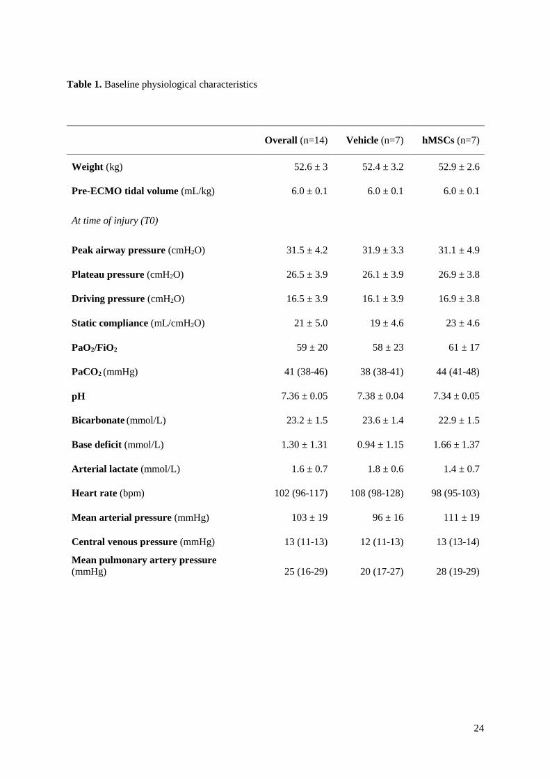
Baseline characteristics at injury (T0) are shown in Table 1 and in supplementary
Table E1. All animals completed the study protocol and were euthanized at T24.
Respiratory variables
The use of ECMO facilitated a lower tidal volume ventilation strategy (4 (4-4) mL/kg
ABW). The median ECMO flow rate was 2.75 L/min (2.5-3.25 L/min), with a sweep gas flow
of 3 L/min (2-3.5 L/min). During VV-ECMO, animals had a median PaO2 of 109 mmHg (94-
131 mmHg) and a PaCO2 of 32 mmHg (30-35 mmHg). There were no significant differences
in these parameters between groups (supplementary Figure E2). Animals were adequately
anticoagulated during ECMO, as measured by activated partial thromboplastin time (aPTT)
ratios. The dose of heparin was not significantly different between groups (supplementary
Figure E2).
Because VV-ECMO controls gas exchange, native lung function was assessed one hour
after cessation of extracorporeal flow and after the performance of a standardized lung
recruitment manoeuvre (T24). As shown in Figure 2 both the PaO2/FiO2 ratio (p = 0.076) and
the oxygenation index (OI) (p = 0.153) were numerically better in the carrier vehicle only
group, the differences were not statistically significant.

Combined MSC therapy and ECMO in experimental ARDS JE Millar et. al.
7
The plateau airway and driving pressures were similar between groups at T24 (Figure
2). Static lung compliance was also similar, both during and after ECMO (Figure 2). The use
of a protocolized recruitment manoeuvre did not improve compliance after cessation of
extracorporeal support in either group (Figure 2).
Hemodynamic variables
This model of acute lung injury was associated with the development of hyperdynamic
shock, which worsened over time (Figure 3). The administration of hMSCs resulted in
significantly lower cumulative vasopressor doses (Figure 3). At T4, mean arterial pressure
(MAP) was significantly higher in the hMSC treated group (p = 0.001), even though these
animals received lower doses of noradrenaline (Figure 3). By T14, MAP was again similar
between groups, although vasopressor requirements continued to be lower in hMSC treated
animals. In addition, there were lower arterial lactate concentrations, higher arterial base
excesses, and lower mean pulmonary artery pressures from 12 hrs (T14) post instillation in the
hMSC group (Figure 3), however these were not statistically significant. Cumulative fluid
balance at T24 was similar in both groups (Vehicle control, 2713 ± 970 mL vs. hMSCs, 2992 ±
1237 mL, p = 0.648).
Histopathology and lung injury
The blinded assessment of lung tissue was conducted by an independent expert
veterinary pathologist. Sections of the right lower lobe were prepared, and a lung injury score
(LIS) was calculated (19). The administration of hMSCs resulted in significantly lower scores
(p = 0.04) (Figure 4), principally mediated by a reduction in neutrophil infiltration.
There were no significant differences in lung wet/dry ratio or bronchoalveolar lavage
(BAL) total protein concentration (Figure 4). BAL fluid inflammatory cell counts are detailed
in supplementary Table E4. There were no significant differences in these counts over time.

Combined MSC therapy and ECMO in experimental ARDS JE Millar et. al.
8
Similarly, there was no difference in lung tissue homogenate gene expression (as assessed by
qPCR) between groups (supplementary Figure E3).
In a post-hoc analysis, pulmonary arterial thrombosis was noted in 5 hMSC treated
animals, but only one animal receiving carrier vehicle alone (p = 0.031).
Inflammatory cytokines
BAL and plasma cytokine concentrations were assessed longitudinally
(supplementary Figure E4 and supplementary Figure E5). In BAL, statistically significant
differences in IL-8 were observed at T3, T14, and T23 (p = 0.013, 0.016, and 0.028 respectively).
In plasma, cytokine trajectories were similar between groups (supplementary Figure E5).
Hematological and biochemical measurements
A summary of hematological and biochemical values are provided in supplementary
Tables E2 and E3. This lung injury model was associated with the development of acute
kidney injury and abnormal liver function, although there were no significant differences in
indices between groups. The administration of hMSCs resulted in a significantly lower
lymphocyte count at T24 (p = 0.047) (supplementary Figure E5).
Cell-ECMO membrane interaction and cell fate
The administration of hMSCs was associated with a significant increase in the trans-
oxygenator pressure gradient, becoming apparent 4 hours after cell delivery (Figure 5). By T23,
the mean pressure gradient in the hMSC group reached 64 ± 37 mmHg vs. 17 ± 9 mmHg in the
vehicle only group. The instillation of carrier vehicle alone was associated with a reduction in
the ECMO pump speed to flow ratio over time, a finding not observed in the hMSC group
(Figure 5). During the study, there were no instances of pump or oxygenator failure requiring

Combined MSC therapy and ECMO in experimental ARDS JE Millar et. al.
9
a component exchange. Likewise, there was no evidence of clotting on the oxygenator surface
by visual examination in either group.
Membrane oxygenators from animals treated with hMSCs were isolated and preserved
at the termination of ECMO, subsequent deconstruction and staining of the fiber bundles (n=7)
revealed adherent cells exhibiting surface markers consistent with those of hMSCs
(supplementary Figure E6). Similar cell populations were not apparent in vehicle only
controls (n=3).
Discussion
We carried out a trial of clinical-grade iPSC-derived hMSCs, given endobronchially,
for acute lung injury in sheep during VV-ECMO. The main findings of this study can be
summarized as follows; (1) with regard to the primary outcome, hMSCs did not improve
oxygenation at 24 hours. (2) hMSCs did not improve pulmonary mechanics but did improve
the severity of histological lung injury and reduced the concentration of bronchoalveolar lavage
IL-8. (3) in spite of endobronchial administration, hMSCs adhered to and impacted the function
of a commercial membrane oxygenator in-vivo - with an increase in the trans-membrane
oxygenator pressure gradient. In addition, more pulmonary arterial thromboses were noted in
hMSC-treated lungs. (4) hMSCs reduced the depth and severity of shock.
This study was conducted in a large animal model of ARDS and ECMO, which
replicates several important clinical features (20). The ‘double-hit’ injury applied in this study
resulted in acute severe hypoxemic respiratory failure consistent with modern criteria for the
use of VV-ECMO (11). To support the severe acute respiratory failure, we employed a
commercial ECMO device which is in widespread clinical use. Additionally, our protocolized
intensive care was consistent with clinical best practice standards (21). A common criticism of
pre-clinical trials of MSCs has been the use of heterogeneous, non-clinical grade cell products

Combined MSC therapy and ECMO in experimental ARDS JE Millar et. al.
10
(1), however, we tested a commercial hMSC product which is under investigation in clinical
trials.
MSCs have been administered to patients with respiratory failure on ECMO (22, 23).
These reports, which include only three patients, did not describe infusion-related adverse
events although failed to fully characterize the interaction between MSCs and the
extracorporeal device. Kocyildrim et al. have conducted the only other pre-clinical study
involving both MSCs and ECMO for respiratory failure (24). Their ovine based, 6-hour pilot
study did not report on the impact of MSC administration on the performance of ECMO.
hMSCs and pulmonary function
In this study, the administration of hMSCs failed to improve oxygenation at T24.
Furthermore, animals receiving hMSCs had a trend for worse oxygenation index values. In a
phase 2a study of 60 patients with ARDS, the intravenous administration of hMSCs did not
significantly improve PaO2/FiO2 ratio, although there was a signal toward improvement in
oxygenation index in a post-hoc analysis (10). Pre-clinical studies of MSCs in ARDS have
reported improvements in oxygenation, although few have produced lung injury as severe (25).
In a recent systematic review of pre-clinical models combining ARDS and ECMO, only four
achieved PaO2/FiO2 values <100 mmHg (20). The degree of lung injury in this model may
explain why oxygenation is impaired in the treated group, despite improvements in
inflammation and lung injury. Emerging research has highlighted the pro-coagulant effects of
transplanted MSCs. These appear to be primarily mediated by MSC expression of tissue factor
(26), but also by the secretion of pro-coagulant microvesicles (27) and by direct enhancement
of platelet deposition (28). In pre-clinical experiments MSCs have been associated with the
development of pulmonary emboli in-vivo (29). In this study, despite the use of heparin, almost
all hMSC animals (n=6) had histological evidence of pulmonary arterial thrombosis at post-
Combined MSC therapy and ECMO in experimental ARDS JE Millar et. al.
11
mortem. The presence of exogenous hMSCs within the disordered pulmonary vasculature may
have contributed to impairments in oxygenation, tempered by the fact that there was no increase
in mean pulmonary artery pressure in treated animals.
Animals receiving hMSCs had improved composite histologic lung injury scores at
post-mortem. The components of the score most influenced by hMSCs were neutrophil
numbers in the alveolar and interstitial space. Multiple studies of MSCs in pre-clinical models
of ARDS have demonstrated their ability to reduce neutrophil infiltration (30) and neutrophil
extracellular trap formation (31). In this study, BAL neutrophil counts did not differ between
groups. This may reflect the technical challenges of obtaining and assessing BAL cell counts.
In a recent porcine model of ARDS and MSC therapy, a reduction in neutrophil infiltration was
correlated with a reduction in BAL IL-8 concentrations (32), a finding confirmed in this study.
hMSCs and the systemic inflammatory response
Multiple pre-clinical models (33) and recent clinical trials (34-37) have examined the
use of MSCs in the treatment of septic shock. Animals receiving hMSCs required less
vasopressor support throughout the experiment to achieve an equivalent or higher MAP. A
similar, early but non-sustained, reduction in vasopressor requirement has previously been
described in a large-animal model of septic shock treated with MCSs (38). hMSCs did not alter
plasma concentrations of pro-inflammatory cytokines over extended time periods in this study,
a finding which has previously been identified in other pre-clinical (39) and clinical studies
(34). A recent Phase I dose escalation study of MSCs in patients with septic shock
demonstrated that the maximum effect of cell therapy on plasma cytokine levels occurred at 4
hours post-administration and declined with time (36). In this study, that time period coincides
with the maximum separation in vasopressor dose, MAP, and levels of IL-1β and IL-6 between

Combined MSC therapy and ECMO in experimental ARDS JE Millar et. al.
12
groups. This may indicate that repeat dosing of MSCs will be required for optimal therapeutic
efficacy.
hMSCs and ECMO
The risk posed by MSCs to membrane oxygenators has been postulated for some time
(1), but has only recently been shown to have an experimental basis. Our group has previously
reported the ability of hMSCs to tightly adhere to the membrane fibers of a commercial
oxygenator. This may have been the result of the known plastic avidity of MSCs (13). Recently,
Cho et al. reported the loss of systemically administered MSCs in an ex-vivo model of veno-
arterial ECMO (15).
Given the emerging signal that systemically administered MSCs may interact with
membrane oxygenators, we decided to test endobronchial instillation in this study. Cardenes et
al. have used 18F-fluorodeoxyglucose labelling to track the fate of both systemically and
endobronchially administered MSCs in an ovine model of ARDS (40). While systemically
administered cells have a wide biodistribution in the first 5 hours, endobronchially
administered cells were retained at the site of instillation. There are key differences in our
approach, including the means of inducing lung injury and its severity.
Limitations
This study has some limitations. First, while our model of injury replicates several
relevant features including severe respiratory and hemodynamic failure, clinical ARDS is
usually caused by infection and develops over several days, often in patients with other
comorbidities (41). Second, MSCs are known to exhibit different functional responses
dependent on the contemporary milieu, which in some circumstances may be detrimental (42).
This study may have modelled only one phase of acute lung injury and so hMSCs may have
exerted an effect which may differ in other phases. Third, while our model extended 21 hours
Combined MSC therapy and ECMO in experimental ARDS JE Millar et. al.
13
post cell or vehicle delivery, this may have been too short a period to observe some beneficial
effects of the intervention. For example, it may be that the favourable effect of hMSCs on
histological injury may have translated to improvements in oxygenation over a longer time
period. Conversely, an extension of the study period in the face of rising trans-oxygenator
pressure gradients in the hMSC group may have ultimately led to circuit failures. Fourth, the
use of a lung recruitment maneuver and the assessment of native lung function off ECMO may
have had several adverse effects and we cannot be certain that these effects did not differ
between groups. This approach was taken due to the challenge of assessing native lung function
during ECMO, particularly where a lower tidal volume ventilatory strategy has been adopted.
The study protocol was designed prior to the publication of the ART randomized controlled
trial (43). Fifth, the addition of an uninjured control group may have provided further insights
into the distribution of hMSCs during VV-ECMO. Finally, the dose and method of delivery of
hMSCs remain a matter of conjecture. Based on the findings of our previous work (14), we
chose not to investigate intravenous administration. Likewise, based on clinical trial
experience, we opted to administer a single, fixed-dose of hMSCs. It is possible that varying
the dose and/or route of administration of hMSCs may alter their efficacy and safety profile
during ECMO.
Conclusion
In a 24-hour, ovine model of ARDS and VV-ECMO, we found that hMSC therapy was
associated with impairment of the membrane oxygenator. The use of cell therapy did not result
in improvements in oxygenation, the primary outcome of this study, but was associated with a
reduction in histological evidence of lung injury and inflammation in the lung. Given these
data we cannot currently recommend the administration of hMSCs during ECMO.
Acknowledgments

Combined MSC therapy and ECMO in experimental ARDS JE Millar et. al.
14
We are grateful for assistance provided during animal experiments by the staff of the
Queensland University of Technology Medical Engineering Research Facility (MERF) and the
University of Queensland Animal Science Precinct (QASP). We extend our thanks to Ms
Arlanna Esguerra-Lallen for providing expert nursing care and to Ms Mengyao Yang, Mr
Matthew Wells, Dr Ai-Ching Boon, Mr Michael Cavaye, and Ms Ashlen Garrett for their
assistance in study preparation, sampling, and laboratory analyses.
Combined MSC therapy and ECMO in experimental ARDS JE Millar et. al.
15
References
1. Millar JE, Fraser JF, McAuley DF. Mesenchymal stromal cells and the acute respiratory
distress syndrome (ARDS): challenges for clinical application. Thorax 2015; 70: 611-
612.
2. Pittenger MF, Mackay AM, Beck SC, Jaiswal RK, Douglas R, Mosca JD, Moorman MA,
Simonetti DW, Craig S, Marshak DR. Multilineage potential of adult human
mesenchymal stem cells. Science 1999; 284: 143-147.
3. Curley GF, McAuley DF. Stem cells for respiratory failure. Curr Opin Crit Care 2015; 21:
42-49.
4. Devaney J, Horie S, Masterson C, Elliman S, Barry F, O'Brien T, Curley GF, O'Toole D,
Laffey JG. Human mesenchymal stromal cells decrease the severity of acute lung injury
induced by E. coli in the rat. Thorax 2015; 70: 625-635.
5. Fang X, Neyrinck AP, Matthay MA, Lee JW. Allogeneic human mesenchymal stem cells
restore epithelial protein permeability in cultured human alveolar type II cells by
secretion of angiopoietin-1. J Biol Chem 2010; 285: 26211-26222.
6. McAuley DF, Curley GF, Hamid UI, Laffey JG, Abbott J, McKenna DH, Fang X, Matthay
MA, Lee JW. Clinical grade allogeneic human mesenchymal stem cells restore alveolar
fluid clearance in human lungs rejected for transplantation. Am J Physiol Lung Cell Mol
Physiol 2014; 306: L809-815.
7. Krasnodembskaya A, Song Y, Fang X, Gupta N, Serikov V, Lee J-W, Matthay MA.
Antibacterial effect of human mesenchymal stem cells is mediated in part from
secretion of the antimicrobial peptide LL-37. Stem Cells 2010; 28: 2229-2238.
8. Jackson MV, Morrison TJ, Doherty DF, McAuley DF, Matthay MA, Kissenpfennig A,
O'Kane CM, Krasnodembskaya AD. Mitochondrial Transfer via Tunneling Nanotubes
is an Important Mechanism by Which Mesenchymal Stem Cells Enhance Macrophage
Combined MSC therapy and ECMO in experimental ARDS JE Millar et. al.
16
Phagocytosis in the In Vitro and In Vivo Models of ARDS. Stem Cells 2016; 34: 2210-
2223.
9. McIntyre LA, Moher D, Fergusson DA, Sullivan KJ, Mei SH, Lalu M, Marshall J, McLeod
M, Griffin G, Grimshaw J, Turgeon A, Avey MT, Rudnicki MA, Jazi M, Fishman J,
Stewart DJ, Canadian Critical Care Translational Biology G. Efficacy of Mesenchymal
Stromal Cell Therapy for Acute Lung Injury in Preclinical Animal Models: A
Systematic Review. PLoS One 2016; 11: e0147170.
10. Matthay MA, Calfee CS, Zhuo H, Thompson BT, Wilson JG, Levitt JE, Rogers AJ, Gotts
JE, Wiener-Kronish JP, Bajwa EK, Donahoe MP, McVerry BJ, Ortiz LA, Exline M,
Christman JW, Abbott J, Delucchi KL, Caballero L, McMillan M, McKenna DH, Liu
KD. Treatment with allogeneic mesenchymal stromal cells for moderate to severe acute
respiratory distress syndrome (START study): a randomised phase 2a safety trial.
Lancet Respir Med 2018; 7(2):154-162.
11. Combes A, Hajage D, Capellier G, Demoule A, Lavoue S, Guervilly C, Da Silva D, Zafrani
L, Tirot P, Veber B, Maury E, Levy B, Cohen Y, Richard C, Kalfon P, Bouadma L,
Mehdaoui H, Beduneau G, Lebreton G, Brochard L, Ferguson ND, Fan E, Slutsky AS,
Brodie D, Mercat A. Extracorporeal Membrane Oxygenation for Severe Acute
Respiratory Distress Syndrome. N Engl J Med; 378: 1965-1975.
12. Hoogduijn MJ, van den Beukel JC, Wiersma LCM, Ijzer J. Morphology and size of stem
cells from mouse and whale: observational study. Br Med J 2013; 347: f6833.
13. Dominici M, Le Blanc K, Mueller I, Slaper-Cortenbach I, Marini F, Krause D, Deans R,
Keating A, Prockop D, Horwitz E. Minimal criteria for defining multipotent
mesenchymal stromal cells. The International Society for Cellular Therapy position
statement. Cytotherapy 2006; 8: 315-317.
Combined MSC therapy and ECMO in experimental ARDS JE Millar et. al.
17
14. Millar JE, von Bahr V, Malfertheiner MV, Ki KK, Redd MA, Bartnikowski N, Suen JY,
McAuley DF, Fraser JF. Administration of mesenchymal stem cells during ECMO
results in a rapid decline in oxygenator performance. Thorax 2018.
15. Cho HJ, Hong H, Kim DW, Lee KS, Han HS, Kim GH, Choi KS, Kim YS, Qayumov M,
Ki KK, Suen J, Fraser J, Jeong IS. Viability of Mesenchymal Stem Cells in an Ex Vivo
Circulation System. ASAIO J 2019. doi: 10.1097/MAT.0000000000001025. [Epub
ahead of print].
16. Millar JE, Fanning JP, McDonald CI, McAuley DF, Fraser JF. The inflammatory response
to extracorporeal membrane oxygenation (ECMO): a review of the pathophysiology.
Crit Care 2016; 20: 387.
17. Council NHaMR. Australian code for the care and use of animals for scientific purposes.
In: Council NHaMR, editor, 8th Edition ed. Canberra; 2013.
18. Kilkenny C, Browne WJ, Cuthill IC, Emerson M, Altman DG. Improving bioscience
research reporting: the ARRIVE guidelines for reporting animal research. Osteoarthr
Cartilage 2012; 20: 256-260.
19. Matute-Bello G, Downey G, Moore BB, Groshong SD, Matthay MA, Slutsky AS, Kuebler
WM. An official American Thoracic Society workshop report: features and
measurements of experimental acute lung injury in animals. Am J Respir Cell Mol Biol
2011; 44: 725-738.
20. Millar JE, Bartnikowski N, von Bahr V, Malfertheiner MV, Obonyo NG, Belliato M, Suen
JY, Combes A, McAuley DF, Lorusso R, Fraser JF. Extracorporeal membrane
oxygenation (ECMO) and the acute respiratory distress syndrome (ARDS): a
systematic review of pre-clinical models. Intensive Care Med Exp 2019; 7: 18.
21. Griffiths MJD, McAuley DF, Perkins GD, Barrett N, Blackwood B, Boyle A, Chee N,
Connolly B, Dark P, Finney S, Salam A, Silversides J, Tarmey N, Wise MP, Baudouin
Combined MSC therapy and ECMO in experimental ARDS JE Millar et. al.
18
SV. Guidelines on the management of acute respiratory distress syndrome. BMJ Open
Respir Res 2019; 6: e000420.
22. Simonson OE, Mougiakakos D, Heldring N, Bassi G, Johansson HJ, Dalen M, Jitschin R,
Rodin S, Corbascio M, El Andaloussi S, Wiklander OP, Nordin JZ, Skog J, Romain C,
Koestler T, Hellgren-Johansson L, Schiller P, Joachimsson PO, Hagglund H, Mattsson
M, Lehtio J, Faridani OR, Sandberg R, Korsgren O, Krampera M, Weiss DJ, Grinnemo
KH, Le Blanc K. In Vivo Effects of Mesenchymal Stromal Cells in Two Patients With
Severe Acute Respiratory Distress Syndrome. Stem Cells Tranl Med 2016; 5: 845.
23. Lin H-C, Wang C-C, Chou H-W, Wu E-T, Lu FL, Ko B-S, Yao M, Wang P-Y, Wu M-H,
Chen Y-S. Airway Delivery of Bone Marrow–Derived Mesenchymal Stem Cells
Reverses Bronchopulmonary Dysplasia Superimposed with Acute Respiratory Distress
Syndrome in an Infant. Cell Med 2018; 10: 2155179018759434.
24. Kocyildirim E, Cardenes N, Ting A, Caceres E, BermUdez C, Rojas M. The Use of GMP-
Produced Bone Marrow-Derived Stem Cells in Combination with Extracorporeal
Membrane Oxygenation in ARDS: An Animal Model. ASAIO J 2017; 63: 324-332.
25. McIntyre LA, Moher D, Fergusson DA, Sullivan KJ, Mei SH, Lalu M, Marshall J, McLeod
M, Griffin G, Grimshaw J, Turgeon A, Avey MT, Rudnicki MA, Jazi M, Fishman J,
Stewart DJ. Efficacy of Mesenchymal Stromal Cell Therapy for Acute Lung Injury in
Preclinical Animal Models: A Systematic Review. PloS One 2016; 11: e0147170.
26. Christy BA, Herzig MC, Montgomery RK, Delavan C, Bynum JA, Reddoch KM, Cap AP.
Procoagulant activity of human mesenchymal stem cells. J Trauma Acute Care Surg
2017; 83: S164-s169.
27. Fiedler T, Rabe M, Mundkowski RG, Oehmcke-Hecht S, Peters K. Adipose-derived
mesenchymal stem cells release microvesicles with procoagulant activity. Int J
Biochem Cell Biol 2018; 100: 49-53.
Combined MSC therapy and ECMO in experimental ARDS JE Millar et. al.
19
28. Gleeson BM, Martin K, Ali MT, Kumar AH, Pillai MG, Kumar SP, O'Sullivan JF, Whelan
D, Stocca A, Khider W, Barry FP, O'Brien T, Caplice NM. Bone Marrow-Derived
Mesenchymal Stem Cells Have Innate Procoagulant Activity and Cause Microvascular
Obstruction Following Intracoronary Delivery: Amelioration by Antithrombin
Therapy. Stem Cells 2015; 33: 2726-2737.
29. Tatsumi K, Ohashi K, Matsubara Y, Kohori A, Ohno T, Kakidachi H, Horii A, Kanegae K,
Utoh R, Iwata T, Okano T. Tissue factor triggers procoagulation in transplanted
mesenchymal stem cells leading to thromboembolism. Biochem Biophys Res Commun
2013; 431: 203-209.
30. Curley GF, Hayes M, Ansari B, Shaw G, Ryan A, Barry F, O'Brien T, O'Toole D, Laffey
JG. Mesenchymal stem cells enhance recovery and repair following ventilator-induced
lung injury in the rat. Thorax 2012; 67: 496-501.
31. Pedrazza L, Cunha AA, Luft C, Nunes NK, Schimitz F, Gassen RB, Breda RV, Donadio
MV, de Souza Wyse AT, Pitrez PMC, Rosa JL, de Oliveira JR. Mesenchymal stem
cells improves survival in LPS-induced acute lung injury acting through inhibition of
NETs formation. J Cell Physiol 2017; 232: 3552-3564.
32. Moodley Y, Sturm M, Shaw K, Shimbori C, Tan DB, Kolb M, Graham R. Human
mesenchymal stem cells attenuate early damage in a ventilated pig model of acute lung
injury. Stem Cell Res 2016; 17: 25-31.
33. Lalu MM, Sullivan KJ, Mei SH, Moher D, Straus A, Fergusson DA, Stewart DJ, Jazi M,
MacLeod M, Winston B, Marshall J, Hutton B, Walley KR, McIntyre L. Evaluating
mesenchymal stem cell therapy for sepsis with preclinical meta-analyses prior to
initiating a first-in-human trial. eLife 2016; 5.
34. McIntyre LA, Stewart DJ, Mei SHJ, Courtman D, Watpool I, Granton J, Marshall J, Dos
Santos C, Walley KR, Winston BW, Schlosser K, Fergusson DA. Cellular
Combined MSC therapy and ECMO in experimental ARDS JE Millar et. al.
20
Immunotherapy for Septic Shock. A Phase I Clinical Trial. Am J Crit Care Med 2018;
197: 337-347.
35. Galstian GM, Parovichnikova EN, Makarova PM, Kuzmina LA, Troitskaya VV,
Gemdzhian E, Drize NI, Savchenko VG. The Results of the Russian Clinical Trial of
Mesenchymal Stromal Cells (MSCs) in Severe Neutropenic Patients (pts) with Septic
Shock (SS) (RUMCESS trial). Blood 2015; 126: 2220.
36. Schlosser K, Wang J-P, Dos Santos C, Walley KR, Marshall J, Fergusson DA, Winston
BW, Granton J, Watpool I, Stewart DJ, McIntyre LA, Mei SHJ, Canadian Critical Care
Trials G, the Canadian Critical Care Translational Biology G. Effects of Mesenchymal
Stem Cell Treatment on Systemic Cytokine Levels in a Phase 1 Dose Escalation Safety
Trial of Septic Shock Patients. Crit Care Med 2019; 47: 918-925.
37. Perlee D, van Vught LA, Scicluna BP, Maag A, Lutter R, Kemper EM, van 't Veer C,
Punchard MA, Gonzalez J, Richard MP, Dalemans W, Lombardo E, de Vos AF, van
der Poll T. Intravenous Infusion of Human Adipose Mesenchymal Stem Cells Modifies
the Host Response to Lipopolysaccharide in Humans: A Randomized, Single-Blind,
Parallel Group, Placebo Controlled Trial. Stem Cells 2018; 36(11):1778-1788.
38. Laroye C, Lemarié J, Boufenzer A, Labroca P, Cunat L, Alauzet C, Groubatch F, Cailac C,
Jolly L, Bensoussan D, Reppel L, Gibot S. Clinical-grade mesenchymal stem cells
derived from umbilical cord improve septic shock in pigs. Intensive Care Med 2018; 6:
24-24.
39. Laroye C, Boufenzer A, Jolly L, Cunat L, Alauzet C, Merlin JL, Yguel C, Bensoussan D,
Reppel L, Gibot S. Bone marrow vs Wharton's jelly mesenchymal stem cells in
experimental sepsis: a comparative study. Stem Cell Res Ther 2019; 10: 192.
40. Cardenes N, Aranda-Valderrama P, Carney JP, Sellares Torres J, Alvarez D, Kocydirim E,
Wolfram Smith JA, Ting AE, Lagazzi L, Yu Z, Mason S, Santos E, Lopresti BJ, Rojas
Combined MSC therapy and ECMO in experimental ARDS JE Millar et. al.
21
M. Cell therapy for ARDS: efficacy of endobronchial versus intravenous administration
and biodistribution of MAPCs in a large animal model. BMJ Open Respir Res 2019; 6:
e000308.
41. Matthay MA, Zemans RL, Zimmerman GA, Arabi YM, Beitler JR, Mercat A, Herridge M,
Randolph AG, Calfee CS. Acute respiratory distress syndrome. Nat Rev Dis Primers
2019; 5: 18.
42. Islam D, Huang Y, Fanelli V, Delsedime L, Wu S, Khang J, Han B, Grassi A, Li M, Xu Y,
Luo A, Wu J, Liu X, McKillop M, Medin J, Qiu H, Zhong N, Liu M, Laffey J, Li Y,
Zhang H. Identification and Modulation of Microenvironment Is Crucial for Effective
Mesenchymal Stromal Cell Therapy in Acute Lung Injury. Am J Crit Care Med 2018;
199: 1214-1224.
43. Cavalcanti AB, Suzumura EA, Laranjeira LN, Paisani DM, Damiani LP, Guimaraes HP,
Romano ER, Regenga MM, Taniguchi LNT, Teixeira C, Pinheiro de Oliveira R,
Machado FR, Diaz-Quijano FA, Filho MSA, Maia IS, Caser EB, Filho WO, Borges
MC, Martins PA, Matsui M, Ospina-Tascon GA, Giancursi TS, Giraldo-Ramirez ND,
Vieira SRR, Assef M, Hasan MS, Szczeklik W, Rios F, Amato MBP, Berwanger O,
Ribeiro de Carvalho CR. Effect of Lung Recruitment and Titrated Positive End-
Expiratory Pressure (PEEP) vs Low PEEP on Mortality in Patients With Acute
Respiratory Distress Syndrome: A Randomized Clinical Trial. JAMA 2017; 318: 1335-
1345.

Combined MSC therapy and ECMO in experimental ARDS JE Millar et. al.
22
Figure Legends
Figure 1. Study schematic.
a Adjusted to maintain pH 7.30-7.45. Permissive hypercapnia was tolerated to a minimum pH of 7.15.
b PaO2 55 – 80 mmHg. If despite an FiO2 of 1.0 oxygenation targets were not met, PEEP was
increased, maintaining plateau pressure ≤ 32 cmH2O.
c Total PEEP (extrinsic PEEP + intrinsic PEEP) did not exceed 20 cmH2O. PEEP was permitted to be
reduced to 5 cmH2O to maintain plateau pressure ≤ 30 cmH2O. If despite a PEEP of 5 cmH2O, plateau
pressure > 30 cmH2O, tidal volume was reduced in 1 mL/kg steps until set at 4 mL/kg.
Figure 2. Oxygenation and respiratory parameters.
a. PaO2/FiO2 ratio. b. Oxygenation index. c. Airway pressures and lung compliance. Data are
presented as mean (± 95% confidence interval). Where error bars intersect the x axis the 95%
CI includes zero.
Figure 3. Hemodynamic variables.
a. Mean arterial pressure and vasopressor dose (mean). b. Mean pulmonary artery pressure. c.
Cumulative vasopressor dose. d. Cardiac index, base deficit, and arterial lactate. Data are
presented as mean (± 95% confidence interval). Where error bars intersect the x axis the 95%
CI includes zero. ** - p < 0.01.
Figure 4. Histopathology and lung injury.
a. Representative images of lung parenchyma. All animals showed evidence of diffuse alveolar
damage however the frequency and degree of injury differed between groups. Panel 1:
extensive alveolar edema with interstitial leukocyte infiltration. Panel 2: marked leukocyte
infiltration within alveolar spaces and larger airways. Panel 3: some loss of alveolar structure
with edema however a reduction in interstitial and alveolar leukocytes. Panel 4: preservation

Combined MSC therapy and ECMO in experimental ARDS JE Millar et. al.
23
of alveolar architecture with few leukocytes in the alveolar spaces. Panel 5 & 6: representative
images of pulmonary arterial and arteriolar emboli (black arrows) in animals receiving hMCSs.
b. Composite lung injury score (LIS), lung wet/dry ratio (right lower lobe). c. BAL total protein
concentration. Data are presented as mean (± 95% confidence interval). * - p = < 0.05.
Figure 5. Cell-ECMO interaction.
a. Trans-membrane oxygenator pressure gradient. b. Pump revolutions per minute (RPM)/flow
ratio. Data are presented as mean (± 95% confidence interval). Where error bars intersect the
x axis the 95% CI includes zero. * - p = < 0.05.
24
Table 1. Baseline physiological characteristics
Overall (n=14) Vehicle (n=7) hMSCs (n=7)
Weight (kg) 52.6 ± 3 52.4 ± 3.2 52.9 ± 2.6
Pre-ECMO tidal volume (mL/kg) 6.0 ± 0.1 6.0 ± 0.1 6.0 ± 0.1
At time of injury (T0)
Peak airway pressure (cmH2O) 31.5 ± 4.2 31.9 ± 3.3 31.1 ± 4.9
Plateau pressure (cmH2O) 26.5 ± 3.9 26.1 ± 3.9 26.9 ± 3.8
Driving pressure (cmH2O) 16.5 ± 3.9 16.1 ± 3.9 16.9 ± 3.8
Static compliance (mL/cmH2O) 21 ± 5.0 19 ± 4.6 23 ± 4.6
PaO2/FiO2 59 ± 20 58 ± 23 61 ± 17
PaCO2 (mmHg) 41 (38-46) 38 (38-41) 44 (41-48)
pH 7.36 ± 0.05 7.38 ± 0.04 7.34 ± 0.05
Bicarbonate (mmol/L) 23.2 ± 1.5 23.6 ± 1.4 22.9 ± 1.5
Base deficit (mmol/L) 1.30 ± 1.31 0.94 ± 1.15 1.66 ± 1.37
Arterial lactate (mmol/L) 1.6 ± 0.7 1.8 ± 0.6 1.4 ± 0.7
Heart rate (bpm) 102 (96-117) 108 (98-128) 98 (95-103)
Mean arterial pressure (mmHg) 103 ± 19 96 ± 16 111 ± 19
Central venous pressure (mmHg) 13 (11-13) 12 (11-13) 13 (13-14)
Mean pulmonary artery pressure
(mmHg)
25 (16-29) 20 (17-27) 28 (19-29)