combhelix® fshd probes english
TRANSCRIPT

Page | 1
CombHeliX® FSHD probes
FSHD-HYB-001
Instructions for use
Version: 04
Date: November 2015
Background information
FSHD (Facio-Scapulo-Humeral Muscular Dystrophy) is a genetic disease with autosomal
dominant transmission defined by a progressive weakening and loss of skeletal muscles
of the face (facio), the shoulder girdle (scapulo) and the upper arms (humeral). In 95 % of
FSHD patients (also referred as FSHD1), a contracted 3.3kb-D4Z4 repeat array (1 to 10
copies) is present on a chromosome 4q35.2 subtelomere [1, 2] whereas the array
contains between 11 and 150 repeats in the control population [3, 4]. In contrast, the
FSHD2 form which is a rare form of FSHD is not linked to D4Z4 repeat array contraction.
D4Z4 repeat arrays are also found on chromosome 10q26.1 subtelomeres, with no
apparent relation to the disease. Chromosomes 4q and 10q subtelomeres may bear the
haplotype A or B, which show several kilobases of divergent sequences. D4Z4 contraction
is only pathogenic when it segregates in cis with the haplotype A on chromosome 4 [5-7].
The FSHD probes enable the determination of the size of the D4Z4 repeat array and the A
or B haplotype on both chromosomes 4q35.2 and 10q26.1 [8].
Intended use
The “CombHeliX® FSHD probes” are designed for the diagnostic of FSHD1 by enabling
the characterization of the 3.3kb-D4Z4 repeat-containing loci on chromosomes 4 and 10
by fluorescent hybridization on combed DNA extracted from blood samples and prepared
according to the Molecular Combing procedures [8]. The “CombHeliX® FSHD probes” are
intended to be used for in vitro diagnostic purpose.
Important note: In addition to the “CombHeliX® FSHD probes”, the diagnostic of FSHD
requires the mandatory use of the CE-IVD “CombHeliX® FSHD Software” for the
EN
GLI
SH
Page | 2
detection of the hybridized “CombHeliX® FSHD probes”, the analysis and the interpretation
of the results (see the Instruction for use for “CombHeliX® FSHD Software”).
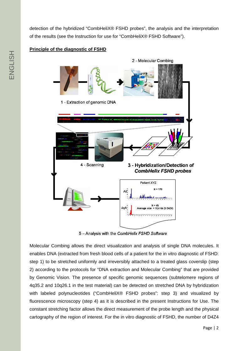
Principle of the diagnostic of FSHD
Molecular Combing allows the direct visualization and analysis of single DNA molecules. It
enables DNA (extracted from fresh blood cells of a patient for the in vitro diagnostic of FSHD:
step 1) to be stretched uniformly and irreversibly attached to a treated glass coverslip (step
2) according to the protocols for “DNA extraction and Molecular Combing” that are provided
by Genomic Vision. The presence of specific genomic sequences (subtelomere regions of
4q35.2 and 10q26.1 in the test material) can be detected on stretched DNA by hybridization
with labeled polynucleotides (“CombHeliX® FSHD probes”: step 3) and visualized by
fluorescence microscopy (step 4) as it is described in the present Instructions for Use. The
constant stretching factor allows the direct measurement of the probe length and the physical
cartography of the region of interest. For the in vitro diagnostic of FSHD, the number of D4Z4
E
NG
LIS
H

Page | 3
repeats in the test material is determined with the “CombHeliX® FSHD Software” (step 5)
as described in the corresponding Instructions for Use provided by Genomic Vision.
Product content and description
“CombHeliX® FSHD probes” set: 5 x 20 µl per vial (2 tests per vial, 10 tests in total). One
test is defined as sufficient for one hybridization experiment of combed DNA on coverslips
(22 x 22 mm area).
The “CombHeliX® FSHD probes” contain fluorescein-, digoxygenin- or/and biotin-labeled
polynucleotides that are intended to be used in combination with specific reagents (see
Reagents and materials recommended but not provided) that allow their detection in
green, blue, red and magenta fluorescent signals.
Labeled polynucleotides are premixed with blocking DNA in formamide-free hybridization
buffer (Saline-Sodium Citrate (SSC), Sodium Dodecyl Sulfate (SDS), Sodium Lauroyl
Sarcosinate and BlockingAid™ blocking solution).
Storage and handling
Upon arrival, the product must be stored between -25°C and -10°C protected from the
light until the expiry date printed on the label. Improper storage of the product can destroy
or impair the performance of the product and consequently the in vitro diagnostic assay
should not be performed from such reagent since it may affect the result of the test.
Handle all reagents and slides containing fluorophores in reduced light to prevent
photobleaching.
Once thawed and prior to opening, the vials should be briefly centrifuged to ensure the
contents are collected at the bottom of the vials.
Once opened, the remaining content of the vial can be frozen again and stored between -
25°C and -10°C up to 6 months.
Warnings and precautions
For in vitro diagnostic use. For professional use only. Carefully read the operating
instructions before use.
The diagnostic performances of the “CombHeliX® FHSD probes” were validated using
coverslips with very long genomic DNA molecules linearly stretched at high density
(picture 5) extracted from fresh blood samples. Using coverslips with genomic DNA at
EN
GLI
SH

Page | 4
lower density (pictures 1-4) or short, wavy or tangled DNA molecules may affect the result of
the test.
Do not use the product after the expiry date.
As some substances contained in this product (in low concentrations and volumes) could be
harmful for health, handle the reagent with care and wear appropriate personal protective
equipment. See the Material Safety Data Sheet (MSDS) for safety information.
Reagents and materials recommended but not provided for the hybridization and the detection of the “CombHeliX® FSHD probes”
IMPORTANT: Since the performance characteristics of the “CombHeliX® FSHD probes”
have been evaluated and validated using the reagents and materials listed below, we
recommend the use of these referenced reagents and materials for an optimal diagnostic
result.
The “CombHeliX® FHSD probes” were validated with the following accompanying reagents
and materials for their detection as green, red, blue and magenta fluorescent signals:
- Green fluorescent signal:
o 1mg/ml anti-fluorescein/Oregon Green® antibody, rabbit IgG fraction
(Invitrogen, Ref: A-889). Ready-to-use.
o 2 mg/mL Alexa Fluor® 488 goat anti-rabbit IgG (H+L) (Invitrogen, Ref: A-
11034). Ready-to-use.
- Red fluorescent signal:
o 1 mg streptavidin, Alexa Fluor® 594 conjugate (Invitrogen, Ref: S-11227). To
be reconstituted (see reagent preparation).
o 0.5 mg biotinylated Anti-Streptavidin (Vector Laboratories, Ref: BA-500). To
be reconstituted (see reagent preparation).
- Blue fluorescent signal:
o 0.5 mg aminomethylcoumarin (AMCA)-IgG Fraction Monoclonal Mouse Anti-
Digoxin (Jackson Immunoresearch, Ref: 415-155-156). To be reconstituted
(see reagent preparation).
E
NG
LIS
H

Page | 5
o 1 mg AMCA-AffiniPure Rat Anti-Mouse IgG (H+L) (Jackson
Immunoresearch, Ref: 415-155-166). To be reconstituted (see reagent
preparation).
o 2 mg/mL Alexa Fluor® 350 Goat Anti-Rat IgG (H+L) (Invitrogen, Ref: A-
21093). Ready-to-use.
Note : Magenta fluorescent signal is the result of the double labeling of the
corresponding probes with digoxygenin and biotin that allows the detection of a co-
localized blue and red signal with the above-mentioned reagents.
- “CombHeliX® FSHD Software” (Contact Genomic Vision for further additional
information).
Other reagents and materials are:
- Autoclaved distilled water
- Deionized formamide
- 20X SSC
- 70%, 90% and 100% ethanol
- BlockAid™ blocking solution
- Tween® 20
- 1X PBS
- Variable micropipette (1 µL - 200 µL)
- Tweezers
- Microscope slide
- Co-denaturation and hybridization instrument (e.g. Hybridizer, Dako)
- Ceramic coverslip tray
- Humidified chamber
- Incubator at 37°C
- 250 mL-beaker
- ImageXpress® Micro System (Molecular Devices)
Preparation of the reagents for the hybridization a nd the detection of the “CombHeliX® FSHD probes”
IMPORTANT: Use autoclaved distilled water for preparation of all stock and working solution.
- Hybridization Washing Buffer (2X SSC solution): mix 100 mL of 20X SSC and 900
mL of distilled water. Store at RT.
Note: The buffer must be warmed at 60°C prior to use.
EN
GLI
SH

Page | 6
- Detection Washing Buffer (2X SSC/1% Tween): Mix 100 mL of 20X SSC and 10 mL
of Tween® 20 with 890 mL of distilled water. To be prepared extemporaneously.
- Reconstitute the lyophilized reagents with autoclaved distilled water as follows:
- Prepare extemporaneously the mix of the different layers of detecting solutions as
follows (for one test):
Volume (µL) First layer:
- 1 mg/mL Streptavidin, Alexa Fluor® 594 conjugate 0,8 - 1 mg/ml anti-fluorescein/Oregon Green® antibody, rabbit IgG
fraction 0,8 - 1,43 mg/mL AMCA-IgG Fraction Monoclonal Mouse Anti-Digoxin 0,8 - BlockAid™ blocking solution 17,6
Second Layer: - 0,5 mg/mL Biotinylated Anti-Streptavidin 0,8 - 2 mg/mL Alexa Fluor® 488 goat anti-rabbit IgG (H+L) 0,8 - 1,33 mg/mL AMCA-AffiniPure Rat Anti-Mouse IgG 0,8 - BlockAid™ blocking solution 17,6
Third layer : - 1 mg/mL Streptavidin, Alexa Fluor® 594 conjugate 0,8 - 2 mg/mL Alexa Fluor® 350 Goat Anti-Rat IgG (H+L) 0,8 - BlockAid™ blocking solution 18,4
Procedure
Hybridization of the CombHeliX® FSHD probes on comb ed DNA
IMPORTANT: all the steps where formamide is used should be performed under a fume
hood.
1. Thaw the vial containing the “CombHeliX® FSHD probes” and the coverslips with
combed DNA on a ceramic coverslip tray for 10 min at room temperature.
2. Dehydrate the combed DNA coverslips by dipping the ceramic coverslip tray 3 min at
room temperature in successive baths of 70%, 90% and 100% ethanol.
3. Air dry the coverslips at room temperature for 10 min protecting from light.
4. For one test, transfer 10 µL of the “CombHeliX® FSHD Probes” in a new microcentrifuge
tube and add 10 µL of deionized formamide to the “CombHeliX® FSHD probes”
5. Mix well and incubate at 37°C for 30 min
Quantity (mg)
Volume of distilled water to be added (mL)
Final concentration
(mg/mL) Biotinylated Anti-Streptavidin 0,5 1 0,5 Streptavidin, Alexa Fluor® 594 conjugate 1 1 1 AMCA-IgG Fraction Monoclonal Mouse Anti-Digoxin 0.5 0,35 1,43 AMCA-AffiniPure Rat Anti-Mouse IgG 1 0,75 1,33
E
NG
LIS
H

Page | 7
6. Pipette 20 µL of the probes/formamide mix on a microscope slide.
7. Avoiding trapped bubbles, set the coverslip on the drop of hybridization solution.
Note: Carefully indicate the side of the coverslip in contact with the hybridization
solution to avoid confusion in the subsequent steps.
Note: Lay down the coverslip carefully using tweezers to avoid bubbles. Do not push
and slide the coverslip once mounted, it will cause scratches on combed coverslips.
Adjust the position of coverslip by gentle touching the corner if needed.
Note: Protect the hybridized slides from light from this step.
8. Co-denature the combed DNA on the coverslip and the probes for 5 min at 90°C in
the humidified chamber of a hands-free co-denaturation and hybridization
instrument.
9. Incubate for 16-20 hrs at 37°C in the humidified chamber of the co-denaturation and
hybridization instrument.
10. Remove the coverslip from the microscope slide and place on a ceramic coverslip
tray in a 250 mL-beaker containing the pre-warmed (60°C) Hybridization Washing
Buffer.
Note: if the coverslip is stuck to the slide, add a drop of Hybridization Washing Buffer
1 around the coverslip and wait until the coverslips floats. When removing coverslip,
slide the coverslips gently until one of corner is out on slide to avoid scratches, then
gently “peel off” the coverslip with tweezers.
11. Wash the coverslip three times in pre-warmed (60°C) Hybridization Washing Buffer
for 5 min each at 60°C.
Note: Do not let the coverslip dry. It must be totally immerged in the solution.
Detection of the CombHeliX® FSHD probes
1. Pipette 20 µL of the first layer of detecting solution on a microscope slide and set the
coverslip on the drop of detecting solution.
Note: Make sure to place the hybridized side of the coverslip in contact with the
detecting solution.
2. Incubate the slide for 20 min in a humidified chamber at 37 °C.
3. Remove the coverslip from the microscope slide and place it on a ceramic coverslip
tray in a 250 mL-beaker containing the Detection Washing Buffer.
4. Wash the coverslip three times in the Detection Washing Buffer for 3 min each at
room temperature with gentle agitation.
5. Pipette 20 µL of the second layer of detecting solution on a microscope slide and set
the coverslip on the drop of detecting solution.
EN
GLI
SH

Page | 8
Note: Make sure to place the hybridized side of the coverslip in contact with the
detecting solution.
6. Repeat steps 2 to 4
7. Pipette 20 µL of the third layer of detecting solution on a microscope slide and set the
coverslip on the drop of detecting solution.
Note: Make sure to place the hybridized side of the coverslip in contact with the
detecting solution.
8. Repeat steps 2 to 4
9. Wash the coverslip in 1xPBS for 3 min at room temperature with gentle agitation.
10. Dehydrate the coverslip by dipping the ceramic coverslip tray 3 min in successive baths
of 70%, 90% and 100% ethanol.
11. Air dry the coverslip at room temperature for 10 min protecting from light.
12. The coverslip can be stored at 4°C protecting from light until observation.
Visualization of the fluorescent signals
For the in vitro diagnostic of FSHD, the hybridized coverslips must be scanned with the
ImageXpress® Micro System, an automated image scanning system from Molecular
Devices, equipped with a 40X objective and the following filters:
The hybridization signals appear as multicolor fluorescent signal arrays. The 3.3kb-D4Z4
repeats appear in green while signals, which distinguish 4q and 10q chromosomes and qA
and qB haplotypes, appear in red, blue and magenta with a specific size, spacing and order
(as indicated in the figure below). The different fluorescent signals that might be detected are
as follows:
• Chromosome 4q35.2 is identified by a centromeric 18kb-red signal, a ̴1.5 kb Dux4
green signal, a 10kb-magenta signal, a 5kb space and a 20kb-blue signal;
• Chromosome 10q26.1 is identified by a centromeric 15kb-blue, a 11kb space, a 10-
kb magenta, a 5kb space and a 20kb-blue signal;
• Haplotype qA appears as a 2 telomeric 6kb red signals
• Haplotype qB appears as a 2 telomeric 6kb blue and red signals. An additional
genomic region of unknown chromosomal localization (Chr Z), which is apparently
unrelated to FSHD, is also detected as 2 blue fluorescent signals surrounding by a
red and a magenta signal.
Fluorophore Excitation Emission
Green 482 ± 35 nm 536 ± 40 nm Red 562 ± 40 nm 624 ± 60 nm Blue 377 ± 50 nm 447 ± 60 nm
E
NG
LIS
H

Page | 9
For the diagnostic of FSHD, the detection, the clas sification and the measurement
of the different fluorescent signals (especially th e length of D4Z4 repeat array) have
to be performed using the “CombHeliX® FSHD software ”. Please refer to the
Instructions for Use of the “ CombHeliX® FSHD software” or contact the manufacturer for
additional information.
Interference
The “CombHeliX® FSHD probes” generates an additional signal on Chr 3 (indicated as
Chr 3 in the figure above) but does not interfere with the results since its specific
fluorescent pattern allows its identification. These signals are not included in the analysis
and in the diagnostic result.
Recommendations and limitations
The “CombHeliX® FSHD probes” are designed for in vitro diagnostic use (according to
the EU directive 98/79/EC) and have to be analyzed with the “CombHeliX® FSHD
Software”. Interpretation of results must be made within the context of the patient’s clinical
history by a qualified pathologist. A medical decision cannot be made based on the
results of this assay alone. The “CombHeliX® FSHD probes” cannot be used for pre-natal
diagnostic or for other diagnostic use (e.g. other muscular disorders). Genomic Vision
does not assume any responsibility for improper application of this product.
Training
Training is mandatory for performing this assay. Genomic Vision will provide training in
specimen preparation, assay procedure, and interpretation of Molecular Combing testing
4qA
chr. 10
chr. 4
511 2010 510qA
15
qA ≠ qB4q ≠10q
5
D4Z4
18 2010 56
Dux4
518 2010 5
4qB6
Dux4
511 2010 510qB
15
Chr 3
1-6 6
6
6 6
6 6
1-6
EN
GLI
SH

Page | 10
of FSHD for inexperienced users. It is also recommended that a laboratory that has
previously received training but now has new personnel performing the assay request
training for the new users.
Performance characteristics
The performance characteristics have been established on 69 patients collected and
analyzed by two independent investigation centers at l’Hôpital La Timone (Marseille, France)
and at the Institute of Human Genetics (Wurzburg, Germany).
The manufacturer ensures that this product meets the performance characteristics
established on the intended sample:
• High accuracy of the number of D4Z4 repeats:
o ± 1 D4Z4 repeat = 89 % ;
o ± 2 D4Z4 repeats = 98 %.
• High sensitivity: >99%. The detection of a contracted 4qA allele (<10 D4Z4 copies)
leads to a FSHD1 positive diagnostic.
• High specificity: >99%. Absence of a contracted 4qA allele (>10 D4Z4 copies) results
to a FSHD1 negative diagnostic.
References
1. van Deutekom, J.C., et al., Hum Mol Genet, 1993. 2(12): p. 2037-42.
2. Wijmenga, C., et al., Nat Genet, 1992. 2(1): p. 26-30.
3. Winokur, S.T., et al., Chromosome Res, 1994. 2(3): p. 225-34.
4. Hewitt, J.E., et al., Hum Mol Genet, 1994. 3(8): p. 1287-95.
5. Lemmers, R.J., et al., Nat Genet, 2002. 32(2): p. 235-6.
6. Lemmers, R.J., et al., Am J Hum Genet, 2004. 75(6): p. 1124-30.
7. van Geel, M., et al., Genomics, 2002. 79(2): p. 210-7.
8. Nguyen, K., et al., Ann Neurol, 2011. 70(4): p. 627-33.
9. Bensimon, A., et al., Science, 1994. 265(5181): p. 2096-8.
10. Lebofsky, R. and A. Bensimon. Brief Funct Genomic Proteomic, 2003. 1(4): p. 385-96.
E
NG
LIS
H

Page | 11
Ordering information and related products
* Products for general laboratory use that have been validated for Molecular Combing but
none specific validation for in vitro diagnostic use has been conducted.
Our experts are available to answer your questions
http://www.genomicvision.com
[email protected] Patent
This product or the use of this product is subject to proprietary rights (EP2007/059299,
IB2009/007197). The Molecular Combing technology and products are covered by
patents (FR2716206, FR 2716263, FR 2737574, FR 2755149) owned by Genomic Vision
S.A.
Trademarks:
CombHelix is a registered trademark of Genomic Vision S.A.
BlockAid is a trademark of Molecular Probes, Inc.
Alexa Fluor and Oregon Green are registered trademarks of Molecular Probes, Inc.
ImageXpress Micro System is a registered trademark of Molecular Devices, LLC.
Tween is a registered trademark of Croda International PLC
Product Content Reference
CombHeliX® FSHD Hybridization kit 10 hybridizations FSH-HYB-001
CombHeliX® FSHD Software 1 permanent license FSH-ASW-001
CombHeliX® DNA Extraction kit * 100 extractions EXT-001
Molecular Combing System * Combing device MCS-001
Teflon Reservoir (for DNA solutions) 1 piece, reusable TEF-001
Coverslip holder 1 piece, for 2 coverslips CLI-001
Disposable Reservoirs * Pack of 10 pieces RES-001
Reservoir Supports for MCS Pack of 2 pieces SUP-001
Reservoir Bench Holder 1 piece, 10 positions POR-001
Silanised Coverslips * Box of 50 pieces COV-001
Genomic Vision S.A. 80-84 rue des meuniers 92220 Bagneux France www.genomicvision.com
EN
GLI
SH
Page | 12
CombHeliX® FSHD probes
FSHD-HYB-001
Notice d’utilisation
Version: 04p
Date: novembre 2015
Informations générales
La dystrophie musculaire facio-scapulo-humérale (FSHD) est une maladie génétique à
transmission autosomale dominante caractérisée par une faiblesse musculaire progressive
associée à la destruction des fibres musculaires des muscles de la face (facio), de la
ceinture scapulaire (scapulo) et des membres supérieurs (huméral). Dans 95% des patients
FSHD (aussi dénommé FSHD1), une réduction du nombre de répétitions d'une séquence de
3,3 kilobases (D4Z4) est présente dans la région subtélomérique du chromosome 4q35 [1, 2]
tandis que cette région contient entre 11 et 50 répétitions D4Z4 dans la population générale
non affectée [3, 4]. En revanche, la forme FSHD2 qui est une forme rare de FSHD n’est pas
associée à une contraction du nombre de répétitions D4Z4. La répétition de la séquence
D4Z4 se retrouve également dans la région subtélomérique du chromosome 10q26.1, sans
aucune relation démontrée avec la maladie. Les régions subtélomérique des chromosomes
4q et 10q possèdent également des séquences particulières, les haplotypes A ou B, qui
diffèrent l’une de l’autre sur plusieurs kilobases. Seule la contraction de la séquence D4Z4
associée en cis à l’haplotype A sur le chromosome 4q est associée au développement de la
maladie [5-7]. Les sondes FSHD ("CombHeliX® FSHD probes") permettent de déterminer la
taille de la répétition D4Z4 et l’haplotype A ou B sur chacun des chromosomes 4q35.2 and
10q26.1 [8].
Indications visées
Les sondes “CombHeliX® FSHD probes” sont conçues pour le diagnostic in vitro de la
FSHD1. L’hybridation de ces sondes fluorescentes sur de l’ADN génomique peigné extrait
F
RA
NÇ
AIS

Page | 13
d’échantillons sanguins de patients [8] permet la mesure du nombre de séquences
répétées D4Z4 présentes dans les régions subtélomériques des chromosomes 4 et 10.
Important: En plus des sondes "CombHeliX® FSHD probes", le diagnostic de FSHD1
exige l'utilisation du logiciel "CombHeliX® FSHD software" pour la détection et l’analyse
des signaux fluorescents issus de l’hybridation des sondes "CombHeliX® FSHD
probes" ainsi que pour l'interprétation des résultats (voir le manuel d’utilisation de
"CombHeliX® FSHD software").
Principes du diagnostic de FSHD1
Le Peignage Moléculaire permet la visualisation directe, ainsi que l’analyse, de molécules
d’ADN individuelles. Il permet d’étirer l’ADN génomique extrait de cellules sanguines de
patients (étape 1) de manière uniforme et irréversible sur une lamelle de verre traitée
(étape 2) selon les protocoles fournis par Genomic Vision. Des séquences génomiques
spécifiques (dans le cas du diagnostic FSHD1, les régions subtélomériques 4q35.2 et
10q26.1) peuvent être détectées sur l’ADN étiré grâce à l’hybridation de sondes
moléculaires marquées (“CombHeliX® FSHD probes”: étape 3) et visualisées à l’aide d’un
FR
AN
ÇA
IS

Page | 14
microscope à épifluorescence (étape 4). Le facteur d’étirement constant de l’ADN (2 kb/µm)
permet de réaliser des mesures directes de la longueur des sondes et de réaliser une
cartographie physique précise de la région génomique d’intérêt. Pour le diagnostic de FSHD,
le nombre de répétitions de la séquence D4Z4 dans l’échantillon est déterminé à l’aide du
logiciel “CombHeliX® FSHD Software” (étape 5) comme décrit dans la notice d’utilisation
correspondante fournie par Genomic Vision.
Contenu et caractéristiques du produit
“CombHeliX® FSHD probes” set: 5 x 20 µl par tube (2 tests par tube, 10 tests au total).
Les sondes “CombHeliX® FSHD probes” sont composées de polynucléotides marqués à la
fluorescéine, à la digoxigénine et/ou la biotine et sont détectées en signaux fluorescents vert,
bleu, rouge ou magenta à l’aide de réactifs spécifiques (voir réactifs et matériels
recommandés mais non fournis).
Les polynucléotides marqués sont fournis dans un tampon de pré-hybridation (Saline-
Sodium Citrate (SSC), Sodium Dodecyl Sulfate (SDS), Sodium Lauroyl Sarcosinate et
BlockingAid™ blocking solution) contenant de l'ADN enrichi en séquences répétées (ADN
Cot-1).
Conservation et manipulation
A réception, le produit doit être stocké entre -25°C et -10°C à l’abri de la lumière jusqu’à la
date d’expiration indiquée sur l’étiquette. Le stockage incorrect du produit peut détruire ou
détériorer ses performances et affecter le résultat du test de diagnostic in vitro.
Manipuler tous les réactifs contenant les fluorochromes et les lamelles hybridées à l’abri de
la lumière pour éviter la décroissance de la fluorescence.
Une fois décongelés et avant ouverture, les tubes doivent être brièvement centrifugés pour
collecter l’ensemble du produit au fond du tube.
Après ouverture, le contenu restant du produit peut être à nouveau congelé et stocké entre -
25°C et -10°C à l’abri de la lumière pour une durée maximale de 6 mois.
Avertissements et précautions
Pour utilisation en diagnostic in vitro . Pour usage professionnel uniquement. Lire
attentivement la notice avant toute utilisation.
Les performances diagnostiques des sondes “CombHeliX® FHSD probes” ont été validées
en utilisant des lamelles sur lesquelles les molécules d’ADN de très haut poids moléculaires
extrait à partir de sang frais sont étirées linéairement (photo 5). L’utilisation de lamelles sur
F
RA
NÇ
AIS

Page | 15
lesquelles les molécules d’ADN génomique sont peignées à plus faible densité (photos 1-
4), fragmentées, ondulées ou emmêlées peut altérer les performances du produits et
affecter le résultat du test de diagnostic in vitro.
Ne pas utiliser le produit après la date d’expiration indiquée sur l’étiquette.
Certaines substances contenues dans ce produit peuvent être nuisibles pour la santé,
manipuler le réactif avec soin et porter les équipements de protection appropriés.
Consulter la fiche de données de sécurité (FDS) pour plus d’informations.
Réactifs et équipements recommandés non fournis
IMPORTANT: Les performances du produit ont été évaluées et validées avec les réactifs
et les équipements mentionnés ci-dessous. Nous recommandons leur utilisation pour un
résultat diagnostic optimal.
Les sondes “CombHeliX® FHSD probes” sont conçues pour être utilisées en
combinaison avec les réactifs suivants pour leur détection :
- Signal fluorescent vert:
o 1mg/ml anti-fluorescein/Oregon Green® antibody, rabbit IgG fraction
(Invitrogen, Ref: A-889). Prêt à l’emploi.
o 2 mg/mL Alexa Fluor® 488 goat anti-rabbit IgG (H+L) (Invitrogen, Ref: A-
11034). Prêt à l’emploi.
- Signal fluorescent rouge:
o 1 mg streptavidin, Alexa Fluor® 594 conjugate (Invitrogen, Ref: S-11227).
A reconstituer (voir ‘”Préparation de réactifs”).
o 0.5 mg biotinylated Anti-Streptavidin (Vector Laboratories, Ref: BA-500). A
reconstituer (voir ‘”Préparation de réactifs”).
- Signal fluorescent bleu:
o 0.5 mg aminomethylcoumarin (AMCA)-IgG Fraction Monoclonal Mouse
Anti-Digoxin (Jackson Immunoresearch, Ref: 415-155-156). A reconstituer
(voir ‘”Préparation de réactifs”).
FR
AN
ÇA
IS

Page | 16
o 1 mg AMCA-AffiniPure Rat Anti-Mouse IgG (H+L) (Jackson Immunoresearch,
Ref: 415-155-166). A reconstituer (voir ‘”Préparation de réactifs”).
o 2 mg/mL Alexa Fluor® 350 Goat Anti-Rat IgG (H+L) (Invitrogen, Ref: A-
21093). Prêt à l’emploi.
Note : le signal fluorescent magenta est le résultat de la colocalisation de signaux
rouges et bleus de polynucléotides marqués à la fois avec de la digoxinénine et la
biotine et révélés avec les réactifs mentionnés ci-dessus.
- “CombHeliX® FSHD Software” (Contacter Genomic Vision pour des informations
supplémentaires).
Autres réactifs et matériels:
- Eau distillée stérile
- Formamide déionisée
- 20X SSC
- Ethanol 70%, 90% et 100%
- BlockAid™ blocking solution
- Tween® 20
- 1X PBS
- Micropipette (1 - 200 µl)
- Pinces
- Lames de microscopie
- Automate de co-dénaturation et d’hybridation
- Portoir de lamelles en céramique
- Chambre humide
- Incubateur à 37°C
- Bécher (250 ml)
- ImageXpress® Micro System (Molecular Devices)
Préparation des réactifs
IMPORTANT: Utiliser de l’eau distillée stérile pour la préparation de toutes les solutions.
- Tampon de lavage 1 (2X SSC solution): mélanger 100 ml de 20X SSC et 900 ml
d’eau. Conserver à température ambiante.
- Note: Le tampon de lavage 1 doit être préchauffé à 60°C avant utilisation.
- Tampon de lavage 2 (2X SSC/1% Tween): mélanger 100 ml de 20X SSC et 10 ml de
Tween® 20 avec 890 ml d’eau. A préparer extemporanément.
F
RA
NÇ
AIS
Page | 17
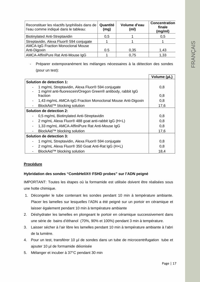
- Préparer extemporanément les mélanges nécessaires à la détection des sondes
(pour un test):
Volume (µL) Solution de detection 1:
- 1 mg/mL Streptavidin, Alexa Fluor® 594 conjugate 0,8 - 1 mg/ml anti-fluorescein/Oregon Green® antibody, rabbit IgG
fraction 0,8 - 1,43 mg/mL AMCA-IgG Fraction Monoclonal Mouse Anti-Digoxin 0,8 - BlockAid™ blocking solution 17,6
Solution de detection 2: - 0,5 mg/mL Biotinylated Anti-Streptavidin 0,8 - 2 mg/mL Alexa Fluor® 488 goat anti-rabbit IgG (H+L) 0,8 - 1,33 mg/mL AMCA-AffiniPure Rat Anti-Mouse IgG 0,8 - BlockAid™ blocking solution 17,6
Solution de detection 3: - 1 mg/mL Streptavidin, Alexa Fluor® 594 conjugate 0,8 - 2 mg/mL Alexa Fluor® 350 Goat Anti-Rat IgG (H+L) 0,8 - BlockAid™ blocking solution 18,4
Procédure
Hybridation des sondes “CombHeliX® FSHD probes” sur l’ADN peigné
IMPORTANT: Toutes les étapes où la formamide est utilisée doivent être réalisées sous
une hotte chimique.
1. Décongeler le tube contenant les sondes pendant 10 min à température ambiante.
Placer les lamelles sur lesquelles l’ADN a été peigné sur un portoir en céramique et
laisser également pendant 10 min à température ambiante
2. Déshydrater les lamelles en plongeant le portoir en céramique successivement dans
une série de bains d’éthanol (70%, 90% et 100%) pendant 3 min à température.
3. Laisser sécher à l’air libre les lamelles pendant 10 min à température ambiante à l’abri
de la lumière.
4. Pour un test, transférer 10 µl de sondes dans un tube de microcentrifugation tube et
ajouter 10 µl de formamide déionisée
5. Mélanger et incuber à 37°C pendant 30 min
Reconstituer les réactifs lyophilisés dans de l’eau comme indiqué dans le tableau:
Quantité (mg)
Volume d’eau (ml)
Concentration finale
(mg/ml) Biotinylated Anti-Streptavidin 0,5 1 0,5 Streptavidin, Alexa Fluor® 594 conjugate 1 1 1 AMCA-IgG Fraction Monoclonal Mouse Anti-Digoxin 0.5 0,35 1,43 AMCA-AffiniPure Rat Anti-Mouse IgG 1 0,75 1,33
FR
AN
ÇA
IS

Page | 18
6. Déposer 20 µl du mélange sondes/formamide sur une lame de microscopie.
7. Recouvrir le mélange sondes/formamide avec la lamelle sur laquelle l’ADN du patient a
été peigné.
Note: Clairement indiquer la face de la lamelle en contact avec le mélange
sondes/formamide pour éviter les confusions lors des étapes suivantes.
Note: déposer la lamelle avec précaution en utilisant des pinces pour éviter la formation
de bulles d’air. Une fois montée, ne pas bouger la lamelle pour éviter la formation de
rayures.
Note: Protéger les lamelles hybridées de la lumière à partir de cette étape.
8. Co-dénaturer l’ADN peigné sur la lamelle et les sondes pendant 5 min à 90°C dans la
chambre humide de l’automate de co-dénaturation et d’hybridation (par exemple,
Hybridizer, Dako).
9. Incuber pendant 16-20 h à 37°C dans la chambre humide de l’automate de co-
dénaturation et d’hybridation.
10. Séparer la lamelle de la lame de microscopie et la placer sur le portoir en céramique.
Mettre le portoir en céramique dans un Bécher de 250 ml contenant le tampon de lavage
1 préchauffé à 60°C.
Note: Si la lamelle reste collée à la lame de microscopie, ajoutez une goutte tampon de
lavage 1 autour de la lamelle et attendre que la lamelle flotte. En enlevant la lamelle,
faites la glisser doucement jusqu'à ce qu'un coin soit en dehors de la lame de
microscopie pour éviter les éraflures, puis détacher doucement la lamelle avec des
pinces.
11. Laver la lamelle trois fois dans le tampon de lavage 1 préchauffé à 60°C pendant 5 min
à 60°C pour chaque lavage.
Note: Ne pas laisser la lamelle sécher. Toujours la maintenir immerger dans la solution
Détection des sondes “CombHeliX® FSHD probes“
1. Déposer 20 µl de la solution de détection 1 sur une lame de microscopie et placer la
face hybridée de la lamelle en contact avec la solution.
2. Placer la lamelle montée dans une chambre humide et incuber pendant 20 min à 37 °C.
3. Séparer la lamelle de la lame de microscopie et la placer sur le portoir en céramique.
Mettre le portoir en céramique dans un Bécher de 250 ml contenant le tampon de lavage
2.
4. Laver la lamelle trois fois dans le tampon de lavage 2 pendant 5 min à température
ambiante sous agitation douce pour chaque lavage.
5. Déposer 20 µl de la solution de détection 2 sur une lame de microscopie et placer la
face hybridée de la lamelle en contact avec la solution.
6. Répéter les étapes 2 à 4.
F
RA
NÇ
AIS

Page | 19
7. Déposer 20 µl de la solution de détection 3 sur une lame de microscopie et placer la
face hybridée de la lamelle en contact avec la solution.
8. Répéter les étapes 2 à 4
9. Laver la lamelle avec du 1xPBS pendant 3 min à température ambiante sous
agitation douce.
10. Déshydrater les lamelles en plongeant le portoir en céramique successivement dans
une série de bains d’éthanol (70%, 90% et 100%) pendant 3 min à température
ambiante.
11. Laisser sécher à l’air libre les lamelles pendant 10 min à température ambiante à
l’abri de la lumière.
12. A cette étape, les lamelles peuvent être soit stockées à 4°C à l’abri de la lumière soit
directement observées à l’aide du système de microscopie à fluorescence.
Visualisation des signaux fluorescents
Pour une visualisation optimale des sondes hybridées, nous recommandons l’utilisation
de l’ImageXpress® Micro System, un système d’imagerie de Molecular Devices, équipé
d’un objectif plan apochromatique x40 et des filtres suivants :
L’hybridation des sondes génère le long de la molécule d’ADN étiré un signal fluorescent
multicolore. Les séquences répétées D4Z4 apparaissent en vert tandis que les signaux,
qui permettent de différencier les chromosomes 4q and 10q ainsi que les haplotypes A et
B apparaissent en rouge, bleu et magenta avec des tailles, des espacements et un ordre
spécifique (comme indiqué dans la figure ci-dessous). Les différents signaux fluorescents
multicolores qui peuvent être détectés sont les suivants :
• Le chromosome 4q35.2 est identifié par un signal centromérique rouge de 18kb
suivi d’un signal vert d’environ 1.5kb (séquence Dux4) puis d’un signal magenta
de 10kb et d’un signal bleu de 20kb séparé par un espacement de 5 kb ;
• Le chromosome 10q26.1 est identifié par un signal centromérique bleu de 15 kb
séparé par 11 kb d’un signal magenta de 10 kb et d’un signal bleu de 20kb
séparé du précédent par un espacement de 5 kb ;
• L’haplotype A apparait comme 2 signaux rouges télomériques de 6kb
• L’haplotype B apparait comme une série composée d’un signal bleu et d’un
signal rouge de de 6kb en position télomérique ;
Fluorophore Excitation Emission
Vert 482 ± 35 nm 536 ± 40 nm Rouge 562 ± 40 nm 624 ± 60 nm Bleu 377 ± 50 nm 447 ± 60 nm
FR
AN
ÇA
IS

Page | 20
• Une région génomique supplémentaire dont la localisation chromosomique est
inconnue (Chr Z), est aussi détectée sous la forme d’un signal fluorescent
multicolore composé de 2 signaux bleus encadrés par un signal rouge et un signal
magenta.
Pour le diagnostic de FSHD1, la détection, la class ification et la mesure de la taille des
différents signaux fluorescents (spécialement la ta ille du signal vert correspondant à
la séquence répétée D4Z4 de 3.3 kb) sont réalisées à l’aide du logiciel “CombHeliX®
FSHD software”. Consulter la notice d’utilisation du logiciel “ CombHeliX® FSHD software”
ou contacter Genomic Vision pour des informations complémentaires.
Interférence
Les sondes “CombHeliX® FSHD probes” génèrent un signal supplémentaire sur le
chromosome 3 (Chr 3 dans la figure ci-dessus) mais qui n’interfère pas avec le résultat
puisque son motif fluorescent spécifique permet son identification. Ces signaux ne sont pas
pris en compte pour l’analyse et le résultat diagnostique.
Recommendations et limitations
Les sondes “CombHeliX® FSHD probes” sont conçues pour le diagnostic in vitro (selon les
termes de la Directive Européenne t98/79/EC) et doivent être analysées avec le logiciel
“CombHeliX® FSHD Software”.
L’interprétation du test doit être effectuée par un professionnel médical qualifié en prenant en
considération les autres informations cliniques et diagnostiques disponibles. Une décision
médicale ne peut pas être prise basée sur les seuls résultats de ce test.
F
RA
NÇ
AIS

Page | 21
Les sondes “CombHeliX® FSHD probes” ne peuvent pas être utilisées pour du
diagnostic prénatal ou pour tout autre utilisation diagnostique. Genomic Vision n'assume
pas la responsabilité d’une utilisation incorrecte de ce produit.
Formation
Une formation préalable spécifique est obligatoire pour la réalisation de ce test. Genomic
Vision fournit la formation pour la préparation des échantillons, la réalisation du test
FSHD1 et l’interprétation des résultats pour les utilisateurs inexpérimentés. Il est
également recommandé que tout nouvel utilisateur d'un laboratoire qui a précédemment
reçu la formation soit formé par Genomic Vision.
Performances du test
Les performances du test ont été établies à partir d’une analyse d’échantillons provenant
de 69 patients collectés et analysés dans deux centres d’investigation indépendants
(l’Hôpital La Timone à Marseille, France et l’Institut de Génétique Humaine à Würzburg,
Allemagne) en comparaison des résultats de Southern Blot.
• Précision du nombre de répétitions D4Z4 :
o ± 1 D4Z4 répétition = 89 % ;
o ± 2 D4Z4 répétitions = 98 %.
• Forte sensibilité: >99%. Tous les allèles 4qA sont détectés. La présence d’un
allèle 4qA contracté (<10 copies de D4Z4) entraîne un diagnostic positif pour
FSHD1
• Forte spécificité: >99%. L’absence d’un allèle 4qA contracté (>10 copies de
D4Z4) entraine un diagnostic négatif pour FSHD1.
Références
1. van Deutekom, J.C., et al., Hum Mol Genet, 1993. 2(12): p. 2037-42.
2. Wijmenga, C., et al., Nat Genet, 1992. 2(1): p. 26-30.
3. Winokur, S.T., et al., Chromosome Res, 1994. 2(3): p. 225-34.
4. Hewitt, J.E., et al., Hum Mol Genet, 1994. 3(8): p. 1287-95.
5. Lemmers, R.J., et al., Nat Genet, 2002. 32(2): p. 235-6.
6. Lemmers, R.J., et al., Am J Hum Genet, 2004. 75(6): p. 1124-30.
7. van Geel, M., et al., Genomics, 2002. 79(2): p. 210-7.
8. Nguyen, K., et al., Ann Neurol, 2011. 70(4): p. 627-33.
9. Bensimon, A., et al., Science, 1994. 265(5181): p. 2096-8.
10. Lebofsky, R. and A. Bensimon. Brief Funct Genomic Proteomic, 2003. 1(4): p. 385-
96.
FR
AN
ÇA
IS

Page | 22
Informations relatives aux produits et commandes :
* Produits à usage général de laboratoire validés pour la technologie de Peignage
Moléculaire mais qui n’ont fait l’objet d’aucune validation spécifique pour des applications de
diagnostic in vitro.
Nos experts sont à votre disposition pour répondre à vos questions
http://www.genomicvision.com
Brevet
Ce produit ou l'utilisation de ce produit sont soumis à des droits de propriété
(EP2007/059299, IB2009/007197). La technologie de Peignage Moléculaire et les produits
dérivés sont couverts par des brevets (FR2716206, 2716263 FR, 2737574 FR, 2755149 FR)
appartenant à Genomic Vision S.A.
Trademarks:
CombHeliX est une marque déposée par Genomic Vision S.A.
BlockAid est une marque déposée par Molecular Probes, Inc.
Alexa Fluor et Oregon Green sont des marques déposées par Molecular Probes, Inc.
ImageXpress Micro System est une marque déposée par Molecular Devices, LLC.
Tween est une marque déposée par Croda International PLC
Produit Conditionnement Référence
CombHeliX® Kit d’Hybridation FSHD Lot de 10 hybridations FSH-HYB-001
CombHeliX® Logiciel d’analyse FSHD 1 licence perpétuelle FSH-ASW-001
CombHeliX® Kit d’Extraction d’ADN * Kit pour 100 extractions EXT-001
Molecular Combing System Appareil de Peignage MCS-001
Réservoir Téflon (pour solutions d’ADN) 1 pièce TEF-001
Pince pour MCS (pour 2 lamelles) 1 pièce CLI-001
Réservoirs à Usage Unique* Sachet de 10 pièces RES-001
Supports de Réservoirs pour MCS Lot de 2 pièces SUP-001
Portoir paillasse (pour 10 réservoirs) 1 pièce POR-001
Lamelles silanisées* Boîte de 50 pièces COV-001
Genomic Vision S.A. 80-84 rue des meuniers 92220 Bagneux France www.genomicvision.com
F
RA
NÇ
AIS