coma final
TRANSCRIPT

Ambo UniversityCollege of Medicine and Health
SciencesDepartment of Medicine
C2 PediatricsSeminar presentation on:The management of comaBy:
Hayelom MichaelHabtamu MokonenHailemariam Bekele
Moderator: Dr. Kissu
Ambo,Ethiopia

Outline• Objective• Introduction• Coma mimicking states• Cause • Pathophysiology• clinical features• Approach to comatose patient• Management of coma

Objective• To define coma and similar clinical
conditions.
• To describe the etiologies of coma
• To describe the pathophysiologic mechanism of coma in terms of the underlying anatomic and physiologic alterations.
• To explain the approach to a comatose patient.
• To explain the management of comatose patient

Introduction
• Consciousness is arousal, in which one is able to interact with the environment, and awareness, with the ability to know "what is going on.
• Is the product of two closely related cerebral functions:– Wakefulness i.e. arousal, alertness.– Content i.e. awareness of self and the env’t.

Cont….
• Coma is a state of unrousable, unconsciousnes without any psychologically understandable response to external stimuli or inner need.
• It is among the most common and striking problems in general medicine.
• There is a continuum of the states of consciousness, coma being the most severe.

Level of consciousness
• Reflects awareness and response to the environment:
• Alert - Appearance of wakefulness, awareness of the self and environment
• Lethargy :mild reduction in alertness -tends to drift of to sleep when not stimulated -when aroused has appropriate response• Obtundation - moderate reduction in alertness.
- It requires touch or voice to maintain arousal. -When aroused is in confusional state. -Usually constant stimulation is required

• Stupor: Deep sleep, patient can be aroused only by vigorous and repetitive stimulation.
-Returns to deep sleep when not continually stimulated.
• Coma: -Sleep like appearance and behaviorally
unresponsive to all external stimuli (Unarousable unresponsiveness, eyes closed)

Coma mimicking states
– Minimally conscious state(MCS)
– Persistently vegetative state(PVS)
– Locked in syndrome(LIS)
– Psychogenic unresponsiveness
– Brain death
• Differentiating coma from related states:
1.purpose full response to stimuli?
2.Brain stem reflex?
3.Sleep wake cycle?
4.EEG?

Minimally conscious state(MCS)
• The patient has rudimentary vocal or motor behaviors, often spontaneous, but some in response to touch, visual stimuli, or command.
• Cardiac arrest with cerebral hypoperfusion and head injuries are the most common causes.

Persistently vegetative state(PVS)
• Signifies an awake but nonresponsive state in a patient who has emerged from coma.
• The vegetative state is characterized by loss of all cognitive functions & the un awareness self &surroundings.
• Reflex & vegetative function, including sleep wake cycles.
• These individuals have spontaneous eye opening with out concurrent awareness.

• Diagnosis criteria for Vegetative state include:
-The absence of awareness self&environment
and inability to interact with others.
-The absence of sustained or reproducible voluntary behavioral responses.
-lack of language comprehension
-Suffiently preserved hypothalamus & brain stem function to maintain life.
-

-Bowel and bladder incontinence
-Variably preserved cranial nerves(e.g. pupillary & gag) and spinal cord reflexes.
NB. The PVS requires that condition has continued for at least 1 month.
Few, if any, meaningful responses to the external and internal environment—in essence, an "awake coma."
• Indicates extensive damage in both cerebral hemispheres, usually 2° to ischemic injury
Locked in syndrome
• An awake patient has no means of producing speech or volitional movement but retains voluntary vertical eye movements and lid elevation, thus allowing the patient to signal with a clear mind.
• The pupils are normally reactive. • The usual cause is an infarction or
hemorrhage of the ventral pons that transects all descending motor (corticospinal and corticobulbar) pathways

Psychogenic unresponsiveness
• Also termed 'pseudocoma‘, this describes a patients who appear to be unconscious and in coma but who are not.
• Oculovestibular testing, will reveal the presence of nystagmus and indicate that the patient has an intact brainstem and cortex.

Brain death
• This is a state of cessation of cerebral function with preservation of cardiac activity and maintenance of somatic function by artificial means.
• It is equivalent to death. • Brain death Criteria: (1) widespread cortical destruction that is reflected by
deep coma and unresponsiveness to all forms of stimulation;
(2) global brainstem damage demonstrated by absent pupillary light reaction and by the loss of oculovestibular and corneal reflexes;
(3) destruction of the medulla, manifested by complete apnea.

Cause
• A more practiced &clinically relevant categorization of CNS insults sever enough to cause coma involves the categorization of as:
-Structural : supra tentorial
Infra tentorial
-Metabolic toxic: common etiologies
uncommon etiologies
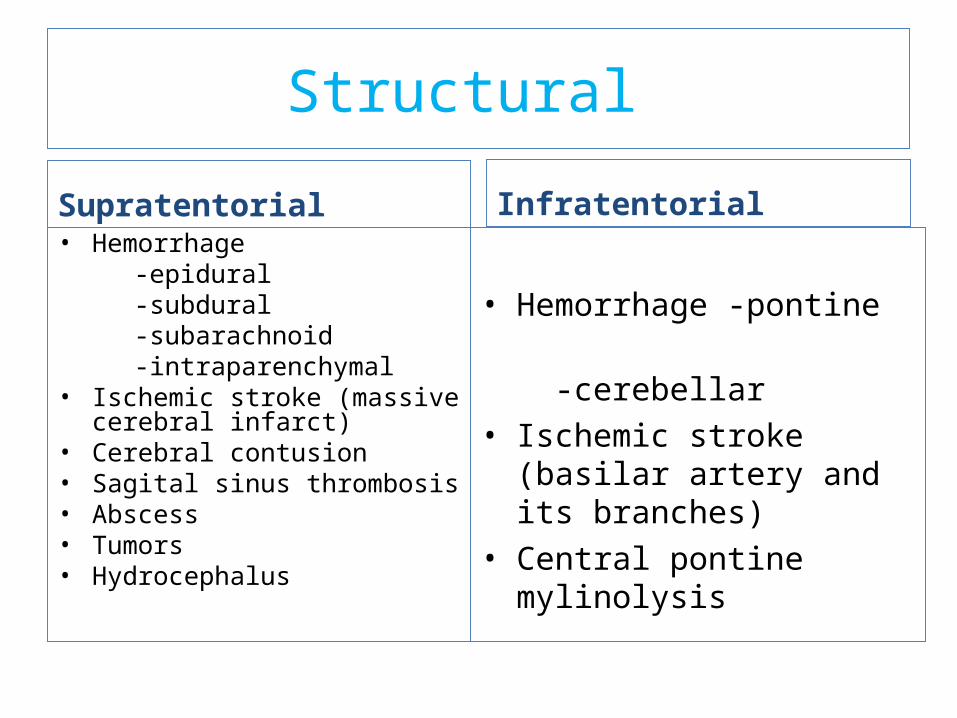
Structural
Supratentorial • Hemorrhage -epidural -subdural -subarachnoid -intraparenchymal• Ischemic stroke (massive
cerebral infarct)• Cerebral contusion• Sagital sinus thrombosis• Abscess • Tumors • Hydrocephalus
Infratentorial
• Hemorrhage -pontine
-cerebellar• Ischemic stroke (basilar
artery and its branches)• Central pontine mylinolysis

Toxic/metabolic
Common
• Drug intoxication (alcohol, benzodiazepines, opiates)
• Hyper-/hyponatremia• Hyper-/hypoglycemia• Hypercarpia• Shock (due to any cause)
Uncommon (easily identifiable)• Hyper-/hypocalcaemia• Hepatic encephalopathy• Uremic encephalopathy• Anoxic encephalopathy• Meningitis/encephalitis• Adrenal crisis• Myxoedema coma• Hyper-/hypothermia• Hypertensive encephalopathy• Post ictal state• Any end stage
neurodegenerative condition• Extreme acid-base disturbance

Cont….Uncommon (not easily identifiable)
• Drug intoxication (TCAs, neuroleptics, antihistamines, anticonvulsants, salisylates, lithium)
• Poisoning (CO, methanol, ethylene glycol, cyanide, heavy metals, organophosphates)
• Thiamin deficiency• Serotonin syndrome• Nonconvulsive status epilepticus• Reversible posterior leukoencephalopathy
syndrome

Pathophysiology
• Almost all instances of diminished alertness can be traced to:
widespread abnormalities of the cerebral hemispheres
reduced activity of a special thalamocortical alerting system termed the reticular activating system (RAS).
• The proper functioning of this system, its ascending projections to the cortex, and the cortex itself are required to maintain alertness and coherence of thought.

Cont…
• The ascending RAS, from the lower border of the pons to the ventromedial thalamus
• The cells of origin of this system occupy a paramedian area in the brainstem

Cont…• It follows that the principal causes of coma
are:
(1) lesions that damage the RAS in the upper midbrain or its projections
(2) destruction of large portions of both cerebral hemispheres
(3) suppression of reticulocerebral function by drugs, toxins, or metabolic derangements such as hypoglycemia, anoxia, uremia, and hepatic failure
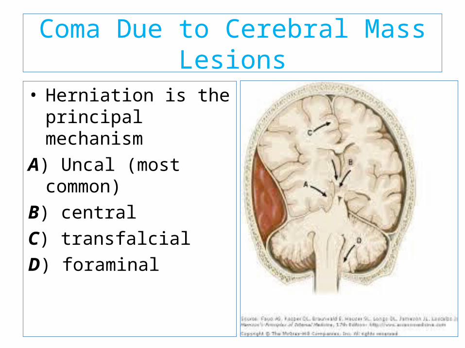
Coma Due to Cerebral Mass Lesions
• Herniation is the principal mechanism
A) Uncal (most common)
B) central
C) transfalcial
D) foraminal

Coma Due to Metabolic Disorders
Interruption of the delivery of energy substrates (e.g., hypoxia, ischemia, hypoglycemia)
Alteration of neuronal excitability (drug and alcohol intoxication, anesthesia, and epilepsy)
• Unlike hypoxia-ischemia, which causes neuronal destruction, most metabolic disorders such as hypoglycemia,hyponatremia,hyperosmolarity, hypercapnia, hypercalcemia, and hepatic and renal failure : - impaired energy supplies,
-changes in ion fluxes across neuronal membranes, and
-neurotransmitter abnormalities.

Toxic (Including Drug–Induced) Coma
• Many drugs and toxins are capable of: depression of nervous system function. producing coma by affecting both the
brainstem nuclei, including the RAS, and the cerebral cortex.
• The combination of cortical and brainstem signs, which occurs in certain drug overdoses, may lead to an incorrect diagnosis of structural brainstem disease.
• Overdose of medications that have atropinic actions produces signs such as dilated pupils, tachycardia, and dry skin; opiate overdose produces pinpoint pupils <1 mm in diameter.

Coma Due to infection
• The most common causes include: Pyogenic menigitis ,TB meninigitis, Cerebral malaria,HIV, encephiltis,
• Widespread structural cerebral damage→ a metabolic disorder of the cortex.
• Hypoxia-ischemia is perhaps the most known →hypoperfusion and oxygen deprivation of the brain.
• Similar bihemispheral damage is produced by disorders that occlude small blood vessels throughout the brain
• Diffuse white matter damage from inflammatory demyelinating diseases causes a similar syndrome of coma.

Clinical features• Sleeplike state from which the patient cannot
be aroused.
• Eyes are closed and remain closed in the face of vigorous stimulation.
• Do not speak.
• Do not arouse to verbal,tactileor noxious stimuli • Motor activity is absent or abnormal and
reflexive rather than purposeful or defensive.
• as opposed to state of transient unconsciousness such as syncope& concussioncoma must last ≥1 hr.

Approach to a patient in Coma
Coma is a medical emergency whose evaluation requires a rapid, comprehensive, and systematic approach.
Early identification of the underlying cause of coma can be crucial for patient management and prognosis.
A. Immediate life supportB. Identification of causesC. Specific therapy

A. Immediate life support
Assessment and maintenance of vital function is the initial step
(ABC of life)
• Maintain the air ways patency and ensure adequate breathing
• Maintain circulation
B. Establishment of cause of coma:
is done by taking a careful history, doing rapid but through
physical examination and investigations.

Patient History
• It is often useful to obtain a history from witnesses, friends or
family members, and emergency medical technicians.
• The patient's personal effects: a Medical Alert bracelet or
necklace and/or a card in the wallet may contain a list of
illnesses and medications.
• Past medical history: looking for disease like diabetes,
hypertension, cirrhosis, chronic renal disease, malignancies
and other diseases.

• History of medications: legal or illicit drugs (sedatives, hypnotics,
narcotics ) and history of drug abuse.
• Details regarding the site where the patient was found
(e.g. the presence of empty drug vials or evidence of fall or trauma),
• If the cause is unknown, Hx assessment should focus on:-
-detail of social and family history including recent travel.
-hx of recent preceding illness e.g. fever, headache.

• Circumstances and rapidity with which change in
mental status developed.
sudden onset suggests intracranial
hemorrhage, seizure, cardiac arrhythmia, trauma,
or intoxication.
gradual onset suggests an infectious process,
metabolic abnormality, or slowly expanding
intracranial mass lesion.
fluctuations suggest subdural hematoma.
A history of preceding headache, double vision, or
nausea suggests increased ICP.

Physical examination
Vital signs: Extremes of BP, pulse or temperature and abnormal
pattern of breathing.
Temperature
Hyperthermia: suggests infection, but is also seen with
inflammatory disorders, environmental or exertional heat
stroke, neuroleptic malignant syndrome, status epilepticus,
hyperthyroidism, and anticholinergic poisoning.
Hypothermia: can occur with infection in infants but is more
often due to drug intoxication, environmental exposure, or
hypothyroidism.

34
Heart rate
• Tachycardia can occur with fever, pain, hypovolemia,
cardiomyopathy, /tachyarrhythmia, and also in status
epilepticus.
• Bradycardia occurs with hypoxemia, hypoxic ischemic
myocardial injury, and with increased ICP
Blood pressure
Hypertension – hypertensive encephalopathy or
hypertensive intracerebral hemorrhage, pain, intoxication,
renal failure, or increased ICP
Hypotension suggests shock, intoxication, or adrenal
insufficiency.

Respirations
Tachypnea can be seen with pulmonary
infections, pain, hypoxia, metabolic acidosis, and
pontine injury.
Slow, irregular, or periodic respirations occur with
metabolic alkalosis, sedative intoxication, and
injury to extrapontine portions of the brainstem.
Irregular breathing-increased ICP

• Head and neck:
Signs of head injury
lacerations or bruising to the head,
Sx of basal skull Fructure
Raccon Sx (around the eyes),
Battle’s sign (behind the ear )
Rhenorrhea & otorrhea(CSF leak from the
nose or ears)

Fundoscopy
• Papilledema may suggests increased ICP.
• Retinal hemorrhages are most commonly
associated with shaken baby syndrome.
Meningismus
• Meningeal irritation or inflammation suggesting
meningitis or subarachnoid hemorrhage.

• Skin:
Look for signs of trauma or injection.
Color- pallor(hemorrhage) ,
cyanosis(inadequate oxygination)
Rash- menengococcemia, bacterial
endocarditis
Sweating- hypoglycemia, shock
Dry & hot skin- heat stroke

General systemic examination: looking for evidences of systemic
illnesses like cirrhosis, chronic renal failure, meningococcemia etc.
Jaundice could indicate liver disease.
A cherry red color, especially of the lips and mucous membranes,
suggests carbon monoxide intoxication.
Pallor, especially with a sallow appearance, may suggest uremia,
myxedema, or severe anemia.
A tongue bitten on the lateral aspect suggests a recent convulsive
seizure.

Neurologic examination:
is to determining whether the pathology is structural or due to
metabolic dysfunction.
• The examiner assesses:
– Level of consciousness(GCS)
– Motor responses
– Brainstem reflexes

Level of consciousness
It can be assessed semi quantitatively using the Glasgow coma
Scale.
Glasgow coma scale:
- Provides easily reproducible and somewhat predictive basic
neurologic exam.
-It is useful as an index of the depth of impaired
consciousness and for prognosis, but does not aid in the
diagnosis of coma.

• Glasgow coma scale is used for adults and older children and
its modification is used in infants and young children (<2
years of age) (PGCS) .

04/17/23 43
ACTIVITY BEST RESPONSE
Adults/Older Children
Infants ( modified GCS ) Score
Eye Opening( E )
SpontaneousTo speechTo painNone
• Spontaneous• To speech• To pain• None
4321
Verbal( V )
Appropriate speech
Confused speechInappropriate
wordsIncomprehensible
or none specific sounds
None
• Appropriat vocalization, smile, or orientation to sound, interacts (coos, babbles), follows object
• Irritable, cries• Cries to pain• Moans to pain• None
5432
1
Motor( M )
Obeys commandsLocalizes painWithdraws to touchDecorticate to painDecerebrate to painNone
• Normal spontaneous movement
• Withdraws to touch• Withdraws to pain• Decorticate to pain• Decerebrate to pain• None
6
54321
Glasgow coma scale

• The Glasgow coma scale (GCS) is scored between
3 and 15, 3 being the worst, and 15 the best.
• Individual elements as well as the sum of the score are
important.
• Hence, the score is expressed in the form "GCS 8 = E2 V3 M3
at 02:30 PM

Significance of GCS
1. To classify/grade altered consciousness
2. Follow up
3. Prediction of prognosis of comatose child
1.clssification
Generally, comas are classified as:
• Severe, with GCS ≤ 8
• Moderate, GCS 9 - 12
• Minor, GCS ≥ 13.

2. Follow up
Used in assessment of response to therapy.
3. Prediction of prognosis
-GCS ≤ 5 on admission, the probability of death is 90%
- GCS ≥ 10, the probability of death is decreasing to 1%

Brain stem reflexes
• helps to localize the cause to specific regions of the
brainstem and/or impending transtentorial
herniation, or a consistent asymmetry between
right- and left-sided responses.
• These are
- pupillary light,
- extraocular movement,
- corneal and respiratory reflexes.

a) Pupillary light response
• Pupillary reactions are examined with a bright, diffuse
light.
• The pupillary reflex depends on intact transmission
within the afferent optic nerve (CNII), the Edinger
Westphal nucleus in the midbrain, and the efferent
oculomotor nerve (CN III).
• Parasympathetic impulses constrict the pupils, while
• sympathetic discharge leads to pupillary dilation.
• During examination size, shape, symmetry and reaction
to light should be noted on both eyes.

• Normally reactive and round pupils of midsize (2.5 to 5 mm) essentially exclude midbrain damage.
• Enlarged (>6mm) and unreactive pupil on one side signifies a compression or stretching of the third nerve from the effects of a mass above.
• Bilaterally dilated and unreactive pupils, indicates severe midbrain damage, usually from compression by a mass.

• Bilaterally small (1 to 2.5 mm) and reactive pupils (not pinpoint) are seen in
metabolic encephalopathies or in deep bilateral hemispheral lesions such as
hydrocephalus or thalamic hemorrhage.
• Very small but reactive pupils (< 1 mm)/pinpoint pupils, characterize
narcotic or barbiturate overdoses but also occur with extensive pontine
hemorrhage.

b) Ocular Movements
The position and spontaneous movements of the eyeballs.
controlled by the cranial nerves III, IV, and VI).
Lid tone is tested by lifting the eyelids :
Resistance to opening the eye lids may suggest hysteric conversion.
Easy eyelid opening with slow closure indicates sever coma.
• Midline deviation suggests frontal/pontine damage.
• Dysconjugate gaze (abduction or adduction) suggests cranial nerve
abnormalities.
• Spontaneous eye movements roving, dipping, bobbing suggest damages
being at different sites.

Occulocephalic reflex
• Elicited by moving the head from side to side or vertically with eyes held
open.
In comatose patient:-
If the eyeballs move to the opposite direction of the head movement =
intact brainstem function (“doll’s eyes” movement is positive.)
If the eyeballs move to the same direction of the head movement=
Brainstem dysfunction

Caloric (occulovestibular) reflex
• This test is performed by irrigating the ear with ice (cold) to
stimulate the vestibular apparatus.
• In patients with intact brain stem the eyes move to the irrigated
ear.

c) Corneal reflex
The corneal reflex tests the sensory function of the
trigeminal nerve and the motor function of the
facial nerve.
• This test assesses the integrity of dorsal midbrain and
pontine.
• It is lost if the reflex connections between the fifth and the
seventh cranial nerves within the pons are damaged.

d) Respiration:
less localizing value in comparison to other brainstem signs.
• Shallow, slow, but regular breathing suggests metabolic or drug
depression.
• Cheyne-Stokes respiration signifies bihemispherical damage or metabolic
suppression, and commonly accompanies light coma.
• Kussmaul breathing usually implies metabolic acidosis but may also occur
with pontomesenephalic lesions and severe pneumonia.
• Agonal gasps are the result of lower brainstem (medullary)
damage and are recognized as the terminal respiratory
pattern of severe brain damage.
3. Motor function /response
Quadriparesis and flaccidity-suggest pontine or medullary damage.
Decorticate posturing: flexion of the elbows and the wrists with supination
of the arms, and extension of the legs, suggests severe bilateral or
unilateral hemispheric or diencephalic lesion (damage above the
midbrain.)

Decerebrate posturing (extension of elbows and the wrist with pronation of the
forearm and extension of the legs) indicates damage to the brainstem( midbrain or
pontine compromise )
Abnormal body movements – seizure, myoclonus may suggest the cause of the
coma is status epelepticus, uremia etc.
DIAGNOSTIC STUDIES
• Studies can be guided by HX and PE, but most patients presenting with coma of unknown etiology require laboratory testing and a neuroimaging study.

Laboratory Testing
• Patients presenting with altered consciousness should undergo a rapid bedside test for blood glucose and basic laboratory testing including:
• Serum electrolytes, calcium, magnesium, glucose
• Arterial blood gas • Liver function tests, ammonia • Complete blood count • Blood urea nitrogen, creatinine • Urine drug screen

• blood and urine tests
• Testing for fungi, rickettsia, mycobacteria, and parasites
• thyroid function tests
• cortisol levels/ carboxyhemoglobin, and coagulation studies

• Neuroimaging — CT is the best initial neuroimaging test. CT quickly detects hydrocephalus, herniation, and mass lesions due to infection, neoplasia, hemorrhage, and edema.

• When lumbar puncture is indicated, a CT is required in the comatose patient to rule out a mass lesion that might precipitate transtentorial herniation as a result of the procedure.

• MRI - provides greater structural detail and is more sensitive for early evidence of encephalitis, infarction, diffuse axonal injury from head injury, petechial hemorrhages, cerebral venous thrombosis, and demyelination.
• Lumbar puncture

• Electroencephalogram : coma of unknown etiology
• It is often the only means of recognizing non convulsive status epilepticus (NCSE), especially in patients who are paralyzed.
Treatment and Prognosis of Coma in Children
• TREATMENT — Early treatment of coma is generally supportive
• An important goal of early treatment is to limit brain injury.
• RX for dangerous etiologies (eg, hypoglycemia, increased ICP, bacterial meningitis) are often initiated empirically

• The primacy of ABCs applies to coma as to other medical emergencies.
Airway• attained by repositioning the child to open
the airway• Patients with GCS <8 are usually unable
to adequately protect their airway and should be intubated.
• If trauma is suspected, the cervical spine should be stabilized with a collar while securing the airway.

• Breathing — O2 saturation should be measured and supplemental O2 provided.
• Adequacy of ventilation should be assessed by examination and arterial blood gases.
• Moderate hyperventilation (target PaCO2 30 to 35 mm Hg) should only be initiated for patients with increased ICP.
• Extreme hyperventilation /aggressive hyperventilation (PaCO2 <30 mmHg) are only justified in patients with transtentorial herniation.

Circulation
Depressed level of consciousness may be an early indicator of poor end-organ perfusion in a patient with shock
Hypotension
• IV administration (NS or LR) and inotropes, if necessary, is essential to deliver oxygen and metabolic substrates to the brain and remove toxic metabolites.
Hypertensive encephalopathy
• The goal of therapy is to lower the DBP to 100 to 110 mmHg (or by a maximum of 25 percent) within two to six hours
• Hypertensive encephalopathy has an excellent prognosis for recovery if ischemia can be avoided

• Glucose — Glucose (2.5 mL/kg of 10 percent dextrose solution) should be administered even before test results are known. If hypoglycemia is revealed, then ongoing monitoring and treatment will be needed.

• Intracranial pressure — When increased ICP is suspected, emergent RX is recommended. Increased ICP is assumed when there is coma after head injury.
• Early interventions to reduce ICP include treating fever, elevating the head of the bed to 30 degrees above horizontal, moderate hyperventilation (target PaCO2 30 to 35 mmHg) and administering mannitol(0.25 to 1 g/kg IV). Neurosurgery should be consulted.
• Seizures — If seizures have occurred, phenytoin or fosphenytoin(15 to 20 mg/kg phenytoin equivalent IV) should be administered.

• Non convulsive status epilepticus should be considered as a diagnosis even when there are no obvious seizure movements.
• If non convulsive seizures are suspected and an electroencephalogram (EEG) is not available, a therapeutic trial of phenytoin or lorazepam (1 to 2 mg IV) is reasonable.

• Infection — Empiric antibiotic and antiviral therapy are recommended
• If bacterial meningitis (eg, ceftriaxone100 mg/kg per day in one or two divided doses, maximum dose 4 g per day, plus vancomycin 60 mg/kg per day in four divided doses)
• Viral encephalitis ( acyclovir 30 to 60 mg/kg per day, in
three divided doses) are among the suspected entities. • Blood cultures should be obtained prior to starting
antibiotics but initiation of therapy should NOT await LP. • Therapy should be continued until these conditions have
been excluded

• Temperature control — Hyperthermia (>38.5 degrees C).
• Fever should be lowered with antipyretics and/or cooling blankets immediately. Shivering, which can contribute to elevated ICP, should be avoided.

• Hypothermia to 32 to 36 degrees has been suggested as a therapy for refractory increased ICP in children with traumatic brain injury - currently not recommended
• May be appropriate for children with o out-of-hospital arrest o persistent coma o ventricular fibrillation or o pulseless ventricular tachycardia

• Acid-base and electrolyte imbalance —resuscitation of patients with cardiovascular compromise should use isotonic solutions only (NS or RL).

• Antidotes — use is recommended only in the setting of known or strongly suspected drug overdose.
• Naloxone(0.1 mg/kg IV in patients up to 20 kg or ≤5 years; maximum 2 mg) - possible opiate ingestion.
• Flumazenil - benzodiazepine overdose, but will render benzodiazepines ineffective in the event of a seizure, so it should also be used with caution.
• Agitation — sedation - should be administered only when the benefits of relieving agitation outweigh the need for close neurologic monitoring by exam.

Management algorithm for infants (≥1 month) and children with suspected
bacterial meningitis
04/17/23 Aproch to comatous child 80

04/17/23 Aproch to comatous child 81

Tb meningitis
04/17/23 Aproch to comatous child 82
• Chemotherapy should be initiated with RHZS in an initial phase for 2 months and RH should be continued for 7 to 10 months in the continuation phase.
• Adjunctive corticosteroid therapy with dexamethasone is recommended for all patients. The recommended regimen is:

04/17/23 Aproch to comatous child 83
• Dexamethasone- a total dose of 8 mg/day for children weighing less than 25 kg and 12 mg/day for children weighing 25 kg or more.
• The initial dose is given for 3 weeks and then decreased gradually during the following 3 weeks.
• Prednisolone- a dose of 2-4mg/kg/day for children for 3 weeks, then tapered of gradually over the following three weeks.

Long term essentials
04/17/23 Aproch to comatous child 84
• Skin care
• Oral hygiene
• Eye care
• Fluids
• Calories
• Sphincters

PROGNOSIS
• The prognosis in coma is etiology specific. • Mass lesions at the fully developed midbrain stage
do poorly even after surgical evacuation. • Sedative drug induced coma has a good
prognosis with proper supportive care.• Coma with purulent meningitis doubles
unfavorable outcome or death. • In hepatic or other metabolic comas, the absence
of pupillary, oculovestibular, and corneal reflexes on admission is a grave prognostic sign.

04/17/23 Aproch to comatous child 86
• Brisk, small-amplitude, mainly vertical eye movements are predictive of a fatal outcome.
• In cardiac arrest patients without seizures, return of pupillary reactivity and purposeful motor movements within the first 72 hours is highly correlated with a favorable outcome.
• Bilateral absence of somatosensory evoked responses in the first week predicts death or a persistent vegetative state.

PX Factors
04/17/23 Aproch to comatous child 87
• GCS
• Age
• Clinical features
• EEG
• MRI
• Serum biomarkers
• GCS - is associated with prognosis in a number of conditions as TBI.
• In some cases, “Age” < 2 years is associated with worse px.

• Clinical features — the presence and severity of certain early complications has been linked to worse outcome
• EEG -a predictor of outcome in coma of certain etiologies.
• Sedative drugs cause EEG abnormalities and make interpretation difficult, particularly for prognosis.

• An isoelectric baseline or a burst suppression pattern during the first week after coma - 100 % specific for poor outcome
• Other EEG patterns associated with poor outcome(but have poor sensitivity and specificity)
o periodic epileptiform dischargeso nonreactive rhythms

MRI• Presence and extent of brain edema,
brainstem injury, and diffuse axonal injury -associated with poorer prognosis in patients with TBI.
• Serum biomarkers — Elevated neuron specific enolase (NSE) levels have been associated with poor outcome after HIE, TBI and other conditions (e.g., encephalitis, Reye's syndrome).

Reference
92
1. Nelson text book of pediatrics 19th ed.
2. Up to date 21.2
3. Harrison 19th ed.
4. Kumar and clark med
5. ceil

93
Thank you!!!