colloids and surfaces b: biointerfaces and surfaces b: biointerfaces 98 (2012) 1–6 contents lists...
TRANSCRIPT
At
JD
a
ARRAA
KSTPC
1
oaaeis
cchmwhsePsc
0h
Colloids and Surfaces B: Biointerfaces 98 (2012) 1– 6
Contents lists available at SciVerse ScienceDirect
Colloids and Surfaces B: Biointerfaces
jou rn al h om epage: www.elsev ier .com/ locate /co lsur fb
ttachment of hydrogel microstructures and proteins to glass viahiol-terminated silanes
eong Hyun Seo, Dong-Sik Shin, Priam Mukundan, Alexander Revzin ∗
epartment of Biomedical Engineering, University of California, Davis, CA 95616, United States
r t i c l e i n f o
rticle history:eceived 30 January 2012eceived in revised form 27 March 2012ccepted 27 March 2012vailable online 27 April 2012
eywords:urface modificationhiol-ene reaction
a b s t r a c t
Micropatterning strategies often call for attachment of non-fouling biomaterials and immobilizationof proteins in order to create biosensing surfaces or to control cell–surface interactions. Our labora-tory has made frequent use of hydrogel photolithography – a micropatterning process for immobilizingpoly(ethylene glycol) (PEG) hydrogel microstructures on glass surfaces. In the present study we exploredthe use of thiolsilane as a coupling layer for both covalent anchoring of hydrogel microstructures andcovalent immobilization of proteins on glass. These new surfaces were compared to acryl-silane func-tionalized glass slides that allowed covalent attachment of gels but only physical adsorption of proteinsas well as surfaces containing a mixture of both functional groups. We observed comparable attach-
EG-hydrogel patterningovalent biomolecular immobilization
ment and retention of hydrogel microstructures on acryl and thiol-terminated silanes. Ellipsometrystudies revealed presence of significantly higher level of proteins on thiol-functionalized glass. Overall,our studies demonstrate that thiol-silane functionalized glass surfaces may be used to create complexmicropatterned surfaces comprised of covalently attached hydrogels and proteins. This simple and effec-tive surface modification strategy will be broadly applicable in cellular engineering and biosensing studiesemploying hydrogel micropatterns.
© 2012 Elsevier B.V. All rights reserved.
. Introduction
Immobilization of biomolecules is an important aspect in devel-ping biosensor and surfaces for cell/tissue engineering. Covalentttachment of biomolecules, surfaces may be functionalized with
variety of chemical groups for example NH2 , SH , COOH , NHSster and epoxide groups [1]. The most common way of introduc-ng these functional groups onto a glass or oxide surface is viaelf-assembly of silanes [2–5].
In addition to their use for biomolecule attachment, silaneoupling agents may also be used for anchoring biomaterialonstructs to surfaces. For example, acrylate-terminate silanesave been used by several groups for attachment of hydrogelicropatterns to glass and silicon surfaces [6–8]. Our lab asell as other groups has utilized poly(ethylene glycol) (PEG)ydrogel micropatterning to create non-fouling domains on theurface and to control cell–surface interactions.[9–14] In thesexperiments, cell attachment to glass regions not covered by
EG gel was promoted by physical adsorption of matrix proteinsuch as collagen or laminin. In other studies, antibodies againstell surface antigens and secreted cytokines were immobilized∗ Corresponding author. Tel.: +1 530 752 2383.E-mail address: [email protected] (A. Revzin).
927-7765/$ – see front matter © 2012 Elsevier B.V. All rights reserved.ttp://dx.doi.org/10.1016/j.colsurfb.2012.03.025
within hydrogel microwells to capture cells and locally detectsecreted cytokine molecules [15]. Therefore, cellular engineeringand biosensing applications would benefit from surfaces that allowrobust anchoring of hydrogels and immobilization of proteins.
Various strategies may be pursued to enable attachment ofbiomaterials and proteins on the same surface. Koh and co-workers in a series of publications described the use of nanoporousmembranes or electrospun polymers as substrates that allow non-covalent immobilization of hydrogels and high loading of proteins[16,17]. These surfaces are quite promising for cell cultivation andsensing, however, are somewhat complex to prepare and maynot be suitable for all applications. Other reported approachesinvolved co-assembly of two different silane molecules (acrylate-and thiol- or amine-terminated silanes), one bearing gel anchor-ing and another protein immobilization chemistries [18,19]. Thisapproach has been used by us recently to create hydrogel microw-ells for capturing immune cells and detecting secreted cytokines[19]. Covalent immobilization of immunoassay components ledto 4-fold improvement in sensitivity of detecting cell-secretedcytokines. However, we reasoned that an optimal coupling layershould avoid the use of multiple silane types and should employ the
same functional groups for gel attachment and protein binding. Theuse of single silane type avoids the amount of optimization requiredfor the surface modification. For example, alkoxysilanes are waterreactive so that the age of individual silanes needs to be taken into
2 urface
afh
tpcibtadseticd
2
2
3ctgpd(OSmN4pt‘ApAwwPgp
2
bsJwtwtwimgsb
J.H. Seo et al. / Colloids and S
ccount prior to modification. The use of single silane also ensuresunctional uniformity of the surface whereas mixture of silanes mayypothetically result in distinct domains of functional groups.
In this paper we explored the use of mercaptopropylrimethoxysilane (abbreviated to thiol-silane throughout thisaper). Thiol-terminated silanes have been used widely inonjunction with hetero-bifunctional crosslinkers for covalentmmobilization of proteins on glass [20,21]. In addition, there haveeen recent reports of photo-initiated thiol-acrylate reaction usedo create biomaterial matrices [22,23]. However, attachment ofcrylated hydrogels to thiol-terminated surfaces has not yet beenemonstrated to the best of our knowledge. This paper demon-trates that self-assembly of thiol-silane on glass provides anxcellent surface coating for attachment of hydrogel microstruc-ures and covalent immobilization of proteins (Fig. 1). Givenncreasing use of hydrogel microstructures and microwells for cellulture, analysis and biosensing the surface modification protocolescribed here will be widely applicable.
. Materials and methods
.1. Materials
Glass slides were obtained from VWR (West Chester, PA).-(acryloxypropyl) trimethoxysilane (acryl-silane) was pur-hased from Gelest, Inc. (Morrisville, PA, USA). 3-Mercaptopropylrimethoxysilane (MW 196.34) (thiol-silane), poly(ethylenelycol)diacrylate (PEG-DA, m.w. 575), 2-hydroxy-2-methyl-ropiophenone (photoinitiator), bovine serum albumin (BSA),imethylsulfoxide (DMSO), methanol, N,N-dimethylformamideDMF), N,N-diisopropylethylamine (DIPEA), toluidine blue, and anhydrous toluene (99.9%) were purchased fromigma–Aldrich (Saint Louis, MO). N-hydroxysuccinimidyl 9-(3-aleimidopropionyl)-amido-4,7-dioxanonanoate (MAL-dPEG2-HS ester) and N-d-biotinamido-N′-(3-maleimidopropionamido)-,7,10-trioxatridecane-1,13-diamine (biotin-dPEG3-MAL) wereurchased from Quanta Biodesign, Ltd. (Powell, OH). For brevity,hese compounds are referred to in the text as ‘biotin linker’ andNHS linker’, respectively. Alexa Fluor-546 conjugated streptavidin,lexa Fluor-488 conjugated neutravidin, and neutravidin wereurchased from Invitrogen (Carlsbad, CA). PE conjugated anti-CD4b was purchased from R&D systems (Minneapolis, MN). Heparinas purchased from Cellsus Ins. (Cincinnati, IA). High moleculareight poly(ethylene glycol)diacrylate (m.w. 3.4 kDa) (high m.w.
EG-DA) was purchased from SunBio Inc. (Anyang, Korea). Syl-ard 184 poly(dimethylsiloxane) (PDMS) and curing agent wereurchased from Dow Corning (Midland, MI).
.2. Silane modification of glass slides
Glass slides were sonicated in ethanol for 10 min to removeulk contaminants. Immediately prior to silanization, glass sub-trates were treated in an oxygen plasma chamber (YES-R3, Sanose, CA) at 300 W for 5 min. For thiol-silane assembly, substrates
ere immersed in thiol-silane diluted to 0.1% (v/v) in anhydrousoluene for 16 h. A similar procedure was followed for acryl-silaneith only a 16 h incubation time required. For surfaces modified
o give a ‘mixed’ silane layer, trimethoxy acryl- and thiol-silanesere prepared to give a concentration of 0.1%, v/v for each silane
n anhydrous toluene for the longer 16 h incubation time. All silane
odification procedures were conducted in a dry nitrogen-purgedlove bag to minimize atmospheric moisture. After incubation,lides were rinsed with fresh toluene, dried under nitrogen andaked at 100 ◦C for 1 h.
s B: Biointerfaces 98 (2012) 1– 6
2.3. Fabrication and staining of PEG hydrogel micropatterns
Photolithographic patterning of PEG hydrogel microstructureswas conducted as previously described. Briefly, a prepolymer solu-tion containing PEG-DA and photoinitiator was spin-coated ontosilanized substrates using Spintech S-100 (Redding, CA) operatedat 850 rpm for 4 s. The PEG-DA prepolymer layer on glass sub-strate was then exposed to UV light at a sample-to-source distanceof 6 cm (giving ∼60 mW cm−2) through a chrome-sodalime pho-tomask for 0.5 s using UV OmniCure series 1000 light source (EXPO,Mississauga, Ontario, Canada). PEG-DA exposed to UV becamecross-linked to itself as well as to acryl and thiol groups on the sur-face while unexposed regions remained unpolymerized and wereeasily dissolved in DI water.
An alternate formulation of PEG hydrogel containing heparinwas also prepared, allowing for staining by toluidine blue O fol-lowing previously reported protocols [23]. In this case, hydrogelswere formed using a high molecular weight PEG-DA (m.w. 3.4 kDa)in combination with a thiolated heparin molecule. The prepolymersolution was spread using a glass coverslip and then exposed toUV for 3 s at a sample-to-source distance of 3 cm. Toluidine blue Ostaining of these heparin-containing hydrogel microstructures wasperformed as described by Gosey et al. [24] After staining, heparin-containing hydrogel micropatterns were visualized using opticalmicroscopy (Zeiss Axiovert 40, Carl Zeiss, NJ, USA). The toluidineblue O staining appears as a deep purple.
2.4. Protein immobilization on silane-modified surfaces
Protein immobilization on uniform and micropatterned sur-faces was characterized by ellipsometry and immunofluorescentstaining. Ellipsometry measurements were made using a Stokesellipsometer LSE (Gaertner Scientific Corporation, Skokie, IL) andproceeded as follows. Silicon substrates containing acryl, thiolor acryl/thiol functional groups were prepared by silanizationprotocols identical to those described above. Subsequently, eachsilane-modified substrate (thiol, acryl, and mixed) was incubatedwith 50 mL of biotin linker in the presence of 100 mM DIPEA in DMFfor 2 h, then washed with DMF and methanol, and dried using nitro-gen gas. As the next step, silicon pieces were immersed in a solutionof 20 �g/mL streptavidin in PBS for 30 min, washed with PBS bufferand DI water. Ellipsometry measurements were made after eachstep in the surface modification protocol, assuming refractive indexof 1.45. Thickness was measured in three locations of the surface toproduce an average thickness per substrate. Three different siliconchips (n = 3) were characterized for each surface type.
Immobilization of proteins in PEG hydrogels proceeded as fol-lows. PEG gels were micropatterned on thiolsilane-modified glassand then incubated for 2 h in a 50 mM solution of NHS or biotinlinker in DIPEA (100 mM) and DMF. Subsequently, surfaces wereimmersed in biotinylated anti-CD4 PE (25 �g/mL). Glass slidescontaining acryl-silane were micropatterned and modified in anidentical manner. To develop a fluorescence signal, micropatternedsurfaces were immersed in neutravidin-Alexa 488 (10 �g/mL).Samples were imaged using a confocal microscope (Zeiss LSM 5Pascal, Carl Zeiss, Inc., Thornwood, NY).
2.5. Stability of PEG-hydrogel adhesion on different surfaces
In order to assess stability over time, PEG-hydogel micropat-terns were fabricated on thiol-, acryl-, and mixed-silane function-alized surfaces under conditions described above. The stability test
was carried out with surfaces containing circular PEG-hydrogelmicropatterns (diameter: 50 �m, center-to-center: 80 �m). Sam-ples were then incubated at 37 ◦C in a 5% CO2 incubator in DMEMcell culture media to test stability of PEG adhesion. The number of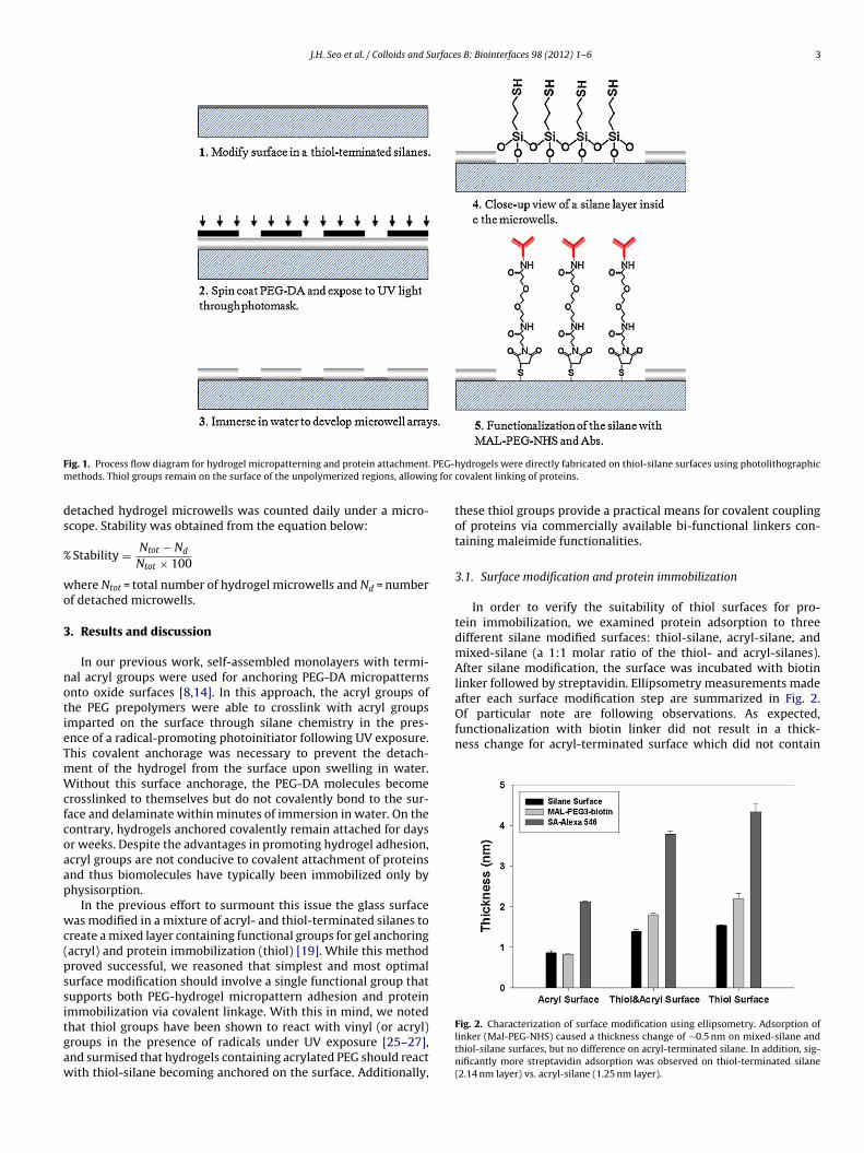
J.H. Seo et al. / Colloids and Surfaces B: Biointerfaces 98 (2012) 1– 6 3
F PEG-hm g for
ds
%
wo
3
notieTmWcfcoaap
wc(pssitgaw
Of particular note are following observations. As expected,functionalization with biotin linker did not result in a thick-ness change for acryl-terminated surface which did not contain
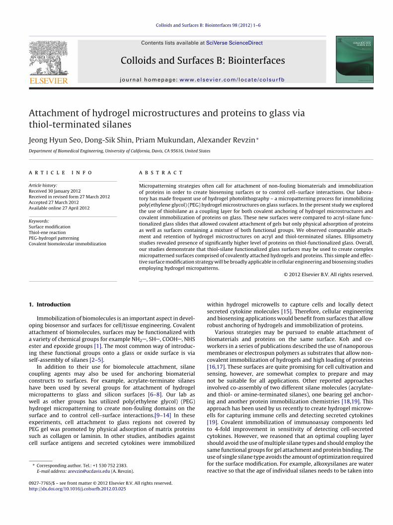
ig. 1. Process flow diagram for hydrogel micropatterning and protein attachment.ethods. Thiol groups remain on the surface of the unpolymerized regions, allowin
etached hydrogel microwells was counted daily under a micro-cope. Stability was obtained from the equation below:
Stability = Ntot − Nd
Ntot × 100
here Ntot = total number of hydrogel microwells and Nd = numberf detached microwells.
. Results and discussion
In our previous work, self-assembled monolayers with termi-al acryl groups were used for anchoring PEG-DA micropatternsnto oxide surfaces [8,14]. In this approach, the acryl groups ofhe PEG prepolymers were able to crosslink with acryl groupsmparted on the surface through silane chemistry in the pres-nce of a radical-promoting photoinitiator following UV exposure.his covalent anchorage was necessary to prevent the detach-ent of the hydrogel from the surface upon swelling in water.ithout this surface anchorage, the PEG-DA molecules become
rosslinked to themselves but do not covalently bond to the sur-ace and delaminate within minutes of immersion in water. On theontrary, hydrogels anchored covalently remain attached for daysr weeks. Despite the advantages in promoting hydrogel adhesion,cryl groups are not conducive to covalent attachment of proteinsnd thus biomolecules have typically been immobilized only byhysisorption.
In the previous effort to surmount this issue the glass surfaceas modified in a mixture of acryl- and thiol-terminated silanes to
reate a mixed layer containing functional groups for gel anchoringacryl) and protein immobilization (thiol) [19]. While this methodroved successful, we reasoned that simplest and most optimalurface modification should involve a single functional group thatupports both PEG-hydrogel micropattern adhesion and proteinmmobilization via covalent linkage. With this in mind, we noted
hat thiol groups have been shown to react with vinyl (or acryl)roups in the presence of radicals under UV exposure [25–27],nd surmised that hydrogels containing acrylated PEG should reactith thiol-silane becoming anchored on the surface. Additionally,ydrogels were directly fabricated on thiol-silane surfaces using photolithographiccovalent linking of proteins.
these thiol groups provide a practical means for covalent couplingof proteins via commercially available bi-functional linkers con-taining maleimide functionalities.
3.1. Surface modification and protein immobilization
In order to verify the suitability of thiol surfaces for pro-tein immobilization, we examined protein adsorption to threedifferent silane modified surfaces: thiol-silane, acryl-silane, andmixed-silane (a 1:1 molar ratio of the thiol- and acryl-silanes).After silane modification, the surface was incubated with biotinlinker followed by streptavidin. Ellipsometry measurements madeafter each surface modification step are summarized in Fig. 2.
Fig. 2. Characterization of surface modification using ellipsometry. Adsorption oflinker (Mal-PEG-NHS) caused a thickness change of ∼0.5 nm on mixed-silane andthiol-silane surfaces, but no difference on acryl-terminated silane. In addition, sig-nificantly more streptavidin adsorption was observed on thiol-terminated silane(2.14 nm layer) vs. acryl-silane (1.25 nm layer).
4 J.H. Seo et al. / Colloids and Surfaces B: Biointerfaces 98 (2012) 1– 6
F Hepart
ma0rtfodpl
3m
othaltPttapdif
eaot
durable for up to 3 days. This timeframe is sufficient for applicationsin biosensing and cell monitoring where these constructs have beenemployed in the past [13,15,28,29].
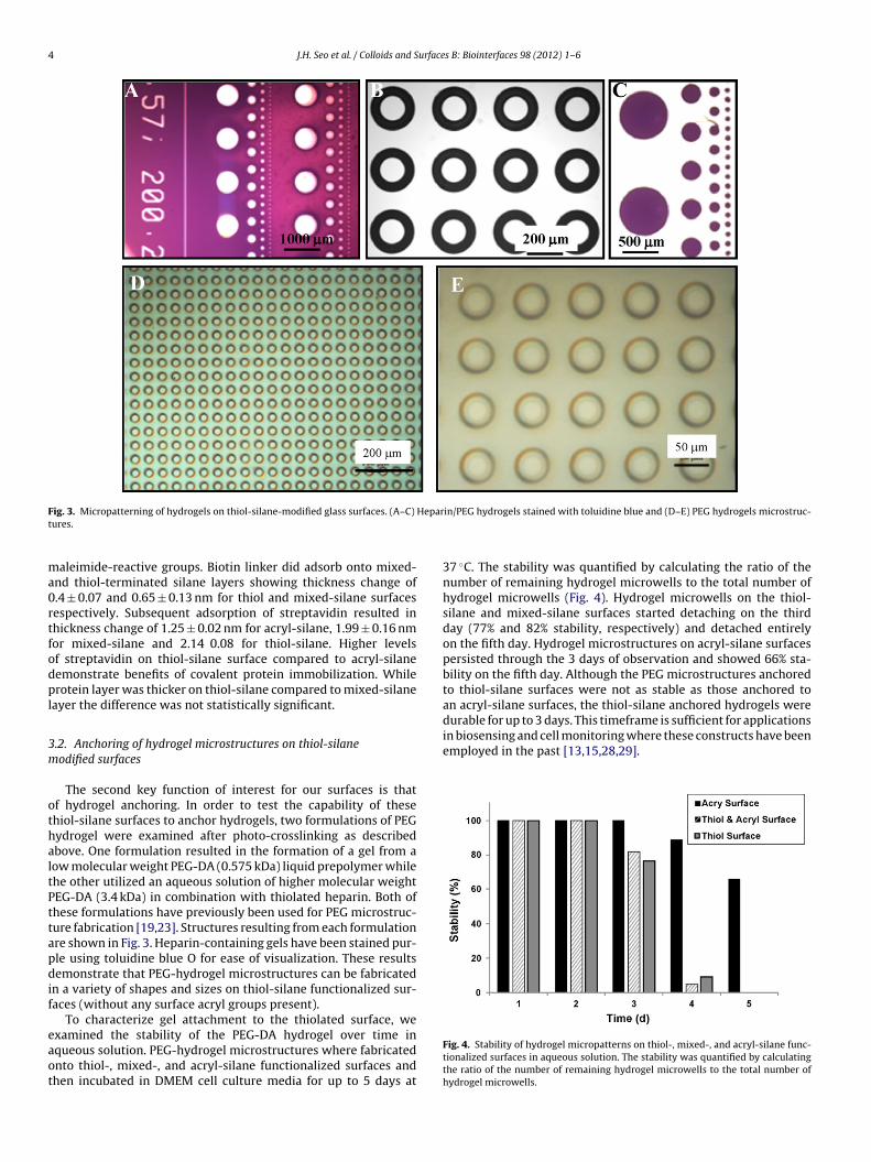
ig. 3. Micropatterning of hydrogels on thiol-silane-modified glass surfaces. (A–C)
ures.
aleimide-reactive groups. Biotin linker did adsorb onto mixed-nd thiol-terminated silane layers showing thickness change of.4 ± 0.07 and 0.65 ± 0.13 nm for thiol and mixed-silane surfacesespectively. Subsequent adsorption of streptavidin resulted inhickness change of 1.25 ± 0.02 nm for acryl-silane, 1.99 ± 0.16 nmor mixed-silane and 2.14 0.08 for thiol-silane. Higher levelsf streptavidin on thiol-silane surface compared to acryl-silaneemonstrate benefits of covalent protein immobilization. Whilerotein layer was thicker on thiol-silane compared to mixed-silane
ayer the difference was not statistically significant.
.2. Anchoring of hydrogel microstructures on thiol-silaneodified surfaces
The second key function of interest for our surfaces is thatf hydrogel anchoring. In order to test the capability of thesehiol-silane surfaces to anchor hydrogels, two formulations of PEGydrogel were examined after photo-crosslinking as describedbove. One formulation resulted in the formation of a gel from aow molecular weight PEG-DA (0.575 kDa) liquid prepolymer whilehe other utilized an aqueous solution of higher molecular weightEG-DA (3.4 kDa) in combination with thiolated heparin. Both ofhese formulations have previously been used for PEG microstruc-ure fabrication [19,23]. Structures resulting from each formulationre shown in Fig. 3. Heparin-containing gels have been stained pur-le using toluidine blue O for ease of visualization. These resultsemonstrate that PEG-hydrogel microstructures can be fabricated
n a variety of shapes and sizes on thiol-silane functionalized sur-aces (without any surface acryl groups present).
To characterize gel attachment to the thiolated surface, we
xamined the stability of the PEG-DA hydrogel over time inqueous solution. PEG-hydrogel microstructures where fabricatednto thiol-, mixed-, and acryl-silane functionalized surfaces andhen incubated in DMEM cell culture media for up to 5 days atin/PEG hydrogels stained with toluidine blue and (D–E) PEG hydrogels microstruc-
37 ◦C. The stability was quantified by calculating the ratio of thenumber of remaining hydrogel microwells to the total number ofhydrogel microwells (Fig. 4). Hydrogel microwells on the thiol-silane and mixed-silane surfaces started detaching on the thirdday (77% and 82% stability, respectively) and detached entirelyon the fifth day. Hydrogel microstructures on acryl-silane surfacespersisted through the 3 days of observation and showed 66% sta-bility on the fifth day. Although the PEG microstructures anchoredto thiol-silane surfaces were not as stable as those anchored toan acryl-silane surfaces, the thiol-silane anchored hydrogels were
Fig. 4. Stability of hydrogel micropatterns on thiol-, mixed-, and acryl-silane func-tionalized surfaces in aqueous solution. The stability was quantified by calculatingthe ratio of the number of remaining hydrogel microwells to the total number ofhydrogel microwells.
J.H. Seo et al. / Colloids and Surfaces B: Biointerfaces 98 (2012) 1– 6 5
F dividm adso
3
issmatnwm(
eoawietooa
4
bgschivpmieaAtiut
[
[
[
[
[
[
[
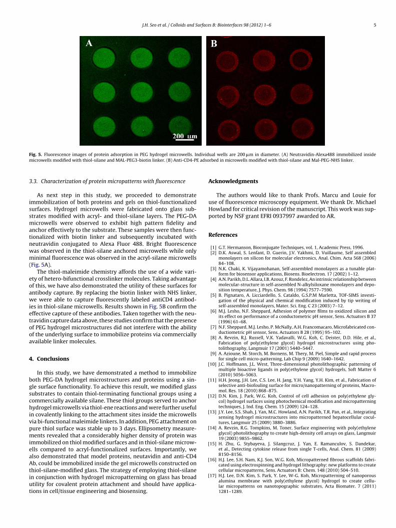
ig. 5. Fluorescence images of protein adsorption in PEG hydrogel microwells. Inicrowells modified with thiol-silane and MAL-PEG3-biotin linker. (B) Anti-CD4-PE
.3. Characterization of protein micropatterns with fluorescence
As next step in this study, we proceeded to demonstratemmobilization of both proteins and gels on thiol-functionalizedurfaces. Hydrogel microwells were fabricated onto glass sub-trates modified with acryl- and thiol-silane layers. The PEG-DAicrowells were observed to exhibit high pattern fidelity and
nchor effectively to the substrate. These samples were then func-ionalized with biotin linker and subsequently incubated witheutravidin conjugated to Alexa Fluor 488. Bright fluorescenceas observed in the thiol-silane anchored microwells while onlyinimal fluorescence was observed in the acryl-silane microwells
Fig. 5A).The thiol-maleimide chemistry affords the use of a wide vari-
ty of hetero-bifunctional crosslinker molecules. Taking advantagef this, we have also demonstrated the utility of these surfaces forntibody capture. By replacing the biotin linker with NHS linker,e were able to capture fluorescently labeled antiCD4 antibod-
es in thiol-silane microwells. Results shown in Fig. 5B confirm theffective capture of these antibodies. Taken together with the neu-ravidin capture data above, these studies confirm that the presencef PEG hydrogel microstructures did not interfere with the abilityf the underlying surface to immobilize proteins via commerciallyvailable linker molecules.
. Conclusions
In this study, we have demonstrated a method to immobilizeoth PEG-DA hydrogel microstructures and proteins using a sin-le surface functionality. To achieve this result, we modified glassubstrates to contain thiol-terminating functional groups using aommercially available silane. These thiol groups served to anchorydrogel microwells via thiol-ene reactions and were further useful
n covalently linking to the attachment sites inside the microwellsia bi-functional maleimide linkers. In addition, PEG attachment onure thiol surface was stable up to 3 days. Ellipsometry measure-ents revealed that a considerably higher density of protein was
mmobilized on thiol modified surfaces and in thiol-silane microw-lls compared to acryl-functionalized surfaces. Importantly, welso demonstrated that model proteins, neutavidin and anti-CD4
b, could be immobilized inside the gel microwells constructed onhiol-silane-modified glass. The strategy of employing thiol-silanen conjunction with hydrogel micropatterning on glass has broadtility for covalent protein attachment and should have applica-ions in cell/tissue engineering and biosensing.
[
ual wells are 200 �m in diameter. (A) Neutravidin-Alexa488 immobilized insiderbed in microwells modified with thiol-silane and Mal-PEG-NHS linker.
Acknowledgments
The authors would like to thank Profs. Marcu and Louie foruse of fluorescence microscopy equipment. We thank Dr. MichaelHowland for critical revision of the manuscript. This work was sup-ported by NSF grant EFRI 0937997 awarded to AR.
References
[1] G.T. Hermanson, Bioconjugate Techniques, vol. 1, Academic Press, 1996.[2] D.K. Aswal, S. Lenfant, D. Guerin, J.V. Yakhmi, D. Vuillaume, Self assembled
monolayers on silicon for molecular electronics, Anal. Chim. Acta 568 (2006)84–108.
[3] N.K. Chaki, K. Vijayamohanan, Self-assembled monolayers as a tunable plat-form for biosensor applications, Biosens. Bioelectron. 17 (2002) 1–12.
[4] A.N. Parikh, D.L. Allara, I.B. Azouz, F. Rondelez, An intrinsic relationship betweenmolecular-structure in self-assembled N-alkylsiloxane monolayers and depo-sition temperature, J. Phys. Chem. 98 (1994) 7577–7590.
[5] B. Pignataro, A. Licciardello, S. Cataldo, G.S.P.M Marletta, TOF-SIMS investi-gation of the physical and chemical modification induced by tip writing ofself-assembled monolayers, Mater. Sci. Eng. C 23 (2003) 7–12.
[6] M.J. Lesho, N.F. Sheppard, Adhesion of polymer films to oxidized silicon andits effect on performance of a conductometric pH sensor, Sens. Actuators B 37(1996) 61–68.
[7] N.F. Sheppard, M.J. Lesho, P. McNally, A.H. Francomacaro, Microfabricated con-ductiometric pH sensor, Sens. Actuators B 28 (1995) 95–102.
[8] A. Revzin, R.J. Russell, V.K. Yadavalli, W.G. Koh, C. Deister, D.D. Hile, et al.,Fabrication of poly(ethylene glycol) hydrogel microstructures using pho-tolithography, Langmuir 17 (2001) 5440–5447.
[9] A. Azioune, M. Storch, M. Bornens, M. Thery, M. Piel, Simple and rapid processfor single cell micro-patterning, Lab Chip 9 (2009) 1640–1642.
10] J.C. Hoffmann, J.L. West, Three-dimensional photolithographic patterning ofmultiple bioactive ligands in poly(ethylene glycol) hydrogels, Soft Matter 6(2010) 5056–5063.
11] H.H. Jeong, J.H. Lee, C.S. Lee, H. Jang, Y.H. Yang, Y.H. Kim, et al., Fabrication ofselective anti-biofouling surface for micro/nanopatterning of proteins, Macro-mol. Res. 18 (2010) 868–875.
12] D.N. Kim, J. Park, W.G. Koh, Control of cell adhesion on poly(ethylene gly-col) hydrogel surfaces using photochemical modification and micropatterningtechniques, J. Ind. Eng. Chem. 15 (2009) 124–128.
13] J.Y. Lee, S.S. Shah, J. Yan, M.C. Howland, A.N. Parikh, T.R. Pan, et al., Integratingsensing hydrogel microstructures into micropatterned hepatocellular cocul-tures, Langmuir 25 (2009) 3880–3886.
14] A. Revzin, R.G. Tompkins, M. Toner, Surface engineering with poly(ethyleneglycol) photolithography to create high-density cell arrays on glass, Langmuir19 (2003) 9855–9862.
15] H. Zhu, G. Stybayeva, J. Silangcruz, J. Yan, E. Ramanculov, S. Dandekar,et al., Detecting cytokine release from single T-cells, Anal. Chem. 81 (2009)8150–8156.
16] H.J. Lee, S.H. Nam, K.J. Son, W.G. Koh, Micropatterned fibrous scaffolds fabri-cated using electrospinning and hydrogel lithography: new platforms to create
cellular micropatterns, Sens. Actuators B: Chem. 148 (2010) 504–510.17] H.J. Lee, D.N. Kim, S. Park, Y. Lee, W-G. Koh, Micropatterning of nanoporousalumina membrane with poly(ethylene glycol) hydrogel to create cellu-lar micropatterns on nanotopographic substrates, Acta Biomater. 7 (2011)1281–1289.

6 urface
[
[
[
[
[
[
[
[
[
[
[
J.H. Seo et al. / Colloids and S
18] K.B. Lee, Y.H. Jung, Z.W. Lee, S. Kim, I.S. Choi, Biospecific anchoring and spa-tially confined germination of bacterial spores in non-biofouling microwells,Biomaterials 28 (2007) 5594–5600.
19] J.H. Seo, L.J. Chen, S.V. Verkhoturov, E.A. Schweikert, A. Revzin, The use ofglass substrates with bi-functional silanes for designing micropatterned cell-secreted cytokine immunoassays, Biomaterials 32 (2011) 5478–5488.
20] S.K. Murthy, A. Sin, R.G. Tompkins, M. Toner, Effect of flow and surface condi-tions on human lymphocyte isolation using microfluidic chambers, Langmuir20 (2004) 11649–11655.
21] A. Sin, S.K. Murthy, A. Revzin, R.G. Tompkins, M. Toner, Enrichment usingantibody-coated microfluidic chambers in shear flow: model mixture of humanlymphocytes, Biotechnol. Bioeng. 91 (2005) 816–826.
22] A.E. Rydholm, C.N. Bowman, K.S. Anseth, Degradable thiol-acrylate pho-topolymers: polymerization and degradation behavior of an in situ formingbiomaterial, Biomaterials 26 (2005) 4495–4506.
23] S.S. Shah, M. Kim, K. Cahill-Thompson, G. Tae, A. Revzin, Micropatterning ofbioactive heparin-based hydrogels, Soft Matter 7 (2011) 3133–3140.
[
s B: Biointerfaces 98 (2012) 1– 6
24] L.L. Gosey, R.M. Howard, F.G. Witebsky, F.P. Ognibene, T.C. Wu, V.J. Gill, et al.,Advantages of a modified toluidine blue-O stain and bronchoalveolar lavage forthe diagnosis of Pneumocystis carinii pneumonia, J. Clin. Microbiol. 22 (1985)803–807.
25] C.R. Becer, R. Hoogenboom, U.S. Schubert, Click chemistry beyond metal-catalyzed cycloaddition, Angew. Chem. Int. Ed. 48 (2009) 4900–4908.
26] J.A. Carioscia, H. Lu, J.W. Stanbury, C.N. Bowman, Thiol-ene oligomers as dentalrestorative materials, Dent. Mater. 21 (2005) 1137–1143.
27] S.P.S. Koo, M.M. Stamenovic, R.A. Prasath, A.J. Inglis, F.E. Du Prez, C. Barner-Kowollik, et al., Limitations of radical thiol-ene reactions for polymer–polymerconjugation, J. Polym. Sci. Part A: Polym. Chem. 48 (2010) 1699–1713.
28] Y. Liu, J. Yan, M.C. Howland, T. Kwa, A. Revzin, Micropatterned aptasensors
for continuous monitoring of cytokine release from human leukocytes, Anal.Chem. 83 (2011) 8286–8292.29] J. Yan, Y. Sun, H. Zhu, L. Marcu, A. Revzin, Enzyme-containing hydrogelmicropatterns serving a dual purpose of cell sequestration and metabolitedetection, Biosens. Bioelectron. 24 (2009), 2604-26102610.