colloidal gold and silver triangular nanoprisms - dicp · relatively few methods that allow one to...
TRANSCRIPT

reviews C. A. Mirkin et al.
646
Nanoprisms
Colloidal Gold and Silver Triangular NanoprismsJill E. Millstone, Sarah J. Hurst, Gabriella S. Metraux, Joshua I. Cutler, and
Chad A. Mirkin*
From the Contents
1. Introduction to Gold and Silver Metal
Nanoparticles . . . . . . . . . . . . . . . . . . 647
2. What is a Triangular Nanoprism?. . . . . 647
3. Photochemical Syntheses of Triangular
Nanoprisms . . . . . . . . . . . . . . . . . . . 649
4. Thermal Syntheses of (or Chemical
Reduction Methods for Producing)
Triangular Nanoprisms . . . . . . . . . . . . 653
5. Mechanisms of Plate-Like Growth . . . . 658
6. Summary and Outlook . . . . . . . . . . . . 661
It is now w
� 2
ell-known that the size, shape, and composition of
nanomaterials can dramatically affect their physical and
chemical properties, and that technologies based on nanoscale
materials have the potential to revolutionize fields ranging from
catalysis to medicine. Among these materials, anisotropic
particles are particularly interesting because the decreased
symmetry of such particles often leads to new and unusual
chemical and physical behavior. Within this class of particles,
triangular Au and Ag nanoprisms stand out due to their
structure- and environment-dependent optical features, their
anisotropic surface energetics, and the emergence of reliable
synthetic methods for producing them in bulk quantities with
control over their edge lengths and thickness. This Review will
describe a variety of solution-basedmethods for synthesizingAu
and Ag triangular prismatic structures, and will address and
discuss proposed mechanisms for their formation.
Frontispiece images reproduced from References [68,71,148] and withpermission from Reference [31]. Copyright 2003, American Chemical Society.
009 Wiley-VCH Verlag GmbH & Co. KGaA, Weinheim small 2009, 5, No. 6, 646–664

Colloidal Gold and Silver Triangular Nanoprisms
[�] Prof. C. A. Mirkin, Dr. J. E. Millstone, Dr. S. J. Hurst, Dr. G. S. Metraux,
J. I. Cutler
Department of Chemistry and
International Institute of Nanotechnology
2145 Sheridan Road, Evanston, IL 60208 (USA)
E-mail: [email protected]
DOI: 10.1002/smll.200801480
1. Introduction to Gold and Silver MetalNanoparticles
Au and Ag nanoparticles have been used in many areas,
including molecular diagnostics,[1–11] catalysis,[12–14] electro-
nics,[13,15] encryption strategies,[16–18] and gene therapy.[19–21]
Therefore, it is no surprise that there has been an explosion of
interest in the development of synthetic methods for preparing
these nanostructures and investigating their size- and shape-
dependent properties. To date, the majority of research has
focused on isotropic (i.e., spherical or pseudospherical)
particles, and many synthetic methods have been developed
for preparing them with moderate to excellent control over
their sizes and size distributions.[22–25] Nanoparticles derived
from these protocols have fostered the study of their
structures[26,27] as well as their optical,[28–31] catalytic,[12–
14,32] and electronic[13,15,33,34] properties. For example, 40-nm-
diameter Au nanoparticles (AuNPs) have a molar absorption
coefficient almost five orders of magnitude higher than a
conventional organic dye absorbing at a similar wavelength
(e.g., 7.66� 109M�1 cm�1 at lmax¼ 528 nm for 40-nm AuNPs vs.
1.16� 105M�1 cm�1 at lmax¼ 530 nm for rhodamine-6G).[35]
AuNPs with diameters less than 10 nm and immobilized within
a metal oxide framework can catalyze CO oxidation or
propylene epoxidation, even at low temperatures (<0 8C),
whereas bulk Au is essentially inactive.[36] In another example,
both Ag and Au nanostructures can significantly influence
fluorescence processes by either enhancing or quenching
fluorophore emission as a function of the distance between
the fluorophore and the metal surface.[37] Bulk samples act
only as quenchers. Yet in the case of Au and Ag, although they
have developed a reasonable understanding of the relationship
between the properties of a particle and its size and
composition, researchers are just beginning to explore the
relationship between the shape of a nanoparticle and its
physical and chemical properties.Many Au and Ag nanoparticle shapes have been observed
by electron microscopy and related methods, including rods
and wires,[38–43] prisms and disks,[44–49] cubes,[50–54] ‘‘dog
bones,’’[55] and hollow structures.[56,57] Overall, there are
relatively few methods that allow one to systematically make
such structures in high yield with control over their
architectural parameters. However, with respect to Au and
Ag, there are three classes of anisotropic structures where
there are reliable methods for making them in high yield with
moderate to excellent control over architectural parameters:
nanorods, ‘‘platonic solids,’’ and triangular prisms.Nanorods can be made by thermal,[42] photochemical,[58]
and electrochemistry-based template[59] methods. In fact,
template-based methods for preparing nanorods marked one
of the first major developments in high-yield, solution-phase
anisotropic metallic nanostructure synthesis.[60] This approach
is extraordinarily useful for synthesizing structures with control
over both rod diameter and length, and these structures have
been investigated in numerous photonic, plasmonic, and
electronic applications.[61] In the last decade, another class
of materials called ‘‘platonic solids,’’ which include cubic- and
icosahedral-shaped particles, have been developed. Methods
for preparing Ag and Au versions of these structures were
small 2009, 5, No. 6, 646–664 � 2009 Wiley-VCH Verlag Gmb
pioneered by both Yang et al. and Xia et al. using different but
complementary polymer-based thermal strategies, wherein the
nanoparticle shape could be controlled by such parameters as
metal precursor to reducing agent ratios and seed particle
type.[62,63] These particles exhibit optical features between 600
and 1000 nm, depending on their morphology, and are another
example of using solution-phase synthetic methods to control
nanoparticle shape and corresponding properties. The third
class of Ag and Au anisotropic nanoparticles to be extensively
investigated is triangular prisms and plate-like nanostructures.
These particles were observed by electron microscopy as
components of complex mixtures as far back as 1951,[64] but our
group developed and reported the first high-yield synthetic
method for this particle type in 2001.[47] Importantly, we were
able to assign the surface plasmon resonance (SPR) bands in
the optical spectra of these colloids by correlating the
experimental data with theoretically predicted values.[47] Since
this initial work, many methods have been developed for
making prismatic structures in high yield and research
conducted on these nanostructures has been extensive. In
particular, research has focused on determining mechanisms
for describing their formation and developing methods to
manipulate their optical features.[42,43,62,65–68]
This review will focus on methods for synthesizing and
characterizing Au and Ag triangular nanoprisms. These
structures are especially interesting because they have
plasmonic features in the visible and IR regions, can be
prepared in high yield, and can be readily functionalized with a
variety of sulfur-containing adsorbates.[47,48,68–73] In Section 2,
nanoprisms are defined and then described in terms of their
common features including dimensions, crystallinity, optical
properties, and surface chemistry. In Sections 3 and 4, both
solution-phase light-mediated syntheses and thermal techni-
ques for making triangular nanoprisms composed of either Au
or Ag are reviewed. Finally, Section 5 summarizes work aimed
at determining a mechanism to describe nanoprism formation.
2. What is a Triangular Nanoprism?
A variety of synthetic routes have been used to generate
prismatic, plate-like nanostructures (also referred to as
nanoprisms, nanotriangles, nanoplates, or nanodisks). How-
ever, despite the differences in the methods used to make
them, the resulting structures share common architectural
elements and possess similar chemical and physical properties.
This section highlights these features and formulates a
definition of structures that are commonly categorized as
nanoprisms. We will discuss these points specifically in relation
to particles composed of pure Au and pure Ag.
From a geometric perspective, prisms can be of any
thickness (i.e., have an arbitrary distance between two parallel
H & Co. KGaA, Weinheim www.small-journal.com 647

reviews C. A. Mirkin et al.
Scheme 1. Illustration of nanoprism dimensions.
Prof. Chad A. Mirkin earned his B. S. at
Dickinson College in 1986 and his Ph. D.
from Pennsylvania State University in 1989.
After an NSF Postdoctoral Fellowship at MIT,
he joined the faculty of Northwestern Uni-
versity where he is currently the Director of
the NU International Institute for Nanotech-
nology and the George B. Rathmann Pro-
fessor of Chemistry, Professor of Medicine,
and Professor of Materials Science and
Engineering. He has authored over 350
manuscripts and 70 patents, founded two
companies (Nanosphere and NanoInk), and
cofounded the journal Small. He has
received numerous awards, including the
NIH Director’s Pioneer Award and the American Chemical Society (ACS) Pure
Chemistry Award.
Figure 1. A) Scanning tunneling spectroscopy (STM) image of hexa-
gonal Au nanoprisms showing the atomic terraces (the image has been
high-pass spatial filtered, and the defect on the nanoprism surface was
caused by a tip crash). B) A high-resolution zoom on the surface of
the nanoprism shown in (A). The corrugation is theffiffiffi
3p
�ffiffiffi
3p
R308molecular lattice characteristic of well-ordered alkanethiol self-
assembled monolayers (SAMs). The dark features (white arrow) are SAM
structural domain boundaries. Reprinted with permission from
Reference [79]. Copyright 2006, American Chemical Society.
648
polygons), but in general, nanoprisms synthesized to date have
been flat, triangular, hexagonal, or circular plates with large
aspect ratios (vide infra) (Scheme 1). This review focuses on
triangular nanoprisms, which exhibit three congruent edge
lengths (‘) and a defined thickness (t). These Au and Ag
nanoprisms typically exhibit edge lengths in the �40 nm to
1mm range and thickness ranging from �5 to 50 nm.
Nanoprism structures with edge lengths as large as several
micrometers have been synthesized, but these have not
exhibited the optical or chemical properties associated with
their smaller analogs.[31,74,75] Technically, triangular nanopr-
isms contain three sharp vertices or ‘‘tips’’ that contribute
significantly to their optical and electronic properties.[31,75]
However, in practice, mixtures of particles with varying
degrees of tip truncation and rounding make up a colloid.
When significant rounding occurs, structures are no longer
described as triangular nanoprisms, and generally are referred
to as nanodisks or in cases of truncation without rounding,
hexagonal nanoprisms. In fact, all colloidal syntheses of
triangular nanoprisms tend to yield some percentage of
nanohexagons or nanodisks, which have either undergone
incomplete transformation to triangular nanoprisms or under-
gone surface reorganization in such a way that they no longer
exhibit the ideal triangular nanoprism structure.
In some cases, nanoprism dimensions can be controlled in
situ by adjusting experimental parameters, including metal ion
and reducing agent ratios,[70] surfactant concentrations,[54]
pH,[71] irradiation wavelength,[73] and seed particle concen-
tration and type.[44,63,68] The edge lengths and thickness of the
nanoprism determine its aspect ratio (‘/t), which can be used to
quantify degree of anisotropy. For example, isotropic
nanoparticles such as pseudospherical particles have an aspect
ratio of one because their dimensions are roughly the same in
all directions. In the case of Au and Ag nanoprisms, their
aspect ratios vary from 5 to over 40.
In general, solution-prepared Au and Ag triangular
nanoprisms are single crystalline with face-centered cubic
(fcc) lattice structures.[47,49,68,71,76–78] This crystallinity differ-
entiates them from lithographically or electrochemically
prepared structures, which are typically polycrystalline. The
www.small-journal.com � 2009 Wiley-VCH Verlag Gm
triangular (or hexagonal or circular) facets of solution-
synthesized nanoprisms are often composed of almost atom-
ically flat {111} crystal faces (Figure 1).[48,79] The edges of the
nanoprisms are typically {110}, {111}, or {100} facets,[47,68,80]
and high-resolution transmission electron microscopy
(HRTEM) analysis suggests that nanoprisms contain a twin
plane parallel to their {111} triangular faces (vide
infra).[47,67,68]
These common architectural elements give rise to a set
of chemical and physical properties that are shared by most
Au and Ag triangular nanoprisms. In particular, these
prisms have SPRs that are tunable throughout the visible
and near-IR (NIR) regions of the spectrum by controlling
nanoprism edge length, thickness, and tip morphology
(Figure 2).[31,47,68,70,71,73,75] These SPRs are generated by the
coherent oscillation of conduction electrons at the surface of
the nanoparticle when they interact with the oscillating
electric field of incident light. The frequency of this oscillation
is not only dependent on the density and effective mass of the
bH & Co. KGaA, Weinheim small 2009, 5, No. 6, 646–664

Colloidal Gold and Silver Triangular Nanoprisms
Figure 2. UV–Vis–NIR spectra and corresponding solutions of Ag
nanoprisms with varying edge length. Labeled vial and spectra numbers
correspond to the wavelength of irradiation used to prepare the
nanostructures. Reprinted from Reference [71].
electrons, but also the size and shape of the charge distribution
(Figure 3).[31] Further, Schatz and coworkers[31,75] have used a
quasistatic approximation model to show that optical proper-
ties of metal nanoparticles are significantly affected by the
wavelength-dependent dielectric constant of the particle and
the dielectric constant of the surrounding medium when the
particle diameter is much smaller than that of the incident
light. The dependence of nanoparticle SPRs on charge
distribution and dielectric properties explains the sensitivity
of their optical features to particle size, shape, and chemical
environment, and is a highly useful feature of Au and Ag
nanoprisms (vide infra).
Figure 3. Orientation-averaged extinction efficiency for triangular
nanoprisms based on a 100-nm-edge dimension with snips of 0, 10,
and 20 nm. The inset shows the shape of a snipped prism. The prism
thickness is 16 nm. Reprinted with permission from Reference [31].
Copyright 2003, American Chemical Society.
small 2009, 5, No. 6, 646–664 � 2009 Wiley-VCH Verlag Gmb
Sufficiently large and thin nanoprisms (aspect ratio
>10)[75] contain both dipole and quadrupole plasmon
resonances that shift in frequency and extinction cross section
as a function of nanoprism size, shape, and dielectric
environment as described above. With spherical particles,
these two modes (quadrupole and dipole) are not distinguish-
able from one another (d< 100 nm),[81,82] however in an
anisotropic particle such as a nanoprism, these modes oscillate
at markedly different frequencies (generally separated by
100–400 nm), and can be resolved experimentally for prisms of
both Au and Ag.[47,48] Roughly, these modes originate from
the degree and direction of polarization of the electron cloud
relative to the incident electric field. In this way, a dipole
plasmon resonance can be described as the electron cloud
surrounding the nanoparticle moving either parallel or
antiparallel to the applied field. For a quadrupole mode, half
of the cloud moves parallel and half moves antiparallel.
Higher-order SPR modes can be obtained through more
complex polarizations, and have been observed for high aspect
ratio nanostructures such as nanorods.[83]
Given the relationship between nanoparticle morphology
and optical features, it follows that these features can be used
to assess the shape, size, and distribution of nanostructures in
solution for structures that exhibit such properties.[48] For
example, as the tips of a nanoprism become rounded, its
optical features become blue-shifted (to shorter wavelengths)
as the electron cloud density changes across the particle sur-
face.[31] Prism thickness, edge length, and dielectric environ-
ment will also red- or blue-shift the SPRs depending on the
change in particle architecture or environment.[31,68–71,73,75]
Of particular interest are the quadrupole plasmon modes
of the nanoprisms, because these modes can only be identified
for colloidal dispersions with sufficiently high concentrations
of nanoprisms that also have relatively narrow particle size
and shape distributions (<approximately 20%).[31,68–71,73,75]
Indeed, one of the best diagnostics for the quality (in terms of
shape and monodispersity) of triangular prisms produced in
a synthetic procedure is the identification, breadth, and
spectral position of the dipole and quadrupole SPRs in the
extinction spectrum of the colloid. Throughout this review,
these optical features, in conjunction with electron micro-
scopy, will be used to compare the various products of Au and
Ag nanoprism syntheses.
3. Photochemical Syntheses of TriangularNanoprisms
A photochemical route was the first reliable and high
yielding method for making solution-phase triangular Ag
nanoprisms.[47] This method, which allowed one to control
edge length with excitation wavelength, allowed researchers to
assign UV–Vis spectral features as a function of prism
architecture.[47] Therefore, our discussion of nanoprisms
begins with photochemical syntheses and related methods
that have been developed to make these nanostructures using
light. Approaches to nanoprism synthesis using irradiation
have been classified by the radiation wavelength employed in
the synthesis.
H & Co. KGaA, Weinheim www.small-journal.com 649

reviews C. A. Mirkin et al.
Figure 4. A) Electron energy loss spectroscopy (EELS) mapping analysis showing the flat-
top morphology of the Ag nanoprisms. Inset shows the EELS intensity over the line scan
(dotted line through triangle axis). B) Stacks of Ag nanoprisms assembled in a top-to-base
manner. C) Electron diffraction analysis of individual Ag nanoprisms. The diffraction pattern is
characteristic of the {111} orientation of an individual Ag nanoprism lying flat on the substrate
with its triangular face perpendicular to the electron beam. On the basis of three-zone axis
analysis (not shown), the crystal structure of the Ag nanoprism was determined to be a
fcc structure. The intense spots in the {111} zone axis are allowed {220} Bragg reflections
(e.g., circled spot, corresponding to the lattice spacing of 1.44 A), and the sharp weak spot in
the center of the triangles formed by the strong spots is indexed as 1/3{422} (e.g., boxed
spot, corresponding to the lattice spacing of 2.50 A). Reprinted with permission from
Reference [47]. Copyright 2001, American Association for the Advancement of Science.
Figure 5. A) Time-dependent UV–Vis spectra showing the conversion of
Ag nanospheres to nanoprisms (a) before irradiation and after (b) 40,
(c) 55, and (d) 70 h of irradiation. B) Corresponding extinction profiles at
670 nm as a function of time. Reprinted with permission from Reference
[47]. Copyright 2001, American Association for the Advancement of
Science.
650
3.1. Visible-Light Methods
This section highlights the use of visible light (300 nm< l<800 nm) to direct and/or drive the growth of prismatic
nanoparticles. In 2001, our group reported a photochemical
reaction in which small Ag nanoparticles (AgNPs; dia-
meter¼ 6–8 nm) could be converted into triangular nanopr-
isms by irradiating a solution containing trisodium citrate and
bis(p-sulfonatophenyl)phenylphosphine dipotassium salt
(BSPP) with fluorescent light.[47] The resulting colloid
contained single crystalline Ag nanoprisms with edge lengths
of �100 nm (Figure 4). The conversion of the nanoparticles to
nanoprisms could be turned on and off simply by turning on or
off the light source. Interestingly, the optical spectrum
displayed SPR bands that had never been observed experi-
mentally. In addition to the in-plane dipole resonance at
670 nm, two new bands: the in-plane quadrupole (440 nm) and
the out-of-plane quadrupole (340 nm), were identified
(Figure 5). In a collaborative effort with our group, Schatz
and coworkers[31,47] calculated the optical signatures of these
nanoprisms and found that the theoretically derived spectra
agreed closely with those obtained experimentally.
The role of light in this photochemical conversion process
was more thoroughly examined in subsequent work,[73] which
demonstrated the effects of photoexciting a AgNP colloid with
wavelengths that overlap the dipole and quadrupole SPR
modes of the final Ag nanoprisms. Interestingly, excitation of a
AgNP colloid with a single wavelength (i.e., 550 nm, dipole
SPR excitation) resulted in a solution of nanoprisms with two
size distributions, designated Type 1 (edge length 70� 12 nm)
and Type 2 (edge length 150� 16 nm). Type 1 and 2
nanoprisms differed only in edge length (by a factor of 2)
whereas their thickness was essentially the same. In contrast,
simultaneous excitation with 550 nm (dipole SPR excitation)
and 450 nm (or 340 nm, quadrupole SPR excitation) light
resulted in Type 1 nanoprisms only. Indeed, the results
indicated that quadrupole or high energy excitation inhibits
www.small-journal.com � 2009 Wiley-VCH Verlag GmbH & Co. KGaA, Weinhe
the fusion of the smaller, Type 1 prisms.
Interestingly, by varying the primary beam
(used for dipole excitation) over the visible
range (450–750 nm), we were able to
generate nanoprisms with edge lengths
ranging from �40 to 120 nm (Figure 6).
The optical properties of the nanoprisms
vary significantly with their dimensions and
thus the colloidal solutions range in color
from red to blue depending on nanoprism
edge length. With this advance, one not
only could prepare these unusual structures
in high yield, but also could have unpre-
cedented control of edge length, one of
their key architectural parameters.
Since the initial report of Ag triangular
nanoprism synthesis, others have con-
firmed the results and significantly
expanded upon their scope. For example,
Brus et al. observed morphological changes
of spherical AgNPs to nanoprisms when
exposed to various wavelengths of visible
light.[84] In this protocol, pseudospherical Ag seeds were
prepared and added to an aqueous growth solution containing
Agþ and trisodium citrate. This mixture was then irradiated
with 457-nm light for several hours (power¼ 0.8 W cm�2). The
UV–Vis spectrum of this solution exhibited three peaks at
338 nm (out-of-plane quadrupole), 400 nm (in-plane quadru-
pole), and 540 nm (in-plane dipole), indicating the formation
of disk-like nanoprisms. TEM analysis showed that these
nanoprisms were single crystalline, approximately 38 nm in
diameter and �10 nm in thickness on average. When this
reaction was monitored over time using TEM, the authors
observed increasing numbers of nanoprisms with increasing
exposure time. Similar to earlier work,[73] the disk diameter of
the final nanoprisms increased with longer excitation wave-
lengths.
im small 2009, 5, No. 6, 646–664
Colloidal Gold and Silver Triangular Nanoprisms
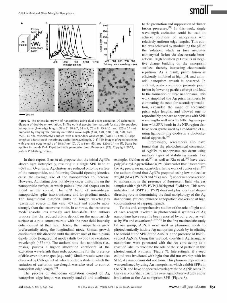
Figure 6. The unimodal growth of nanoprisms using dual-beam excitation. A) Schematic
diagram of dual-beam excitation. B) The optical spectra (normalized) for six different-sized
nanoprisms (1–6 edge length: 38�7, 50� 7, 62�9, 72�8, 95� 11, and 120� 14 nm)
prepared by varying the primary excitation wavelength (450, 490, 520, 550, 650, and
750�40 nm, respectively) coupled with a secondary wavelength (340�10 nm). C) Edge
length as a function of the primary excitation wavelength. D–F) TEM images of Ag nanoprisms
with average edge lengths of 38�7 nm (D), 72�8 nm (E), and 120�14 nm (F). Scale bar
applies to panels D–F. Reprinted with permission from Reference [73]. Copyright 2003,
Nature Publishing Group.
In their report, Brus et al. propose that the initial AgNPs
absorb light isotropically, resulting in a single SPR band at
�395 nm. Over time, Ag clusters are reduced onto the surface
of the nanoparticle, and following Ostwald ripening kinetics,
cause the average size of the nanoparticles to increase.
However, Ag plating does not always occur uniformly on the
nanoparticle surface, at which point ellipsoidal shapes can be
found in the colloid. The SPR band of nonisotropic
nanoparticles splits into transverse and longitudinal modes.
The longitudinal plasmon shifts to longer wavelengths
(excitation source in this case, 457 nm) and absorbs more
strongly than the transverse mode. In contrast, the transverse
mode absorbs less strongly and blue-shifts. The authors
propose that the reduced atoms deposit on the nanoparticle
surface at a rate commensurate with the near-field intensity
enhancement at that face. Hence, the nanoparticles grow
preferentially along the longitudinal mode. Crystal growth
continues in this direction until the absorbance of the in-plane
dipole mode (longitudinal mode) shifts beyond the excitation
wavelength (457 nm). The authors note that nanodisks (i.e.,
prisms) possess a higher absorption coefficient at the
excitation wavelength than rods, accounting for the presence
of disks over other shapes (e.g., rods). Similar results were also
observed by Callegari et al. who reported a study in which the
variation of excitation wavelength was also used to control
nanoprism edge length.[85]
The process of dual-beam excitation control of Ag
nanoprism edge length was recently studied and attributed
small 2009, 5, No. 6, 646–664 � 2009 Wiley-VCH Verlag GmbH & Co. KGaA, Weinheim
to the promotion and suppression of cluster
fusion processes.[71] In this work, single
wavelength excitation could be used to
achieve solutions of nanoprisms with
relatively uniform edge lengths. This con-
trol was achieved by modulating the pH of
the solution, which in turn mediates
nanocrystal fusion via electrostatic inter-
actions. High solution pH results in nega-
tive charge building on the nanoprism
surface, thereby increasing electrostatic
repulsion. As a result, prism fusion is
efficiently inhibited at high pH, and unim-
odal nanoprism growth is observed. In
contrast, acidic conditions promote prism
fusion by lowering particle charge and lead
to the formation of large nanoprisms. This
work simplified the Ag prism synthesis by
eliminating the need for secondary irradia-
tion, expanded the range of accessible
prism edge lengths, and allowed one to
reproducibly prepare nanoprisms with SPR
wavelengths well into the NIR. Ag nanopr-
isms with SPR bands in the NIR region also
have been synthesized by Liz-Marzan et al.
using light-emitting diodes in a photoche-
mical approach.[86]
Interestingly, researchers also have
found that the photochemical conversion
of AgNPs to nanoprisms can occur using
multiple types of stabilizing agents. For
example, Gehlen et al.[87] as well as Xia et al.[88] have used
poly(N-vinyl-2-pyrrolidone) (PVP) instead of BSPP to stabilize
the Ag precursor nanoparticles. In the work of Junior et al.,[87]
the authors found that AgNPs prepared using low molecular
weight (MW) PVP (29 and 55 kg mol�1) underwent conversion
to nanoprisms in the presence of fluorescent light whereas
samples with high MW PVP (1300 kg mol�1) did not. This work
indicates that BSPP (or PVP) does not play a critical shape-
directing role in determining the final morphology of the Ag
nanoprisms, yet can influence nanoparticle conversion at high
concentrations of capping ligands.
To this end, comprehensive studies of the role of light and
of each reagent involved in photochemical synthesis of Ag
nanoprisms have recently been reported by our group as well
as by Wu and coworkers.[72,89,90] In the first mechanistic study
by our group, AuNPs were used as plasmonic seeds to
photochemically initiate Ag nanoprism growth by irradiating
the colloid at the SPR of the AuNPs in the presence of BSPP-
capped AgNPs. Using this method, core/shell Ag triangular
nanoprisms were generated with the Au core acting as a
reaction label to elucidate the role of the seed particle in this
photochemical synthesis (Figure 7). Interestingly, if a seed
colloid was irradiated with light that did not overlap with its
SPR, Ag nanoprisms did not form. This plasmon dependence
was confirmed by using Au nanoprisms, which exhibit SPRs in
the NIR, and have no spectral overlap with the AgNP seeds. In
this case, core/shell structures were again observed only under
irradiation at the Au nanoprism SPR (Figure 8).
www.small-journal.com 651

reviews C. A. Mirkin et al.
Figure 7. A) A TEM image of the Au@Ag core/shell nanoprisms (average
edge length of 70�6 nm) synthesized by irradiation with 550-nm light.
The inset shows the side view of a core/shell nanoprism. B) Extinction
spectrum of the Au@Ag core/shell colloidal nanoprisms after
centrifugation. C) A HRTEM image of the {111} face of the Au@Ag core/
shell nanoprisms. The hexagonal lattice shows a spacing of 1.44 A,
indexed as {220} of fcc Ag. Reprinted from Reference [72].
Figure 8. Representative TEM images of Au@Ag core/shell nanoprisms
with a Au prism core. The scale bar is the same for all images. Reprinted
from Reference [72].
652
Building on this work, investigations were also made
into the chemical role of each reagent in the synthesis, and a
three-step growth mechanism was proposed.[89] During the
initial stage of photomediated Ag nanoprism growth, a AgNP
colloid is prepared by NaBH4 reduction of AgNO3 in the
presence of trisodium citrate and BSPP. The resulting mixture
Scheme 2. Proposed photomediated growth pathway of Ag nanoprisms from spherical
nanoparticles. Reprinted with permission from Reference [89]. Copyright 2008, American
Chemical Society.
exhibits an extinction maximum at 395 nm
and absorbs light throughout the visible
range. The photochemical reaction induced
by plasmon excitation of these particles has
been proposed by several groups to be the
charge transfer between adsorbates on the
surface of the seed particle and ‘‘hot’’ holes
that are likely produced by plasmon
decay.[84,89–91] Specifically, these reactions
involve the reduction of Agþ by trisodium
www.small-journal.com � 2009 Wiley-VCH Verlag Gm
citrate on the Ag particle surface and oxidative dissolution of
small Ag particles by O2.
Ag0þ 1
2O2þH2O ! Agþþ2OH� (1)
BSPP increases the solubility of Agþ by complexing them
and thereby acts as a buffer to keep the concentration of Agþ
at approximately 20mM (as determined by inductively coupled
mass spectrometry, ICP-MS). The Ag particles then serve as
photocatalysts and, under plasmon excitation, facilitate Agþ
reduction by citrate (Scheme 2). This is evidenced by the
oxidation of citrate into 1,3-acetonedicarboxylate and its
further decomposition into acetoacetate and CO2, which was
monitored by 1H-NMR spectroscopy.[89]
A mechanism for the subsequent growth of these isotropic
particles into small and then larger nanoprism structures was
proposed based on several empirical observations. First, after
only 30 min of irradiating the Ag colloid, Ag triangular
nanoprisms can be observed by TEM. While many mechan-
isms may be responsible for this growth pattern, a possible
pathway involves dipole SPR excitation-induced ultrafast
charge separation on the nanoparticle surface,[31] which may
produce face-selective Agþ reduction as first postulated by
Brus et al. (vide supra).[84] This theory is consistent with our
recent observations using Au particles as plasmon reaction
labels,[72] as well as the observations of others that show
inhomogeneous Ag shell growth at early stages of photo-
chemical synthesis.[84] Further growth by dipole plasmon
excitation favors the formation of sharp-tipped Ag nanoprisms
because excitation of the dipole SPR localizes energy at the
tips of the prism structure, while in-plane quadrupole
excitation produces truncated prism growth by localizing
energy on the edge of the nanoprism and facilitating Ag
deposition at those sites. This work provided significant insight
into photochemical routes for preparing Ag nanoprisms, and
provided a straightforward, self-consistent way to tailor both
the architectural parameters and spectroscopic features of the
Ag nanoprisms. Remarkably, in an independent study, Wu
et al. arrived at an almost identical mechanism for prism
growth.[90] In their work, they show that the reaction is first
order in seed concentration, which indicates that seed particle
fusion is unlikely to occur during the Ag nanoprism growth
process. Importantly, the authors also report that at low
illumination power (<10 mW cm�2) the photochemical
processes are rate-limiting, but at higher illumination power
(>50 mW cm�2) a thermal process is rate-limiting. This
illumination power dependence was confirmed by the linear
dependence of prism formation at illumination less than
bH & Co. KGaA, Weinheim small 2009, 5, No. 6, 646–664

Colloidal Gold and Silver Triangular Nanoprisms
10 mW cm�2 and a sublinear dependence with illumination
intensities greater than 50 mW cm�2.[90]
3.2. Ultraviolet Light and Radiolysis
In addition to methods that use visible light, several
techniques have now been developed that use UV light
(l <400 nm) to prepare nanoprisms. Some of these techniques
use UV light as an energy source to promote heating, and this
process often leads to fusion or fragmentation of nanoparticles
in solution.[73,92–94] Other syntheses use UV light for radiolytic
generation of radicals[95] that can, in turn, reduce metal ions to
metals. In general, syntheses using these types of electromag-
netic radiation often produce nanostructures of many different
shapes, including prismatic ones. However, these syntheses are
important because they highlight ways in which light can be
used to produce nanoprisms that are not mediated by SPRs.
In 2003, Jiang et al. prepared Ag nanostructures with a
variety of morphologies, including ribbons and prisms, using
UV irradiation.[96] In a typical synthesis, an aqueous solution
containing AgNO3 and a capping ligand (nicotinic acid, formic
acid, or pyridine) was exposed to UV light for 2 min, followed
by boiling for several minutes. Ag nanoribbons were
generated as the primary product when nicotinic acid was
used as the particle surface capping agent, whereas pyridine or
formic acid resulted in the formation of polycrystalline Ag
nanoprisms. To describe the growth of these structures, the
authors propose that organic molecules cap specific faces of
growing AgNPs and direct their final morphology during the
heating phase of the synthesis, and that specifically the number
of pyridyl groups of the capping ligand dictates the final shape
of the nanostructures. For example, pyridine (containing one
pyridyl group) results in the observed prismatic nanostruc-
tures, whereas nicotinic acid (two pyridyl groups) yields
nanoribbons and 2,20-dipyridylamine (with three pyridyl
groups) generates long, wire-like structures. This description
can be called the ‘‘face-blocking theory,’’ which postulates that
a given capping ligand or surfactant has a preferential affinity
for one crystal face over another based on surface energetics
and/or arrangement of surface atoms.
In a different use of light, Tsuji et al. formed both
nanoprisms and nanorods by exposing an aqueous solution of
AgNPs to a Nd:YAG laser without the use of molecular
stabilizers.[97] Initially, AgNPs (�20-nm diameter) were
generated by ablation of a Ag metal plate in pure water with
the fundamental harmonic (1064 nm) of a Nd:YAG laser
(12 mJ pulse�1) for 10 min. The Ag plate was then removed and
the colloid was subsequently subjected to the third harmonic
(355 nm, 50–100 mJ cm�2) of the Nd:YAG laser for an
additional 10 min. The final colloid was composed of Ag
triangular nanoprisms or nanorods. The nanoprisms were
found to be single crystalline and had a broad size distribution
with edge lengths ranging from 100 to 300 nm. This synthesis is
of significant interest in the context of face-blocking mechan-
isms. Here, no capping ligand or surface passivating moiety was
intentionally used, which indicates that there may be multiple
ways to effect plate-like growth of noble metal nanoparticles.
Delcourt and coworkers reported that Ag nanoprisms also
could be prepared via radiolysis in the presence of an organic
small 2009, 5, No. 6, 646–664 � 2009 Wiley-VCH Verlag Gmb
complexing agent such as ethylenediaminetetraacetic acid
(EDTA).[98,99] In a typical synthesis, nanoprisms were
obtained when a solution of Ag2SO4, EDTA, and 2-propanol
was subjected to 10 krad of radiation for several days. The final
nanoprisms are single crystals (triangular face bound by {110}
planes) with average edge lengths between 100 and 150 nm and
thickness of 10 nm. Interestingly, the thickness of the prisms is
approximately the same as that of the initial particles, suggest-
ing that crystal growth occurs predominantly in the {110} and
{100} directions. As with the methodologies previously des-
cribed in this section, the authors conclude that light is necessary
only for radiolytically reducing Agþ (via the decomposition of
2-propanol to form organic radicals) to form the initial AgNP
seeds, which undergo ligand-directed growth (e.g., by a face-
blocking mechanism) to form the final nanoprisms.
3.3. Summary of Photochemical Routes
Research thus far has shown that a variety of radiation
wavelengths can be used to generate nanoprisms. Depending
on wavelength, the proposed mechanisms of formation
differ, but have some common elements. These mechanisms
involve crystal face-blocking[77,96] and anisotropic surface
energetics that create preferential growth on various
crystal facets,[67,84,89] as well as photoinduced redox pro-
cesses.[71,89,100] For the processes that use SPR-excitation-
mediated methods, a significant degree of particle size control
has been demonstrated and the mechanistic underpinnings of
the reaction have been evaluated. These syntheses are efficient
and reliable, and the ability to tailor architectural parameters
such as thickness and edge length allow the researcher to
envision numerous applications based upon them.
4. Thermal Syntheses of (or ChemicalReduction Methods for Producing)Triangular Nanoprisms
Although the first reported, high-yielding synthesis of
triangular nanoprisms followed a photochemical mechanism,
it was not long before comparable syntheses were developed
using thermal methodologies. For these methods, the central
synthetic approach dates back to early protocols designed to
produce pseudospherical nanostructures[25,64] where methods
follow a general formula: metal ions are reduced by a given
chemical reducing agent in the presence of a capping agent
(generally a surfactant, polymer, or small molecule) to form
small nanoparticles. These nanoparticles subsequently grow at
a specific temperature and pH to form larger structures. In this
section, nanoprisms that have been prepared in both aqueous
and organic environments will be reviewed. Those processes
that are mediated through the addition of biological molecules
or in a biological host are highlighted as a subset of synthetic
schemes carried out in the aqueous phase.
4.1. Thermal Syntheses in Aqueous Media
One of the first observations of spectroscopically identifi-
able nanoprisms from a thermal synthesis was made by
H & Co. KGaA, Weinheim www.small-journal.com 653

reviews C. A. Mirkin et al.
Figure 9. A) UV–Vis spectrum measured from a dilute solution containing the particles shown
in (B). C) Electron diffraction pattern from a single Au nanotriangle with the electron beam
perpendicular to the {111} plane. The spot array indicates the [111] direction. Reprinted with
permission from Reference [101]. Copyright 2002, American Chemical Society.
Figure 10. A) TEM image of Au spherical and triangular nanoparticles.
B) Zoomed-in image. The inset shows the electron diffraction pattern of
the top of a single prism. C) Histogram of nanoprism edge lengths. D)
Atomic force microscopy (AFM) image of nanoprisms on mica (tapping
mode). Inset: height profile along the dashed lines. Reprinted with
permission from Reference [48]. Copyright 2005, American Chemical
Society.
654
Liz-Marzan et al.[101] The preparation of these nanostructures
involved the formation of a Au sol using salicylic acid and
HAuCl4 in the presence of NaOH, followed by heating. The
resulting nanoparticle solution contained a mixture of plate-
like nanostructures and pseudospherical nanoparticles, and
the extinction spectrum from this mixture showed two distinct
bands corresponding to the SPRs from the two types of
particles (Figure 9). The band in the visible region was
assigned to the dipole plasmon resonance of the pseudo-
spherical nanoparticles, and the NIR band was assigned to the
SPR of the triangular nanoprisms. However, the NIR SPR
band observed from the nanostructures produced in this
synthesis was relatively broad as compared with later work,
and implied a large size and shape distribution of the
anisotropic nanostructures in solution. Similar optical spectra
were later observed for Au nanoprisms made by Norman et al.
using Na2S reduction of HAuCl4,[102] and by Sastry et al. using
a biological methodology[49] (discussed in detail in Section
4.1.1).
Since that time, methods for producing high quality Au
nanoprisms, which exhibit higher-order plasmon resonance
modes, have been developed.[44,48] Our group has used a seed-
mediated, surfactant-based system that produces a mixture
of Au nanoprisms and pseudospherical nanoparticles, each
with relatively narrow size distributions (nanoprism edge
length: 144� 30 nm, nanoparticle diameter: 35� 2 nm)
(Figure 10).[44,48] This method involves the use of nanoparticle
seeds generated by rapidly reducing HAuCl4 with NaBH4 in
the presence of trisodium citrate. These seeds are 4–6 nm in
diameter, and are serially added to growth solutions that
contain the cationic surfactant cetyltrimethylammonium
bromide (CTAB), NaOH, HAuCl4, and ascorbic acid. The
resulting nanoprism solution exhibits distinct optical features
that have been assigned to the dipole and quadrupole plasmon
resonances of the Au nanoprisms, and the dipole SPR of
pseudospherical nanoparticles that form concomitantly. These
observations marked the first time that the quadrupole SPR
was experimentally identified for a colloidal solution of Au
nanoprisms. This synthetic method was subsequently used to
control the edge length of Au nanoprisms between 100 and
300 nm by using the nanoprisms themselves as seeds.[68]
Higher-order plasmon modes also have been observed from
prisms produced from a synthesis described by Yun et al.
wherein PVP is used as a capping ligand and shape directing
moiety (Figure 11).[44]
www.small-journal.com � 2009 Wiley-VCH Verlag GmbH & Co. KGaA, Weinhe
The characterization of Au nanoprism
optical properties evolved with the
improvement of synthetic procedures for
generating such structures. Conversely, the
optical signatures of Ag nanoprisms were
initially identified from photochemically
generated nanostructures,[47] and these
spectra were benchmark references for
the development of thermal syntheses of
similar products. Interestingly, Ag plate-
like nanostructures were observed from
thermal processes as early as 1999 using the
bacteria Pseudomonas stutzeri AG259 (see
Section 4.1.1 for detailed description),[103]
however Carroll and coworkers[45,104] reported one of the first
high-yielding thermal syntheses to prepare Ag nanoprisms
using a seeding methodology. In this synthesis, small AgNP
seeds (�15 nm) were prepared by reducing AgNO3 with
NaBH4 in the presence of sodium citrate. These particles were
then grown by serial addition of the seed particles into growth
solutions containing Agþ, ascorbic acid, and CTAB, in a
manner similar to the method described for the synthesis of
both Au nanoprisms and nanorods.[43,48] The resulting AgNP
mixture was subsequently aged for 24 h to produce a mixture
of truncated nanoprisms, nanodisks, short nanorods, and
polyhedral nanoparticles. Centrifugation-based separation
methods were used to prepare colloids composed primarily
of Ag triangular prisms (78%). In subsequent work, Chen and
Carroll showed that many of the same factors that influence
the seed-mediated thermal synthesis of Au anisotropic
nanoparticles, also influence the growth of Ag nanoprisms.
These factors include metal ion to reducing agent ratios, seed
concentrations, and bromide ion concentrations.[46]
im small 2009, 5, No. 6, 646–664

Colloidal Gold and Silver Triangular Nanoprisms
Figure 12. TEM images of stacked Ag nanoprisms showing the effect of
NaBH4 concentration on nanoprism thickness; A) 0.30 mM, B) 0.80 mM.
Scale bars for both images correspond to 50 nm. Reprinted from
Reference [70].
Figure 11. A–D) Field emission scanning electron microscopy (FESEM)
images of Au nanoplates with edge lengths of varying size; scale bar is
1mm in all cases. E) UV–Vis–NIR absorption spectra of the samples in
panels (A–D). Spectra 1–4 were obtained from the corresponding
samples A–D. F) Aspect ratio (width/thickness) as a function of the
molar ratio of PVP to Au. Reprinted with permission from Reference [44].
Copyright 2005, American Chemical Society.
Xia et al. also have developed methods for preparing Ag
nanoprisms using a combination of thermal and photochemical
methods in aqueous solution.[88] In this work, AgNP seeds
(d< 5 nm) are prepared by NaBH4 reduction of AgNO3 in the
presence of PVP and sodium citrate. The resulting colloid
(which is yellow and has a narrow UV–Vis band at �400 nm) is
then refluxed in ambient laboratory light for 10 h. After this
process, the mixture is almost completely converted into
triangular nanoprisms (�95%) and wire-like nanostructures
(�5%). In contrast to previous thermal synthesis of Ag
nanoprisms, these triangular nanostructures exhibited very
little tip rounding, as evidenced by a red-shift in the nanoprism
SPR bands consistent with theoretical predictions.[31] Interest-
ingly, the authors found that both light and heat were necessary
for prism formation in this synthesis, where, possibly through
an SPR-mediated preferential metal ion deposition mechan-
ism,[84] light initiates the formation of small prismatic seeds that
then grow via thermal processes into larger structures.
Controlling nanoprism thickness has been more challen-
ging than controlling edge length. There is only one
photochemical approach[89] and one thermal method reported
thus far.[70] The thermal approach involves the reduction of
small 2009, 5, No. 6, 646–664 � 2009 Wiley-VCH Verlag Gmb
AgNO3 with NaBH4 in the presence of trisodium citrate, PVP,
and H2O2 at room temperature. The thickness (and to a lesser
degree, the edge length) of the final Ag nanoprisms was
dependent on the concentration of NaBH4 and varied from �8
(using 0.3 mM aqueous NaBH4) to �4 nm (using 0.8 mM
aqueous solution of NaBH4). Electron microscopy and
spectroscopic and theoretical studies showed that the varia-
tions in thickness, not edge length, were responsible for the
large differences observed in the optical spectra of the various
samples (Figure 12).
While optical spectra are an exceptionally powerful
nanoparticle characterization tool, work on nanoprism thick-
ness highlights that multiple structural variables (edge length,
thickness, and degree of truncation) ultimately dictate the
corresponding optical properties.[31,75] For this reason, it is
impossible to determine the exact dimensions of the
nanoprisms based only on the optical properties of the colloid.
For example, tip truncation, shorter nanoprism edge length, or
increased nanoprism thickness all lead to a blue-shift in the in-
plane dipole SPR. In this case, UV–Vis spectroscopy cannot
reveal which of these architectural parameters is causing the
change in the optical properties, and emphasizes the
complementary role of extinction spectra to electron micro-
scopy or surface probe techniques in characterizing noble
metal nanostructures.
4.1.1. Biological Thermal Syntheses
Among the aqueous methods for preparing Ag and Au
nanoprisms, a few syntheses have been developed that generate
plate-like nanomaterials based on a combination of biological
organisms, environments, and molecules. For example, Klaus
et al. have synthesized Ag nanoprisms in the bacterium, P.
stutzeri AG259,[103] which is an organism known to accumulate
metal ions in its intracellular space. In these experiments,
bacteria were grown on agar substrates containing 50 mM
AgNO3. These metal ions were then reduced in either the
growth medium or within the bacteria where they ultimately
formed nanoprisms that accumulated in the periplasm of the
organism. TEM and energy dispersive X-ray spectroscopy
(EDS) analysis showed that the triangular faces of the
nanoprisms, like the previously described triangular
Au nanoprisms, were {111} planes. Nanoparticles (including
nanoprisms) were most often found at the poles of the bacteria,
and each cell generally contained less than five nanoprism
H & Co. KGaA, Weinheim www.small-journal.com 655
reviews C. A. Mirkin et al.
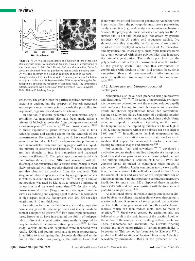
Figure 13. A) UV–Vis spectra recorded as a function of time of reaction
of lemongrass extract with aqueous Au ions; curves 1–5 correspond to
spectra recorded 1, 90, 160, 220, and 340 min after reaction. Curve 6:
spectrum obtained from the purified Au nanotriangle solution; inset:
UV–Vis–NIR spectrum of a solution-cast film of purified Au nano-
triangles obtained by reaction of AuCl4�–lemongrass extract solution
on a quartz substrate. B) Representative TEM image of triangular Au
nanoprisms obtained by reduction of aqueous AuCl4� by lemongrass
extract. Reprinted with permission from Reference [49]. Copyright
2004, Nature Publishing Group.
656
structures. The driving force for particle localization within the
bacteria is unclear, but the prospect of bacteria-generated
anisotropic nanostructures points towards the possibility for
large-scale, organism-based synthetic schemes.
In addition to bacteria-generated Ag nanoprisms, single-
crystalline Au nanoprisms also have been made using a
mixture of biological molecules from the aqueous extract of
lemongrass plants,[49] aloe vera,[105] and brown seaweed.[106]
In these experiments, plant extracts were used as both
reducing agents and capping agents for the synthesis of Au
nanostructures. For example, Sastry et al. have used lemon-
grass extract to reduce HAuCl4, and propose that preliminary
seed nanoparticles form and then aggregate within a liquid-
like mixture of aldehydes and ketones.[49] These aggregates
are then thought to fuse into nanoprisms and truncated
nanoprisms (Figure 13). The optical spectrum associated with
this mixture shows a broad NIR band associated with the
anisotropic nanostructures and a visible band, which is most
likely associated with the pseudospherical nanoparticles that
are also observed as products from this synthesis. This
assignment is based upon work done by our group and others
as well as calculations by Schatz et al.[75] Finally, a similar
methodology was used by Liu et al. to produce a mixture of
nanoprisms and truncated nanoprisms.[106] In this study,
brown seaweed extract (Sargassum sp.) was again found to
serve as a reducing and capping agent to direct the formation
of high aspect ratio Au nanoprisms with 200–800-nm-edge
lengths and 8–10-nm thickness.
In addition to these methodologies, several groups also
have investigated the use of proteins and nucleic acids to
control nanoparticle growth.[107] For anisotropic nanostruc-
tures, Brown et al. have investigated the ability of polypep-
tides to direct Au crystallization using methods inspired by
enzyme-mediated biomineralization processes.[107] In this
study, various amino acid sequences were incubated with
AuCl3, KOH, and sodium ascorbate at room temperature.
In addition to investigating the biomolecule-directed synth-
esis of other AuNP morphologies, the authors found that
www.small-journal.com � 2009 Wiley-VCH Verlag Gm
there were two critical factors for generating Au nanoprisms
in particular. First, the polypeptide must have a pre-existing
catalytic function (e.g., acid catalysis or a similar mechanism).
Second, the polypeptide must possess an affinity for the Au
surface that is not thiol-based (e.g., not driven by cysteine
residues). Of the 50 amino acid sequences studied, five
showed the ability to modify the formation of Au crystals,
of which three displayed increased rates of Au nucleation
and crystallization. Interestingly, anisotropic nanostructures
were only observed with those polypeptides that increased
the rate of crystallization. The authors postulate that the
polypeptides create a low pH environment near the surface
of the growing crystal, and that this environment in
combination with blocking of the {111} crystals faces, yields
nanoprisms. Shao et al. have reported a similar preparative
route to synthesize Au nanoprisms that relies on amino
acids.[108]
4.1.2. Microwave- and Ultrasound-AssistedTechniques
Nanoprisms also have been prepared using microwaves
and ultrasound.[77,92–94,109] In terms of nanoparticle synthesis,
microwaves are believed to heat the reaction solution rapidly
and uniformly leading to more homogeneous nucleation
events and shorter crystallization times than conventional
heating (e.g., by hot plate). Sonication of a colloidal solution
results in acoustic cavitation, during which time bubbles form,
grow, and implode in solution. Depending on the power,
the temperature of the imploding bubbles can be as high as
1 000 K and the pressure within the bubbles can be as high as
1 800 atm.[92–94] In addition to the high temperatures and
pressures created, cavitation can also create shockwaves in
solution that impact the nanoparticle surface, sometimes
leading to unusual shapes and structures.[92]
For example, Tsuji and coworkers[109,110] developed a
method to prepare single crystalline triangular Au nanoprisms
(and truncated nanoprisms) via a microwave polyol method.
The authors subjected a solution of HAuCl4, PVP, and
ethylene glycol to pulsed or continuous wave modes of
microwave irradiation. Under continuous microwave irradia-
tion, the temperature of the colloid increased to 196 8C over
the course of 1 min and was held at this temperature for an
additional minute. Samples exposed to continuous microwave
irradiation for more than 120 s displayed three extinction
bands (545, 590, and 645 nm) consistent with the formation of
plate-like nanoparticles.[44,48]
As mentioned above, ultrasonic energy can cause cavita-
tion bubbles to collapse, creating shockwaves throughout a
reaction solution. Researchers have proposed that cavitation
can lead to the decomposition of water or other molecules into
radicals, which can then reduce metal ions to metal in
solution.[92–94] Shockwaves created by cavitation also are
believed to result in the rapid impact of the reaction liquid on
the surface of the nanoparticles, resulting in their dissolution.
These phenomena can accelerate the Ostwald ripening
process and allow nanoparticles of various morphologies to
be generated. This method has been used by Zhu et al.[92] to
produce colloidal solutions of Ag nanoplates using AgNO3 in
N,N-dimethylformamide (DMF) in the presence of PVP
bH & Co. KGaA, Weinheim small 2009, 5, No. 6, 646–664

Colloidal Gold and Silver Triangular Nanoprisms
Figure 14. Time evolution of UV–Vis spectra during the formation of Ag
nanosprisms in DMF. Reprinted with permission from Reference [100].
Copyright 2002, American Chemical Society.
where DMF can be used as both solvent and reducing agent for
metal nanoparticle synthesis as follows
HCONMe2þ2AgþþH2O ! 2Ag0þMe2NCOOHþ 2Hþ (2)
For these methods, the molar ratio of PVP to AgNO3 was
a key factor in determining their final morphology, where
ratios between 0.1 and 0.3 were optimal for nanoprism
formation.
Similarly, Cai et al. have developed an ultrasonication
route to prepare Au nanoprisms in solution,[77] although the
overall yield of prism particles was low. In a typical
experiment, HAuCl4 and PVP are combined in ethylene
glycol under oxygen-free conditions and subjected to ultra-
sonication (frequency¼ 45� 2.5 kHz, power¼ 2.4 W cm�2)
for various periods of time. Here again, the ethylene glycol
is believed to serve a dual role as solvent and reducing
agent. Interestingly, the authors found that the formation of
Au nanoprisms is time-dependent. The final colloid is
composed primarily of 6–10-nm-thick nanoprisms and trun-
cated nanoprisms with 30–40-nm-edge lengths, as well as a
small number of spherical nanoparticles. Aging of the
nanoprism colloid for one week resulted in an overall increase
of the average edge length of the nanoprisms from 30–40 to
70–90 nm. This observation was corroborated by a significant
red-shift of the in-plane dipole SPR band from 690 to 760 nm.
The authors propose that adsorption of PVP to the {111}
crystal faces, in conjunction with the mild reaction conditions,
are the primary factors influencing nanoprism formation and
morphology.
4.2. Thermal Syntheses in Organic Media
There has also been significant progress in the develop-
ment of organic phase syntheses for triangular nanoprisms. In
contrast to the aqueous methods described previously (which
are typically conducted at room temperature or under
physiological conditions), many of the organic protocols
require elevated temperatures (e.g., reflux conditions). A
particularly interesting aspect of these synthetic approaches is
that often the solvent and/or surfactant acts as both a capping
ligand and reducing agent.
An early work in thermal organic synthesis of nanoprisms
was reported by Liz-Marzan et al. where Ag nanoprisms were
prepared by boiling DMF and reducing Agþ in the presence of
PVP.[100] The authors postulate that DMF acts as both the
solvent and reducing agent.[111] The authors found that if the
concentration of Ag ions was increased relative to the
concentration of PVP, particles with anisotropic shapes
(mainly nanoprisms) were observed. After purification by
centrifugation, the nanoprisms could be largely isolated from
the pseudospherical nanoparticles, and the optical signatures
of the Ag nanoprisms could be observed. The optical spectrum
is consistent with that observed for photochemically generated
nanostructures: the in-plane dipole resonance (�770 nm), the
in-plane and out-of-plane quadrupole resonances (�470 and
340 nm, respectively), and the weak out-of-plane dipole
resonance (�410 nm) with deviations explained by the
imperfect triangular shape of the prisms.
small 2009, 5, No. 6, 646–664 � 2009 Wiley-VCH Verlag Gmb
Interestingly, this work showed that the optical signatures
of nanoprisms were very sensitive to the refractive index of the
surrounding medium. When these Ag nanoprisms were
transferred from DMF to water, the in-plane dipole resonance
blue-shifted �40 nm and the out-of-plane quadrupole reso-
nance shifted �2 nm. This effect has also been observed by
others in the context of surface-immobilized metal nanos-
tructures.[69,112,113] The authors also demonstrated a degree of
size control based on the reflux time of the nanoparticles in
DMF, where longer reflux times led to larger nanoprism
structures (Figure 14). In a separate report, Ag nanoprisms
have been made in a similar fashion using formamide as both a
solvent and reducing agent in the presence of poly(ethylene
glycol) (PEG) at room temperature. In this report, the authors
found that in the presence of a 1:1 polymer mixture of PEG
and PVP, a mixture of nanoprisms and nanospheres could be
prepared.[76]
4.3. Summary of Thermal Syntheses of (or ChemicalReduction Methods for Producing) TriangularNanoprisms
It is clear that nanoprisms can be formed in a wide variety
of media under relatively mild reaction conditions, and that
these prisms exhibit common optical and crystallographic
features. However, synthetic challenges for thermal synthesis
remain. There are still very few methods for controlling
nanoprism thickness, and the driving forces behind the growth
of either triangular, hexagonal, or disk-like nanoprisms are
still not fully understood. What stands out among the many
thermal methods for preparing nanoprism structures is the
wide variety of chemical conditions used to achieve the same
nanoparticle architecture. While yield, size, and monodisper-
sity of nanoprisms vary from synthesis to synthesis, the
consistent observation of plate-like growth drives one to
consider the common themes and critical factors in these
sometimes disparate approaches. In the following section, an
overview of commonly proposed plate-like growth mechan-
isms are presented in order to provide current ideas about the
shape evolution of Au and Ag anisotropic nanoparticles.
H & Co. KGaA, Weinheim www.small-journal.com 657

reviews C. A. Mirkin et al.
658
5. Mechanisms of Plate-Like Growth
At first glance, there is little overlap between the
chemistries involved in each preparative route for nanoprisms.
Indeed, each synthetic scheme generates nanoprisms with
different compositions, yields, sizes, and size distributions.
However, upon closer inspection a central theme emerges
within most syntheses: mediated reduction of metal ions onto
nanoparticle seeds. Although the experimental details differ
(e.g., temperature, pH, surfactant/capping ligands, reducing
agents), each methodology involves two general steps:
i) nucleation of nanoparticle seeds and ii) crystal growth of
seeds by mediated reduction of metal ions. In the nucleation
stage, metal ions are reduced via thermal or photochemical
means to generate small metal nanoparticles. In a subsequent
growth step, these nanoparticle seeds are combined with metal
ions and reducing agents and exhibit additional crystal growth
until the final structure is obtained. Yet, such a general scheme
oversimplifies the complex issue of crystal nucleation and
growth. Typically, mechanisms for nanoprism formation can
be broken down into crystallographic and redox chemistry
arguments. While aspects of these theories overlap, here they
are treated as distinct components that control nanoprism
formation through a delicate interplay between the two.
5.1. Crystallographic Arguments
Crystallographic mechanisms can be described as mechan-
isms that use the crystal structure of the original seed particle,
crystal face-blocking mechanisms, and/or crystal facet surface
energetics to explain the preferential growth of a nanoprism
structure. For the seed nanoparticle, it has often been
postulated that the original structure of the seed dictates
the final morphology of the nanostructure by limiting the
number and variety of crystal facets available for
growth.[41,114,115] In the case of face-blocking mechanisms,
as discussed previously, these processes selectively block one
crystal face from metal ion reduction and thereby promote
growth of other facets. In the case of crystal facet surface
energetics, due to the coordination number and therefore
chemical reactivity of the surface atoms, certain crystal facets
exhibit higher surface energies and higher chemical reactiv-
ities than others (e.g., sAu(111) <sAu(110)<sAu(100)).[116,117] To
explore this topic, theoretical models and experimental results
Scheme 3. Ag halide model for a single twinned plane. Alternating sides contain A- and B-type
faces. The reentrant grooves of the A-type faces causes rapid growth that is arrested when the
face grows itself out, leaving a triangular prism with slow-growing B-type faces. Adapted from
Reference [67].
in plate-like Ag halide crystals (rock salt
structure, composed of two interpene-
trating fcc lattices) are discussed and
parallels can be drawn between the
formation of these structures and the
plate-like growth of Ag and Au (both fcc
metals) nanoprisms.
For the past two centuries, the
photosensitivity and photoreactivity of
plate-like Ag halide crystals (i.e., AgBr,
AgI) have been exploited in a variety of
photographic film and memory storage
applications.[118–122] In an effort to
improve the current technology, a variety
of studies in both the scientific and patent
www.small-journal.com � 2009 Wiley-VCH Verlag Gm
literature have focused on developing an understanding of the
crystallization processes occurring in the formation of plate-
like Ag halide (primarily AgBr) crystals. From these reports, it
is generally believed that plate-like crystal growth can only
occur when the initial nanoparticle seeds contain one or more
parallel twin planes.[123–126] Although AgBr nanoparticle
seeds are often described as spherical structures, on the atomic
scale they are bound by the {111} and {100} faces, which are the
most stable AgBr faces in the absence of capping ligands.
During the initial stages of nucleation, the AgBr seeds
undergo a process called twinning, in which stacking faults are
formed within the crystal matrix. Due to their atomic
symmetry, coalescence between two {100} faces will not
generate the low-energy stacking faults (twins) required for
plate-like crystal growth. In contrast, coalescence between two
{111} facets (oriented at a 608 rotation relative to one another)
yields stacking faults, which can lead to crystal growth normal
to the {111} crystal planes.[67]
Crystal twinning that leads to plate-like structures was
proposed by Berriman and Herz to account for the plate-like
morphology of Ag bromide crystals.[127] Hamilton and
Seidensticker later supported this hypothesis experimentally
in their report that plate-like germanium crystals possess two
or more twin planes parallel to their major {111} facets.[128]
Twinned seeds are believed to be formed from coalescence
events between two unstable {111} crystal faces.[122,129] This
was demonstrated experimentally by Antoniades and Wey,
who showed that the rate of addition of Ag precursor
(AgNO3) as well as the concentration of the reducing agent
(gelatin) control the coalescence events that lead to twinned
AgBr seeds (which ultimately lead to plate-like AgBr
crystals).[119] Hence, although coalescence is responsible for
the formation of twins, several papers have found that other
experimental factors including capping ligand and reducing
agent (gelatin in both cases), concentration, pH, and
temperature are all key parameters in controlling the degree
of crystal twinning in solution.[123]
Twinned crystal seeds are believed to set the stage for
plate-like morphologies by providing low-energy reentrant
grooves favoring lateral crystal growth (Scheme 3). Jagan-
nathan et al. demonstrate experimentally and theoretically
that plate-like crystal growth is propagated by the formation of
two twin planes parallel to their major {111} crystal faces.[124]
This atomic arrangement initially results in {111} faceted
bH & Co. KGaA, Weinheim small 2009, 5, No. 6, 646–664

Colloidal Gold and Silver Triangular Nanoprisms
Figure 15. A) TEM image of the Ag nanodisk taken in side view, showing
the contrast from (111) stacking faults and a preferential growth along
the stacking faults. B) A typical selected area electron diffraction (SAED)
pattern of a Ag nanodisk at 100 kV in the [011] orientation (side view).
Reprinted with permission from Reference [80]. Copyright 2003,
American Chemical Society.
reentrant grooves on the sides of the crystal plates, providing
nucleation sites for adsorption of new crystal layers and
driving plate-like crystal growth. The authors calculated that
the probability of adsorption at the reentrant groove is
�50 times greater than adsorption at a (non-twinned) surface
site. Preferential crystallization at reentrant grooves can also
be rationalized using a nearest neighbor argument: an isolated
atom can form four nearest neighbor bonds in a reentrant
groove and only three on a {111} face. The increased bonding
strength and coordination number thus results in preferential
adsorption at reentrant grooves over the {111} faces. Ming
et al. and Sunagawa et al. observed similar crystallization
events using Monte Carlo simulations.[126,130] Interestingly,
the reentrant grooves are regenerated as new layers of atoms
deposit on them, making them permanent preferential regions
of lateral growth. Growth continues until the adsorption units
(AgBr32� and other species) are exhausted and yields the final
nanoprisms where the major faces are bound by the {111}
crystal planes.
Anisotropic crystal growth of Au or Ag derived from
twinned crystal seeds was most recently addressed in a
comprehensive article written by Lofton and Sigmund, who
extended these crystallization arguments to plate- and needle-
like nanostructures composed of Ag and Au.[67] Theoretical
and experimental results have shown that Ag halide crystals
(NaCl structure) and metals (fcc structure) are bound by the
{111} crystal faces.[131,132] In their paper, the authors argue that
the crystal structure of the seed particle ultimately dictates the
final morphology of the crystal. This is seemingly in contrast to
many papers published by other groups that argue that
preferential adsorption of capping ligands or surfactants
directs the formation of rod- or plate-like growth. Lofton and
Sigmund point out that such surface passivation (or ‘‘crystal-
face poisoning’’) models are unlikely given that identical
nanoparticle shapes can be attained via drastically different
methods and chemical environments. Indeed, the various
methodologies highlighted in this discussion support their
conclusions. However, recent work with halide ions presents
an interesting counterpoint (see Section 5.2).[133–135]
Although there are no clear answers about the parameters
that control the degree and arrangement of stacking faults in
crystal seeds, the role of crystal twinning in directing the final
architecture of nanostructures may be a crucial element. In
most of the examples for preparing Ag and Au nanoprism
crystals cited in this review, the initial seed nanoparticles are
prepared by chemical reduction of metal precursors (e.g.,
AgNO3 or HAuCl4) in the presence of one or more capping
molecules. Generally, fast reduction of the metal ions (e.g.,
accomplished by rapid addition of strong reducing agents to
metal salts) results in small, pseudospherical nanoparti-
cles.[136–139] The surfaces of the nanoparticles typically exhibit
a mixture of {111} and {100} planes. To minimize their overall
energy, nanoparticle seeds will undergo twinning to form a
twinned icosahedron or decahedron. Interestingly, the shape
of the small nanoparticles (<5 nm) can fluctuate, and studying
their morphology and crystal structure can be difficult. The
chemical environment can also cause morphological and
crystal structure changes in the nanoparticles. For example,
Xia et al. recently reported that addition of Fe3þ or O2/Cl� to a
small 2009, 5, No. 6, 646–664 � 2009 Wiley-VCH Verlag Gmb
AgNP colloid comprised of twinned crystals results in rapid
etching of the crystals.[114] After 24 h, a second nucleation
stage occurred to yield single-crystalline AgNP seeds.
The sensitivity of small nanoparticles to experimental and
environmental conditions makes their characterization via
electron microscopy or optical techniques difficult. In spite of
these limitations, some HRTEM studies have been performed
on nanoparticle seeds, but have not yet been able to
distinguish seed crystal structure as the driving force of
anisotropic crystal growth. HRTEM data from Pileni et al.
suggest that stacking faults parallel to the {111} crystal planes
are responsible for plate-like growth of Ag nanostructures
(nanodisks) (Figure 15).[80] Indeed, crystal twin planes parallel
to the major {111} faces are frequently observed in the final
disks (in both the TEM and diffraction images), as well as
during the early stages of nanodisk growth. The authors claim
that varying the degree of crystal twinning is critical to
controlling the final morphology of the nanostructures,
although no detail is given as to possible experimental
methods to do so. Similarly, Murphy et al. have reported that
AuNP seeds containing fivefold twinning exhibited growth to
form Au nanorods whereas single crystalline seeds did not.[140]
More recently, Xia and coworkers have demonstrated that
the crystal structure of AgNPs prepared by a polyol method
can be modified based on the nature of the salt added to the
reaction mixture.[53,115] In 2005, the authors reported that
addition of NaCl or HCl to a solution of AgNO3, PVP, and
H & Co. KGaA, Weinheim www.small-journal.com 659

reviews C. A. Mirkin et al.
Scheme 4. Illustration of ‘‘surfactant-templating’’ or ‘‘face-blocking’’
growth theories. Here, the circle attached to the black curve represents
an ampiphilic surfactant (e.g., CTAB) that forms a bilayer on the
nanocrystal surface and blocks metal ion reduction at that site.
660
ethylene glycol yield single crystalline nanoparticle seeds upon
30 min of refluxing (160 8C).[53] Additional refluxing of the
mixture yielded single crystalline Ag nanocubes. In their more
recent report, the authors found that replacing NaCl with
NaBr resulted in the formation of AgNP seeds (containing a
single twin plane after 1.5 h of reflux). After 5 h of refluxing,
these twinned seeds (diameter �15 nm) yielded Ag right
bipyramids (edge length �150 nm). Interestingly, the authors
note that the seed nanoparticles contain a single twin plane
that may allow growth of the right bipyramids.
Liu and Guyot-Sionnest have reported a comprehensive
study of the effect of seed crystal structure on the final
nanostructure morphology.[41] In their report, the authors
prepared AuNP seeds via two methods and examined the
crystal structure of each type. Seeds I were prepared via the
synthesis outlined by Nikoobakht and El-Sayed,[40] whereas
seeds II were synthesized via that described by Murphy
et al.[38] HRTEM analysis revealed that the two preparative
routes yielded seeds with very different crystal structures.
Seeds I were single crystalline (diameter �1.5 nm) whereas
seeds II were twinned with pentagonal symmetry. Each batch
of seeds were subsequently exposed to a Ag(I)-assisted growth
solution (composed of HAuCl4, AgNO3, CTAB, HCl, and
L-ascorbic acid) and their final morphology evaluated.
Interestingly, Au nanostructures derived from seeds I under-
went 1D growth to form Au nanorods with various aspect
ratios. In contrast, seeds II displayed growth to form
bipyramidal structures in high yield. The authors conclude
that the seed structure is, in fact, governing the shape and
crystal structure of the final nanostructures.
Although these reports demonstrate that stacking faults
are present in the final structure, they do not reveal what types
of crystal twins lead to plate-like (or other anisotropic)
morphologies. Indeed, unless anisotropic growth of the initial
seed particles is observed in situ, it is difficult to predict what
nanoparticle morphology will lead to plate-like crystal growth.
In addition, a detailed analysis of the experimental parameters
that control crystal twinning (e.g., number of twins and
orientation) has yet to be reported. Such studies are critical to
a better understanding of plate-like crystal growth.
5.2. Chemical Methods and Redox ChemistryArguments
Despite an argument that the crystal structure of the
nanoparticle seed is a determining factor in plate-like growth,
Table 1. Shapes of Au particles and corresponding reaction conditions. ReChemical Society.
[CTAB] [M] [Au]seed [M] [Au3þ] [M] [AA] [
1.6�10�2 1.25�10�8 2.0� 10�4 6.0� 1
1.6�10�2 1.25�10�8 2.0� 10�4 3.0� 1
1.6�10�2 1.25�10�7 2.0� 10�4 6.0� 1
1.6�10�2 1.25�10�8 4.0� 10�4 6.4� 1
9.5�10�2 1.25�10�7 4.0� 10�4 6.0� 1
1.6�10�2 1.25�10�8 4.0� 10�4 1.2� 1
5.0�10�2 6.25�10�7 5.0� 10�4 3.0� 1
9.5�10�2 2.5�10�7 4.0� 10�4 6.4� 1
[a] 6.0� 10�5M AgNO3 was also used in this synthesis.
www.small-journal.com � 2009 Wiley-VCH Verlag Gm
it is also true that the same seed particles can, in some cases,
yield various morphologies depending on reaction conditions
such as surfactant concentration, metal ion concentration,
reducing agent concentration, and metal or halide ion
additives.[40,42,54,96,133,141–143] That different nanoparticle
morphologies can be obtained from the same nanoparticle
seed indicates that the reduction method and chemical
environment for crystal growth can also be critical factors
in determining the final shape and size of a nanoparticle. For
example, Scheme 4 illustrates ‘‘face-blocking’’ that bridges
crystallographic and redox chemistry theories. In this
mechanism, surfactant selectively adsorbs to the most
favorable crystal facet where ‘‘favorable’’ is determined by
either surface reactivity (as dictated by crystal facet) or surface
charge (as dictated by capping ligand).[26,143] It is thought that
once bound, these capping molecules significantly or com-
pletely block reduction of metal ions onto the surface of the
growing nanocrystal. This explanation is common to the vast
majority of proposed mechanisms for the role of a particular
capping ligand.[43,62,65] One representative study is the work of
Sau and Murphy, which showed that the morphology and
dimension of AuNPs produced in a particular aqueous thermal
methodology depended strongly on the concentrations of the
seed particles and CTAB, in addition to the concentration of
Au ions (Au3þ) and reducing agent (ascorbic acid).[54] All of
the above factors were found to be interdependent, and gave
rise to a variety of shapes depending on combination (Table 1).
For example, high surfactant concentrations produced
rectangular nanorod structures, whereas lower surfactant
concentrations of surfactant mixture produced pentagonally
printed with permission from Reference [54]. Copyright 2004, American
M] Shape/profile Dimension [nm] % Yield
0�3 Cube 66 �85
0�3 Hexagon 70 �80
0�3 Triangle 35 �80
0�4 Cube [a] 90 �70
0�3 Tetrapod [a] 30 �70
0�2 Star 66 �50
0�3 Tetrapod 293 �75
0�4 Branched [a] 174 �95
bH & Co. KGaA, Weinheim small 2009, 5, No. 6, 646–664
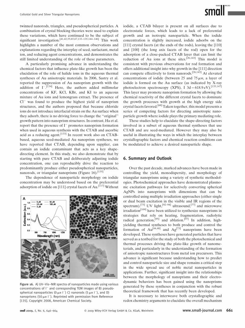
Colloidal Gold and Silver Triangular Nanoprisms
twinned nanorods, triangles, and pseudospherical particles. A
combination of crystal blocking theories were used to explain
these variations, which have continued to be the subject of
significant investigation.[41,42,55,65,67,133–135,144–146] This work
highlights a number of the most common observations and
explanations regarding the interplay of seed, surfactant, metal
ion, and reducing agent concentrations, and demonstrates the
still limited understanding of the role of these parameters.
A particularly promising advance in understanding the
chemical factors that influence plate-like growth has been the
elucidation of the role of halide ions in the aqueous thermal
syntheses of Au anisotropic materials. In 2006, Sastry et al.
reported the suppression of Au nanoprism growth with the
addition of I�.[134] Here, the authors added millimolar
concentrations of KF, KCl, KBr, and KI to an aqueous
mixture of Au ions and lemongrass extract. The presence of
Cl� was found to produce the highest yield of nanoprism
structures, and the authors proposed that because chloride
ions do not introduce interfacial strain on the Au surface when
they adsorb, there is no driving force to change the ‘‘original’’
growth pattern into nanoprism structures. In contrast, Ha et al.
report that the presence of I� promotes nanoprism formation
when used in aqueous synthesis with the CTAB and ascorbic
acid as a reducing agent.[133] In recent work also on CTAB-
based, aqueous seed-mediated Au nanoprism syntheses, we
have reported that CTAB, depending upon supplier, can
contain an iodide contaminant that acts as a key shape-
directing element. In this study, we also demonstrate that by
starting with pure CTAB and deliberately adjusting iodide
concentration, one can reproducibly drive the reaction to
predominantly produce either pseudospherical nanoparticles,
nanorods, or triangular nanoprisms (Figure 16).[135]
The dependence of nanoparticle morphology on iodide
concentration may be understood based on the preferential
adsorption of iodide on {111} crystal facets of Au.[117] Without
Figure 16. A) UV–Vis–NIR spectra of nanoparticles made using various
concentrations of I� and corresponding TEM images of B) pseudo-
spherical nanoparticles (0mM I�) C) nanorods (5mM I�), and D)
nanoprisms (50mM I�). Reprinted with permission from Reference
[135]. Copyright 2008, American Chemical Society.
small 2009, 5, No. 6, 646–664 � 2009 Wiley-VCH Verlag Gmb
iodide, a CTAB bilayer is present on all surfaces due to
electrostatic forces, which leads to a lack of preferential
growth and an isotropic nanoparticle. When the iodide
concentration is slightly increased, iodide adsorbs on the
{111} crystal facets (at the ends of the rods), leaving the {110}
and {100} (the long axis facets of the rod) open for the
adsorption of a close-packed CTAB layer that can limit the
reduction of Au ions at these sites.[26,143] This model is
consistent with previous observations for rod formation and
offers additional insight into why growth in the [111] direction
can compete effectively to form nanorods.[26,143] At elevated
concentrations of iodide (between 25 and 75mM, a layer of
iodide is formed on the Au surface (as indicated by X-ray
photoelectron spectroscopy (XPS), I 3d¼ 618.9 eV).[135,147]
This layer may promote nanoprism formation by allowing the
chemical reactivity of the different crystal facets to dominate
the growth processes with growth at the high energy side
crystal facets favored.[68] Taken together, this model presents a
series of competing factors for directing anisotropic nano-
particle growth where iodide plays the primary mediating role.
These studies help to elucidate the shape-directing factors
involved in a subset of aqueous thermal syntheses that use
CTAB and are seed-mediated. However they may also be
useful in illustrating the ways in which the interplay between
crystallographic factors and chemical reaction conditions can
be modulated to achieve a desired nanoparticle shape.
6. Summary and Outlook
Over the past decade, marked advances have been made in
controlling the yield, monodispersity, and morphology of
triangular nanoprisms using a variety of synthetic methodol-
ogies. Photochemical approaches have demonstrated plasmo-
nic excitation pathways for selectively converting spherical
AgNPs into nanoprisms with dimensions that can be
controlled using multiple irradiation approaches (either single
or dual beam excitation in the visible and IR regions of the
spectrum).[73] UV light,[92–94] ultrasound,[77] and microwave
irradiation[109] have been utilized to synthesize nanoprisms in
strategies that rely on heating, fragmentation, radiolytic
radical generation,[95] and ablation.[97] In addition, high-
yielding thermal syntheses to both produce and control the
formation of Au[44,48] and Ag[73] nanoprisms have been
developed. These syntheses have generated particles that have
served as a testbed for the study of both the photochemical and
thermal processes driving the plate-like growth of nanome-
terials, and particularly in the understanding of the formation
of anisotropic nanostructures from metal ion precursors. This
advance is significant because understanding how to predict
and control nanoparticle size and shape remains a critical step
in the wide spread use of noble metal nanoparticles in
applications. Further, significant insight into the relationships
between the morphology of nanoprisms and their electro-
dynamic behaviors has been gained using the nanoprisms
generated by these syntheses in conjunction with the robust
theoretical framework that has recently been developed.
It is necessary to interweave both crystallographic and
redox chemistry arguments to elucidate the overall mechanism
H & Co. KGaA, Weinheim www.small-journal.com 661

reviews C. A. Mirkin et al.
662
of nanoprism formation. Crystallographic arguments stem
from the observation that the faces of the nanoprism twinned
planes differ in their crystallographic assignment[68] ({111},
{100}, etc.) and that the seeds from which they are formed are
multifaceted and likely twinned.[67,136–139] It has therefore been
hypothesized that seed particle morphology determines the
final structure of the anisotropic particle. Researchers have
shown that by using seed particles with a particular morphology
(e.g., multifaceted or twinned), the final architecture of
anisotropic nanoparticles can be controlled in some synthetic
approaches.[41] On the other hand, one also can tune the
morphology of the resulting anisotropic structures by modulat-
ing the chemical and redox environment of the initial seed
particle in situ. A change in seed concentration,[63,90] surfac-
tant,[54] pH,[71] temperature,[88] or metal ion[70] (and even
dopant concentrations[135]) canhave drasticeffects onthe shape
and size of the anisotropic product. These combined observa-
tions suggest that through further investigation of the mechan-
istic driving forces of plate-like growth it may soon become
possible to direct nanocrystal growth simply by characterizing
the crystal structure of a seed particle and regulating the
reduction of metal ions onto the surface of that seed using
well-understood face-blocking strategies. These mechanistic
principles could serve as a set of design rules for the synthesis
of novel anisotropic nanostructures with desired architecture
and properties, but also could allow for unique nanostruc-
tures beyond nanoprisms, rods, and cubes to be generated
and examined.
Work is still required to fully understand and completely
control anisotropic growth and to realize the potential of
anisotropic structures in novel applications. The discovery and
subsequent research efforts made concerning triangular
nanoprisms have vastly improved the fundamental under-
standing of the underlying dynamics of formation for
anisotropic particles of all structures, and triangular nanopr-
isms may soon be the first such materials to be implemented in
interesting and vital applications such as optics, electronics,
catalysis, and biomedicine. However, the underlying work
already completed and the continued, substantial effort to
probe the fundamental aspects of the synthesis and properties
of nanoprisms will be essential to realizing these applications
and paving the way for the use of other similar structures in
nanoscience and technology.
Acknowledgements
C. A. M. acknowledges the ONR, DARPA, AFRL, and NSF-MRSEC
for their support of this work. J. E. M. is grateful to
Northwestern University for a Presidential Fellowship.
Keywords:anisotropic materials . crystal growth . gold . nanoprisms .silver
[1] A. J. Haes, L. Chang, W. L. Klein, R. P. Van Duyne, J. Am. Chem. Soc.
2005, 127, 2264.
[2] Y. C. Cao, R. Jin, C. A. Mirkin, Science 2002, 297, 1536.
www.small-journal.com � 2009 Wiley-VCH Verlag Gm
[3] D. G. Georganopoulou, L. Chang, J. M. Nam, C. S. Thaxton, E. J.
Mufson, W. L. Klein, C. A. Mirkin, Proc. Natl. Acad. Sci. USA 2005,
102, 2273.
[4] C. S. Thaxton, D. G. Georganopoulou, C. A. Mirkin, Clin. Chim. Acta
2006, 363, 120.
[5] H. D. Hill, C. A. Mirkin, Nat. Protoc. 2006, 1, 324.
[6] D. S. Seferos, D. A. Giljohann, H. D. Hill, A. E. Prigodich, C. A.
Mirkin, J. Am. Chem. Soc. 2007, 129, 15477.
[7] H. D. Hill, R. A. Vega, C. A. Mirkin, Anal. Chem. 2007, 79, 9218.
[8] R. Elghanian, J. J. Storhoff, R. C. Mucic, R. L. Letsinger, C. A. Mirkin,
Science 1997, 277, 1078.
[9] T. A. Taton, C. A. Mirkin, R. L. Letsinger, Science 2000, 289, 1757.
[10] S. J. Park, T. A. Taton, C. A. Mirkin, Science 2002, 295, 1503.
[11] J. M. Nam, C. S. Thaxton, C. A. Mirkin, Science 2003, 301, 1884.
[12] Y. Shiraishi, N. Toshima, J. Mol. Catal. 1999, 141, 187.
[13] G. A. Somorjai, F. Tao, J. Y. Park, Top. Catal. 2008, 47, 1.
[14] D. Astruc, F. Lu, J. R. Aranzaes, Angew. Chem. Int. Ed. 2005, 44,
7852.
[15] G. Schmid, B. Corain, Eur. J. Inorg. Chem. 2003, 2003. 3081.
[16] J. Perez-Juste, A. Mulvaney, L. M. Liz-Marzan, Int. J. Nanotechnol.
2007, 4, 215.
[17] S. P. Liao, N. C. Seeman, Science 2004, 306, 2072.
[18] L. Qin, M. J. Banholzer, J. E. Millstone, C. A. Mirkin, Nano Lett.
2007, 7, 3849.
[19] N. L. Rosi, D. A. Giljohann, C. S. Thaxton, A. K. R. Lytton-Jean, M. S.
Han, C. A. Mirkin, Science 2006, 312, 1027.
[20] J. D. Hood, M. Bednarski, R. Frausto, S. Guccione, R. A. Reisfeld, R.
Xiang, D. A. Cheresh, Science 2002, 296, 2404.
[21] L. Brannon-Peppas, J. O. Blanchette, Adv. Drug Delivery Rev.
2004, 56, 1649.
[22] G. Frens, Nat. Phys. Sci. 1973, 241, 20.
[23] M. Brust, M. Walker, D. Bethell, D. J. Schiffrin, R. Whyman, Chem.
Commun. 1994, 801.
[24] I. Hussain, S. Graham, Z. X. Wang, B. Tan, D. C. Sherrington, S. P.
Rannard, A. I. Cooper, M. Brust, J. Am. Chem. Soc. 2005, 127,
16398.
[25] K. R. Brown, D. G. Walter, M. J. Natan, Chem. Mater. 2000, 12,
306.
[26] Z. L. Wang, R. P. Gao, B. Nikoobakht, M. A. El-Sayed, J. Phys.
Chem. B 2000, 104, 5417.
[27] P. D. Jadzinsky, G. Calero, C. J. Ackerson, D. A. Bushnell, R. D.
Kornberg, Science 2007, 318, 430.
[28] U. Kreibig, L. Genzel, Surf. Sci. 1985, 156, 678.
[29] A. Henglein, Isr. J. Chem. 1993, 33, 77.
[30] S. Link, M. B. Mohamed, M. A. El-Sayed, J. Phys. Chem. B 1999,
103, 3073.
[31] K. L. Kelly, E. Coronado, L. L. Zhao, G. C. Schatz, J. Phys. Chem. B
2003, 107, 668.
[32] R. Narayanan, M. A. El-Sayed, J. Phys. Chem. 2005, 109, 12663.
[33] A. N. Shipway, E. Katz, I. Willner, ChemPhysChem. 2000, 1, 18.
[34] M. S. Gudiksen, L. J. Lauhon, J. Wang, D. C. Smith, C. M. Lieber,
Nature 2002, 415, 617.
[35] P. K. Jain, K. S. Lee, I. H. El-Sayed, M. A. El-Sayed, J. Phys. Chem. B
2006, 110, 7238.
[36] M. Haruta, Cattech 2002, 6, 102.
[37] J. R. Lakowicz, Anal. Biochem. 2005, 337, 171.
[38] N. R. Jana, L. Gearheart, C. J. Murphy, J. Phys. Chem. B 2001, 105,
4065.
[39] K. K. Caswell, C. M. Bender, C. J. Murphy, Nano Lett. 2003, 3, 667.
[40] B. Nikoobakht, M. A. El-Sayed, Chem. Mater. 2003, 15, 1957.
[41] M. Liu, P. Guyot-Sionnest, J. Phys. Chem. B 2005, 109, 22192.
[42] J. Perez-Juste, I. Pastoriza-Santos, L. M. Liz-Marzan, P. Mulvaney,
Coord. Chem. Rev. 2005, 249, 1870.
[43] C. J. Murphy, A. M. Gole, S. E. Hunyadi, C. J. Orendorff, Inorg.
Chem. 2006, 45, 7544.
[44] C. S. Ah, Y. J. Yun, H. J. Park, W.-J. Kim, D. H. Ha, W. S. Yun, Chem.
Mater. 2005, 17, 5558.
bH & Co. KGaA, Weinheim small 2009, 5, No. 6, 646–664

Colloidal Gold and Silver Triangular Nanoprisms
[45] S. Chen, D. L. Carroll, Nano Lett. 2002, 2, 1003.
[46] S. Chen, D. L. Carroll, J. Phys. Chem. B 2004, 108, 5500.
[47] R. Jin, Y. Cao, C. A. Mirkin, K. L. Kelly, G. C. Schatz, J. G. Zheng,
Science 2001, 294, 1901.
[48] J. E. Millstone, S. Park, K. L. Shuford, L. Qin, G. C. Schatz, C. A.
Mirkin, J. Am. Chem. Soc. 2005, 127, 5312.
[49] S. S. Shankar, A. Rai, B. Ankamwar, A. Singh, A. Ahmad, M.
Sastry, Nat. Mater. 2004, 3, 482.
[50] R. C. Jin, S. Egusa, N. F. Scherer, J. Am. Chem. Soc. 2004, 126,
9900.
[51] B. Wiley, T. Herricks, Y. Sun, Y. Xia, Nano Lett. 2004, 4, 1733.
[52] D. Yu, V. W.-W. Yam, J. Am. Chem. Soc. 2004, 126, 13200.
[53] S. H. Im, Y. T. Lee, B. Wiley, Y. Xia, Angew. Chem. Int. Ed. 2005, 44,
2154.
[54] T. K. Sau, C. J. Murphy, J. Am. Chem. Soc. 2004, 126, 8648.
[55] C. G. Wang, T. T. Wang, Z. F. Ma, Z. M. Su, Nanotechnology 2005,
16, 2555.
[56] T. Cassagneau, F. Caruso, Adv. Mater. 2002, 14, 732.
[57] G. S. Metraux, Y. C. Cao, R. Jin, C. A. Mirkin, Nano Lett. 2003, 3,
519.
[58] F. Kim, J. H. Song, P. Yang, J. Am. Chem. Soc. 2002, 124, 14316.
[59] C. R. Martin, Science 1994, 266, 1961.
[60] C. K. Preston, M. Moskovits, J. Phys. Chem. 1993, 97, 8495.
[61] S. J. Hurst, E. K. Payne, L. Qin, C. A. Mirkin, Angew. Chem. Int. Ed.
2006, 45, 2672.
[62] A. R. Tao, S. Habas, P. D. Yang, Small 2008, 4, 310.
[63] B. Wiley, Y. Sun, J. Chen, H. Cang, Z.-Y. Li, X. Li, Y. Xia, MRS Bull.
2005, 30, 356.
[64] J. Turkevich, P. C. Stevenson, J. Hillier, Faraday Discuss. 1951, 11,
55.
[65] C. Burda, X. Chen, R. Narayanan, M. A. El-Sayed, Chem. Rev. 2005,
105, 1025.
[66] L. M. Liz-Marzan, Mater. Today 2004, 7, 26.
[67] C. Lofton, W. Sigmund, Adv. Funct. Mater. 2005, 15, 1197.
[68] J. E. Millstone, G. S. Metraux, C. A. Mirkin, Adv. Funct. Mater.
2006, 16, 1209.
[69] C. Xue, Z. Li, C. A. Mirkin, Small 2005, 1, 513.
[70] G. S. Metraux, C. A. Mirkin, Adv. Mater. 2005, 17, 412.
[71] C. Xue, C. A. Mirkin, Angew. Chem. Int. Ed. 2007, 46, 2036.
[72] C. Xue, J. E. Millstone, S. Y. Li, C. A. Mirkin, Angew. Chem. Int. Ed.
2007, 46, 8436.
[73] R. Jin, Y. C. Cao, E. Hao, G. S. Metraux, G. C. Schatz, C. A. Mirkin,
Nature 2003, 425, 487.
[74] Y. L. Luo, Mater. Lett. 2007, 61, 1346.
[75] K. L. Shuford, M. A. Ratner, G. C. Schatz, J. Chem. Phys. 2005, 123,
114713.
[76] A. Sarkar, S. Kapoor, T. Mukherjee, J. Colloid Interface Sci. 2005,
287, 496.
[77] C. Li, W. Cai, Y. Li, J. Hu, P. Liu, J. Phys. Chem. B 2006, 110,
1546.
[78] Y. A. Sun, Y. N. Xia, Adv. Mater. 2003, 15, 695.
[79] D. H. Dahanayaka, J. X. Wang, S. Hossain, L. A. Bumm, J. Am.
Chem. Soc. 2006, 128, 6052.
[80] V. Germain, J. Li, D. Ingert, Z. L. Wang, M. P. Pileni, J. Phys. Chem. B
2003, 107, 8717.
[81] P. Mulvaney, Langmuir 1996, 12, 788.
[82] J. Rodriguez-Fernandez, J. Perez-Juste, F. J. Garcia de Abajo, L. M.
Liz-Marzan, Langmuir 2006, 22, 7007.
[83] E. K. Payne, K. L. Shuford, S. Park, G. C. Schatz, C. A. Mirkin, J.
Phys. Chem. B 2006, 110, 2150.
[84] M. Maillard, P. Huang, L. Brus, Nano Lett. 2003, 3, 1611.
[85] A. Callegari, D. Tonti, M. Chergui, Nano Lett. 2003, 3, 1565.
[86] V. Bastys, I. Pastoriza-Santos, B. Rodrıguez-Gonzalez, R.
Vaisnoras, L. M. Liz-Marzan, Adv. Funct. Mater. 2006, 16, 766.
[87] A. M. Junior, H. P. Moises de Oliveira, M. H. Gehlen, Photochem.
Photobiol. Sci. 2003, 2, 921.
[88] Y. Sun, B. Mayers, Y. Xia, Nano Lett. 2003, 3, 675.
small 2009, 5, No. 6, 646–664 � 2009 Wiley-VCH Verlag Gmb
[89] C. Xue, G. S. Metraux, J. E. Millstone, C. A. Mirkin, J. Am. Chem.
Soc. 2008, 130, 8337.
[90] X. Wu, P. L. Redmond, H. Liu, Y. Chen, M. Steigerwald, L. Brus, J.
Am. Chem. Soc. 2008, 130, 9500.
[91] P. L. Redmond, X. Wu, L. Brus, J. Phys. Chem. C 2007, 111, 8942.
[92] L.-P. Jiang, S. Xu, J.-M. Zhu, J.-R. Zhang, J.-J. Zhu, H.-Y. Chen, Inorg.
Chem. 2004, 43, 5877.
[93] K. Okitsu, A. Yue, S. Tanabe, H. Matsumoto, Y. Yobiko, Langmuir
2001, 17, 7717.
[94] K. Okitsu, A. Yue, S. Tanabe, H. Matsumoto, Y. Yobiko, Langmuir
2007, 23, 13244.
[95] Y. Takeuchi, T. Ida, K. Kimura, J. Phys. Chem. B 1997, 101, 1322.
[96] Z. Xie, Z. Wang, Y. Ke, Z. Zha, C. Jiang, Chem. Lett. 2003, 32, 686.
[97] T. Tsuji, T. Higuchi, M. Tsuji, Chem. Lett. 2005, 34, 476.
[98] S. Remita, M. Mostafavi, M. O. Delcourt, New J. Chem. 1994, 18,
581.
[99] J. Belloni, M. Mostafavi, H. Remita, J.-L. Marignier, M.-O. Delcourt,
New J. Chem. 1998, 22, 1239.
[100] I. Pastoriza-Santos, L. M. Liz-Marzan, Nano Lett. 2002, 2, 903.
[101] N. Malikova, I. Pastoriza-Santos, M. Schierhorn, N. A. Kotov, L. M.
Liz-Marzan, Langmuir 2002, 18, 3694.
[102] T.J. Norman, Jr., C. D. Grant, D. Magana, J. Z. Zhang, J. Liu, D. Cao,
F. Bridges, A. Van Buuren, J. Phys. Chem. B 2002, 106, 7005.
[103] T. Klaus, R. Joerger, E. Olsson, C. G. Granqvist, Proc. Natl. Acad.
Sci. USA 1999, 96, 13611.
[104] S. Chen, Z. Fan, D. L. Carroll, J. Phys. Chem. B 2002, 106, 10777.
[105] S. P. Chandran, M. Chaudhary, R. Pasricha, A. Ahmad, M. Sastry,
Biotechnol. Prog. 2006, 22, 577.
[106] B. Liu, J. Xie, J. Y. Lee, Y. P. Ting, J. P. Chen, J. Phys. Chem. B 2005,
109, 15256.
[107] S. Brown, M. Sarikaya, E. Johnson, J. Mol. Biol. 2000, 299, 725.
[108] Y. Shao, Y. Jin, S. Dong, Chem. Commun. 2004, 1104.
[109] M. Tsuji, M. Hashimoto, Y. Nishizawa, T. Tsuji, Chem. Lett. 2003,
32, 1114.
[110] M. Tsuji, M. Hashimoto, Y. Nishizawa, M. Kubokawa, T. Tsuji,
Chem. Eur. J. 2005, 11, 440.
[111] I. Pastoriza-Santos, L. M. Liz-Marzan, Langmuir 2002, 18, 2888.
[112] T. R. Jensen, M. L. Duval, K. L. Kelly, A. A. Lazarides, G. C. Schatz,
R. P. Van Duyne, J. Phys. Chem. B 1999, 103, 9846.
[113] A. J. Haes, R. P. Van Duyne, J. Am. Chem. Soc. 2002, 124, 10596.
[114] B. Wiley, Y. Sun, Y. Xia, Langmuir 2005, 21, 8077.
[115] B. J. Wiley, Y. Xiong, Z.-Y. Li, Y. Yin, Y. Xia, Nano Lett. 2006, 6, 765.
[116] J. C. Love, L. A. Estroff, J. K. Kriebel, R. G. Nuzzo, G. M. Whitesides,
Chem. Rev. 2005, 105, 1103.
[117] O. M. Magnussen, Chem. Rev. 2002, 102, 679.
[118] S. Chen, S. Jagannathan, R. V. Mehta, R. Jagannathan, A. E.
Taddei, J. Imaging Sci. Technol. 1998, 42, 399.
[119] M. G. Antoniades, J. S. Wey, J. Imaging Sci. Technol. 1995, 39,
323.
[120] M. G. Antoniades, J. S. Wey, J. Imaging Sci. 1998, 42, 393.
[121] J. E. Maskasky, J. Imaging Sci. 1986, 30, 247.
[122] C. T. Mumaw, E. F. Haugh, J. Imaging Sci. 1986, 30, 198.
[123] R. Jagannathan, J. Imaging Sci. 1991, 35, 104.
[124] R. Jagannathan, R. V. Mehta, J. A. Timmons, D. L. Black, Phys. Rev.
B 1993, 48, 13261.
[125] N.-B. Ming, J. Cryst. Growth 1993, 128, 104.
[126] N.-B. Ming, H. Li, J. Cryst. Growth 1991, 115, 199.
[127] R. W. Berriman, R. H. Herz, Nature 1957, 180, 293.
[128] D. R. Hamilton, R. G. Seidensticker, J. Appl. Phys. 1960, 31, 1165.
[129] Y. Hosoya, S. Urabe, J. Imaging Sci. Technol. 1998, 42, 487.
[130] N.-B. Ming, K. Tsukamoto, I. Sunagawa, J. Cryst. Growth 1988, 91,
11.
[131] J.-M. Jin, N.-B. Ming, Solid State Commun. 1989, 70, 759.
[132] P. Bennema, G. Bogels, D. Bollen, T. Mussig, H. Meekes, Imaging
Sci. J. 2001, 49, 1.
[133] T. H. Ha, H.-J. Koo, B. H. Chung, J. Phys. Chem. C 2007, 111, 1123.
[134] A. Rai, A. Singh, A. Ahmad, M. Sastry, Langmuir 2006, 22, 736.
H & Co. KGaA, Weinheim www.small-journal.com 663

reviews C. A. Mirkin et al.
664
[135] J. E. Millstone, W. Wei, M. R. Jones, H. Yoo, C. A. Mirkin, Nano Lett.
2008, 8, 2526.
[136] A. Henglein, M. Giersig, J. Phys. Chem. B 1999, 103, 9533.
[137] D. L. Van Hyning, W. G. Klemperer, C. F. Zukoski, Langmuir 2001,
17, 3128.
[138] D. L. Van Hyning, W. G. Klemperer, C. F. Zukoski, Langmuir 2001,
17, 3120.
[139] D. L. Van Hyning, C. F. Zukoski, Langmuir 1998, 14, 7034.
[140] C. J. Johnson, E. Dujardin, S. A. Davis, C. J. Murphy, S. Mann, J.
Mater. Chem. 2002, 12, 1765.
[141] N. R. Jana, L. Gearheart, C. J. Murphy, Adv. Mater. 2001, 13,
1389.
[142] J. X. Gao, C. M. Bender, C. J. Murphy, Langmuir 2003, 19, 9065.
[143] B. Nikoobakht, M. A. El-Sayed, Langmuir 2001, 17, 6368.
www.small-journal.com � 2009 Wiley-VCH Verlag Gm
[144] A. J. Mieszawska, F. P. Zamborini, Chem. Mater. 2005, 17,
3415.
[145] R. G. Sanedrin, D. G. Georganopoulou, S. Park, C. A. Mirkin, Adv.
Mater. 2005, 17, 1027.
[146] D. K. Smith, B. A. Korgel, Langmuir 2008, 24, 644.
[147] B. G. Bravo, S. L. Michelhaugh, M. P. Soriaga, I. Villegas, D. W.
Suggs, J. L. Stickney, J. Phys. Chem. 1991, 95, 5245.
[148] C. Xue, X. Chen, S. J. Hurst, C. A. Mirkin, Adv. Mater. 2007, 19,
4071.
bH & Co. KGaA, Weinheim
Received: October 6, 2008Revised: November 25, 2008
small 2009, 5, No. 6, 646–664