collecting cancer data stomach and esophagusweb1.sph.emory.edu/gccs/naaccr_webinars/20121004/stom...
TRANSCRIPT

10/10/2012
1
Collecting Cancer DataStomach and Esophagus
NAACCR 2012‐2013 Webinar Series
QUESTIONS
Fabulous Prizes

10/10/2012
2
The PresentersShannon Vann, CTR
Jim Hofferkamp, CTR
Collecting Cancer Data: Stomach & Esophagus
• Agenda– Overview– Collaborative Stage Data Collection System– Diagnosis & Treatment
Key Statistics: Esophagus
• Estimated new cases and deaths from esophageal cancer in the United States in 2012:– New cases: 17,460 (13,950 in men and 3,510 in women)
– Deaths: 15,070 (12,040 in men and 3,030 in women)
American Cancer Society Cancer Facts and Figures 2012

10/10/2012
3
Key Statistics: Esophagus• Risk Factors
– Obesity– Gastroesophageal reflux and Barrett’s esophagus– Smoking– Alcohol
• Rates and Trends 1999‐2008– Significant increase among
• White men (1.8% per year• White women ( 2.1% per year)• Hispanic men (2.8% per year)
American Cancer Society Cancer Facts and Figures 2012
Key Statistics: Esophagus
Stage 5‐Year Relative Survival Rate
Localized 37%
Regional 18%
Distant 3%
• These survival rates for esophageal cancer do not separate squamous cell carcinomas from adenocarcinomas. • Adenocarcinomas are generally thought to have a
slightly better prognosis overall.
http://www.cancer.org/Cancer/EsophagusCancer/DetailedGuide/esophagus‐cancer‐survival‐rates
Key Statistics: Esophagus
• Squamous Cell Carcinoma– Endemic in Asia, southern and eastern Africa, Northern France
• Adenocarcinoma– Increased prevalence among white men– Gradually increasing in all ethnic backgrounds

10/10/2012
4
Key Statistics‐Stomach
• Estimated new cases and deaths from stomach cancer in the United States in 2012:– New cases: 21,320 (13,020 in men and 8,300 in women)
– Deaths: 10,540 (6,190 in men and 4,350 in women)
• Estimated to be the 4th most common cancer worldwide
Key Statistics: Stomach
• Risk Factors– Helicobacter pylori (H. Pylori)– Smoking– High salt intake– Heavy alcohol use
Key Statistic: Stomach
• Adenocarcinoma of the distal half of the stomach has been decreasing in the United States since the 1930s
• The incidence of cancer of the cardia and gastroesophageal junction has been rapidly rising in the last 20 years

10/10/2012
5
Histology• Squamous Cell Carcinoma– Typically found in the upper two thirds of the esophagus.
• Adenocarcinoma– Usually forms in the lower third of the esophagus, near the stomach.
Z‐Line
Barrett’s Esophagus• Repeated exposure to acidic stomach contents washing back (refluxing) through the lower esophageal sphincter may cause squamous cells to be replaced by glandular cells resembling those cells in the stomach.
Z‐Line
High Grade Dysplasia/Ca In Situ
Per AJCC Manual• High grade dysplasia
includes all non invasive neoplastic epithelia formally called carcinoma in situ.
• Carcinoma in situ no longer used for columnar mucosae anywhere in the digestive tract.
Per Standard Setters• Please discuss this issue
with your cancer committee and/or pathologists. – If they feel these cases should
be reported as carcinoma in situ, please do so.
– If they do not feel these cases should be picked up as carcinoma in situ, do not report them to SEER, CoC, or your state registry (unless they indicate otherwise).

10/10/2012
6
Proximal vs. Distal• Proximal‐ Towards
the incisors• Distal‐Away from the
incisors• This is the same for
the entire GI tract Proximal
Distal
Topography Cervical Esophagus (C15.0)• Part of the
esophagus within the neck
• Between the hypopharynx superiorly and the sternal notch inferiorly
Cervical Esophagus
TopographyThoracic Esophagus C15.1• Upper
• Between the sternal notch and the Azygos vein
• Mid• Between the azygos vein and the pulmonary vein
Upper Thoracic
Mid Thoracic
10/10/2012
7
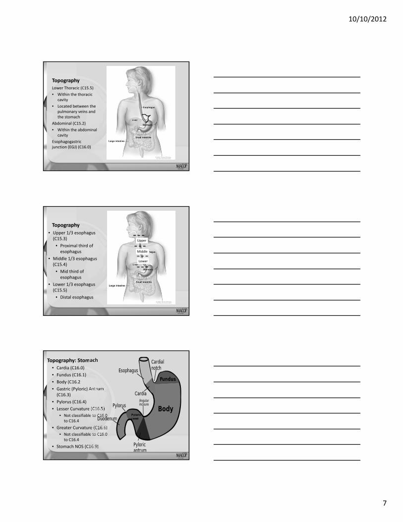
TopographyLower Thoracic (C15.5)• Within the thoracic
cavity• Located between the
pulmonary veins and the stomach
Abdominal (C15.2)• Within the abdominal
cavityEsophagogastric junction (EGJ) (C16.0)
Topography• Upper 1/3 esophagus (C15.3)
• Proximal third of esophagus
• Middle 1/3 esophagus (C15.4)
• Mid third of esophagus
• Lower 1/3 esophagus (C15.5)
• Distal esophagus
Upper
Middle
Lower
Topography: Stomach• Cardia (C16.0)• Fundus (C16.1)• Body (C16.2• Gastric (Pyloric) Antrum (C16.3)
• Pylorus (C16.4)• Lesser Curvature (C16.5)
• Not classifiable to C16.0 to C16.4
• Greater Curvature (C16.6)• Not classifiable to C16.0
to C16.4• Stomach NOS (C16.9)
10/10/2012
8
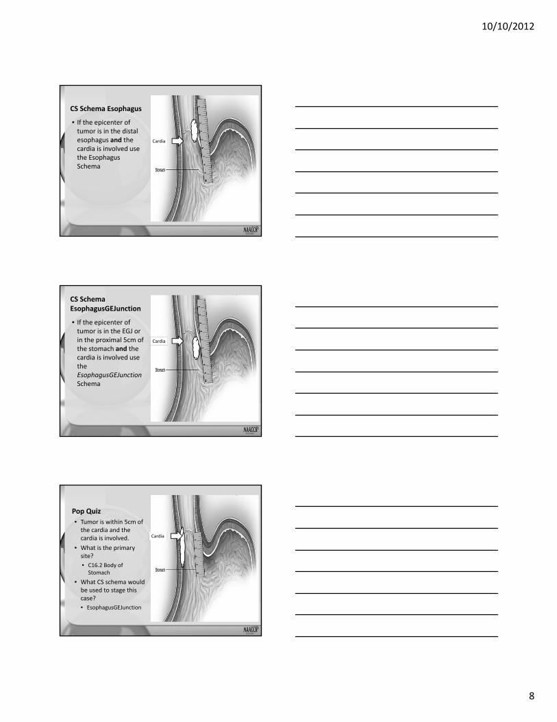
CS Schema Esophagus
• If the epicenter of tumor is in the distal esophagus and the cardia is involved use the Esophagus Schema
Cardia
CS Schema EsophagusGEJunction
• If the epicenter of tumor is in the EGJ or in the proximal 5cm of the stomach and the cardia is involved use the EsophagusGEJunctionSchema
Cardia
Pop Quiz• Tumor is within 5cm of the cardia and the cardia is involved.
• What is the primary site?• C16.2 Body of Stomach
• What CS schema would be used to stage this case? • EsophagusGEJunction
Cardia
10/10/2012
9
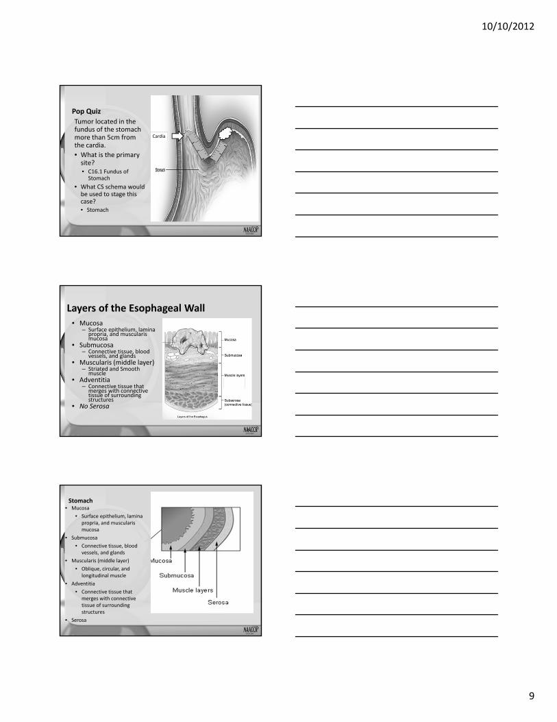
Pop QuizTumor located in the fundus of the stomach more than 5cm from the cardia.• What is the primary site?• C16.1 Fundus of Stomach
• What CS schema would be used to stage this case? • Stomach
Cardia
Layers of the Esophageal Wall• Mucosa
– Surface epithelium, lamina propria, and muscularis mucosa
• Submucosa – Connective tissue, blood
vessels, and glands • Muscularis (middle layer)
– Striated and Smooth muscle
• Adventitia – Connective tissue that
merges with connective tissue of surrounding structures
• No Serosa
26
Stomach• Mucosa
• Surface epithelium, lamina propria, and muscularis mucosa
• Submucosa • Connective tissue, blood
vessels, and glands • Muscularis (middle layer)
• Oblique, circular, and longitudinal muscle
• Adventitia • Connective tissue that
merges with connective tissue of surrounding structures
• Serosa

10/10/2012
10
Rugae• Rugae a series of ridges produced by folding of the wall of an organ.
• Allows the stomach expand when needed.
Linitis Plastica• Spreads to the muscles of the stomach wall and makes it thicker and more rigid.
Lymphatics of the Esophagus• Drainage is intramural
and longitudinal• Concentration of
lymphatic channels in the submucosa and lamina propria
• The anatomic site of the cancer and the nodes to which the site drains may not be the same.

10/10/2012
11
Lymphatics of the Esophagus• Regional nodes extend
from the paraesophageal cervical nodes to the celiac nodes
• Staging of lymph nodes is different in the AJCC 6th edition and the AJCC 7th edition
Lymphatics of the Stomach• Greater curvature
• Greater omental• Pyloric• Pancreaticoduodenal
• Pancreatic and Splenic Area
• Peripancreatic• Splenic
• Lesser curvature• Lesser omental• Left gastric• Celiac
Distant Metastasis: Esophagus• The most common sites
are:• Liver• Lungs• Pleura
• In the AJCC 6th edition the cervical lymph nodes were distant for primaries of the thoracic esophagus. • This is not true for
AJCC 7th edition

10/10/2012
12
Grade• For Esophagus and EGJ, grade is required to derive AJCC TNM stages 0‐IIA for both squamous cell carcinoma and adenocarcinoma
• Grade is not required to derive AJCC TNM stage for Stomach
• Standard four grade grading system– Well differentiated– Moderately differentiated– Poorly differentiated– Undifferentiated
34
Grade
• C T1a N0 M0 G1 Stage IA– Treatment options include
• Esophagectomy • Endoscopic mucosal resection • Other ablative technique
• C T1a N0 M0 G2‐3 Stage IB– Esophagectomy
35
QUIZ

10/10/2012
13
COLLABORATIVE STAGE DATA COLLECTION SYSTEM (CS) V02.04
Esophagus & Stomach
CS Schemas: V02.04
• Esophagus: C15.0‐C15.5, C15.8‐C15.9• Esophagus GE Junction: C16.0, C16.1, C16.2• Stomach: C16.1‐C16.6, C16.8‐C16.9
ESOPHAGUS CS SCHEMA

10/10/2012
14
CS Tumor Size: Esophagus
• Code 998: Circumferential– Takes precedence over statement of tumor size
CS Extension: Esophagus
• Code 000: In situ; high grade dysplasia• Codes 100‐170; 300: Invades lamina propria, muscularis mucosae, or submucosa
• Codes 200‐210: Invades muscularis propria• Codes 400‐450: Invades adventitia• Codes 615‐820: Invades adjacent structures
Pop Quiz
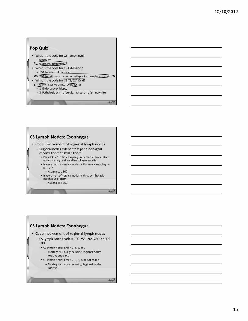
• Esophagogastroduodenoscopy (EGD) & biopsy: Circumferential 6 cm lesion in middle third of esophagus with squamous cell carcinoma invading submucosa.
• CT scan: Extensive wall thickening from lesion of the mid esophagus extending into the aorta compatible with known malignancy.
10/10/2012
15
Pop Quiz• What is the code for CS Tumor Size?
– 060: 6 cm– 998: Circumferential
• What is the code for CS Extension?– 160: Invades submucosa– 730: Intrathoracic, upper or mid‐portion, esophagus: aorta
• What is the code for CS TS/EXT Eval?– 0: Noninvasive clinical evidence– 1: Endoscopy or biopsy– 3: Pathologic exam of surgical resection of primary site
CS Lymph Nodes: Esophagus• Code involvement of regional lymph nodes
– Regional nodes extend from periesophageal cervical nodes to celiac nodes
• Per AJCC 7th Edition esophagus chapter authors celiac nodes are regional for all esophagus subsites
• Involvement of cervical nodes with cervical esophagus primary
– Assign code 100• Involvement of cervical nodes with upper thoracic esophagus primary
– Assign code 250
CS Lymph Nodes: Esophagus• Code involvement of regional lymph nodes
– CS Lymph Nodes code = 100‐255, 265‐280, or 305‐500
• CS Lymph Nodes Eval = 0, 1, 5, or 9– N category is assigned using Regional Nodes Positive and SSF1
• CS Lymph Nodes Eval = 2, 3, 6, 8, or not coded– N category is assigned using Regional Nodes Positive
10/10/2012
16
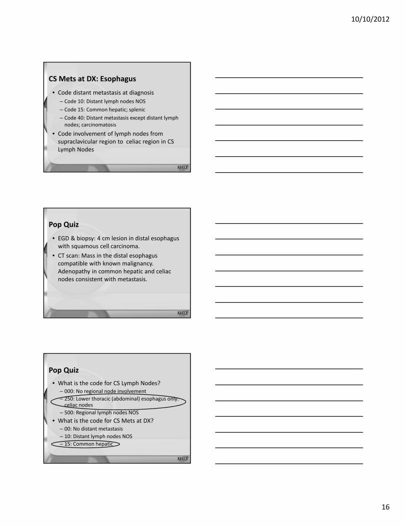
CS Mets at DX: Esophagus
• Code distant metastasis at diagnosis– Code 10: Distant lymph nodes NOS– Code 15: Common hepatic; splenic– Code 40: Distant metastasis except distant lymph nodes; carcinomatosis
• Code involvement of lymph nodes from supraclavicular region to celiac region in CS Lymph Nodes
Pop Quiz
• EGD & biopsy: 4 cm lesion in distal esophagus with squamous cell carcinoma.
• CT scan: Mass in the distal esophagus compatible with known malignancy. Adenopathy in common hepatic and celiac nodes consistent with metastasis.
Pop Quiz• What is the code for CS Lymph Nodes?
– 000: No regional node involvement– 250: Lower thoracic (abdominal) esophagus only: celiac nodes
– 500: Regional lymph nodes NOS• What is the code for CS Mets at DX?
– 00: No distant metastasis– 10: Distant lymph nodes NOS– 15: Common hepatic
10/10/2012
17
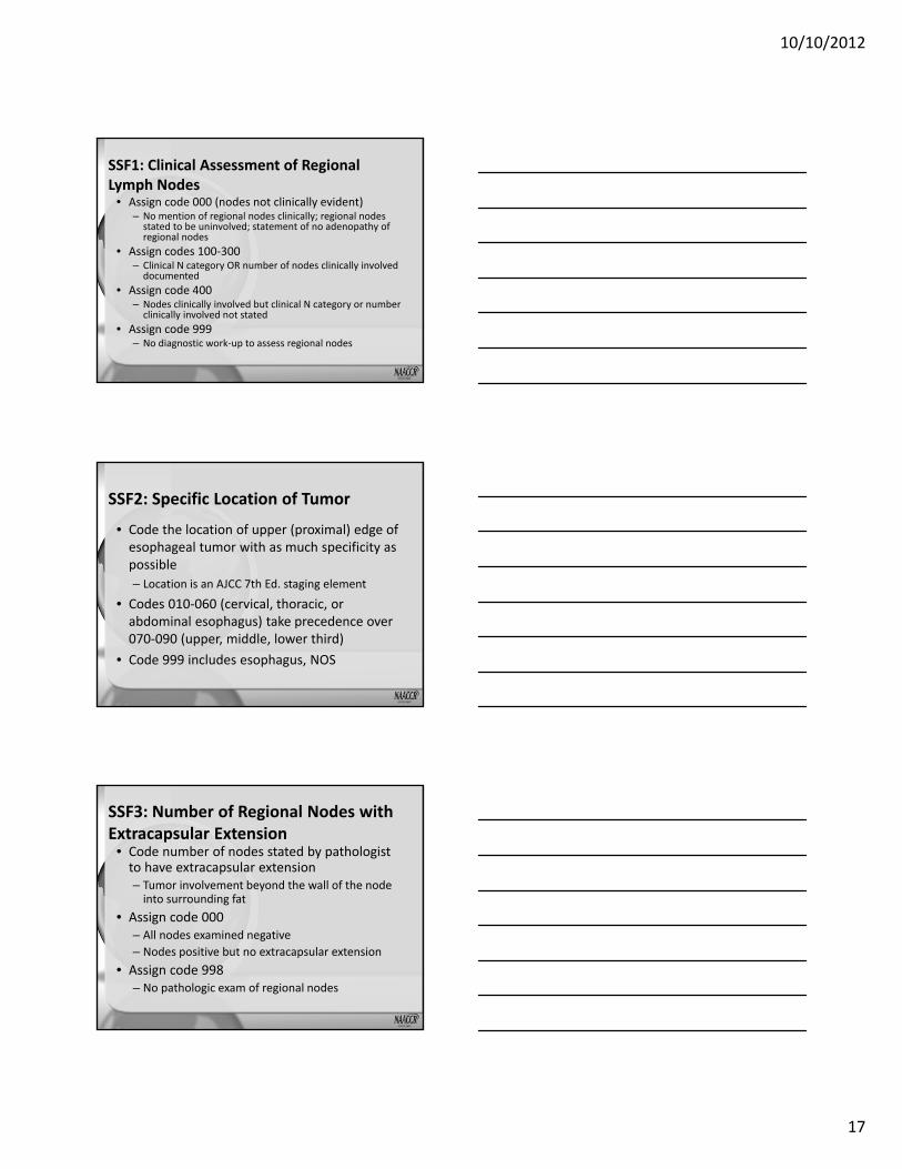
SSF1: Clinical Assessment of Regional Lymph Nodes
• Assign code 000 (nodes not clinically evident)– No mention of regional nodes clinically; regional nodes stated to be uninvolved; statement of no adenopathy of regional nodes
• Assign codes 100‐300– Clinical N category OR number of nodes clinically involved documented
• Assign code 400– Nodes clinically involved but clinical N category or number clinically involved not stated
• Assign code 999– No diagnostic work‐up to assess regional nodes
SSF2: Specific Location of Tumor
• Code the location of upper (proximal) edge of esophageal tumor with as much specificity as possible– Location is an AJCC 7th Ed. staging element
• Codes 010‐060 (cervical, thoracic, or abdominal esophagus) take precedence over 070‐090 (upper, middle, lower third)
• Code 999 includes esophagus, NOS
SSF3: Number of Regional Nodes with Extracapsular Extension
• Code number of nodes stated by pathologist to have extracapsular extension– Tumor involvement beyond the wall of the node into surrounding fat
• Assign code 000– All nodes examined negative– Nodes positive but no extracapsular extension
• Assign code 998– No pathologic exam of regional nodes

10/10/2012
18
SSF4 and SSF5• In SSF4
– Record distance from incisors (front teeth) to proximal (upper) edge of esophageal tumor to nearest cm
– Calculate distance to proximal edge if distance to distal edge and tumor length is known
• In SSF5– Record distance from incisors (front teeth) to distal (lower) edge of esophageal tumor to nearest cm
– Calculate distance to distal edge if distance to proximal edge and tumor length is known
Question
• Q: If a patient has multiple tumors of the esophagus determined to be a single primary, is the largest tumor used to code tumor location information in SSF2, SSF4, and SSF5?
• A: In that situation, code SSF2, SSF4, and SSF5 from the most invasive tumor.
Pop Quiz• EGD & biopsy: 1 cm tumor of cervical esophagus; well differentiated squamous cell carcinoma.
• Endoscopic ultrasound (EUS): Tumor confined to upper esophagus; cervical node adenopathy.
• Esophagectomy and lymph node dissection: 1 cm mass of cervical esophagus, squamous cell carcinoma, invading submucosa; 3 of 6 cervical nodes with metastasis but no extracapsular extension.
• Staging form: Clinical T1 N1; Pathologic T1b N2
10/10/2012
19
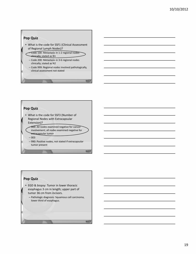
Pop Quiz
• What is the code for SSF1 (Clinical Assessment of Regional Lymph Nodes)?– Code 100: Metastasis in 1‐2 regional nodes clinically; stated as N1
– Code 200: Metastasis in 3‐6 regional nodes clinically; stated as N2
– Code 999: Regional nodes involved pathologically, clinical assessment not stated
Pop Quiz
• What is the code for SSF3 (Number of Regional Nodes with Extracapsular Extension)?– 000: All nodes examined negative for cancer involvement; all nodes examined negative for extracapsular tumor
– 003– 990: Positive nodes, not stated if extracapsular tumor present
Pop Quiz
• EGD & biopsy: Tumor in lower thoracic esophagus 3 cm in length; upper part of tumor 36 cm from incisors.– Pathologic diagnosis: Squamous cell carcinoma, lower third of esophagus.

10/10/2012
20
Pop Quiz• What is the code for SSF2 (Specific Location of Tumor)?– Code 050: Stated as lower thoracic esophagus– Code 090: Stated as lower third
• What is the code for SSF4 (Distance to Proximal Edge of Tumor)?– 036– 039
• What is the code for SSF5 (Distance to Distal Edge of Tumor)?– 036– 039
ESOPHAGUS GE JUNCTION CS SCHEMA
CS Tumor Size: Esophagus GE Junction
• Code 998: Diffuse; widespread; three‐fourths or more; linitis plastica– Takes precedence over statement of tumor size
10/10/2012
21
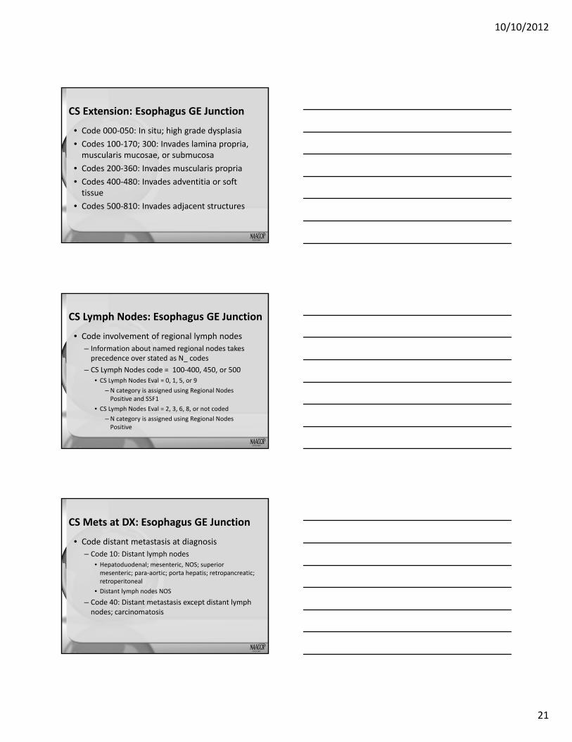
CS Extension: Esophagus GE Junction
• Code 000‐050: In situ; high grade dysplasia• Codes 100‐170; 300: Invades lamina propria, muscularis mucosae, or submucosa
• Codes 200‐360: Invades muscularis propria• Codes 400‐480: Invades adventitia or soft tissue
• Codes 500‐810: Invades adjacent structures
CS Lymph Nodes: Esophagus GE Junction
• Code involvement of regional lymph nodes– Information about named regional nodes takes precedence over stated as N_ codes
– CS Lymph Nodes code = 100‐400, 450, or 500• CS Lymph Nodes Eval = 0, 1, 5, or 9
– N category is assigned using Regional Nodes Positive and SSF1
• CS Lymph Nodes Eval = 2, 3, 6, 8, or not coded– N category is assigned using Regional Nodes Positive
CS Mets at DX: Esophagus GE Junction
• Code distant metastasis at diagnosis– Code 10: Distant lymph nodes
• Hepatoduodenal; mesenteric, NOS; superior mesenteric; para‐aortic; porta hepatis; retropancreatic; retroperitoneal
• Distant lymph nodes NOS
– Code 40: Distant metastasis except distant lymph nodes; carcinomatosis

10/10/2012
22
Pop Quiz
• EGD with biopsy: Mixed squamous cell and adenocarcinoma of EGJ extending through the wall
• EUS: 3 cm tumor of EGJ; gastrohepatic and hepatoduodenal nodes compatible with malignancy
Pop Quiz• What is the code for CS Lymph Nodes?
– Code 000: No regional node involvement– Code 100: Gastrohepatic– Code 400: Hepatic– Code 500: Regional nodes NOS
• What is the code for CS Mets at DX?– Code 00: No distant metastasis– Code 10: Distant lymph nodes including hepatoduodenal
Site‐Specific FactorsEsophagus GE Junction
• SSF1: Clinical Assessment of Regional Lymph Nodes
• SSF3: Number of Regional Lymph Nodes with Extracapsular Tumor
• SSF4: Distance to Proximal Edge of Tumor from Incisors
• SSF5: Distance to Distal Edge of Tumor from Incisors

10/10/2012
23
CS SSF25: Schema DiscriminatorEsophagus GE Junction & Stomach SchemasCode Description Schema000 No involvement of esophagus or
EGJStomach
020 Esophagus or EGJ involved AND distance of tumor midpoint from EGJ 5 cm or less
EsophagusGEJunction
030 Esophagus or EGJ involved AND distance of tumor midpoint from EGJ more than 5 cm
Stomach
040 Esophagus or EGJ involved AND distance of tumor midpoint from EGJ unknown
EsophagusGEJunction
CS SSF25: Schema DiscriminatorEsophagus GE Junction & Stomach SchemasCode Description SchemaCode Description Schema060 Esophagus/EGJ involved AND
distance of tumor midpoint from EGJ more than 5 cm from EGJAND physician stages case using esophagus definitions OREsophagus/EGJ involvement unknown AND distance of tumor midpoint from EGJ more than 5 cm or unknown AND physician stages case using esophagus definition
EsophagusGEJunction
CS SSF25: Schema DiscriminatorEsophagus GE Junction & Stomach SchemasCode Description Schema981 Primary site coded to C16.3 ‐
C16.9Stomach
982 Primary site coded to C16.0 EsophagusGEJunction999 Involvement of esophagus/EGJ
unknown, or no informationNot documented in patient record
Stomach

10/10/2012
24
Pop Quiz• Upper gastrectomy: Adenocarcinoma of EGJ, 3 cm, involves stomach body intraluminally and invades muscularis mucosa.
• What is the code for SSF25?– Code 020: Esophagus or EGJ involved & distance of tumor midpoint from EGJ 5 cm or less
– Code 982: Primary site coded to C16.0• What is the code for CS Extension
– Code 120: Invades muscularis mucosae– Code 800: Further contiguous extension
STOMACH CS SCHEMA
CS Tumor Size: Stomach
• Code 998: Diffuse; widespread; three‐fourths or more; linitis plastica– Takes precedence over statement of tumor size

10/10/2012
25
CS Extension: Stomach• Code 000‐050: In situ; noninvasive• Codes 100‐180; 300: Invades lamina propria, muscularis mucosae, or submucosa
• Codes 200‐390: Invades muscularis propria• Codes 400‐480: Penetrates subserosal connective tissue without invasion of (serosa) visceral peritoneum or adjacent structures
• Codes 505‐810: Invades serosa or adjacent structures
CS Lymph Nodes: Stomach• Code involvement of regional lymph nodes
– Code metastatic nodules in fat adjacent to gastric carcinoma without evidence of residual lymph node tissue in CS Lymph Nodes
– CS Lymph Nodes code = 110‐500 or 800• CS Lymph Nodes Eval = 0, 1, 5, or 9
– N category is assigned using Regional Nodes Positive and SSF1
• CS Lymph Nodes Eval = 2, 3, 6, 8, or not coded– N category is assigned using Regional Nodes Positive
CS Mets at DX: Stomach• Code distant metastasis at diagnosis
– Code 10: Distant lymph nodes• Mesenteric, NOS; inferior mesenteric; superior mesenteric; para‐aortic; porta hepatis; retropancreatic; retroperitoneal
• For all subsites EXCEPT lessercurvature: hepatoduodenal
• Distant lymph nodes NOS– Code 40: Distant metastasis except distant lymph nodes; carcinomatosis; Krukenberg tumor; malignant peritoneal cytology
10/10/2012
26
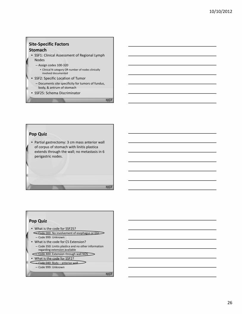
Site‐Specific FactorsStomach
• SSF1: Clinical Assessment of Regional Lymph Nodes– Assign codes 100‐320
• Clinical N category OR number of nodes clinically involved documented
• SSF2: Specific Location of Tumor– Documents site specificity for tumors of fundus, body, & antrum of stomach
• SSF25: Schema Discriminator
Pop Quiz
• Partial gastrectomy: 3 cm mass anterior wall of corpus of stomach with linitis plastica extends through the wall; no metastasis in 6 perigastric nodes.
Pop Quiz• What is the code for SSF25?
– Code 000: No involvement of esophagus or EGJ– Code 999: Unknown
• What is the code for CS Extension?– Code 350: Linitis plastica and no other information regarding extension available
– Code 400: Extension through wall NOS• What is the code for SSF2?
– Code 040: Body – anterior wall– Code 999: Unknown
10/10/2012
27
Site‐Specific FactorsStomach
• SSF13: Carcinoembryonic Antingen (CEA)– Record clinician’s interpretation of highest CEA test result prior to treatment
• Elevated, normal, borderline– Use same test coded in SSF14
• SSF14: CEA Lab Value– Record lab value in ng/ml of highest CEA test result prior to treatment
– Use same test coded in SSF14• SSF15: Carbohydrate Antigen 19‐9 (CA 19‐9) Lab Value
– Record lab value in U/ml of highest CA 19‐9 test result prior to treatment
QUIZ
DIAGNOSIS & TREATMENTEsophagus and Stomach
10/10/2012
28
Endoscopy
• Diagnostic– Determine the presence, location, and circumference of tumor
– Biopsies to confirm disease
Endoscopy
• Staging‐Endoscopic Ultrasound (EUS)– Determine the depth of tumor invasion (T)
• Hypoechoic (dark) expansion can be done to better visualize the depth of invasion into the layers of the esophagus or stomach
– Mediastinal and perigastric lymph nodes are readily seen and biopsied by EUS (N)
– Signs of distant spread may be identified (M)
Endoscopy
• Treatment– Endoscopic Mucosal Resection (EMR) – Ablation
• Barrett’s• High‐grade dysplasia• Invasive tumors confined to the lamina propria or muscularis mucosa
10/10/2012
29
Endoscopic Mucosal Resection• A cap on the end of the scope pulls the mucosal layer into the cap.
• A snare is put around the polyp that is created.
• Using current and the snare the polyp is removed.
• The tissue is sent to pathology.– Codes as 27‐Excisional biopsy NOShttp://www.youtube.com/watch?v=Sv7eARxui7s
Endoscopic Ablation• Radiofrequency Ablation
– High frequency alternating current is used to create heat that destroys the tissue
– Code as 10‐local tumor destruction, NOS• Cryosurgery
– Liquid nitrogen is applied to the area to be treated– May take several treatments– Code as 13‐Cryosurger7
• Photodynamic Therapy– Photosensitizing agent is injected into the patient– Photosensitizer is activated when light of a specific wave length.
– Codes as 11 Photodynamic therapy (PDT)
Surgery Codes‐Esophagus
• 50 Esophagectomy, NOS WITH laryngectomy and/or gastrectomy, NOS[SEER Note: Codes 50‐55 include partial esophagectomy, total esophagectomy, or esophagectomy, NOS.]– 51 WITH laryngectomy– 52 WITH gastrectomy, NOS– 53 Partial gastrectomy– 54 Total gastrectomy– 55 Combination of 51 WITH any of 52–54

10/10/2012
30
Unresectable Disease
Esophagus• T4 tumors with involvement
of the heart, great vessels, trachea or adjacent organs
• Multi station or bulky lymphadenopathy
• EGJ primary and supraclavicular node involvment
• Distant metastasis
Stomach• Peritoneal involvement• Distant metastasis• Invasion or encasement of
major blood vessels
Surgery‐Esophagus• Partial Esophagectomy
– Removal of a section of the esophagus.• Esophagus is reconstructed using another organ such as the stomach or large intestine.
– Code 30• Esophagogastrectomy
– Removal of a section of the esophagus and the fundus of the stomach.
– Stomach is surgically attached to the remaining esophagus.Code 53
• At least 15 lymph nodes should be removed for adequate nodal staging
Surgery‐Esophagus• Ivor‐Lewis esophagogastrectomy (laparotomy & right thoracotomy)
• McKeown esophagogastrectomy (right thoracotomy & laparotomy & cervical anastomosis)

10/10/2012
31
Surgery‐Stomach
• Distal subtotal gastrectomy (32) is the preferred approach for distal gastric cancers
• Proximal gastrectomy (33) and total gastrectomy (40) are both indicated for proximal gastric cancers
• Esophagectomy, NOS WITH laryngectomy and/or gastrectomy, NOS
Treatment by Stage‐Esophagus
• Tis‐EMR or Ablation• T1a
– EMR or Ablation– Esophagectomy
• T1b N0‐Esophagectomy
Treatment by Stage‐Esophagus• T2‐T4a any N
– Preoperative chemoradiation– Definitive chemoradiation
• Preferred for cervical esophagus– Preoperative chemotherapy
• Only for adenocarcinoma of distal esophagus or EGJ– Esophagectomy
• Low risk lesions less than 2cm and well differentiated
• T4b‐Definitive chemoradiation

10/10/2012
32
Treatment by Stage‐Stomach
• Tis or T1a‐EMR or Surgery• T1b N0‐Surgery• T2 or higher and any N
– Surgery or– Preoperative chemotherapy– Preoperative chemoradiation
• M1‐Palliative therapy
QUIZ
QUESTIONS?

10/10/2012
33
Coming up!
• 11/1/12Collecting Cancer Data: Uterus
• 12/6/12Collecting Cancer Data: Pharynx
• Certificate phrase:– NAACCR 2012– http://www.surveygizmo.com/s3/1031627/Stomach‐and‐Esophagus
97
And the fabulous prize winners are…
THANK YOU!