collagen membranes of dermal and pericardial origin in
TRANSCRIPT

OR I G I N A L A R T I C L E
Collagen membranes of dermal and pericardial origin—In vivoevolvement of vascularization over time
Michael Dau1 | Lisann Volprich1 | Eberhard Grambow2 | Brigitte Vollmar3 |
Bernhard Frerich1 | Bilal Al-Nawas4 | Peer W. Kämmerer1,4
1Department of Oral, Maxillofacial Plastic
Surgery, University Medical Center Rostock,
Rostock, Germany
2Department for General-, Visceral-, Vascular-
and Transplantation Surgery, University
Medical Center Rostock, Rostock, Germany
3Institute for Experimental Surgery, University
Medical Center Rostock, Rostock, Germany
4Department of Oral, Maxillofacial Plastic
Surgery, University Medical Center Mainz,
Mainz, Germany
Correspondence
Peer W. Kämmerer, Department of Oral,
Maxillofacial Plastic Surgery, University
Medical Center Mainz, Augustusplatz 2, 55131
Mainz, Germany.
Email: [email protected]
Funding information
DGI (Deutsche Gesellschaft für Implantologie
im Zahn-, Mund- und Kieferbereich e.V.,
Munich, Germany)
Abstract
Aim of the study was to compare the evolvement of vascularization over time of collagen
membranes (CMs) of dermal and pericardial origin in an in vivo animal study. Twenty-
eight mice underwent implantation of three commercially available CM derived from por-
cine dermis (homogenous structure: CM1 (Control 1) and bilayer structure: CM2 [Control
2]), from porcine pericardium (CM3; Test 1) as well as CM3 sprayed with silica-enhanced
nanostructured hydroxyapatite (CM4, Test 2). After 3, 6, 9, and 12 days, intravital fluores-
cence microscopy was conducted for determination of capillary diameter, density, flow,
and length. At Day 12, samples were examined immunohistologically for expression of
fibroblast growth factor receptor 4 (FGFR4), CD11b, CD68, αSMA, and CD34. In all CM,
intravital fluorescence microscopy over time showed increasing values for all parameters
with the highest levels in CM4 and the lowest values in CM1. Significant lower amounts
of FGFR4, CD11b, and CD68 were detected in CM4 when compared to CM2 (p < .05).
In contrast to CM3, lower values of αSMA and higher numbers of CD34 positive-marked
vessels were observed in CM4 (p < .05). In conclusion, dermal bilayer as well as pericardial
CM seem to have a higher vascularization rate than dermal homogenous CM. Additional
coating of pericardial CM with a silica-enhanced hydroxyapatite increases the speed of
vascularization as well as biological remodeling processes.
K E YWORD S
angiogenesis, collagen membrane, dermal, dorsal skinfold chamber, guided bone regeneration,
nanocrystalline hydroxyapatite, pericardial, silicon dioxide, vascularization
1 | INTRODUCTION
Guided bone regeneration (GBR) is a commonly used procedure for
regeneration of osseous defects of the jaws. Here, nonresorbable and
resorbable barrier membranes are thought to exclude nonosteogenic tis-
sue and to stabilize the grafted defect (Elgali, Omar, Dahlin, &
Thomsen, 2017; Kämmerer et al., 2017). Nonresorbable membranes have
to be removed during a second surgical intervention and are susceptible
to higher complication rates in terms of premature exposure and infection
(Chiapasco & Zaniboni, 2009), that could be caused by lack of vascular
supply through the membrane (Elgali et al., 2017). Especially for horizontal
defects, resorbable collagen-based membranes (CM) offer similar clinical
success but less complication rates plus degradation within the host tissue
(Keestra, Barry, Jong, & Wahl, 2016; Naenni et al., 2017; Pabst &
Kämmerer, 2020). Besides, experimental findings suggest that CM may
also have an active role in promoting osseous regeneration next to the
passive barrier function (Elgali et al., 2017; Liu & Kerns, 2014). For exam-
ple, the overlying CM may have an additional influence on graft
Received: 13 November 2019 Revised: 29 March 2020 Accepted: 4 April 2020
DOI: 10.1002/jbm.a.36989
This is an open access article under the terms of the Creative Commons Attribution License, which permits use, distribution and reproduction in any medium,
provided the original work is properly cited.
© 2020 The Authors. Journal of Biomedical Materials Research Part A published by Wiley Periodicals, Inc.
2368 J Biomed Mater Res. 2020;108:2368–2378.wileyonlinelibrary.com/journal/jbma
vascularization that is known to have a pivotal role for successful tissue
integration (Koerdt, Ristow, Wannhoff, Kubler, & Reuther, 2014). In
accordance, improvement of vascularization of CM could be of high inter-
est. As CMs are derived from different tissues and their degradation as
well as tissue integration depends on the source (e.g., dermis and pericar-
dium) (Bunyaratavej & Wang, 2001), it is possible than they have a differ-
ent biological behavior over time that has not been evaluated so far.
Further modification of CM with enamel matrix, respectively, platelet-
rich fibrin resulted in a significantly enhanced migration of endothelial cells
(Park et al., 2018) and an increased angiogenesis (Blatt et al., 2020). A
combination of CM with recombinant platelet-derived growth factor
increased early bone growth in a rabbit model (Kämmerer et al., 2013) and
improved soft tissue healing (Amorfini, Migliorati, Signori, Silvestrini-
Biavati, & Benedicenti, 2014). However, also membranes for GBR modi-
fied with incorporated bone substitute materials (BSMs) such as hydroxy-
apatite or beta-tricalcium phosphate have shown to promote the activity
of stromal and osteoblastic cells both in vitro and in vivo (Li et al., 2011;
Shim et al., 2013; Teng et al., 2009). Besides, the BSM allows the mem-
brane to withstand static pressure to a greater extend (Anderud
et al., 2014).Though, there are also vast differences regarding the biologi-
cal behavior of BSM (Dau et al., 2016; Kyyak et al., 2020). For example,
synthetic silicium dioxide-enhanced nanostructured hydroxyapatite seems
to have a high biocompatibility and vascularization rate in vitro and in vivo
(Abshagen, Schrodi, Gerber, & Vollmar, 2009; Ghanaati et al., 2013) and
could be an interesting candidate for additional coating of a CM.
Regardless the observed differences in vascularization of various
CM, most study results based on histomorphometric analyses
(Ghanaati, 2012; Rothamel et al., 2005; Rothamel et al., 2014; Schwarz,
Rothamel, Herten, Sager, & Becker, 2006; Silva et al., 2017) at defined
time points using a combination of light microscopy and immunohisto-
chemical staining. Intravital fluorescence microscopy in a dorsal skin fold
chamber model (Sckell & Leunig, 2009; Sckell & Leunig, 2016) provides
in vivo analyses of vascularization including the evolvement over time.
Therefore, the aim of this study was to investigate and compare
vascularization parameters of CM of porcine dermal and pericardial ori-
gin using intravital fluorescence microscopy over time and final
(immuno)histomorphometry using an in vivo dorsal skin fold mouse
model. In addition, a pericardial CM additionally sprayed with synthetic
silicium dioxide-enhanced nanostructured hydroxyapatite was tested.
The null hypothesis was that there are no differences in vascularization
over time as well as at the final endpoint between the different CMs.
2 | MATERIALS AND METHODS
2.1 | Study materials
Four different porcine CMs, two of dermal origin and two of pericar-
dial origin, were used in the experiment:
• CM1 (CollProtect, Botiss biomaterials GmbH, Zossen, Germany) is
derived from porcine dermis and consists of a homogenous, dense
and porous 3D network of collagen bundles.
• CM2 (Bio-Gide, Geistlich Pharma AG, Wolhusen, Switzerland), also
from porcine dermal origin, has a bilayer structure consisting of a
compact outer layer and a porous inner layer of collagen fiber
bundles.
• CM3 (Jason, Botiss biomaterials GmbH) originates from porcine
pericardium and consists of differently orientated collagen fibers.
• CM4 consists of the porcine pericardium membrane CM3 that was
additionally sprayed with a silicium dioxide-enhanched nanostruc-
tured synthetic hydroxyapatite (NanoBone S39, Artoss GmbH,
Rostock, Germany). For this purpose, the spin-spray coating was
conducted using a custom-made two-substance nozzle in a laminar
flow box (Herasafe HSP12 Safety Cabinets class 2, Kendro Labora-
tory Products GmbH, Hanau, Germany) with a coating time of
0.5 s. Subsequently, the sprayed membranes were dried under oil-
free airflow as described before (Adam et al., 2014).
2.2 | Animal model and procedures
An established animal mouse dorsal skinfold chamber model was used
in the present study. The experiment was approved by the State
Office of Agriculture, Food Safety and Fishery Mecklenburg-Vor-
pommern, Germany (LVL M-V/TSD/7221.3–1.1-040/16) and
followed the National Institutes of Health guidelines for the care
and use of laboratory animals as well the ARRIVE Guidelines for
Reporting Animal Research (Kilkenny, Browne, Cuthill, Emerson, &
Altman, 2010). Twenty-eight male C57BL/6 Tyr mice underwent
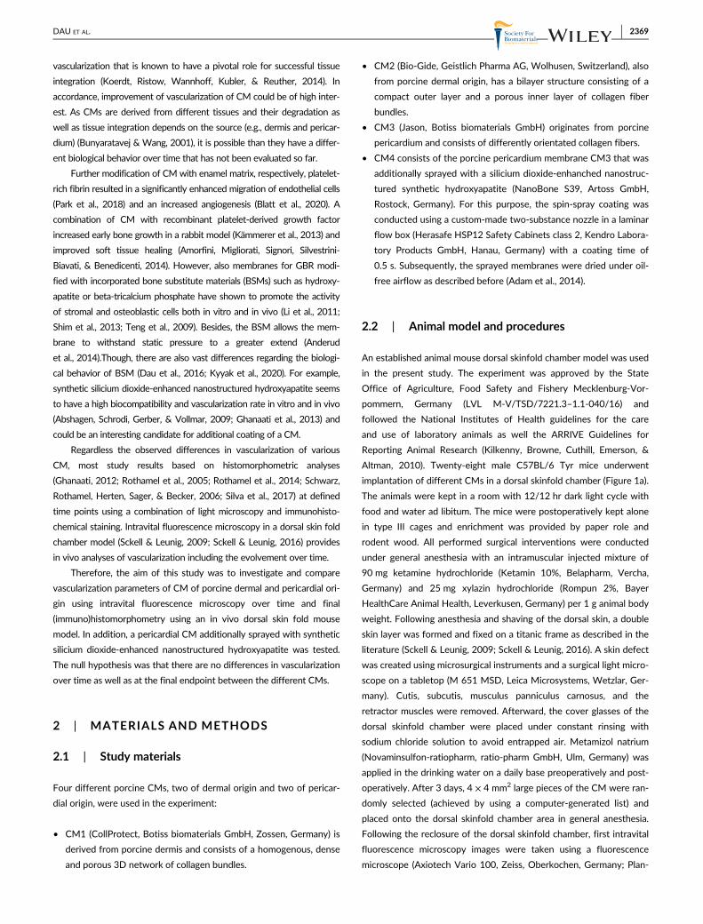
implantation of different CMs in a dorsal skinfold chamber (Figure 1a).
The animals were kept in a room with 12/12 hr dark light cycle with
food and water ad libitum. The mice were postoperatively kept alone
in type III cages and enrichment was provided by paper role and
rodent wood. All performed surgical interventions were conducted
under general anesthesia with an intramuscular injected mixture of
90 mg ketamine hydrochloride (Ketamin 10%, Belapharm, Vercha,
Germany) and 25 mg xylazin hydrochloride (Rompun 2%, Bayer
HealthCare Animal Health, Leverkusen, Germany) per 1 g animal body
weight. Following anesthesia and shaving of the dorsal skin, a double
skin layer was formed and fixed on a titanic frame as described in the
literature (Sckell & Leunig, 2009; Sckell & Leunig, 2016). A skin defect
was created using microsurgical instruments and a surgical light micro-
scope on a tabletop (M 651 MSD, Leica Microsystems, Wetzlar, Ger-
many). Cutis, subcutis, musculus panniculus carnosus, and the
retractor muscles were removed. Afterward, the cover glasses of the
dorsal skinfold chamber were placed under constant rinsing with
sodium chloride solution to avoid entrapped air. Metamizol natrium
(Novaminsulfon-ratiopharm, ratio-pharm GmbH, Ulm, Germany) was
applied in the drinking water on a daily base preoperatively and post-
operatively. After 3 days, 4 × 4 mm2 large pieces of the CM were ran-
domly selected (achieved by using a computer-generated list) and
placed onto the dorsal skinfold chamber area in general anesthesia.
Following the reclosure of the dorsal skinfold chamber, first intravital
fluorescence microscopy images were taken using a fluorescence
microscope (Axiotech Vario 100, Zeiss, Oberkochen, Germany; Plan-
DAU ET AL. 2369
NEOFLUAR ×20/0.50 W; Zeiss) after retrobulbar injection of fluores-
cein isothiocyanate-dextran (2% FITC-Dextran average mol wt
150,000 D, Sigma-Aldrich, Taufkirchen, Germany). All images were
captured with a video camera (FK 6990 IQ-S, Pieper, Schwerte, Ger-
many) connected to a S-VHS recorder (AG-7350; Panasonic Germany,
Hamburg, Germany) and saved using a DVD recorder (DMR-EX99V,
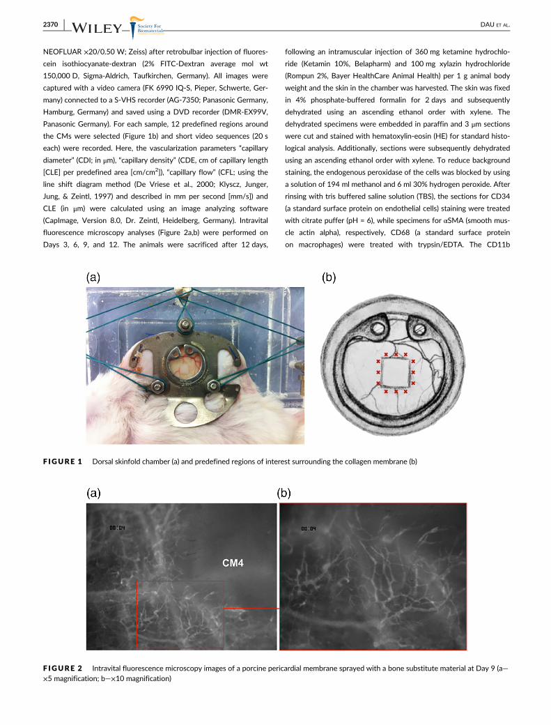
Panasonic Germany). For each sample, 12 predefined regions around
the CMs were selected (Figure 1b) and short video sequences (20 s
each) were recorded. Here, the vascularization parameters “capillary
diameter” (CDI; in μm), “capillary density” (CDE, cm of capillary length
[CLE] per predefined area [cm/cm2]), “capillary flow” (CFL; using the
line shift diagram method (De Vriese et al., 2000; Klyscz, Junger,
Jung, & Zeintl, 1997) and described in mm per second [mm/s]) and
CLE (in μm) were calculated using an image analyzing software
(CapImage, Version 8.0, Dr. Zeintl, Heidelberg, Germany). Intravital
fluorescence microscopy analyses (Figure 2a,b) were performed on
Days 3, 6, 9, and 12. The animals were sacrificed after 12 days,
following an intramuscular injection of 360 mg ketamine hydrochlo-
ride (Ketamin 10%, Belapharm) and 100 mg xylazin hydrochloride
(Rompun 2%, Bayer HealthCare Animal Health) per 1 g animal body
weight and the skin in the chamber was harvested. The skin was fixed
in 4% phosphate-buffered formalin for 2 days and subsequently
dehydrated using an ascending ethanol order with xylene. The
dehydrated specimens were embedded in paraffin and 3 μm sections
were cut and stained with hematoxylin-eosin (HE) for standard histo-
logical analysis. Additionally, sections were subsequently dehydrated
using an ascending ethanol order with xylene. To reduce background
staining, the endogenous peroxidase of the cells was blocked by using
a solution of 194 ml methanol and 6 ml 30% hydrogen peroxide. After
rinsing with tris buffered saline solution (TBS), the sections for CD34
(a standard surface protein on endothelial cells) staining were treated
with citrate puffer (pH = 6), while specimens for αSMA (smooth mus-
cle actin alpha), respectively, CD68 (a standard surface protein
on macrophages) were treated with trypsin/EDTA. The CD11b
F IGURE 1 Dorsal skinfold chamber (a) and predefined regions of interest surrounding the collagen membrane (b)
F IGURE 2 Intravital fluorescence microscopy images of a porcine pericardial membrane sprayed with a bone substitute material at Day 9 (a—×5 magnification; b—×10 magnification)
2370 DAU ET AL.

(a standard surface protein on monocytes) and Fibroblast growth fac-
tor receptor 4 (FGFR4) staining required no antigen demasking. After
rerinsing with TBS, all sections were blocked using bovine serum albu-
min. Depending on the antibody markers, the sections were incubated
with the respective antibodies for detection of CD68 (Anti-CD68
antibody 1:200, Abcam, Cambridge, UK), CD11b (Anti-CD11b anti-
body 1:250, Abcam), CD34 (Anti-CD34 antibody 1:200, Abcam),
αSMA (Anti-alpha smooth muscle Actin antibody 1:500, Abcam), and
FGFR4 (Anti-FGFR4 antibody 1:50, Abcam) and rinsed again with
TBS. Following incubation of the slides in a humidity chamber,
another rerinsing with TBS was performed and in preparation of the
antibody linking, all sections were incubated for 30 min with horse-
radish peroxidase (Labeled Polymer-HRP, EnVision Kit, Dako/Agilent
Technologies, Santa Clara, CA). After rinsing with TBS, dia-
minobenzidine staining and rerinsing with TBS, the sections were
counterstained with hemalaun. Using an ascending ethanol order and
xylene, all sections were dehydrated and the slides were covered with
a rapid mounting medium for microscopy (Entellan, Sigma-Aldrich,
St. Louis, MO).
2.3 | Histological analysis
Histological analysis was performed using a light microscope (Axio
Imager.M2 microscope, Zeiss) together with its software (Zen 2.0,
Zeiss). All histological figures were captured with a camera (AxioCam
MRc5 camera, Zeiss), connected to an automatic scanning table (M-
686K011, Wienecke & Sinske GmbH, Gleichen, Germany).
2.4 | Quantitative analyses
In each animal, the height of each membrane was separated into three
sections of the same size (A—top layer, B—middle layer, and C—
ground layer) as shown in Figure 3a. Within each section, six fields of
views (size 128px × 128px) were randomly selected and analyzed.
The areas of CD68-, CD11b-, αSMA-, and FGFR4-positive marked
cells were calculated as sum of areas (Adobe Photoshop CS6, Adobe
Systems Inc., San Jose, CA), the values were set in relation to the
predefined field and the resulting mean ratio (percentage rate) was
calculated for each section. Additionally, CD34 positive-marked ves-
sels (Figure 3b,c) in each predefined field of view were counted and
reported as number of vessels of each section.
2.5 | Statistics
Analyses were performed with SPSS statistical package version 20.0
(SPSS Inc., Chicago, IL). With an intended n = 7 per group and time,
the sample size was higher as compared to other studies evaluating
the biological properties of different membranes (Fujioka-Kobayashi
et al., 2017; Mahajan et al., 2018; Talebi Ardakani, Hajizadeh, &
Yadegari, 2018). Results were expressed as arithmetic means ± SEM.
Before testing for statistical significances, all variables were evaluated
for normal distribution via Shapiro–Wilk test. Two-way analysis of
variance (ANOVA) for membranes (first way) and time (second way)
followed by subsequent repeated measures within one group and
analyses between four groups at each time point were conducted via
ANOVA (parametric data), respectively, Kruskal–Wallis rank sum test
(nonparametric data). Depending on the presence or the absence of
normal distribution, Student's t test, respectively, Mann–Whitney test
for continuous variables were used for pairwise comparison at each
time point. p-Values <.05 were considered as significant. Bar and line
charts with error bars (SEM) were used for data illustration.
3 | RESULTS
3.1 | Intravital vascularization analysis
For all animals (n = 28; n = 7 samples per group [homogenous dermal
membrane [CM1]: n = 7; bilayer dermal membrane [CM2]: n = 7; peri-
cardial membrane [CM3]: n = 7 and pericardial membrane coated with
BSM [CM4]: n = 7]), postoperative healing was uneventful.
F IGURE 3 Trisection of integrated collagen membrane (a—top layer, b—middle layer, and c—ground layer) (a) and images of predefinedregions with CD34 positive-marked vessels (b: negative control without any antibody and c: positive control)
DAU ET AL. 2371

Additionally, 140 intravital fluorescence microscopy video clips per
region of interest (n = 7 for each group at each time point) were ana-
lyzed. In total, an increase of all vascularization parameters was seen
in all CM over time (Tables 1 and 2).
3.2 | Group comparisons of intravitalvascularization analysis
3.2.1 | Three days
The pericardial membrane coated with BSM (CM4) showed the
highest values for capillary diameter (CDI, 11-fold); CDE (ninefold);
CFL (12-fold); and CLE (ninefold) when compared to the homogenous
dermal membrane (CM1; all: p ≤ .034) and the pericardial membrane
without coating (CM3; CDI: 10-fold, CDE: 30-fold, CFL: 13-fold, CLE:
ninefold; all: p ≤ .029; Figure 4; Tables 1 and 2).
3.2.2 | Six days
At this time point, CM4 also had the highest vascularization values
regarding CDI (10-fold), CDE (28-fold), CFL (18-fold), and CLE
(30-fold) when compared to CM1 (all: p < .001). The direct compari-
son of CM4 with CM2/CM3 showed significant higher values in all
parameters (all: p ≤ .021). Additionally, higher values of CDE, CDI, and
CFL were detected in CM3 when compared to CM1 (all p ≤ .034;
Figure 4; Tables 1 and 2).
3.2.3 | Nine days
Similar to the previous time points, significant higher values in all vas-
cularization parameters were seen in the modified pericardial (CM4)
versus the homogenous dermal membrane (CM1). In brief, CDI was
increased 12 times, CDE 25 times, CFL 1.5 times and CLE 12 times
(all values: p < .001). Also, all vascularization parameters except for
CLF were significantly increased in the nonmodified pericardial mem-
brane (CM3) versus CM1 (all: p ≤ .003) and—except for CLE—in CM3
versus the bilayer dermal membrane (all: p ≤ .031). For CDI and CLE,
higher values were found in CM4 when compared to CM3 (p = .004,
respectively, p = .008; Figure 4; Tables 1 and 2).
3.2.4 | Twelve days
CM4 still showed the highest values of all groups regarding CDI
(3.5-fold), CDE (33-fold), CFL (1.4-fold), and CLE (ninefold) compared
to the lowest values seen for CM1 (all: p < .001). All vascularization
parameters were also significantly lower in CM1 when compared to
all other groups (all: p ≤ .003; Figure 4; Tables 1 and 2).
3.3 | Descriptive histological analysis
All CMs were well tolerated and new blood vessels were observed. In
the HE-stained samples, no hints for inflammatory reactions were
found and all CMs were still visible in all groups after 12 days.
TABLE 1 Mean ± SEM for measurement of intravital fluorescence microscopy (all values rounded to one, respectively, two decimal digits)
MembraneVascularizationparameter
Day
3 6 9 12 p-Valueb
CM1 CDI (μm) 0.09 ± 0.09 0.24 ± 0.17 1.42 ± 0.45 1.60 ± 0.50 .003
CDE (cm/cm2) 0.38 ± 0.38 0.50 ± 0.36 1.59 ± 0.65 3.06 ± 1.13 .004
CFL (mm/s) 0.003 ± 0.003 0.004 ± 0.004 0.08 ± 0.04 0.08 ± 0.03 .004
CLE (μm) 12.1 ± 12.1 15.9 ± 11.3 99.9 ± 52.4 239.9 ± 99.7 .004
CM2 CDI (μm) 0.62 ± 0.31 0.78 ± 0.31 2.40 ± 0.49 4.07 ± 0.57 <.001
CDE (cm/cm2) 2.78 ± 1.45 4.32 ± 2.04 14.80 ± 3.54 33.91 ± 6.06 <.001
CFL (mm/s) 0.01 ± 0.07 0.03 ± 0.02 0.05 ± 0.02 0.09 ± 0.02 <.001
CLE (μm) 85.0 ± 44.0 131.4 ± 61.6 413.1 ± 107.1 1,033.3 ± 190.0 <.001
CM3 CDI (μm) 0.10 ± 0.10 0.89 ± 0.33 4.08 ± 0.63 4.87 ± 0.64 <.001
CDE (cm/cm2) 0.11 ± 0.11 2.49 ± 1.40 10.77 ± 2.94 34.86 ± 9.03 <.001
CFL (mm/s) a 0.06 ± 0.03 0.13 ± 0.04 0.11 ± 0.03 <.001
CLE (μm) 1.7 ± 1.7 66.5 ± 42.3 397.9 ± 99.0 1,129.4 ± 283.3 <.001
CM4 CDI (μm) 0.98 ± 0.39 2.45 ± 0.51 5.49 ± 0.66 5.15 ± 0.65 <.001
CDE (cm/cm2) 3.26 ± 1.45 14.79 ± 3.89 38.19 ± 6.96 69.61 ± 10.72 <.001
CFL (mm/s) 0.04 ± 0.02 0.07 ± 0.02 0.13 ± 0.03 0.11 ± 0.02 <.001
CLE (μm) 103.0 ± 45.7 467.1 ± 122.7 1,195.9 ± 215.4 2,184.9 ± 399.5 <.001
Abbreviations: CDE, capillary density; CDI, capillary diameter; CFL, capillary flow; CLE, capillary length.aNo measurable values found.bKruskal–Wallis test for differences in each group over time.
2372 DAU ET AL.

3.4 | Quantitative histological analyses
Analyses of immunohistochemical staining (CD68, CD11b, αSMA,
FGFR4, respectively, CD34) in Sections A, B, and C did not reveal any
marked differences between the sections. Therefore, data of
Sections A, B, and C were summarized and mean values for each sam-
ple were generated.
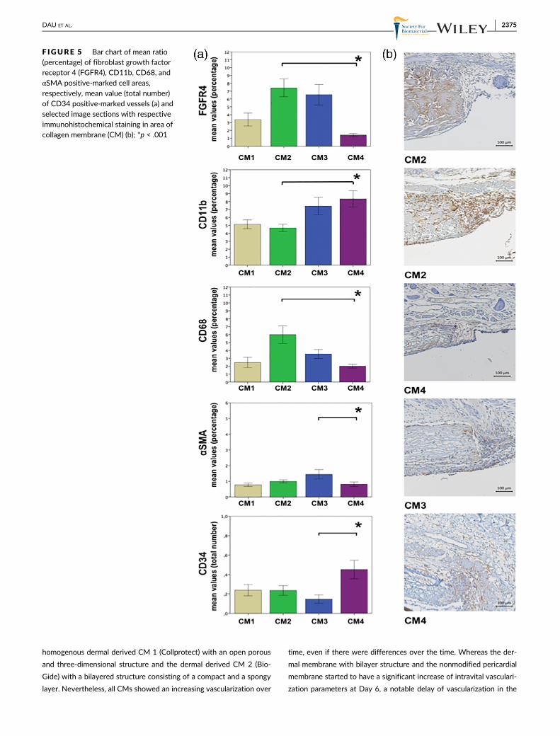
The pericardial membrane coated with BSM (CM4) showed the
lowest level of FGFR4-positive and CD68-positive cells when com-
pared to the bilayer dermal membrane (CM2; p < .001). For CD11b,
CM4 had significant higher values when compared to CM2 (p < .001).
CM3 showed the lowest number of CD34-positive vessels when com-
pared to the other membranes with a significant difference to CM4
(all: p < .004). Additionally, the highest values of αSMA-positive cells
were seen in CM3 when compared to CM4 (all: p < .04; Table 3 and
Figure 5).
4 | DISCUSSION
Whereas the beneficial effect of CMs in GBR in terms of bone regen-
eration has been proven in various studies, there is a lack of literature
on the active effects of and within the respective membranes. In
accordance, the membrane has traditionally been considered to be a
passive barrier only. However, more recent research gave evidence
that CMs actively promote bone formation and bone-remodeling
genes (Turri et al., 2016). Therefore, the aim of the study was to
examine the chronological pattern of vascularization of two porcine
dermal and two porcine pericardial CMs. It was shown, that there are
vast differences in the speed of vascularization over the time and the
extend of vascularization at the defined endpoint. In brief, the modifi-
cation of the pericardial CM with silica-enhanced nanostructured
hydroxyapatite resulted in the fastest and most pronounced vasculari-
zation of all tested membranes whereas the least activity was seen for
the dermal homogenous membrane. Therefore, the null hypothesis
was rejected. This is in accordance to the literature showing that the
addition of glass nanoparticles to resorbable membranes had a posi-
tive effect on cellular metabolic activity as well as on mineralization
and bone regeneration (Hong et al., 2010; Mota et al., 2012; Tirri
et al., 2008). Although to the best of our knowledge, this is the first
in vivo research covering the respective evolvement over time. These
different biological behaviors might influence the outcome of GBR as
membranes should support an early transmembraneous angiogenesis
without early degradation in order to promote osseous regeneration
(Rothamel et al., 2005; Schwarz et al., 2008). In accordance, the pore
size and structure of the GBR-membrane influences the degree of
bone regeneration within the defect since this determines diffusion of
nutrients and bioactive substances (Oh, Kim, Kim, & Lee, 2006) and a
total occlusive barrier was associated with a slow bone-tissue regen-
eration rate (Lundgren, Lundgren, & Taylor, 1998).
Both pericardial membranes (Jason with and without NanoBone)
used in this study consist of variable oriented collagen fibers creating
a comb-like structure that is characterized by multidirectional linking
(Elgali et al., 2017; Ortolani et al., 2015). This is different to the
TABLE 2 Mean ± SEM for measurement of intravital fluorescence microscopy (all values rounded to one, respectively, two decimal digits)
Day Vascularization parameter
Membrane
CM1 CM2 CM3 CM4 p-Valueb
3 CDI (μm) 0.09 ± 0.09 0.62 ± 0.31 0.10 ± 0.10 0.98 ± 0.39 .035
CDE (cm/cm2) 0.38 ± 0.38 2.78 ± 1.45 0.11 ± 0.11 3.26 ± 1.45 .035
CFL (mm/s) 0.003 ± 0.003 0.01 ± 0.07 a 0.04 ± 0.02 .026
CLE (μm) 12.1 ± 12.1 85.0 ± 44.0 1.7 ± 1.7 103.0 ± 45.7 .035
6 CDI (μm) 0.24 ± 0.17 0.78 ± 0.31 0.89 ± 0.33 2.45 ± 0.51 <.001
CDE (cm/cm2) 0.50 ± 0.36 4.32 ± 2.04 2.49 ± 1.40 14.79 ± 3.89 <.001
CFL (mm/s) 0.004 ± 0.004 0.03 ± 0.02 0.06 ± 0.03 0.07 ± 0.02 <.001
CLE (μm) 15.9 ± 11.3 131.4 ± 61.6 66.5 ± 42.3 467.1 ± 122.7 <.001
9 CDI (μm) 1.42 ± 0.45 2.40 ± 0.49 4.08 ± 0.63 5.49 ± 0.66 <.001
CDE (cm/cm2) 1.59 ± 0.65 14.80 ± 3.54 10.77 ± 2.94 38.19 ± 6.96 <.001
CFL (mm/s) 0.08 ± 0.04 0.05 ± 0.02 0.13 ± 0.04 0.13 ± 0.03 <.001
CLE (μm) 99.9 ± 52.4 413.1 ± 107.1 397.9 ± 99.0 1,195.9 ± 215.4 <.001
12 CDI (μm) 1.60 ± 0.50 4.07 ± 0.57 4.87 ± 0.64 5.15 ± 0.65 <.001
CDE (cm/cm2) 3.06 ± 1.13 33.91 ± 6.06 34.86 ± 9.03 69.61 ± 10.72 <.001
CFL (mm/s) 0.08 ± 0.03 0.09 ± 0.02 0.11 ± 0.03 0.11 ± 0.02 <.001
CLE (μm) 239.9 ± 99.7 1,033.3 ± 190.0 1,129.4 ± 283.3 2,184.9 ± 399.5 <.001
Abbreviations: CDE, capillary density; CDI, capillary diameter; CFL, capillary flow; CLE, capillary length; CM, collagen membrane.aNo measurable values found.bKruskal–Wallis test for differences between groups CM1, CM2, CM3, respectively, CM4 at each time point.
DAU ET AL. 2373
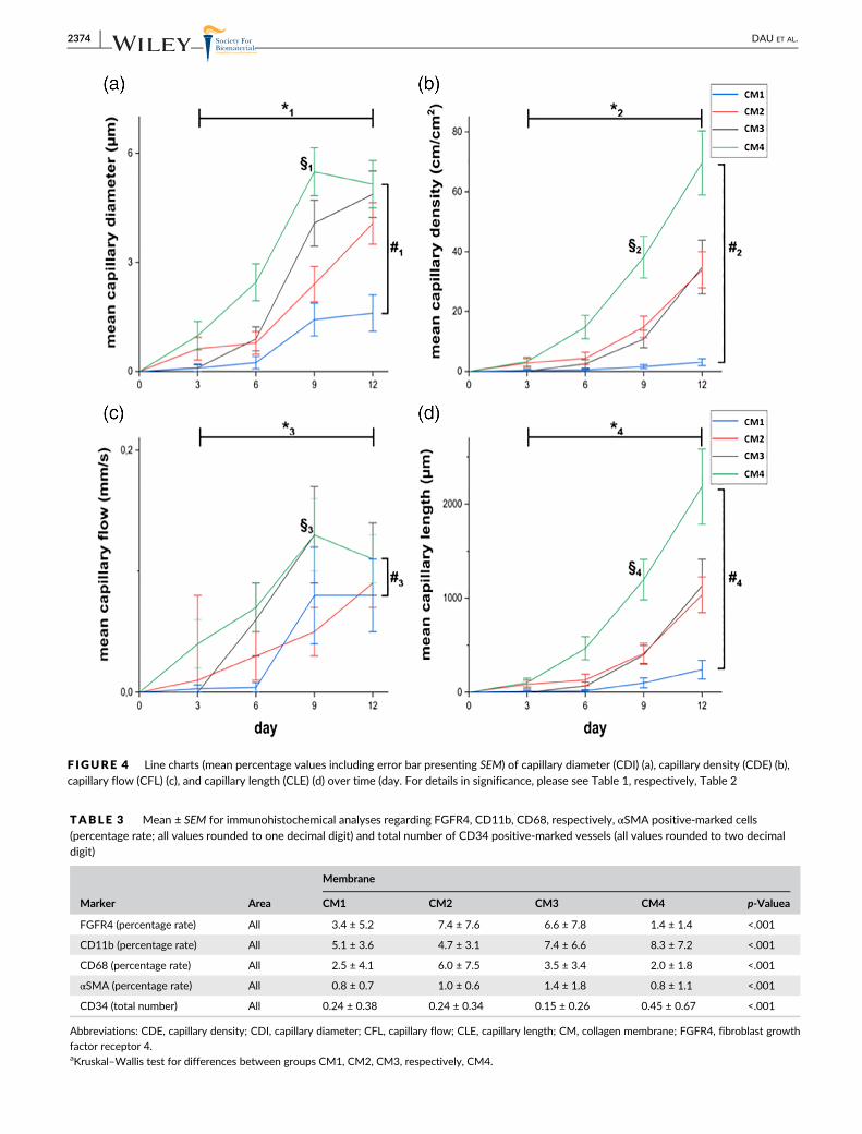
F IGURE 4 Line charts (mean percentage values including error bar presenting SEM) of capillary diameter (CDI) (a), capillary density (CDE) (b),capillary flow (CFL) (c), and capillary length (CLE) (d) over time (day. For details in significance, please see Table 1, respectively, Table 2
TABLE 3 Mean ± SEM for immunohistochemical analyses regarding FGFR4, CD11b, CD68, respectively, αSMA positive-marked cells(percentage rate; all values rounded to one decimal digit) and total number of CD34 positive-marked vessels (all values rounded to two decimaldigit)
Marker Area
Membrane
CM1 CM2 CM3 CM4 p-Valuea
FGFR4 (percentage rate) All 3.4 ± 5.2 7.4 ± 7.6 6.6 ± 7.8 1.4 ± 1.4 <.001
CD11b (percentage rate) All 5.1 ± 3.6 4.7 ± 3.1 7.4 ± 6.6 8.3 ± 7.2 <.001
CD68 (percentage rate) All 2.5 ± 4.1 6.0 ± 7.5 3.5 ± 3.4 2.0 ± 1.8 <.001
αSMA (percentage rate) All 0.8 ± 0.7 1.0 ± 0.6 1.4 ± 1.8 0.8 ± 1.1 <.001
CD34 (total number) All 0.24 ± 0.38 0.24 ± 0.34 0.15 ± 0.26 0.45 ± 0.67 <.001
Abbreviations: CDE, capillary density; CDI, capillary diameter; CFL, capillary flow; CLE, capillary length; CM, collagen membrane; FGFR4, fibroblast growth
factor receptor 4.aKruskal–Wallis test for differences between groups CM1, CM2, CM3, respectively, CM4.
2374 DAU ET AL.
homogenous dermal derived CM 1 (Collprotect) with an open porous
and three-dimensional structure and the dermal derived CM 2 (Bio-
Gide) with a bilayered structure consisting of a compact and a spongy
layer. Nevertheless, all CMs showed an increasing vascularization over
time, even if there were differences over the time. Whereas the der-
mal membrane with bilayer structure and the nonmodified pericardial
membrane started to have a significant increase of intravital vasculari-
zation parameters at Day 6, a notable delay of vascularization in the
F IGURE 5 Bar chart of mean ratio(percentage) of fibroblast growth factorreceptor 4 (FGFR4), CD11b, CD68, andαSMA positive-marked cell areas,respectively, mean value (total number)of CD34 positive-marked vessels (a) andselected image sections with respectiveimmunohistochemical staining in area ofcollagen membrane (CM) (b); *p < .001
DAU ET AL. 2375

homogenous CM was detected. This is supported by increased
FGFR4- and αSMA-values when compared to the homogenous dermal
membrane. Analogue to this, significant differences in angiogenetic pat-
tern were described between various CMs already (Schwarz
et al., 2006), even though these results were based on immuno-
histomorphometrical analyses only. In contrast to these findings only a
mild vascularization of the porcine dermal bilayer membrane was seen
within 6 weeks in a subcutaneous animal model (Ghanaati, 2012), that
could be attributed to the different test approach.
In the present study, an increase of CD68-positive cells and an
increase of other vascularization parameters were seen in the dermal
bilayer and in the nonmodified pericardial membrane. When com-
pared to the homogenous dermal membrane with significant lower
vascularization parameters, this might indicate an association between
vascularization and reorganization events.
The porcine pericardial membrane coated with synthetic BSM
showed even higher vascularization parameters starting earlier at Day
3 even if CD68-values after 12 days were lower when compared to the
dermal bilayer membrane. Together with the lower expression of FGFR4
and higher numbers CD34-marked vessels, this might be an indication
for a combination of faster vascularization and faster reorganization.
Modification of a CM with a BSM may possibly enhance the
respective mechanical properties (Anderud et al., 2014; Chu, Deng,
Man, & Qu, 2017) and there is evidence for a beneficial influence of
such a membrane on promotion of bone regeneration (Chu
et al., 2017; Li et al., 2011; Teng et al., 2009). In accordance, this
material might improve known success factors of barrier membranes
in GBR such as space maintaining capacity, cell occlusiveness and
bioactivation of friendly materials (Caballe-Serrano et al., 2018). As
angiogenesis and vascularization is known to be central for successful
GBR-procedures, its improvement is crucial for an enhanced bone
regeneration. Even so, the clinical impact of such a modification
should be analyzed in future research in order to optimize the biologi-
cal behavior of CMs and to identify optimal material combinations.
5 | CONCLUSION
Structural different CMs from porcine origin show different biological
behaviors in terms of vascularization over time. In accordance, CMs of
dermal origin with a bilayer structure as well as from pericardial origin
were associated with a faster vascularization when compared to
homogenous CMs. Additional spraying of a pericardial CM with a sil-
icium dioxide enriched nanostructured synthetic hydroxyapatite sig-
nificantly increased diameter, density and length of surrounding
vessels over time. Together with the observed high levels of CD11b
and CD34 marked vessels as well as a low amount of FGFR4, this
might be a hint for an even faster vascularization and biological remo-
deling process.
ACKNOWLEDGMENTS
The authors thank Cornelia Ganz for providing the modified CMs used
in the experiment and Daniel Wolter for excellent technical
assistance. This research received a specific grant from DGI (Deutsche
Gesellschaft für Implantologie im Zahn-, Mund- und Kieferbereich e.
V., Munich, Germany). No additional funding was made.
CONFLICT OF INTEREST
The authors declare that they have no conflict of interests and finan-
cial interests related to any products involved in this study.
ETHICS STATEMENT
All applicable international, national, and/or institutional guidelines for
the care and use of animals were followed.
ORCID
Peer W. Kämmerer https://orcid.org/0000-0002-1671-3764
REFERENCES
Abshagen, K., Schrodi, I., Gerber, T., & Vollmar, B. (2009). In vivo analysis
of biocompatibility and vascularization of the synthetic bone grafting
substitute NanoBone. Journal of Biomedical Materials Research. Part A,
91, 557–566.Adam, M., Ganz, C., Xu, W., Sarajian, H. R., Gotz, W., & Gerber, T. (2014). In
vivo and in vitro investigations of a nanostructured coating material—A
preclinical study. International Journal of Nanomedicine, 9, 975–984.Amorfini, L., Migliorati, M., Signori, A., Silvestrini-Biavati, A., &
Benedicenti, S. (2014). Block allograft technique versus standard
guided bone regeneration: A randomized clinical trial. Clinical Implant
Dentistry and Related Research, 16, 655–667.Anderud, J., Jimbo, R., Abrahamsson, P., Isaksson, S. G., Adolfsson, E.,
Malmstrom, J., … Wennerberg, A. (2014). Guided bone augmentation
using a ceramic space-maintaining device. Oral Surgery, Oral Medicine,
Oral Pathology, Oral Radiology, 118, 532–538.Blatt, S., Burkhardt, V., Kämmerer, P. W., Pabst, A. M., Sagheb, K., Heller, M.,
… Schiegnitz, E. (2020). Biofunctionalization of porcine-derived collagen
matrices with platelet rich fibrin: Influence on angiogenesis in vitro and
in vivo. Clinical Oral Investigations. [Epub ahead of print].
Bunyaratavej, P., & Wang, H. L. (2001). Collagen membranes: A review.
Journal of Periodontology, 72, 215–229.Caballe-Serrano, J., Munar-Frau, A., Ortiz-Puigpelat, O., Soto-Penaloza, D.,
Penarrocha, M., & Hernandez-Alfaro, F. (2018). On the search of the
ideal barrier membrane for guided bone regeneration. Journal of Clini-
cal and Experimental Dentistry, 10, e477–e483.Chiapasco, M., & Zaniboni, M. (2009). Clinical outcomes of GBR proce-
dures to correct peri-implant dehiscences and fenestrations: A system-
atic review. Clinical Oral Implants Research, 20(Suppl 4), 113–123.Chu, C., Deng, J., Man, Y., & Qu, Y. (2017). Evaluation of
nanohydroxyapaptite (nano-HA) coated epigallocatechin-3-gallate
(EGCG) cross-linked collagen membranes. Materials Science &
Engineering. C, Materials for Biological Applications, 78, 258–264.Dau, M., Kämmerer, P. W., Henkel, K. O., Gerber, T., Frerich, B., &
Gundlach, K. K. (2016). Bone formation in mono cortical mandibular
critical size defects after augmentation with two synthetic nanostruc-
tured and one xenogenous hydroxyapatite bone substitute—In vivo
animal study. Clinical Oral Implants Research, 27, 597–603.De Vriese, A. S., Verbeuren, T. J., Vallez, M. O., Lameire, N. H., De
Buyzere, M., & Vanhoutte, P. M. (2000). Off-line analysis of red blood
cell velocity in renal arterioles. Journal of Vascular Research, 37, 26–31.Elgali, I., Omar, O., Dahlin, C., & Thomsen, P. (2017). Guided bone regener-
ation: Materials and biological mechanisms revisited. European Journal
of Oral Sciences, 125, 315–337.Fujioka-Kobayashi, M., Schaler, B., Shirakata, Y., Nakamura, T.,
Noguchi, K., Zhang, Y., & Miron, R. J. (2017). Comparison of two
2376 DAU ET AL.

porcine collagen membranes combined with rhBMP-2 and rhBMP-9
on osteoblast behavior in vitro. The International Journal of Oral & Max-
illofacial Implants, 32, e221–e230.Ghanaati, S. (2012). Non-cross-linked porcine-based collagen I-III mem-
branes do not require high vascularization rates for their integration
within the implantation bed: A paradigm shift. Acta Biomaterialia, 8,
3061–3072.Ghanaati, S., Barbeck, M., Lorenz, J., Stuebinger, S., Seitz, O., Landes, C., …
Sader, R. A. (2013). Synthetic bone substitute material comparable
with xenogeneic material for bone tissue regeneration in oral cancer
patients: First and preliminary histological, histomorphometrical and
clinical results. Annals of Maxillofacial Surgery, 3, 126–138.Hong, K. S., Kim, E. C., Bang, S. H., Chung, C. H., Lee, Y. I., Hyun, J. K., …
Kim, H. W. (2010). Bone regeneration by bioactive hybrid membrane
containing FGF2 within rat calvarium. Journal of Biomedical Materials
Research. Part A, 94, 1187–1194.Kämmerer, P. W., Palarie, V., Schiegnitz, E., Nacu, V., Draenert, F. G., & Al-
Nawas, B. (2013). Influence of a collagen membrane and recombinant
platelet-derived growth factor on vertical bone augmentation in
implant-fixed deproteinized bovine bone—Animal pilot study. Clinical
Oral Implants Research, 24, 1222–1230.Kämmerer, P. W., Scholz, M., Baudisch, M., Liese, J., Wegner, K.,
Frerich, B., & Lang, H. (2017). Guided bone regeneration using collagen
scaffolds, growth factors, and periodontal ligament stem cells for treat-
ment of peri-implant bone defects in vivo. Stem Cells International,
2017, 3548435.
Keestra, J. A., Barry, O., Jong, L., & Wahl, G. (2016). Long-term effects of
vertical bone augmentation: A systematic review. Journal of Applied
Oral Science, 24, 3–17.Kilkenny, C., Browne, W. J., Cuthill, I. C., Emerson, M., & Altman, D. G.
(2010). Improving bioscience research reporting: The ARRIVE guide-
lines for reporting animal research. Journal of Pharmacology and
Pharmacotherapeutics, 1, 94–99.Klyscz, T., Junger, M., Jung, F., & Zeintl, H. (1997). Cap image—A new kind of
computer-assisted video image analysis system for dynamic capillary
microscopy. Biomedizinische Technik. Biomedical Engineering, 42, 168–175.Koerdt, S., Ristow, O., Wannhoff, A., Kubler, A. C., & Reuther, T. (2014).
Expression of growth factors during the healing process of alveolar ridge
augmentation procedures using autogenous bone grafts in combination
with GTR and an anorganic bovine bone substitute: An immunohisto-
chemical study in the sheep. Clinical Oral Investigations, 18, 179–188.Kyyak, S., Blatt, S., Pabst, A., Thiem, D., Al-Nawas, B., & Kämmerer, P. W.
(2020). Combination of an allogenic and a xenogenic bone substitute
material with injectable platelet-rich fibrin—A comparative in vitro
study. Journal of Biomaterials Applications, 088532822091440. [Epub
ahead of print].
Li, J., Man, Y., Zuo, Y., Zhang, L., Huang, C., Liu, M., & Li, Y. (2011). In vitro
and in vivo evaluation of a nHA/PA66 composite membrane for
guided bone regeneration. Journal of Biomaterials Science. Polymer Edi-
tion, 22, 263–275.Liu, J., & Kerns, D. G. (2014). Mechanisms of guided bone regeneration: A
review. The Open Dentistry Journal, 8, 56–65.Lundgren, A., Lundgren, D., & Taylor, A. (1998). Influence of barrier
occlusiveness on guided bone augmentation. An experimental study in
the rat. Clinical Oral Implants Research, 9, 251–260.Mahajan, R., Khinda, P., Shewale, A., Ghotra, K., Bhasin, M. T., & Bhasin, P.
(2018). Comparative efficacy of placental membrane and healiguide in
treatment of gingival recession using guided tissue regeneration. Jour-
nal of Indian Society of Periodontology, 22, 513–522.Mota, J., Yu, N., Caridade, S. G., Luz, G. M., Gomes, M. E., Reis, R. L., …
Mano, J. F. (2012). Chitosan/bioactive glass nanoparticle composite
membranes for periodontal regeneration. Acta Biomaterialia, 8,
4173–4180.
Naenni, N., Schneider, D., Jung, R. E., Husler, J., Hammerle, C. H. F., &
Thoma, D. S. (2017). Randomized clinical study assessing two mem-
branes for guided bone regeneration of peri-implant bone defects:
Clinical and histological outcomes at 6 months. Clinical Oral Implants
Research, 28, 1309–1317.Oh, S. H., Kim, J. H., Kim, J. M., & Lee, J. H. (2006). Asymmetrically porous
PLGA/Pluronic F127 membrane for effective guided bone regenera-
tion. Journal of Biomaterials Science. Polymer Edition, 17, 1375–1387.Ortolani, E., Quadrini, F., Bellisario, D., Santo, L., Polimeni, A., &
Santarsiero, A. (2015). Mechanical qualification of collagen membranes
used in dentistry. Annali dell'Istituto Superiore di Sanità, 51, 229–235.Pabst, A., & Kämmerer, P. W. (2020). Collagen matrices: Opportunities and
perspectives in oral hard and soft tissue regeneration. Quintessence
International, 51, 318–327.Park, J. S., Pabst, A. M., Ackermann, M., Moergel, M., Jung, J., & Kasaj, A.
(2018). Biofunctionalization of porcine-derived collagen matrix using
enamel matrix derivative and platelet-rich fibrin: Influence on mature
endothelial cell characteristics in vitro. Clinical Oral Investigations, 22,
909–917.Rothamel, D., Benner, M., Fienitz, T., Happe, A., Kreppel, M.,
Nickenig, H. J., & Zoller, J. E. (2014). Biodegradation pattern and tissue
integration of native and cross-linked porcine collagen soft tissue aug-
mentation matrices—An experimental study in the rat. Head & Face
Medicine, 10, 10.
Rothamel, D., Schwarz, F., Sager, M., Herten, M., Sculean, A., & Becker, J.
(2005). Biodegradation of differently cross-linked collagen mem-
branes: An experimental study in the rat. Clinical Oral Implants
Research, 16, 369–378.Schwarz, F., Rothamel, D., Herten, M., Sager, M., & Becker, J. (2006).
Angiogenesis pattern of native and cross-linked collagen membranes:
An immunohistochemical study in the rat. Clinical Oral Implants
Research, 17, 403–409.Schwarz, F., Rothamel, D., Herten, M., Wustefeld, M., Sager, M.,
Ferrari, D., & Becker, J. (2008). Immunohistochemical characterization
of guided bone regeneration at a dehiscence-type defect using differ-
ent barrier membranes: An experimental study in dogs. Clinical Oral
Implants Research, 19, 402–415.Sckell, A., & Leunig, M. (2009). The dorsal skinfold chamber: Studying
angiogenesis by intravital microscopy. Methods in Molecular Biology,
467, 305–317.Sckell, A., & Leunig, M. (2016). Dorsal skinfold chamber preparation in
mice: Studying angiogenesis by intravital microscopy. Methods in
Molecular Biology, 1430, 251–263.Shim, J. H., Huh, J. B., Park, J. Y., Jeon, Y. C., Kang, S. S., Kim, J. Y., …
Cho, D. W. (2013). Fabrication of blended polycaprolactone/poly(lac-
tic-co-glycolic acid)/beta-tricalcium phosphate thin membrane using
solid freeform fabrication technology for guided bone regeneration.
Tissue Engineering. Part A, 19, 317–328.Silva, E. C., Omonte, S. V., Martins, A. G., de Castro, H. H., Gomes, H. E.,
Zenobio, E. G., … Souza, P. E. (2017). Hyaluronic acid on collagen
membranes: An experimental study in rats. Archives of Oral Biology, 73,
214–222.Talebi Ardakani, M. R., Hajizadeh, F., & Yadegari, Z. (2018). Comparison of
attachment and proliferation of human gingival fibroblasts on different
collagen membranes. Annals of Maxillofacial Surgery, 8, 218–223.Teng, S. H., Lee, E. J., Yoon, B. H., Shin, D. S., Kim, H. E., & Oh, J. S. (2009).
Chitosan/nanohydroxyapatite composite membranes via dynamic fil-
tration for guided bone regeneration. Journal of Biomedical Materials
Research. Part A, 88, 569–580.Tirri, T., Rich, J., Wolke, J., Seppala, J., Yli-Urpo, A., & Narhi, T. O. (2008).
Bioactive glass induced in vitro apatite formation on composite GBR
membranes. Journal of Materials Science. Materials in Medicine, 19,
2919–2923.
DAU ET AL. 2377

Turri, A., Elgali, I., Vazirisani, F., Johansson, A., Emanuelsson, L., Dahlin, C., …Omar, O. (2016). Guided bone regeneration is promoted by the molecu-
lar events in the membrane compartment. Biomaterials, 84, 167–183.
SUPPORTING INFORMATION
Additional supporting information may be found online in the
Supporting Information section at the end of this article.
How to cite this article: Dau M, Volprich L, Grambow E, et al.
Collagen membranes of dermal and pericardial origin—In vivo
evolvement of vascularization over time. J Biomed Mater Res.
2020;108:2368–2378. https://doi.org/10.1002/jbm.a.36989
2378 DAU ET AL.