cognitive neuroscience: tracking the time course of object...
TRANSCRIPT

Cognitive Neuroscience NeuroReport
0959-4965 # Lippincott Williams & Wilkins
Tracking the time courseof object categorizationusing event-relatedpotentials
James Tanaka,CA Phan Luu,1
Matthias Weisbrod2 and Markus Kiefer3
Department of Psychology, Oberlin College,Oberlin, OH 44074; 1Department of Psychology,University of Oregon, OR 44074, USA;2Department of Psychiatry, University ofHeidelberg D69115 Heidelberg; 3Department ofPsychology, University of Ulm, D8970 UlmGermany
CACorresponding Author
OBJECT categorization processes were investigated bymeasuring event-related potentials while subjects cate-gorized objects at the superordinate (e.g. animal), basic(e.g. dog) and subordinate (e.g. beagle) levels of abstrac-tion. An enhanced negative de¯ection (N1) was foundat posterior recording sites for subordinate level cate-gorizations compared with basic level categorizationsand was interpreted as a marker of increased visualanalysis. In contrast, superordinate level categorizationsproduced a larger frontal negativity relative to basiclevel categorizations and were interpreted as an indica-tor of increased semantic processing. These results sug-gest a neurophysiological basis for the separatecognitive processes responsible for subordinate andsuperordinate object categorizations. NeuroReport10:829±835 # 1999 Lippincott Williams & Wilkins.
Key words: Event-related potentials; Object recognition;Semantics; Visual perception
Introduction
In object naming, the same object can be referred toby many names. For example, the object dog can becalled an animal at a general (superordinate) level ofabstraction, a dog at an intermediate (basic) level,and a golden retriever at a speci®c (subordinate)level. While any object can be categorized at multi-ple levels of abstraction, it has been argued that onelevel, referred to as the basic level, occupies aprivileged position in the object hierarchy. The basiclevel is de®ned as the most inclusive level at whichcategory members share a common shape. Membersof the same basic level category (e.g. dog) tend toshare the same structural features and, therefore,bear a physical resemblance to one another. Thespecial status of basic level categories in objectrecognition has been established in naming andcategory veri®cation tasks where people are fastestto categorize objects at the basic level (e.g. apple,chair, dog) and slower to categorize objects atsuperordinate (e.g. fruit, furniture, animal) and sub-ordinate (e.g. Macintosh apple, kitchen chair, beagle)levels of abstraction [1±3]. Based on these data, ithas been suggested that the basic level is the point atwhich the perceived object stimulus ®rst makescontact with an object representation stored inmemory [1].
According to current object categorization theory,
subsequent activation of categories superordinateand subordinate to the basic level rely on distinctcognitive processes. Relative to basic level categor-izations, it has been claimed that superordinate levelcategorizations require abstract semantic processing.Some support for this view comes from featurelisting studies in which a higher proportion offunctional and behavioral properties of the object(e.g. eat, breathe, reproduce) are mentioned forsuperordinate level concepts (e.g. animal) [3±5].Other studies have shown that the times requiredfor superordinate level categorization of basic levelwords are highly correlated with superordinate levelcategorization of basic level pictures [1]. This latterresult indicates that categorizations at the super-ordinate level are not directly bound to the formatof the stimulus (i.e. word or picture), but are basedon the abstract semantic association between basicand superordinate level concepts.
Subordinate level categorization, on the otherhand, has been linked to perceptual processes. Forexample, in a feature listing task, a large number ofperceptual features (e.g. small size, brown for spar-row) and modi®ed part features (e.g. long beak forwoodpecker) are mentioned for subordinate levelobjects, suggesting that this level of categorizationrequires a ®ner grain of perceptual analysis. Accord-ingly, it has been shown that in an object recogni-tion task, short exposures of the picture stimulus
NeuroReport 10, 829±835 (1999)
Vol 10 No 4 17 March 1999 829
(i.e. 75 ms) interfered with subordinate level categor-izations but not basic or superordinate decisions[1,6]. These experiments suggest that subordinatecategorizations demand an increased amount ofvisual analysis.
If different cognitive operations are recruited tocategorize objects at different levels of abstractionthen the brain areas associated with those operationsshould be differentially engaged in an object cate-gorization task. Consistent with this view, prefrontalbrain areas are more active when subjects categor-ized objects at the superordinate level than when thesame objects were categorized at the basic level [7].Presumably, prefrontal activation re¯ects the acces-sing of semantic information necessary for super-ordinate level judgments. In contrast, otherneuroimaging studies have shown that subordinatelevel categorization activated fusiform and inferiortemporal gyri (FIT) regions as well as the temporalpoles [8], areas associated with visual object recogni-tion. Thus, the above studies suggest that super-ordinate and subordinate level categorizations aredissociable with respect to their neural substrates.
Object categorization processes have not beendirectly examined using ERP methods. However,related electrophysioloigcal studies have indicatedthat semantic and visual processes can be dissociatedwith respect to their time course and topographiclocations. As an indicator of semantic processes, ithas been reported that left frontal areas are differen-tially activated as early as 200 ms after stimulusonset in tasks involving noun±verb associations [9].As an index of visual analysis, an enhanced earlypositive component and negative component areelicited when subjects must selectively attend to aone-stimulus dimension (e.g. color) while ignoringother stimulus dimensions (e.g. shape) [10,11].Although the exact source of the visual attentionalcomponents cannot be precisely determined fromscalp recordings, both the early positive and negativecomponents appear to originate in posterior, visualbrain areas (e.g. occipital, occipital±temporal areas)[10].
In the current experiment, the temporal dynamicsinvolved in object categorization are directly evalu-ated. Following the standard category veri®cationparadigm, subjects categorized objects at superordi-nate (e.g. animal), basic (e.g. bird) and subordinate(e.g. robin) levels of categorization while wearing ahigh density electrode net that monitored electro-physiological activity at 64 scalp sites. If visual andsemantic processes are localizable to different neuralsubstrates, brain areas associated with visual process-ing (i.e. posterior areas) should be differentiallyactivated when subjects categorize objects at thesubordinate level of abstraction. In contrast, brain
regions associated with semantic processing (i.e.frontal areas) should be differentially activated whensubjects categorize objects at the superordinate level.
Materials and Methods
Subjects: Twenty-eight right-handed undergraduatestudents from the University of Oregon participatedin this study. All subjects were native English speak-ers and had normal or corrected-to-normal vision.Subjects were paid for their participation.
Materials: Stimuli for the experiment consisted of40 color pictures from four natural (animal, plant,fruit, vegetable) and six artifactual (tool, furniture,musical instrument, sports equipment, vehicle,weapon) categories. These objects were judged astypical exemplars of their category according to theBattig and Montague [12] normative ratings. Pic-tures were taken primarily from McMillan's VisualDictionary [13] and digitized with a MicroTek ZScanner. Each image was scaled to ®t within a100 3 100 pixel array. Left-facing and right-facingversions of each picture were generated and pre-sented randomly in the experiment.
Procedure: Subjects were tested in an electrically-shielded booth. After subjects read a list of the 40objects included in the study, they were seated infront of the computer monitor with their headstabilized in a chin rest. From the subjects' viewingdistance of 60 cm, the picture stimuli subtended avisual angle of ,0.58 in the horizontal and verticaldimensions. At the beginning of each trial, a ®xationpoint (a plus sign symbol) appeared on the computermonitor for a random interval that varied between1000 and 1500 ms. The ®xation point was replacedby a category name (superordinate, basic or subordi-nate) for 255 ms and was then replaced by the®xation point for 570 ms. The picture stimulus wasthen presented for 255 ms and was replaced by the®xation point for 735 ms. At the end of the trial, the`true/false' screen appeared prompting subjects fortheir response. If the picture matched the categoryword, subjects were instructed to press the `true'key; otherwise they were to press the `false' key. Inthe false condition, foil category labels were drawnfrom a contrast category that was at the same levelof abstraction as the target. For example, the cate-gory labels furniture, saw, and claw hammer pre-ceded the picture of the ball peen hammer in thesuperordinate, basic, and subordinate false trials,respectively. EEG was recorded for 2 s during eachtrial, beginning 195 ms prior to the onset of thecategory word and was terminated prior to subjectresponse at the onset of the true/false screen. The
830 Vol 10 No 4 17 March 1999
NeuroReport J. Tanaka et al.

inter-trial interval was randomly varied from 2 to2.5 s. Each of the 40 picture stimuli were tested inthree category conditions (superordinate, basic, sub-ordinate) and two response conditions (true, false)yielding a total of 240 randomly presented trials.
EEG recording: EEG was recorded with the Geo-desic Sensor Net [14] with an array of 57 Ag/AgClsponge sensors placed on the scalp surface with anadditional seven Ag/AgCl surface sensors placed ateach infra-orbital, external canthus, and mastoidsites and at the glabella. The impedance of allelectrodes was below 40 kÙ (10±40; k� 87). Allrecordings were referenced to the right mastoid.EEG recordings from the 64 sites were ampli®edand ®ltered with 0.01±50 Hz (3 dB attenuation)bandpass and a 60 Hz notch ®lter. The signals weresampled at 125 samples/s and were digitized with a16 bit analog-to-digital converter (National Instru-ments NB-MIO 16). Editing of the EEG for move-ment, eye movement and blink artifacts wasperformed by a computerized artifact detectionalgorithm. Trials containing blinks (30 ìV differencebetween vertical EOG channels) were omitted fromanalysis. Channels were also eliminated if theychanged by . 50 ìV between samples or exceeded� 100 ìV.
The artifact-free EEG was then averaged for eachsubject across all correct trials. The activity of allrecorded sites was average referenced in which theactivity of a given site was expressed as the differ-ence between the site and the average of all otherrecording sites [15,16]. Although the underside ofthe brain is poorly represented by this method,analyses with the 64-channel Geodesic Sensor Netshow that the average reference closely approximatesthe distance-weighted average reference [16]. Theaverage reference transformation provides a refer-ence independent estimation of scalp voltage. Theaveraged epochs were digitally ®ltered with a 30 Hzlowpass to remove any artifacts due to the 67 Hzrefresh rate of the monitor. EEG was baseline-corrected for the 195 ms interval prior to the pre-sentation of the category word and 50 ms intervalprior to the picture stimulus. A ®nal grand averagewas obtained by averaging across the subjectaverages for each experimental condition.
Results
During the word epoch (0±1020 ms), an enhancedpositivity was produced by subordinate level wordsat the central recording sites relative to the basic andsuperordinate level words. However, at the end ofthe word epoch and prior to the picure epoch(1020±2000 ms), EEG activity returned to baseline
levels and no differences were found between thesuperordinate, basic and subordinate level categoryconditions ( P , 0.10). Because the focus on thisstudy was on processing during the picture, nofurther analyses were performed on word epochERPs.
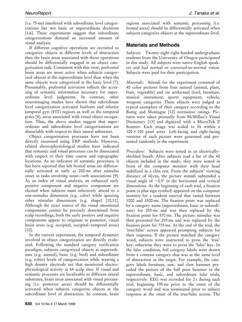
For analysis of the ERPs to the picture stimulus,waveforms were baseline adjusted 100 ms prior tothe onset of the picture. Figure 1 shows the averagedERP waveform for superordinate, basic and subordi-nate conditions collapsed across true and falseresponses during the picture epoch. Beginning,100 ms after the onset of the picture stimulus, apositive de¯ection (P1) was demonstrated across allthree category conditions. The P1 was followed by anegative de¯ection in the posterior channels (N1),140 ms after onset of the picture stimulus. Whilethe N1 was detected in all three category conditions,subordinate level categorizations generated an en-hanced N1 amplitude in left posterior channelsrelative to basic and superordinate level, as shown inFig. 1. As the topographic voltage map of Fig. 2ashows, the difference between subordinate and basiclevel activation peaked about 160 ms after the pic-ture was presented.
An analysis of variance performed on the latenciesof the N1 component was performed for super-ordinate, basic and subordinate level categorizationsof the posterior channels (channels 16, 20, 22, 23, 25,26, 30, 31, 34) [17]. The ANOVA showed nodifferences across the three category conditions withrespect to their latencies ( P . 0.10). Using a latencywindow of 139±179 ms, the mean amplitudes of theN1 component were submitted to a three-wayANOVA with the factors of category level (super-ordinate, basic, subordinate), response (true, false)and laterality (left, right) as within-subject variables.The signi®cant effect of category level (F(2,26)�15.30, MSe� 51,080,131, P , 0.001) demonstratedthe differences in ERP produced by superordinate,basic and subordinate level categorizations. Criti-cally, direct comparisons revealed that the subordi-nate categorizations were signi®cantly more negativethan basic and superordinate categorizations ( P ,0.001). Laterality interacted with category (F(2,52)�4.40, MSe� 8,421,253, P , 0.02), such that subordi-nate categorizations produced a greater negativede¯ection in the left hemisphere than in the righthemisphere. No other main effects or interactionswere signi®cant.
A brain electrical source analysis (BESA) [18] wasapplied to further explore possible sources of theERP effect attributable to subordinate categoriza-tion. Dipoles were ®tted on the difference ERPsbetween the subordinate and basic level conditionswithin the same time window of 139±179 ms post-
Vol 10 No 4 17 March 1999 831
Tracking the time course of object categorization using ERP NeuroReport
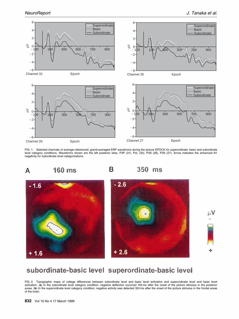
FIG. 1. Selected channels of average-referenced, grand-averaged ERP waveforms during the picture EPOCH for superordinate, basic and subordinatelevel category conditions. Waveforms shown are the left posterior sites, P3P (31), PzL (35), P3A (26), P3S (27). Arrow indicates the enhanced N1negativity for subordinate level categorizations.
6
4
2
0
24
26
µV
22
2100 100 300 500 700 900
SuperordinateBasicSubordinate
EpochChannel 31
6
4
2
0
24
26
µV
22
2100 100 300 500 700 900
SuperordinateBasicSubordinate
EpochChannel 35
6
4
2
0
24
26
µV
22
SuperordinateBasicSubordinate
EpochChannel 26
2100 100 300 500 700 900
6
4
2
0
24
26
µV
22
SuperordinateBasicSubordinate
EpochChannel 27
2100 100 300 500 700 900
FIG. 2. Topographic maps of voltage differences between subordinate level and basic level activation and superordinate level and basic levelactivation. (a) In the subordinate level category condition, negative de¯ection occurred 164 ms after the onset of the picture stimulus in the posteriorareas. (b) In the superordinate level category condition, negative activity was detected 324 ms after the onset of the picture stimulus in the frontal areasof the brain.
832 Vol 10 No 4 17 March 1999
NeuroReport J. Tanaka et al.
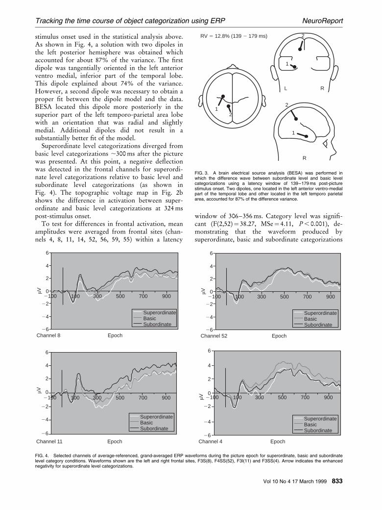
stimulus onset used in the statistical analysis above.As shown in Fig. 4, a solution with two dipoles inthe left posterior hemisphere was obtained whichaccounted for about 87% of the variance. The ®rstdipole was tangentially oriented in the left anteriorventro medial, inferior part of the temporal lobe.This dipole explained about 74% of the variance.However, a second dipole was necessary to obtain aproper ®t between the dipole model and the data.BESA located this dipole more posteriorly in thesuperior part of the left temporo-parietal area lobewith an orientation that was radial and slightlymedial. Additional dipoles did not result in asubstantially better ®t of the model.
Superordinate level categorizations diverged frombasic level categorizations ,300 ms after the picturewas presented. At this point, a negative de¯ectionwas detected in the frontal channels for superordi-nate level categorizations relative to basic level andsubordinate level categorizations (as shown inFig. 4). The topographic voltage map in Fig. 2bshows the difference in activation between super-ordinate and basic level categorizations at 324 mspost-stimulus onset.
To test for differences in frontal activation, meanamplitudes were averaged from frontal sites (chan-nels 4, 8, 11, 14, 52, 56, 59, 55) within a latency
window of 306±356 ms. Category level was signi®-cant (F(2,52)� 38.27, MSe� 4.11, P , 0.001), de-monstrating that the waveform produced bysuperordinate, basic and subordinate categorizations
12
R
L R
2
2
1
1
RV 5 12.8% (139 2 179 ms)
FIG. 3. A brain electrical source analysis (BESA) was performed inwhich the difference wave between subordinate level and basic levelcategorizations using a latency window of 139±179 ms post-picturestimulus onset. Two dipoles, one located in the left anterior ventro-medialpart of the temporal lobe and other located in the left temporo parietalarea, accounted for 87% of the difference variance.
FIG. 4. Selected channels of average-referenced, grand-averaged ERP waveforms during the picture epoch for superordinate, basic and subordinatelevel category conditions. Waveforms shown are the left and right frontal sites, F3S(8), F4SS(52), F3l(11) and F3SS(4). Arrow indicates the enhancednegativity for superordinate level categorizations.
6
4
2
0
24
26
µV
22
2100 100 300 500 700 900
SuperordinateBasicSubordinate
EpochChannel 8
6
4
2
0
24
26
µV
22
2100 100 300 500 700 900
SuperordinateBasicSubordinate
EpochChannel 52
6
4
2
0
24
26
µV
22
2100 100 300 500 700 900
SuperordinateBasicSubordinate
EpochChannel 11
6
4
2
0
24
26
µV
22
2100 100 300 500 700 900
SuperordinateBasicSubordinate
EpochChannel 4
Vol 10 No 4 17 March 1999 833
Tracking the time course of object categorization using ERP NeuroReport
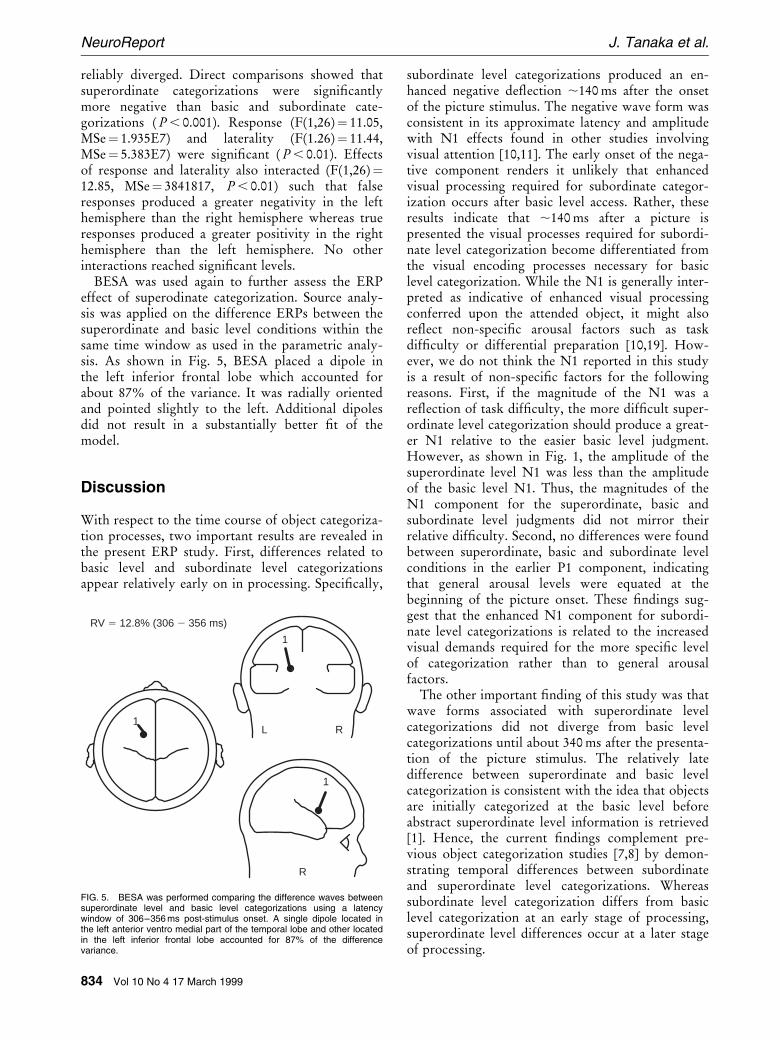
reliably diverged. Direct comparisons showed thatsuperordinate categorizations were signi®cantlymore negative than basic and subordinate cate-gorizations ( P , 0.001). Response (F(1,26)� 11.05,MSe� 1.935E7) and laterality (F(1.26)� 11.44,MSe� 5.383E7) were signi®cant ( P , 0.01). Effectsof response and laterality also interacted (F(1,26)�12.85, MSe� 3841817, P , 0.01) such that falseresponses produced a greater negativity in the lefthemisphere than the right hemisphere whereas trueresponses produced a greater positivity in the righthemisphere than the left hemisphere. No otherinteractions reached signi®cant levels.
BESA was used again to further assess the ERPeffect of superodinate categorization. Source analy-sis was applied on the difference ERPs between thesuperordinate and basic level conditions within thesame time window as used in the parametric analy-sis. As shown in Fig. 5, BESA placed a dipole inthe left inferior frontal lobe which accounted forabout 87% of the variance. It was radially orientedand pointed slightly to the left. Additional dipolesdid not result in a substantially better ®t of themodel.
Discussion
With respect to the time course of object categoriza-tion processes, two important results are revealed inthe present ERP study. First, differences related tobasic level and subordinate level categorizationsappear relatively early on in processing. Speci®cally,
subordinate level categorizations produced an en-hanced negative de¯ection ,140 ms after the onsetof the picture stimulus. The negative wave form wasconsistent in its approximate latency and amplitudewith N1 effects found in other studies involvingvisual attention [10,11]. The early onset of the nega-tive component renders it unlikely that enhancedvisual processing required for subordinate categor-ization occurs after basic level access. Rather, theseresults indicate that ,140 ms after a picture ispresented the visual processes required for subordi-nate level categorization become differentiated fromthe visual encoding processes necessary for basiclevel categorization. While the N1 is generally inter-preted as indicative of enhanced visual processingconferred upon the attended object, it might alsore¯ect non-speci®c arousal factors such as taskdif®culty or differential preparation [10,19]. How-ever, we do not think the N1 reported in this studyis a result of non-speci®c factors for the followingreasons. First, if the magnitude of the N1 was are¯ection of task dif®culty, the more dif®cult super-ordinate level categorization should produce a great-er N1 relative to the easier basic level judgment.However, as shown in Fig. 1, the amplitude of thesuperordinate level N1 was less than the amplitudeof the basic level N1. Thus, the magnitudes of theN1 component for the superordinate, basic andsubordinate level judgments did not mirror theirrelative dif®culty. Second, no differences were foundbetween superordinate, basic and subordinate levelconditions in the earlier P1 component, indicatingthat general arousal levels were equated at thebeginning of the picture onset. These ®ndings sug-gest that the enhanced N1 component for subordi-nate level categorizations is related to the increasedvisual demands required for the more speci®c levelof categorization rather than to general arousalfactors.
The other important ®nding of this study was thatwave forms associated with superordinate levelcategorizations did not diverge from basic levelcategorizations until about 340 ms after the presenta-tion of the picture stimulus. The relatively latedifference between superordinate and basic levelcategorization is consistent with the idea that objectsare initially categorized at the basic level beforeabstract superordinate level information is retrieved[1]. Hence, the current ®ndings complement pre-vious object categorization studies [7,8] by demon-strating temporal differences between subordinateand superordinate level categorizations. Whereassubordinate level categorization differs from basiclevel categorization at an early stage of processing,superordinate level differences occur at a later stageof processing.
1
R
L R
1
1
RV 5 12.8% (306 2 356 ms)
FIG. 5. BESA was performed comparing the difference waves betweensuperordinate level and basic level categorizations using a latencywindow of 306±356 ms post-stimulus onset. A single dipole located inthe left anterior ventro medial part of the temporal lobe and other locatedin the left inferior frontal lobe accounted for 87% of the differencevariance.
834 Vol 10 No 4 17 March 1999
NeuroReport J. Tanaka et al.
Conclusion
The present study demonstrates that subordinatelevel and superordinate levels of categorization aretemporally and spatially separable. About 140 msafter a picture is presented the left posterior regionsof the brain are differentially engaged when anobject is categorized at the subordinate level ofabstraction. In contrast, about 350 ms after pictureonset, frontal brain areas are activated when anobject is categorized at the superordinate level.Because the picture stimuli were held constant in thesuperordinate and subordinate level category condi-tions, ERP differences must stem from differences incognitive processes needed to categorize pictures atdifferent levels of abstraction rather than differencesin the stimuli themselves. The current results indi-cate that the cognitive separability of object categor-ization processes may be maintained and facilitatedby separable neural systems.
References
1. Jolicoeur P, Gluck MA and Kosslyn SM. Cogn Psychol 16, 243±275 (1984).2. Murphy G and Brownell H. J Exp Psychol Learn Mem Cogn 11, 70±84 (1985).3. Rosch E, Mervis C, Gray W et al. Cogn Psychol 8, 382±439 (1976).
4. Tanaka JW and Taylor M. Cogn Psychol 23, 457±482 (1991).5. Tversky B and Hemenway K. J Exp Psychol Gen 113, 169±193 (1984).6. Kiefer M. Poodle, dog or animal? Speci®city of object names as a result of ¯exible
concept-word activation. In: R. Meyer-Klabunde C. von Stutterheim (Hrsg.),Conceptual and semantic knowledge in language production. Proceedings of aWorkshop of the Special Collaborative Research Program 245 ``Language andSituation'' (Vol. 2, pp. 186±204), 1996: Heidelberg: University of Heidelberg..
7. Kosslyn S, Alpert N and Thompson W. Hum Brain Mapp 3, 107±132 (1995).8. Gauthier I, Anderson A, Tarr M et al. Curr Biol 7, 645±651 (1997).9. Snyder A, Abdullaev Y, Posner M and Raichle M. Proc Natl Acad Sci USA 92,
1689±1693 (1995).10. Mangun G and Hillyard S. Mechanisms and models of selective attention. In:
Coles M and Mangun G, eds. Electrophysiology of the Mind. Oxford: OxfordUniversity Press, 1996: 40±85.
11. Van Voorhis S and Hillyard S. Percept Psychophys 22, 54±62 (1977).12. Battig W and Montague W. J Exp Psychol Monogr 80, 1±46 (1969).13. MacMillan Visual Dictionary. New York: MacMillan, 1992.14. Tucker D. Electroencephalogr Clin Neurophysiol 87, 154±163 (1993).15. Bertrand O, Perrin F and Pernier J. Electroencephalogr Clin Neurophysiol 62,
462±464 (1985).16. Tucker D, Liotti M, Potts G et al. Hum Brain Mapp 1, 134±152 (1994).17. Curran T, Tucker D, Kutas M and Posner M. Electroencephalogr Clin Neurophy-
siol 88, 188±209 (1993).18. Scherg M. Fundamentals of dipole source potential analysis. In: F Grandori, M
Hoke, and G Romani, eds. Auditory Evoked Magnetic Fields and ElectricPotentials. Advances in Audiology. Basel: Karger, 1990: 40±69.
19. Hillyard S and Picton T. Event-related potentials and selective informationprocessing in man. In: Desmedt J, ed. Cogn Components in Cerebral Event-Related Potentials and Selective Attention. Progress in Clinical NeurophysiologyVol. 6. Basel: Karger, 1979: 1±52.
ACKNOWLEDGEMENTS: We are grateful to Michael Posner and the Institute ofCognitive and Decision Sciences at the University of Oregon for their support and TimCurran for his technical assistance. This research was supported by NIH grant R15HD30433 and a Keck Foundation Faculty Research Grant award to J.T.
Received 22 December 1998;accepted 19 January 1999
Vol 10 No 4 17 March 1999 835
Tracking the time course of object categorization using ERP NeuroReport