cognitive decline 2015: evaluation and treatment of memory ... · dementia: a practical guide for...
TRANSCRIPT

Cognitive Decline 2015: Evaluation and Treatment of Memory Loss, Alzheimer’s, & Dementia 9/2015 update
Andrew E. Budson, M.D. VA Boston Healthcare System
Boston University Alzheimer’s Disease Center Brigham and Women’s Hospital
[email protected] www.thebrainlab.org
Learning objectives: 1. Understand the Alzheimer’s disease pathophysiologic process. 2. Use the new diagnostic criteria to diagnose Alzheimer’s disease dementia and mild cognitive
impairment (MCI) due to the Alzheimer’s disease pathophysiologic process. 3. Distinguish Alzheimer’s disease from other common causes of cognitive impairment and
dementia. 4. Treat Alzheimer’s disease and other common causes of cognitive impairment and dementia. I. Dementia Prevalence
A. Increases geometrically with age 1. 5-10% of individuals > age 65 2. 50% of those > age 85
B. Alzheimer’s disease is by far the most common form of dementia, affecting about 7 out of every 10 patients.
C. Distribution of pathology in Alzheimer’s disease:

Cognitive Decline 2015: Evaluation and Treatment of Memory Loss, Alzheimer’s, & Dementia 9/2015 update
II. Evaluation (to diagnose Alzheimer’s disease vs. other dementia). For additional information see Sections I & II of Budson & Solomon, Memory Loss, Alzheimer’s Disease, and Dementia: A Practical Guide for Clinicians, Philadelphia: Elsevier Inc., 2016.
A. Structure of appointment: • With both patient & caregiver: brief hx per patient’s perspective, PMH, All, Meds,
SHx including education & occupation, FHx (25-40% of AD patients have at least one other afflicted relative) (10 min)
• Caregiver alone: History per caregiver’s perspective. Key questions: (1) What was the first symptom suggesting impairment, and (2) when did it occur? (3) What was the pace of the decline? Was it gradual or stepwise? (4) What cognitive areas are currently impaired & (5) which are the most prominent? (15 min)
• Patient alone: Physical, Neurological & Cognitive exam. (20 min) • Both patient & caregiver: Assessment, further work-up and/or treatment plan. (15
min) • Do in two visits if necessary. (An extra 10 minutes each on the history and
cognitive exam is worth at least as much diagnostically as a PET or SPECT scan, neuropsychological testing, expert referral, etc., and is much more cost effective.)
B. History: (Although classically in neurology exam tells where, and history tells what, history often tells where as well as what in dementia.) ****Need to talk with caregiver/child/spouse alone****
General: Gradual and insidious onset over months to years, not stepwise (ask about when retired and why; keep checkbook; do taxes; continue community participation; etc.)
Hippocampal: Inability to learn new information, with striking preservation of older memories initially (repeats self; needs to be told information multiple times; misses appointments).
Temporal: Word finding difficulties. Disruption in the semantic storage and retrieval of linguistic information (anomia, not just for names of people; empty speech).
Parietal: Visuo-spatial deficits (difficulty planning routes; gets lost; cannot draw intersecting pentagons).
Frontal: (late) Dysexecutive syndrome (disinhibition; aggression; agitation; also much worse memory, attention and other cognitive functions).
C. General Exam: Check for cervical bruits; look for signs of systemic disease (COPD, liver failure, etc.)
D. Neuro exam: Inconsistent: focal signs suggesting strokes, subdural fluid collections, tumors,
etc.; signs suggesting Parkinson’s (rigidity, tremor, etc.), PSP (no downgaze) or other neurodegenerative disease. (Note: some patients have both PD and AD.)
Supportive (early): none Supportive (mid to late): Brisk reflexes, extensor plantars, snout, grasp,
palmomental reflexes. (These are, however, not sensitive or specific.) E. Cognitive exam: Use one simple global cognitive exam to evaluate cognitive function. Use the MMSE—but copyrights held by Psychological Assessments Resources.
Cognitive Decline 2015: Evaluation and Treatment of Memory Loss, Alzheimer’s, & Dementia 9/2015 update
Use the Montreal Cognitive Assessment (MoCA)—my new favorite—test & instructions below.
Useful for: • Establishing the pattern of deficits • Evaluation of drug effects (typical increases of 2-3 points are seen on both MoCA
& MMSE) • Annual comparisons (typical yearly decline of 2-3 points/year are seen on both
MoCA & MMSE).
Pattern of deficits on the MoCA: Delayed Recall tests new learning which is always impaired in early AD and MCI
due to AD. Orientation tests recent memory, which is generally impaired in early AD,
particularly the date. Other tests, including Visuospatial/Executive, Naming, Attention, Language, &
Abstraction typically become more impaired as patients progress from early to moderate AD. These tests should be more or less intact in patients with MCI due to AD.
Other tests. Useful if the pattern from the MoCA is unclear:
Word Fluency: Intact individuals generate more words to categories (animals, vegetables, fruits; 12-15 or > for each), than letters (F, A, S; 10-12 or > for each); early AD patients show opposite pattern, i.e. can generate more words to letters than categories. (This is the only test that uses the patient as their own control.)
Instructions: “Tell me all the words that you can think of in 1 minute that begin with a certain letter. You cannot use names or different forms of the same words. For example, if the letter was ‘R’ you could not say Richard or Roger or Rochester, because those are names. You could say ‘run’ but then you could not say ‘runs,’ ‘running,’ or ‘ran’ because those are different forms of the same word. The first letter is F….” Prompt patients if they do not give any words for any 15 second block. For efficiency, only do as many letters as needed to establish pattern or normality. For categories: “Now we’re going to do the same thing only different. I want you to tell me all the words you can think of that are all in the same category (which I will give you). The words can begin with any letters. You can say both big subcategories, as well as small individual items. For example, if the category was ‘furniture,’ you could not only say ‘tables,’ ‘desks,’ and ‘chairs,’ but also ‘armchair,’ ‘high chair,’ ‘rocker,’ ‘recliner,’ etc. The first category is ‘animals’….” Again, prompt patients after no response for 15 seconds, and only do as many categories as necessary to either establish a pattern or normality.
Attention: Simple (always intact in early AD): Digit span forwards, 1 to 20, months forwards, registration (remembering words etc. w/o distraction for 30-60 seconds).
Attention: Complex (may be impaired in early AD): Digit span backwards, 20 to 1, months backwards, calculations.
Memory (always impaired in early AD): (note: registration must be intact) drilled word span (= to 1 less than digit span forwards), story “Bill and Tom went fishing…”, others. If they don’t get the words on free recall, check cued recall and recognition.
Remote Memory (often intact in early AD): Presidents, personal information.

Cognitive Decline 2015: Evaluation and Treatment of Memory Loss, Alzheimer’s, & Dementia 9/2015 update
Language (usually intact early, except for naming): naming high and low frequency objects (watch & band, pencil & point), writing sentence, comprehension, repetition, (empty speech late).
Visuospatial (may be impaired in early AD): copy figure F. Laboratory tests: TSH, B12, Vit D. Also screen for encephalopathy / delirium: CBC,
lytes, BUN, Cr, glucose, LFTs, Ca, alb. If clinically indicated: RPR, Lyme titre. G. Imaging: MR is better for judging atrophy (esp. hippocampal), better for small vessel
disease, better for evaluating for unusual conditions. CT is adequate, and better for agitated patients. (Everyone deserves at least one image in the course of their disease. E.g. a subdural on top of AD may explain the patient’s current presentation—AD patients don’t remember their falls!)
H. Additional w/u: Behavioral Neurology Evaluation: (1.5-2 hrs over 1-2 visits with neurologist who
has additional training in dementias.) Review history, imaging, interviews with patient & caregiver, general and neurological examinations, 20-30 minute cognitive exam. Especially important for complicated patients or those seeking second opinion of specialist. (Will follow patient if you desire.)
Neuropsychological Evaluation: (1-1.5 hrs with neuropsychologist (Ph.D.) + 3-6 hrs with technician for cognitive testing, over 1-3 visits.) Helpful if after your own thorough w/u including 15-20 min cognitive exam it is necessary to better characterize the existing deficit to make a diagnosis. E.g. to help determine the contributions of depression to a memory disorder, or to help evaluate someone who has an above average IQ and educational background such that although they perform normally on your office tests, you still suspect they are impaired. [Caution: all neuropsychologists are NOT equal when it comes to diagnosing dementia; only refer your patients to ones with experience in dementias.]
Brain FDG PET or 99Tc SPECT: Nuclear medicine tests. Useful for confirming diagnosis of atypical dementia, or AD in a young patient (< age 65). In AD Expect temporal and parietal hypoperfusion. Moderate sensitivity and specificity. In correct clinical setting, areas of medial temporal, temporal, or parietal hypoperfusion suggests AD regardless of the official interpretation. PET and SPECT scans (unlike MRI) will often show significant changes from one year to the next, making them a useful follow-up test in the setting of previous negative work-up. In frontotemporal dementia, PET and SPECT shows abnormalities in frontal lobes; in corticobasal degeneration there are abnormalities in parietal lobes; in Lewy body disease there are abnormalities similar to AD but also in occipital lobes; in primary progressive aphasia abnormalities are in left perisylvian areas. Some pathologies yield multifocal patterns: e.g., Lyme disease, cerebral vasculitis.
Florbetapir (Amyvid), flutemetamol (Vizamyl), or florbetaben (Neuraceq) PET: Nuclear medicine tests. Useful for confirming diagnosis of AD in a young patient (< age 65) or at any age when being sure that the patient has AD would significantly alter prognosis or treatment. About 90% sensitivity and specificity.
CSF Aβ & tau: laboratory study. Useful for confirming diagnosis of AD in a young patient (< age 65) or at any age when being sure that the patient has AD

Cognitive Decline 2015: Evaluation and Treatment of Memory Loss, Alzheimer’s, & Dementia 9/2015 update
would significantly alter prognosis or treatment. About 85% sensitivity and specificity.
I. Apply new criteria for AD and MCI due to AD (see review from Budson & Solomon, 2012, at end of handout)
III. Treatment For additional information see Section III of Budson & Solomon, Memory Loss, Alzheimer’s Disease, and Dementia: A Practical Guide for Clinicians, Philadelphia: Elsevier Inc., 2016.
A. D/c or change anticholinergic agents, sedatives, etc. B. To enhance cognition:
1. donepezil (generic & as Aricept). Cholinesterase inhibitor. Main side effects are: anorexia, nausea, & diarrhea (occur infrequently, <1 out of 10), also vivid dreams. (Additionally, need to use non-cholinergic paralytic agent for anesthesia; i.e. no succinyl choline) Start with 5mg QD, increase to 10 mg after 4-6 weeks if tolerated. 23 mg tablet available for moderate to severe AD; data suggests it may improve cognition but side-effects (anorexia, nausea, vomiting, diarrhea) also more common. Low income assistance program available. Produces a noticeable improvement in most patients. FDA approved for mild, moderate, and severe AD.
2. rivastigmine (Exelon). Cholinesterase inhibitor. Side-effects are more than donepezil in capsule form, but rivastigmine is now available in a QD patch which has comparable efficacy and fewer side-effects than any drug in this class. Start 4.6 mg/24 hr patch; can increase to 9.5 mg/24 hr patch after one month, and 13.3 mg/24 hr patch after that. Note: 13.3 mg/24 hr patch has more side-effects than 9.5 mg/24 hr patch; most patients do best with the 9.5 mg/24 hr patch. The rivastigmine patch is FDA approved for mild to moderate AD and mild to moderate dementia associated with Parkinson’s disease (Lewy Body Dementia).
3. galantamine (now generic, formerly Razadyne, formerly Reminyl). Cholinesterase inhibitor. Similar to Donepezil. Immediate release (IR) and extended release (ER) formulations available; ER is both easier to use and has fewer side-effects (due to lower serum peak levels). ER: Start with 8 mg QD and increase after 4 weeks to 16 mg QD. Can also go to 24 mg QD. IR: 4 mg bid and increase after 4 weeks to 8 mg bid. Can also go to 12 mg bid.
References for cholinesterase inhibitors: • Improves cognition, participation in activities of daily living, & global
function in mild to moderate patients with AD: • Donepezil: Neurology 1998;50:136 • Rivastigmine: BMJ 1999;318:633 • Rivastigmine patch: Int J Geriatr Psychiatry 2007;22:456 • Galantamine: Neurology 2000;54:2261
• Improves cognition & behavior in mild to moderate and moderate to severe patients with AD • Donepezil: Neurology 2001;57:613 & Lancet 2006;367:1057 • Donepezil 23 mg: Clin Ther. 2010 Jul;32(7):1234-51. • Galantamine: Neurology 2000;54:2269

Cognitive Decline 2015: Evaluation and Treatment of Memory Loss, Alzheimer’s, & Dementia 9/2015 update
• Delay to nursing home placement by up to 2 years: • Donepezil: J Am Ger Soc 2003;51:937
• Reduces healthcare expenditures because treatment costs are offset by reductions in other healthcare expenditures. • Galantamine: Neurology 2000;57:972 • Donepezil: Dementia and Geriatric Cognitive Disorders 2003;15:44
• Reduces caregiver time by over 1 hour per day • Galantamine: Int J Geriatr Psychiatry 2003;18:942
• Mild Cognitive Impairment • Donepezil: Neurology 2004;63:651 & NEJM 2005;352:23
• Vascular Dementia • Galantamine: Lancet 2002;359:1283 • Donepezil: Neurology 2003;61:479
• Lewy Body Dementia • Rivastigmine: Lancet 2000;356:2031
• Neuropsychiatric inventory meta-analysis • JAMA 2003;289:210
• Responders and non-responders • 25-30% show an improvement equivalent to a 1 year reversal of symptoms • 50-60% show an improvement equivalent to a 6-month reversal of
symptoms • 10-15% show either less than a 6-month reversal of symptoms or no
significant improvement • NEJM 2004;351:56.
• How long to use them? • Studies suggest at least 4 to 5 years (CNS Drugs 2004;18:757) • Recommend: continue as long as there is quality of life to preserve. • In patients with moderate or severe Alzheimer’s disease, continued
treatment with donepezil was associated with cognitive benefits that exceeded the minimum clinically important difference and with significant functional benefits over the course of 12 months. (NEJM 2012; 366: 893-903)
See Figure below for Treatment outcomes curves. Treatment expectations for cholinesterase inhibitors: •Small but noticeable improvements: –Less time spent looking for keys, glasses, etc. –Repeats self less often –Dwells in past less –Easier time keeping track of conversation –More engaged, outgoing •Will decline over time even if the cholinesterase is working.

Cognitive Decline 2015: Evaluation and Treatment of Memory Loss, Alzheimer’s, & Dementia 9/2015 update
Treatment side effects: •Gastrointestinal effects –anorexia –nausea/vomiting –diarrhea •Vivid dreams –take in AM or earlier PM dose •Other cholinergic symptoms –rarely slows heart rate -muscle cramps -increased salivation -rhinorrhea -can exacerbate existing ulcers (but not known to cause them) 4. huperzine A (Cerebra). Available w/o prescription as a nutritional product.
Cholinesterase inhibitor. Similar (perhaps fewer) side effects. 100 mcg of huperzine A BID is equivalent to 5 mg donepezil QHS. Effects are somewhat less impressive. (Obviously, this drug should not be used with other cholinesterase inhibitors.) http://www.nutrapharm.com/
5. Memantine (Namenda). Two mechanisms: 1. Uncompetitive antagonist at the NMDA glutamate receptor. 2. Dopamine agonist. Approved for use in moderate to severe patients (MMSE 15/Blessed 15 or worse). Works well with cholinesterase inhibitors—both are better than either one alone. Improves both cognition and behavior—particularly agitation. Side-effects uncommon, < 1
TTrreeaattmmeenntt OOuuttccoommeess
Time
Function
No effect
Treatment
Slowed progression
Disease arrest
Symptomatic benefit
Cognitive Decline 2015: Evaluation and Treatment of Memory Loss, Alzheimer’s, & Dementia 9/2015 update
out of 10. Main side-effect is drowsiness, confusion, and dizziness which is dose related, often transient, and worse in milder patients. A few patients have experienced changes in blood pressure. Old pills are 10 mg. Start at half a pill, then increase weekly by half a pill: ½ qd, then ½ bid, then ½ in AM and 1 in PM, then 1 bid. Can prescribe memantine “titration pack” disp #1 sig. Use as directed, followed by memantine 10 mg BID disp #60. New pills are 7 mg extended release. Start 7 mg QAM, then increase by 7 mg QAM weekly until 28 mg QAM. Latest retrospective data analysis suggest that combination therapy, memantine plus a cholinesterase inhibitor, is superior to either medication alone. • References for memantine:
o N Engl J Med 2003; 348:1333 o Int J Geriatr Psychiatry 1999;14:135 o JAMA 2004; 291:317 o Alzheimer Dis Assoc Disord. 2008 Jul-Sep;22(3):209-21
6. Gingko Biloba. Doesn’t work: JAMA 2002;288:835. 7. Ritalin. A useful stimulant when attention or encoding deficits are prominent,
or when fatigue, somnolence, and poor energy are issues. Also great when there is a problem with napping during the day and consequent wandering at night—much better to use a stimulant in the morning than a sleeper at night. I use the 20 mg of the sustained release formulation. Can also use Concerta 18 mg or modafinil 100 mg.
8. On-going clinical trials. C. To slow down disease progression:
1. Lower homocysteine: Folate, B6, B12 (NEJM 2002;346:476) Can use Folgard (Folate 0.8 mg, B6 10 mg, B12 115 mcg)
2. Statins 3. Clinical trials.
D. Managing Agitaion. For additional information see Section IV of Budson & Solomon, Memory Loss, Alzheimer’s Disease, and Dementia: A Practical Guide for Clinicians, Philadelphia: Elsevier Inc., 2016. Try to determine the underlying cause of agitation:
1. Agitation is often due to anxiety i. Start with sertraline (Zoloft) 50 to 100 mg or citalopram (Celexa) 20 to
40 mg (others not as good.) 2. Manage sleep cycle disturbances
i. Limit naps ii. Methylphenidate (Ritalin) SR 20 mg or modafanil (Provigil) in AM if
needed. 3. Daytime agitation
i. Risperidone (Risperdal) start 0.25 mg QD 4. Nighttime agitation
i. Trazodone start 50 mg QHS ii. Quetiapine (Seroquel) start 25 mg QHS

Cognitive Decline 2015: Evaluation and Treatment of Memory Loss, Alzheimer’s, & Dementia 9/2015 update
5. Studies underway to evaluate prazosin & carbamazepine. (Corbett A, Smith J, Creese B, Ballard C. Treatment of behavioral and psychological symptoms of Alzheimer's disease. Curr Treat Options Neurol. 2012)
6. Apr;14(2):113-25. 7. Inform families of cardiovascular risk of neuroleptics along with side-effects
of drowsiness and Parkinsonism. 8. Refer to psychiatry if needed.
E. Pseudobulbar Affect 1. What it is: Crying, laughing, or showing other strong emotions for little or no
reason. Crying out of proportion to mood is particularly common in AD and vascular dementia.
2. New Medication FDA approved to treat pseudobulbar affect: i. 20 mg Dextromethorphan HBr / 10 mg Quinidine sulfate (Nuedexta)
ii. 1 capsule daily x 7 days, then 1 capsule Q12H iii. Common adverse reactions: diarrhea, dizziness, cough, vomiting,
asthenia, peripheral edema, UTI, influenza, GGT, & flatulence iv. Serious side-effects & contraindications mainly related to quinidine
F. Social Work referral often helpful for helping patients and families deal with diagnosis, day programs, nursing home and other long term care placement.
G. Early on, Cognitive Occupational Therapy can be especially helpful for those with a bit of insight, to provide alternative strategies.
H. Driving. For additional information see Section V of Budson & Solomon, Memory Loss, Alzheimer’s Disease, and Dementia: A Practical Guide for Clinicians, Philadelphia: Elsevier Inc., 2016.
1. Patients with very mild AD have accident rates similar to 16 to 19 year old drivers (Neurology 2000;54:2205)
2. Have family members ride as passengers; when they feel uncomfortable patient is not safe to drive.
3. Rehabilitation hospitals have driving evaluations ($250 to $500) I. Non-pharmacologic approaches
1. Aerobic exercise has been proven to improve memory in healthy older adults and patients with mild Alzheimer’s disease likely related to increased production of growth factors in the brain which stimulates hippocampal stem cells.
2. Social activities have also been shown to improve cognition in healthy older adults and patients with mild Alzheimer’s disease
3. Use of habit can be helpful. Habit learning (procedural memory, learning by doing) is intact in mild Alzheimer’s disease and therefore these patients can learn new routines and improve their lives.
J. Alzheimer’s Association: www.alz.org, is a great resource for patients and families.

Practical Neurology 2012;12:88–96. doi:10.1136/practneurol-2011-000145
REVIEW
88
AbstractFour articles in the journal Alzheimer’s and Dementia in 2011 describe new criteria for Alzheimer’s disease (AD) dementia and mild cognitive impairment (MCI) due to the AD pathophysiological process (MCI due to AD), as well as the underlying rationale for them. The new criteria also include preclinical AD criteria but these are intended purely for research purposes. The new criteria emphasise that the AD pathophysiological process starts years and perhaps decades before clinical symptoms, and that biomarkers can detect amyloid β deposition and the effects of neurodegeneration in the brain. The criteria are recommendations based upon consensus meetings and will require future validation. Nonetheless, the authors believe that they are immediately helpful to the practising clinician, providing more accurate and specifi c guidelines for the diagnosis of AD dementia and MCI due to AD. As new diagnostic tools and treatments for AD become available, diagnoses using these criteria will enable patients with AD dementia, MCI due to AD and eventually preclinical AD to receive the best possible care.
Rationale for new criteriaThe National Institute on Aging and the Alzheimer’s Association work group on diagnostic guidelines for Alzheimer’s disease have updated the 1984 Alzheimer’s disease cri-teria in four articles in the journal Alzheimer’s and Dementia in 2011. In these, they describe new clinical crite-ria for Alzheimer’s disease (AD) and mild cognitive impairment (MCI) due to the AD pathophysiological proc-ess (MCI due to AD).1 Although these groups were not the first to update the 1984 criteria (see papers by Dubois
and colleagues),2 3 they currently have the greatest consensus in the scientific AD community and are likely to have a lasting impact.
The new criteria were developed due to several factors that have changed since 1984:(1) The AD pathophysiological process
likely starts years before cognitive changes and decades before onset of clinical dementia4 5 (figure 1). The concept of the ‘AD pathophysiological process’ is thus separated from ‘AD dementia.’
(2) Many patients whose cognition is not normal for age do not meet criteria for dementia.
(3) Other causes of dementia are more likely mistaken for AD than are thyroid disorders and B12 deficiency.
(4) Genetics of AD are better understood.(5) Biomarkers of AD are available in some
centres.(6) New criteria are needed for research.(7) Specific treatments for the AD
pathophysiological process are being developed; when these treatments are available it will be critical to know if patients have that process.
There are three stages of AD:Preclinical AD requires measureable ■
changes in biomarkers and/or poor performance on challenging cognitive tests.MCI due to AD manifests the first ■
clinical changes. Patients and families notice mild changes in memory and other cognitive abilities; these changes can be detected through careful evaluation, but do not interfere with day-to-day activities.
1Center for Translational Cognitive Neuroscience, VA Boston Healthcare System, Boston, Massachusetts, USA, and Boston University Alzheimer’s Disease Center, Boston University School of Medicine, Boston, Massachusetts, USA2The Memory Clinic, Bennington, Vermont, USA3Department of Psychology, Program in Neuroscience, Williams College, Williamstown Massachusetts, USA
Correspondence toAndrew E Budson, Center for Translational Cognitive Neuroscience, VA Boston Healthcare System, 1400 VFW Parkway, West Roxbury MA 02132, USA; [email protected]
Received 24 October 2011Accepted 31 January 2012
New diagnostic criteria for Alzheimer’s disease and mild cognitive impairment for the practical neurologist
Andrew E Budson,1,2 Paul R Solomon2,3
04_practneurol-2011-000145.indd 8804_practneurol-2011-000145.indd 88 3/10/2012 5:44:15 PM3/10/2012 5:44:15 PM

Practical Neurology 2012;12:88–96. doi:10.1136/practneurol-2011-000145
REVIEW
89
Dementia due to AD is characterized by changes ■
in two or more aspects of cognition and behaviour that interfere with function in everyday life.
One model of AD is that many factors combine to cause the accumulation of amyloid β (Aβ) in the brain, which in turn produces synaptic dys-function, tangle formation, and neuronal death, ultimately leading to cognitive decline (Sperling et al,)5 (figure 2). Because events in this sequence likely take years or decades, there must be a prior ‘preclinical’ stage of AD (figure 3).
Table 1 presents several currently used biomarkers of Aβ deposition or neurodegeneration. Markers of Aβ deposition include low cerebrospinal fluid
(CSF) Aβ42 and positive positron emission tomog-raphy (PET) amyloid imaging. Markers of neuro-degeneration include elevated CSF tau (both total and hyperphosphorylated tau), decreased metab-olism in temporal and parietal cortex on 18fluro-deoxyglucose (FDG) PET, and atrophy on MRI in temporal (medial, basal, and lateral) and medial parietal cortex. In clinical practice, one tends to divide the biomarkers by how they are obtained: structural MRI, PET, and CSF studies.
Although volumetric MRI analyses are not rou-tinely available, we encourage all clinicians to look for qualitative patterns of atrophy in tempo-ral (medial, basal, and lateral) and medial parietal cortex.6
FDG PET scans show decreased metabolism in temporal and parietal cortex when the AD pathophysiological process has caused neurode-generation.6 FDG PET scans are available to the clinician now (and are covered by Medicare in the USA). We do not, however, recommend using these scans routinely when the history, physical examination, cognitive testing, and structural imaging are all consistent with AD: it is simply not necessary.6 However, when one suspects an atypical neurodegenerative disease or the patient is younger than 66 years of age (when the prevalence of AD is similar to that of many other aetiologies), an FDG PET scan can help to distinguish AD from another disorder (such as
Figure 1 Postulated temporal lag between the deposition of amyloid β in amyloid plaques from an autopsy series and the development of clinical Alzheimer’s disease (AD) dementia based upon three epidemiological studies (from Sperling et al).5
Figure 2 Hypothetical model of Alzheimer’s disease (AD) pathophysiological cascade sequence (from Sperling et al).5
04_practneurol-2011-000145.indd 8904_practneurol-2011-000145.indd 89 3/10/2012 5:44:16 PM3/10/2012 5:44:16 PM

Practical Neurology 2012;12:88–96. doi:10.1136/practneurol-2011-000145
REVIEW
90
dementia with Lewy bodies or frontotemporal dementia).
Several compounds that can identify Aβ depo-sition using PET are currently being used for research and/or are under commercial develop-ment, including Pittsburgh compound B and florbetapir. Because amyloid accumulation may occur years before clinical symptoms, PET identi-fication of Aβ raises the possibility of identifying and treating patients with AD years before symp-toms emerge—once a drug has been proven to be disease-modifying.
Standard CSF biomarkers for AD are Aβ42, total tau, and hyperphosphorylated tau. When all three markers are combined, the accuracy of the diagnosis is the highest, with sensitivity and specificity of 85–90%. Although CSF analysis is already commercially available (http://www.ath-enadiagnostics.com) and some clinicians use it to aid diagnosis, we view this test as promising but not yet ready for routine clinical practice.6
Guided by these theoretical underpinnings, the new criteria are presented for all cause dementia, AD dementia, MCI due to AD, and—for research only—preclinical AD. (See figure 4 for an over-all flow chart for the evaluation of a patient with cognitive impairment.)
Table 1 Putative biomarkers for the AD pathophysiological process currently being used
(1) Markers of amyloid-β (Aβ) protein deposition in the brain
a. Low CSF Aβ42
b. Positive PET amyloid imaging
(2) Markers of downstream neurodegeneration
a. Elevated CSF tau (total and phosphorylated)
b. Decreased metabolism in temporal and parietal cortex on 18fl urodeoxyglucose PET
c. Atrophy on MRI in temporal (medial, basal, and lateral) and medial parietal cortex
AD, Alzheimer’s disease; CSF, cerebrospinal fl uid; PET, positron emission tomography.
Figure 3 Model of the clinical course of Alzheimer’s disease (from Sperling et al).5 MCI, mild cognitive impairment.
Figure 4 Flow chart for the evaluation of a patient with cognitive impairment leading to the diagnosis of mild cognitive impairment (MCI) and dementia using the new guidelines, with references to the tables (T) and steps (S) in the text.
04_practneurol-2011-000145.indd 9004_practneurol-2011-000145.indd 90 3/10/2012 5:44:17 PM3/10/2012 5:44:17 PM

Practical Neurology 2012;12:88–96. doi:10.1136/practneurol-2011-000145
REVIEW
91
Table 2 Clinical and cognitive evaluation for all cause dementia and AD
Guideline Procedures
Step 1: criteria for ‘all cause dementia’Interferes with the ability to function at work or with usual abilities and History and observationRepresents a decline from previous ability and Evidence of changes in functioning reported by either patient and/or ■
informant or observed by clinicianCannot be explained by delirium or major psychiatric disorder
Presence of cognitive impairment History, observation, neuropsychological testingHistory-taking from a knowledgeable informant ■
Objective mental status testing and/or neuropsychological testing ■
Neuropsychological testing is recommended when history and mental ■
status testing cannot provide a confi dent diagnosisThe cognitive or behavioural impairment involves a minimum of two domains
History, observation, neuropsychological testingImpaired ability to acquire/remember new information (eg, repeating ■
questions, forgetting events or appointments, becoming lost in familiar places)Impaired reasoning and handling of complex tasks, poor judgement (eg, ■
inability to handle fi nances, poor decision making)Impaired visuospatial abilities (eg, diffi culty recognising faces or common ■
objects)Impaired language function (speaking, reading, writing; eg, diffi culty ■
thinking of common words while speaking, hesitations in speech)Changes in personality, behaviour, comportment (eg, agitation, apathy, ■
social withdrawal)Difference between MCI and dementia History and observation
The fundamental difference between diagnoses of dementia versus MCI ■
depends upon whether or not there is a signifi cant change in the ability to function at work or in daily activities. This will necessarily require clinical judgment based upon the information provided by the patient and a knowledgeable informant.
Step 2: criteria for ‘probable AD dementia’Meets criteria for dementia See criteria above for dementia, step 1Insidious onset: symptoms have a gradual onset over months or years, not sudden over hours or days.
HistoryFrom patient and knowledgeable informant ■
Clear cut history of worsening of cognition History, serial neuropsychological testingFrom patient and knowledgeable informant ■
Initial cognitive defi cits are evident and most prominent in one of the following categories History, neuropsychological testing
Amnestic presentation – the most common presentation ■ Amnestic presentationNon-amnestic presentations ■ Impairment of learning and recall of recently learned information ■
(1) Language presentation Defi cit in at least one other cognitive area ■
Non-amnestic presentations(2) Visuospatial presentation Language: most prominent defi cits are word fi nding, but should also be ■
defi cits in other cognitive areas(3) Executive dysfunction Visuospatial: most prominent defi cits are spatial cognition, but should ■
also be defi cits in other cognitive areasExecutive: most prominent defi cits are reasoning, judgment and problem ■
solving, but should also be defi cits in other cognitive areasDiagnosis of AD should not be made when there is evidence of another dementing illness
History, neuropsychological testing, imaging studies, laboratory studiesDisorders to rule out include:
Vascular cognitive impairment/vascular dementia ■
Dementia with Lewy bodies ■
Frontal-temporal dementia – behavioural variant ■
Primary progressive aphasia ■
Evidence of neurological disease or non-neurological condition or ■
medication that could have a substantial effect on cognition
Step 3: criteria for ‘probable AD dementia with increased level of certainty’
Meets criteria for AD dementia See criteria above for AD dementia, step 2
Probable AD dementia with documented decline History, serial neuropsychological testingEvidence of progressive cognitive decline on subsequent evaluations from
knowledgeable informant or ■
cognitive testing (either formal neuropsychological evaluation or ■
standardised mental status examinations)
Continue
04_practneurol-2011-000145.indd 9104_practneurol-2011-000145.indd 91 3/10/2012 5:44:18 PM3/10/2012 5:44:18 PM

Practical Neurology 2012;12:88–96. doi:10.1136/practneurol-2011-000145
REVIEW
92
Criteria for all cause dementia and ADThe new criteria propose four possible classifi-cations of dementia caused by AD: (1) probable AD dementia, (2) probable AD dementia with increased level of certainty, (3) possible AD demen-tia and (4) probable or possible AD dementia with evidence of AD pathophysiological process.7
The new criteria suggest a four-step approach to diagnosing dementia due to AD (table 2). Step 1 determines that dementia is present, step 2 determines that the dementia is due to AD, step 3 provides an increased level of certainty to the
diagnosis and step 4 evaluates the biomarker probability of AD aetiology. The authors do not advocate obtaining biomarkers for routine clinical
Table 2 Continue
Guideline Procedures
Probable AD dementia in a carrier of a causative AD genetic mutation Laboratory studiesPresence of an early-onset familial genetic mutation
APP, PSEN1, or PSEN2 ■
(Note that the apolipoprotein E ε4 allele was not considered specifi c enough to meet criteria)
Step 4: evaluate the ‘biomarker probability of AD aetiology’
Evaluate for atrophy of temporal (medial, basal, and lateral) and medial parietal cortex and other biomarkers when available and clinically useful
BiomarkersAlthough the use of biomarkers is not recommended routinely, they are ■
available to the clinician when desired
There are two categories of biomarkers, those associated with A ■ β protein deposition and those associated with downstream neurodegeneration (see table 1)
We recommend routine review of CT and MRI patterns of atrophy, a ■
marker of downstream neurodegeneration
Presence of one biomarker category makes the ‘biomarker probability ■
of AD aetiology’ ‘intermediate;’ both categories must be positive for a ‘high’ probability. The ‘lowest’ probability is present if both categories are negative
Note: patients who would have met criteria under the 1984 guidelines would also meet criteria under the current guidelines.Aβ, amyloid β; AD, Alzheimer’s disease; APP, amyloid precursor protein; MCI, mild cognitive impairment; PSEN, presenilin.
Table 3 Clinical and cognitive evaluation for possible AD
Guideline Procedures
Criteria for ‘possible AD dementia’ History, neuropsychological testing, imaging studies, laboratory studies
Atypical course Meets the core clinical criteria in terms of the nature of the cognitive defi cits for AD dementia, but either
has a sudden onset of cognitive impairment or ■
demonstrates insuffi cient historical detail or objective cognitive ■
documentation of progressive decline
Aetiologically mixed presentation History, neuropsychological testing, imaging studies, laboratory studiesMeet all core clinical criteria for AD dementia but has evidence of
(a) concomitant cerebrovascular disease, defi ned by a history of stroke ■
temporally related to the onset or worsening of cognitive impairment; or the presence of multiple or extensive infarcts or severe white matter hyperintensity burden; or
(b) features of dementia with Lewy bodies other than the dementia itself; or ■
(c) evidence for another neurological disease or a non-neurological ■
medical comorbidity or medication use that could have a substantial effect on cognition
AD, Alzheimer’s disease.
Table 4 Criteria for dementia unlikely to be due to AD
(1) Does not meet clinical criteria for AD dementia
(2) Regardless of meeting clinical criteria for probable or possible AD dementia
a. There is suffi cient evidence for an alternative diagnosis such as HIV dementia, dementia of Huntington’s disease or others that rarely overlap with AD
b. Biomarkers for both Aβ and neuronal degeneration are negative
Aβ, amyloid β; AD, Alzheimer’s disease. Adapted from McKhann et al.7
04_practneurol-2011-000145.indd 9204_practneurol-2011-000145.indd 92 3/10/2012 5:44:18 PM3/10/2012 5:44:18 PM

Practical Neurology 2012;12:88–96. doi:10.1136/practneurol-2011-000145
REVIEW
93
Table 5 Clinical and cognitive evaluation for MCI due to AD
Guideline Procedures
Step 1: establish clinical and cognitive criteria: determine that the clinical and cognitive syndrome is consistent with MCI and the patient is not demented
Concern regarding a change in cognition History and observation
Concern of a ■ change in cognition from prior level
Reported by patient and/or informant or observed by clinician ■
Objective evidence of impairment in one of more areas of cognition (eg, memory, attention, language, visuospatial skills, executive function)
Neurocognitive testing
Impairment in episodic memory (learning and retention of new ■
information such as word lists), the most common symptom and best predictor of progression to AD dementia
Other cognitive areas should also be evaluated ■
Sample battery: Rey Auditory Verbal Learning Test (memory), the Trail ■
Making Test Parts A and B (executive function), the Boston Naming Test, letter and category fl uency (language), fi gure copying (spatial skills) and digit span forward (attention) (see Budson and Solomon,6 for discussion of these and other tests)
Patients with MCI typically score 1–1.5 SD below the mean on ■
cognitive tests
Note that cognitive assessments are infl uenced by age, education, ■
motivation, and cultural variation. Not all tests provide normative data taking these factors into account
Evaluation by a neuropsychologist is appropriate and helpful in these ■
patients with mild defi cits. Brief or informal offi ce testing may not be sensitive enough to detect defi cits.
Preservation of independence in functional abilities History, questionnaires
MCI patients ■ maintain independence of function in daily life although they may experience more diffi culty or take longer in carrying out complex tasks (eg, balancing the books, household projects, meal planning and preparation)
Interviews with friends or family will usually detect these changes ■
Standardised and validated scales completed by family or friends can be ■
helpful (see Budson and Solomon,6 for a discussion of specifi c scales)
Not demented History, observation, questionnaires
There is no signifi cant impairment in occupational or social function ■
Step 2: examine aetiology of MCI consistent with AD pathophysiological process: determine the likely primary cause of signs and symptoms
Rule out other possible causes of cognitive decline History, neurocognitive testing, imaging and laboratory studies
History and testing may be consistent with various clinical phenotypes ■
Possibilities include: vascular, Lewy body, other degenerative disease, traumatic, depression, medical comorbidities, mixed dementia, other (see Budson and Solomon,6 for complete list and description of the various disorders)
CT and MRI may show vascular infarcts and patterns of atrophy ■
Laboratory studies (eg, B ■12, TSH, Lyme titre) may fi nd other causes of
cognitive defi cits
Provide evidence of longitudinal decline in cognition History, serial neuropsychological testing
Documentation of progressive cognitive decline increases the ■
probability of MCI due to AD
Decline can be determined by history and/or neuropsychological testing ■
Report history consistent with AD genetic factors Genotyping
Although genotyping is not part of the routine workup for MCI or AD, ■
if an autosomal dominant form of the gene is known to be present (ie, mutation in APP, PS1, PS2), then the development of MCI is highly likely to be the prodrome of AD
The vast majority of these cases develop early onset AD in the patient’s ■
40s or 50s
The presence of one or two ■ ε4 alleles in the apolipoprotein E increases the risk for late onset AD
Evaluate for atrophy of temporal (medial, basal and lateral) and medial parietal cortex and other biomarkers when available and clinically useful
Biomarkers
See table 2 step 4 ■
AD, Alzheimer’s disease; APP, amyloid precursor protein; MCI, mild cognitive impairment; TSH, Thyroid Stimulating Hormone.
04_practneurol-2011-000145.indd 9304_practneurol-2011-000145.indd 93 3/10/2012 5:44:18 PM3/10/2012 5:44:18 PM

Practical Neurology 2012;12:88–96. doi:10.1136/practneurol-2011-000145
REVIEW
94
purposes at present, although they note that they may be used when available and deemed appro-priate by the clinician.
Biomarkers enable the diagnosis of ‘probable AD dementia with evidence of the AD patho-physiological process’ (table 1). If one of the two biomarker categories is positive, the ‘biomarker probability of AD aetiology’ rises to ‘intermedi-ate,’ and if both categories are positive the prob-ability becomes ‘high’.
‘Possible AD’ is used instead of ‘probable AD’ if the cognitive deficits look like AD but there is an atypical course (either sudden onset or no definite decline) or evidence of a mixed aetiology. Thus, the patient might meet the criteria for probable AD dementia but there is also evidence of significant vascular disease, features of dementia with Lewy bodies or other disease, or condition that could be contributing to the patient’s dementia (table 3).
The next category is pathophysiologically proven AD, consisting simply of the unchanged criteria of patients meeting both the clinical and neuropathological criteria for AD. Finally, the authors discuss the criteria for dementia unlikely to be due to AD (table 4).
Criteria for MCI due to ADMCI due to AD refers to the symptomatic phase of the AD pathophysiological process before the individual develops the functional impair-ment that defines dementia. The guidelines pre-sented by Albert et al recognise that everyone
who eventually develops AD goes through a transitional period of mild but detectable cogni-tive impairment.8 However, not everyone who is diagnosed with MCI goes on to develop AD. MCI can be due to several disorders, including vascu-lar dementia, frontotemporal dementia, dementia with Lewy bodies, and others. Furthermore, dis-tinguishing between normal cognition and MCI, and between MCI and dementia needs clinical judgment.
The criteria for MCI due to AD include:excluding patients with other causes of MCI, ■
including extensive vascular disease, frontotemporal dementia, and dementia with Lewy bodiesincluding patients with increasing cognitive decline ■
over timeincluding patients with mutations associated with ■
early-onset familial AD (amyloid precursor protein, presenilin 1 or presenilin 2).
First, criteria are presented for the clinical and cognitive syndrome of MCI, and second, criteria are presented regarding the aetiology of the MCI syndrome being consistent with AD (table 5).
Criteria for MCI due to AD incorporat-ing biomarkers (table 1) are next presented. Biomarkers of Aβ protein deposition may help determine aetiology, and markers of neurodegen-eration may aid prognosis. If one of these two biomarker categories is positive, the ‘biomarker probability of AD aetiology’ rises to ‘interme-diate’; both categories must be positive for the
Figure 5 Model of how the different stages of Alzheimer’s disease may be detected by changes in various biological, cognitive and clinical markers (from Sperling et al).5 CSF, cerebrospinal fl uid; FDG, 18fl urodeoxyglucose; fMRI, functional Magnetic Resonance Imaging; MCI, mild cognitive impairment; PET, positron emission tomography.
04_practneurol-2011-000145.indd 9404_practneurol-2011-000145.indd 94 3/10/2012 5:44:18 PM3/10/2012 5:44:18 PM
Practical Neurology 2012;12:88–96. doi:10.1136/practneurol-2011-000145
REVIEW
95
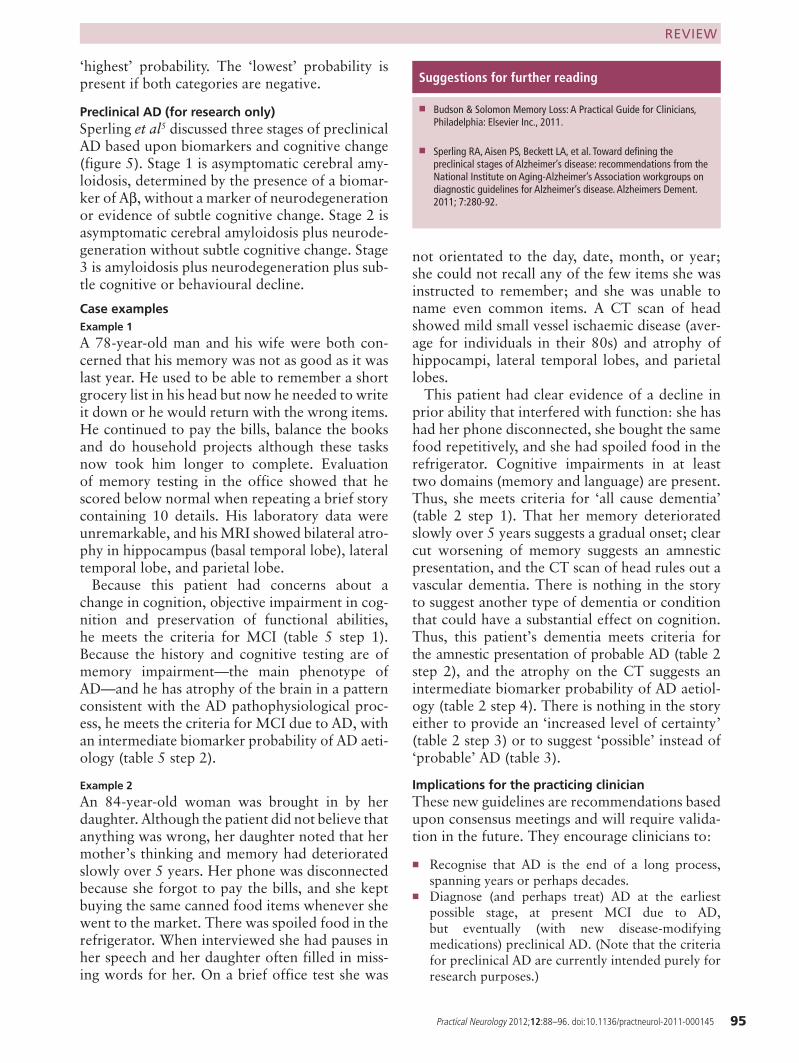
not orientated to the day, date, month, or year; she could not recall any of the few items she was instructed to remember; and she was unable to name even common items. A CT scan of head showed mild small vessel ischaemic disease (aver-age for individuals in their 80s) and atrophy of hippocampi, lateral temporal lobes, and parietal lobes.
This patient had clear evidence of a decline in prior ability that interfered with function: she has had her phone disconnected, she bought the same food repetitively, and she had spoiled food in the refrigerator. Cognitive impairments in at least two domains (memory and language) are present. Thus, she meets criteria for ‘all cause dementia’ (table 2 step 1). That her memory deteriorated slowly over 5 years suggests a gradual onset; clear cut worsening of memory suggests an amnestic presentation, and the CT scan of head rules out a vascular dementia. There is nothing in the story to suggest another type of dementia or condition that could have a substantial effect on cognition. Thus, this patient’s dementia meets criteria for the amnestic presentation of probable AD (table 2 step 2), and the atrophy on the CT suggests an intermediate biomarker probability of AD aetiol-ogy (table 2 step 4). There is nothing in the story either to provide an ‘increased level of certainty’ (table 2 step 3) or to suggest ‘possible’ instead of ‘probable’ AD (table 3).
Implications for the practicing clinicianThese new guidelines are recommendations based upon consensus meetings and will require valida-tion in the future. They encourage clinicians to:
Recognise that AD is the end of a long process, ■
spanning years or perhaps decades.Diagnose (and perhaps treat) AD at the earliest ■
possible stage, at present MCI due to AD, but eventually (with new disease-modifying medications) preclinical AD. (Note that the criteria for preclinical AD are currently intended purely for research purposes.)
‘highest’ probability. The ‘lowest’ probability is present if both categories are negative.
Preclinical AD (for research only)Sperling et al5 discussed three stages of preclinical AD based upon biomarkers and cognitive change (figure 5). Stage 1 is asymptomatic cerebral amy-loidosis, determined by the presence of a biomar-ker of Aβ, without a marker of neurodegeneration or evidence of subtle cognitive change. Stage 2 is asymptomatic cerebral amyloidosis plus neurode-generation without subtle cognitive change. Stage 3 is amyloidosis plus neurodegeneration plus sub-tle cognitive or behavioural decline.
Case examplesExample 1
A 78-year-old man and his wife were both con-cerned that his memory was not as good as it was last year. He used to be able to remember a short grocery list in his head but now he needed to write it down or he would return with the wrong items. He continued to pay the bills, balance the books and do household projects although these tasks now took him longer to complete. Evaluation of memory testing in the office showed that he scored below normal when repeating a brief story containing 10 details. His laboratory data were unremarkable, and his MRI showed bilateral atro-phy in hippocampus (basal temporal lobe), lateral temporal lobe, and parietal lobe.
Because this patient had concerns about a change in cognition, objective impairment in cog-nition and preservation of functional abilities, he meets the criteria for MCI (table 5 step 1). Because the history and cognitive testing are of memory impairment—the main phenotype of AD—and he has atrophy of the brain in a pattern consistent with the AD pathophysiological proc-ess, he meets the criteria for MCI due to AD, with an intermediate biomarker probability of AD aeti-ology (table 5 step 2).
Example 2
An 84-year-old woman was brought in by her daughter. Although the patient did not believe that anything was wrong, her daughter noted that her mother’s thinking and memory had deteriorated slowly over 5 years. Her phone was disconnected because she forgot to pay the bills, and she kept buying the same canned food items whenever she went to the market. There was spoiled food in the refrigerator. When interviewed she had pauses in her speech and her daughter often filled in miss-ing words for her. On a brief office test she was
■ Budson & Solomon Memory Loss: A Practical Guide for Clinicians, Philadelphia: Elsevier Inc., 2011.
■ Sperling RA, Aisen PS, Beckett LA, et al. Toward defi ning the preclinical stages of Alzheimer’s disease: recommendations from the National Institute on Aging-Alzheimer’s Association workgroups on diagnostic guidelines for Alzheimer’s disease. Alzheimers Dement. 2011; 7:280-92.
Suggestions for further reading
04_practneurol-2011-000145.indd 9504_practneurol-2011-000145.indd 95 3/10/2012 5:44:19 PM3/10/2012 5:44:19 PM

Practical Neurology 2012;12:88–96. doi:10.1136/practneurol-2011-000145
REVIEW
96
Consider using biomarkers in the diagnosis of all ■
stages of AD. Our current recommendation is to use biomarkers for those cases that present diagnostic quandaries.6
Evaluate patients with cognitive impairment ■
and dementia to determine aetiology, with special attention to amnestic and non-amnestic presentations of AD.Remember that because of the ageing population, ■
numbers of patients with all stages of AD will likely triple in the next 50 years.
Contributors AEB wrote the first draft of the manuscript. PRS created the tables and extensively revised the manuscript.
Acknowledgements This work was supported by National Institute on Ageing grant P30 AG13846. This material is also the result of work supported with resources and the use of facilities at the VA Boston Healthcare System.
Funding Funding was provided by the National Institute on Ageing.
Competing interests None.
Provenance and peer review Commissioned; externally peer reviewed. We are grateful to Jonathan Schott, London, UK, for reviewing this paper.
References 1. Jack CR Jr, Albert MS, Knopman DS, et al. Introduction
to the recommendations from the National Institute on
Aging-Alzheimer’s Association workgroups on diagnostic guidelines for Alzheimer’s disease. Alzheimers Dement 2011;7:257–62.
2. Dubois B, Feldman HH, Jacova C, et al. Revising the definition of Alzheimer’s disease: a new lexicon. Lancet Neurol 2010;9:1118–27.
3. Dubois B, Feldman HH, Jacova C, et al. Research criteria for the diagnosis of Alzheimer’s disease: revising the NINCDS-ADRDA criteria. Lancet Neurol 2007;6:734–46.
4. Jack CR Jr, Knopman DS, Jagust WJ, et al. Hypothetical model of dynamic biomarkers of the Alzheimer’s pathological cascade. Lancet Neurol 2010;9:119–28.
5. Sperling RA, Aisen PS, Beckett LA, et al. Toward defining the preclinical stages of Alzheimer’s disease: recommendations from the National Institute on Aging-Alzheimer’s Association workgroups on diagnostic guidelines for Alzheimer’s disease. Alzheimers Dement 2011;7:280–92.
6. Budson AE, Solomon PR. Memory Loss: A Practical Guide for Clinicians, Philadelphia: Elsevier 2011.
7. McKhann GM, Knopman DS, Chertkow H, et al. The diagnosis of dementia due to Alzheimer’s disease: recommendations from the National Institute on Aging-Alzheimer’s Association workgroups on diagnostic guidelines for Alzheimer’s disease. Alzheimers Dement 2011;7:263–9.
8. Albert MS, DeKosky ST, Dickson D, et al. The diagnosis of mild cognitive impairment due to Alzheimer’s disease: recommendations from the National Institute on Aging-Alzheimer’s Association workgroups on diagnostic guidelines for Alzheimer’s disease. Alzheimers Dement 2011;7:270–9.
04_practneurol-2011-000145.indd 9604_practneurol-2011-000145.indd 96 3/10/2012 5:44:20 PM3/10/2012 5:44:20 PM

POINTS
TOTAL
M E M O R Y
N A M I N G
VISUOSPATIAL / EXECUTIVE
ATTENTION
LANGUAGE
ABSTRACTION
DELAYED RECALL
ORIENTATION
Read list of words, subject must repeat them. Do 2 trials, even if 1st trial is successful. Do a recall after 5 minutes.
Subject has to repeat them in the forward order [ ] 2 1 8 5 4Subject has to repeat them in the backward order [ ] 7 4 2
Read list of letters. The subject must tap with his hand at each letter A. No points if ≥ 2 errors
[ ] F B A C M N A A J K L B A F A K D E A A A J A M O F A A B
Serial 7 subtraction starting at 100 [ ] 93 [ ] 86 [ ] 79 [ ] 72 [ ] 65
Repeat : I only know that John is the one to help today. [ ]The cat always hid under the couch when dogs were in the room. [ ]
Similarity between e.g. banana - orange = fruit [ ] train – bicycle [ ] watch - ruler
Draw CLOCK (Ten past eleven)Copy cube
__/5
__/3
Nopoints
1st trial
2nd trial
FACE VELVET CHURCH DAISY RED
__/5
__/2
__/1
__/3
__/2Fluency / Name maximum number of words in one minute that begin with the letter F _____ [ ] (N ≥ 11 words) __/1
__/2
__/6
__/30
B
Begin
End
5
E
1
A
2
4 3
C
D
Read list of digits (1 digit/ sec.).
NAME :Education :
Sex :Date of birth :
DATE :
© Z.Nasreddine MD Version 7.1 www.mocatest.org Normal ≥ 26 / 30
Add 1 point if ≤ 12 yr edu
MONTREAL COGNITIVE ASSESSMENT (MOCA)
[ ] Date [ ] Month [ ] Year [ ] Day [ ] Place [ ] City
[ ]Contour
[ ][ ] [ ]Numbers
[ ]Hands
[ ] [ ] [ ]
4 or 5 correct subtractions: 3 pts , 2 or 3 correct: 2 pts , 1 correct: 1 pt , 0 correct: 0 pt
( 3 points )
Category cue
Points for UNCUEDrecall onlyWITH NO CUE
Optional
Has to recall words
Multiple choice cue
FACE VELVET CHURCH DAISY RED[ ] [ ] [ ] [ ] [ ]
Administered by: ___________________________________________________

MoCA Version November 12, 2004 © Z. Nasreddine MD
www.mocatest.org
1
Montreal Cognitive Assessment
(MoCA)
Administration and Scoring Instructions The Montreal Cognitive Assessment (MoCA) was designed as a rapid screening instrument for mild cognitive dysfunction. It assesses different cognitive domains: attention and concentration, executive functions, memory, language, visuoconstructional skills, conceptual thinking, calculations, and orientation. Time to administer the MoCA is approximately 10 minutes. The total possible score is 30 points; a score of 26 or above is considered normal. 1. Alternating Trail Making:
Administration: The examiner instructs the subject: "Please draw a line, going from a number to a letter in ascending order. Begin here [point to (1)] and draw a line from 1 then to A then to 2 and so on. End here [point to (E)]."
Scoring: Allocate one point if the subject successfully draws the following pattern: 1 −A- 2- B- 3- C- 4- D- 5- E, without drawing any lines that cross. Any error that is not immediately self-corrected earns a score of 0.
2. Visuoconstructional Skills (Cube):
Administration: The examiner gives the following instructions, pointing to the cube: “Copy this drawing as accurately as you can, in the space below”.
Scoring: One point is allocated for a correctly executed drawing.
• Drawing must be three-dimensional • All lines are drawn • No line is added • Lines are relatively parallel and their length is similar (rectangular prisms are
accepted) A point is not assigned if any of the above-criteria are not met.
3. Visuoconstructional Skills (Clock):
Administration: Indicate the right third of the space and give the following instructions: “Draw a clock. Put in all the numbers and set the time to 10 after 11”.
Scoring: One point is allocated for each of the following three criteria:
Contour (1 pt.): the clock face must be a circle with only minor distortion acceptable (e.g., slight imperfection on closing the circle);
Numbers (1 pt.): all clock numbers must be present with no additional numbers; numbers must be in the correct order and placed in the approximate quadrants on the clock face; Roman numerals are acceptable; numbers can be placed outside the circle contour;
Hands (1 pt.): there must be two hands jointly indicating the correct time; the hour hand must be clearly shorter than the minute hand; hands must be centred within the clock face with their junction close to the clock centre.
A point is not assigned for a given element if any of the above-criteria are not met.
MoCA Version November 12, 2004 © Z. Nasreddine MD
www.mocatest.org
2
4. Naming:
Administration: Beginning on the left, point to each figure and say: “Tell me the name of this animal”.
Scoring: One point each is given for the following responses: (1) camel or dromedary, (2)
lion, (3) rhinoceros or rhino. 5. Memory:
Administration: The examiner reads a list of 5 words at a rate of one per second, giving the following instructions: “This is a memory test. I am going to read a list of words that you will have to remember now and later on. Listen carefully. When I am through, tell me as many words as you can remember. It doesn’t matter in what order you say them”. Mark a check in the allocated space for each word the subject produces on this first trial. When the subject indicates that (s)he has finished (has recalled all words), or can recall no more words, read the list a second time with the following instructions: “I am going to read the same list for a second time. Try to remember and tell me as many words as you can, including words you said the first time.” Put a check in the allocated space for each word the subject recalls after the second trial. At the end of the second trial, inform the subject that (s)he will be asked to recall these words again by saying, “I will ask you to recall those words again at the end of the test.”
Scoring: No points are given for Trials One and Two. 6. Attention:
Forward Digit Span: Administration: Give the following instruction: “I am going to say some numbers and when I am through, repeat them to me exactly as I said them”. Read the five number sequence at a rate of one digit per second. Backward Digit Span: Administration: Give the following instruction: “Now I am going to say some more numbers, but when I am through you must repeat them to me in the backwards order.” Read the three number sequence at a rate of one digit per second.
Scoring: Allocate one point for each sequence correctly repeated, (N.B.: the correct response for the backwards trial is 2-4-7).
Vigilance: Administration: The examiner reads the list of letters at a rate of one per second, after giving the following instruction: “I am going to read a sequence of letters. Every time I say the letter A, tap your hand once. If I say a different letter, do not tap your hand”.
Scoring: Give one point if there is zero to one errors (an error is a tap on a wrong letter or a failure to tap on letter A).
MoCA Version November 12, 2004 © Z. Nasreddine MD
www.mocatest.org
3
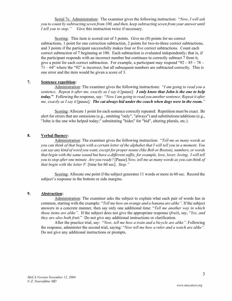
Serial 7s: Administration: The examiner gives the following instruction: “Now, I will ask you to count by subtracting seven from 100, and then, keep subtracting seven from your answer until I tell you to stop.” Give this instruction twice if necessary.
Scoring: This item is scored out of 3 points. Give no (0) points for no correct subtractions, 1 point for one correction subtraction, 2 points for two-to-three correct subtractions, and 3 points if the participant successfully makes four or five correct subtractions. Count each correct subtraction of 7 beginning at 100. Each subtraction is evaluated independently; that is, if the participant responds with an incorrect number but continues to correctly subtract 7 from it, give a point for each correct subtraction. For example, a participant may respond “92 – 85 – 78 – 71 – 64” where the “92” is incorrect, but all subsequent numbers are subtracted correctly. This is one error and the item would be given a score of 3.
7. Sentence repetition:
Administration: The examiner gives the following instructions: “I am going to read you a sentence. Repeat it after me, exactly as I say it [pause]: I only know that John is the one to help today.” Following the response, say: “Now I am going to read you another sentence. Repeat it after me, exactly as I say it [pause]: The cat always hid under the couch when dogs were in the room.”
Scoring: Allocate 1 point for each sentence correctly repeated. Repetition must be exact. Be alert for errors that are omissions (e.g., omitting "only", "always") and substitutions/additions (e.g., "John is the one who helped today;" substituting "hides" for "hid", altering plurals, etc.).
8. Verbal fluency: Administration: The examiner gives the following instruction: “Tell me as many words as you can think of that begin with a certain letter of the alphabet that I will tell you in a moment. You can say any kind of word you want, except for proper nouns (like Bob or Boston), numbers, or words that begin with the same sound but have a different suffix, for example, love, lover, loving. I will tell you to stop after one minute. Are you ready? [Pause] Now, tell me as many words as you can think of that begin with the letter F. [time for 60 sec]. Stop.”
Scoring: Allocate one point if the subject generates 11 words or more in 60 sec. Record the subject’s response in the bottom or side margins.
9. Abstraction:
Administration: The examiner asks the subject to explain what each pair of words has in common, starting with the example: “Tell me how an orange and a banana are alike”. If the subject answers in a concrete manner, then say only one additional time: “Tell me another way in which those items are alike”. If the subject does not give the appropriate response (fruit), say, “Yes, and they are also both fruit.” Do not give any additional instructions or clarification. After the practice trial, say: “Now, tell me how a train and a bicycle are alike”. Following the response, administer the second trial, saying: “Now tell me how a ruler and a watch are alike”. Do not give any additional instructions or prompts.

MoCA Version November 12, 2004 © Z. Nasreddine MD
www.mocatest.org
4
Scoring: Only the last two item pairs are scored. Give 1 point to each item pair correctly answered. The following responses are acceptable: Train-bicycle = means of transportation, means of travelling, you take trips in both; Ruler-watch = measuring instruments, used to measure. The following responses are not acceptable: Train-bicycle = they have wheels; Ruler-watch = they have numbers.
10. Delayed recall:
Administration: The examiner gives the following instruction: “I read some words to you earlier, which I asked you to remember. Tell me as many of those words as you can remember. Make a check mark ( ) for each of the words correctly recalled spontaneously without any cues, in the allocated space. Scoring: Allocate 1 point for each word recalled freely without any cues. Optional: Following the delayed free recall trial, prompt the subject with the semantic category cue provided below for any word not recalled. Make a check mark ( ) in the allocated space if the subject remembered the word with the help of a category or multiple-choice cue. Prompt all non-recalled words in this manner. If the subject does not recall the word after the category cue, give him/her a multiple choice trial, using the following example instruction, “Which of the following words do you think it was, NOSE, FACE, or HAND?” Use the following category and/or multiple-choice cues for each word, when appropriate: FACE: category cue: part of the body multiple choice: nose, face, hand VELVET: category cue: type of fabric multiple choice: denim, cotton, velvet CHURCH: category cue: type of building multiple choice: church, school, hospital DAISY: category cue: type of flower multiple choice: rose, daisy, tulip RED: category cue: a colour multiple choice: red, blue, green Scoring: No points are allocated for words recalled with a cue. A cue is used for clinical information purposes only and can give the test interpreter additional information about the type of memory disorder. For memory deficits due to retrieval failures, performance can be improved with a cue. For memory deficits due to encoding failures, performance does not improve with a cue.
11. Orientation:
Administration: The examiner gives the following instructions: “Tell me the date today”. If the subject does not give a complete answer, then prompt accordingly by saying: “Tell me the [year, month, exact date, and day of the week].” Then say: “Now, tell me the name of this place, and which city it is in.” Scoring: Give one point for each item correctly answered. The subject must tell the exact date and the exact place (name of hospital, clinic, office). No points are allocated if subject makes an error of one day for the day and date. TOTAL SCORE: Sum all subscores listed on the right-hand side. Add one point for an individual who has 12 years or fewer of formal education, for a possible maximum of 30 points. A final total score of 26 and above is considered normal.

AD8 Dementia Screening Interview Patient ID#:__________ CS ID#:___________ Date:___________
NO,
No change
Remember, “Yes, a change” indicates that there has been a change in the last several years caused by cognitive (thinking and memory) problems.
YES,
A change
N/A,
Don’t know
1. Problems with judgment (e.g., problems making decisions, bad financial decisions, problems with thinking)
Adapted from Galvin JE et al, The AD8, a brief informant interview to detect dementia, Neurology 2005:65:559-564Copyright 2005. The AD8 is a copyrighted instrument of the Alzheimer’s Disease Research Center, Washington University, St. Louis, Missouri. All Rights Reserved.
2. Less interest in hobbies/activities
3. Repeats the same things over and over (questions, stories, or statements)
4. Trouble learning how to use a tool, appliance, or gadget (e.g., VCR, computer, microwave, remote control)
5. Forgets correct month or year
6. Trouble handling complicated financial affairs (e.g., balancing checkbook, income taxes, paying bills)
7. Trouble remembering appointments
8. Daily problems with thinking and/or memory
TOTAL AD8 SCORE

The AD8 Administration and Scoring Guidelines A spontaneous self-correction is allowed for all responses without counting as an error. The questions are given to the respondent on a clipboard for self–administration or can be read aloud to the respondent either in person or over the phone. It is preferable to administer the AD8 to an informant, if available. If an informant is not available, the AD8 may be administered to the patient. When administered to an informant, specifically ask the respondent to rate change in the patient. When administered to the patient, specifically ask the patient to rate changes in his/her ability for each of the items, without attributing causality. If read aloud to the respondent, it is important for the clinician to carefully read the phrase as worded and give emphasis to note changes due to cognitive problems (not physical problems). There should be a one second delay between individual items. No timeframe for change is required. The final score is a sum of the number items marked “Yes, A change”. Interpretation of the AD8 (Adapted from Galvin JE et al, The AD8, a brief informant interview to detect dementia, Neurology 2005:65:559-564) A screening test in itself is insufficient to diagnose a dementing disorder. The AD8 is, however, quite sensitive to detecting early cognitive changes associated many common dementing illness including Alzheimer disease, vascular dementia, Lewy body dementia and frontotemporal dementia. Scores in the impaired range (see below) indicate a need for further assessment. Scores in the “normal” range suggest that a dementing disorder is unlikely, but a very early disease process cannot be ruled out. More advanced assessment may be warranted in cases where other objective evidence of impairment exists. Based on clinical research findings from 995 individuals included in the development and validation samples, the following cut points are provided:
• 0 – 1: Normal cognition
1.00.80.60.40.20.0
• 2 or greater: Cognitive impairment is likely to be present
1 - Specificity
1.0
0.8
0.6
0.4
0.2
0.0
Sens
itivi
ty
Reciever Operator Characteristics (ROC) curve for AD8
Administered to either the informant (preferable) or the patient, the AD8 has the following properties:
• Sensitiv ity >84% • Specific ity >80% • Positive Predictive Value > 85% • Negative Predictive Value > 70% • Area under the Curve: 0.908; 95%CI: 0.888-
0.925