cognitive control reflects context monitoring, not motoric ... · cognitive control reflects...
TRANSCRIPT
Cognitive Control Reflects Context Monitoring, NotMotoric Stopping, in Response InhibitionChristopher H. Chatham1*, Eric D. Claus2, Albert Kim3, Tim Curran3, Marie T. Banich4, Yuko Munakata3
1 Department of Cognitive, Linguistic and Psychological Sciences, Brown University, Providence, Rhode Island, United States of America, 2 The Mind Research Network,
Albuquerque, New Mexico, United States of America, 3 Department of Psychology and Neuroscience, University of Colorado, Boulder, Colorado, United States of America,
4 Institute of Cognitive Science, University of Colorado, Boulder, Colorado, United States of America
Abstract
The inhibition of unwanted behaviors is considered an effortful and controlled ability. However, inhibition also requires thedetection of contexts indicating that old behaviors may be inappropriate – in other words, inhibition requires the ability tomonitor context in the service of goals, which we refer to as context-monitoring. Using behavioral, neuroimaging,electrophysiological and computational approaches, we tested whether motoric stopping per se is the cognitively-controlled process supporting response inhibition, or whether context-monitoring may fill this role. Our results demonstratethat inhibition does not require control mechanisms beyond those involved in context-monitoring, and that such controlmechanisms are the same regardless of stopping demands. These results challenge dominant accounts of inhibitory control,which posit that motoric stopping is the cognitively-controlled process of response inhibition, and clarify emerging debateson the frontal substrates of response inhibition by replacing the centrality of controlled mechanisms for motoric stoppingwith context-monitoring.
Citation: Chatham CH, Claus ED, Kim A, Curran T, Banich MT, et al. (2012) Cognitive Control Reflects Context Monitoring, Not Motoric Stopping, in ResponseInhibition. PLoS ONE 7(2): e31546. doi:10.1371/journal.pone.0031546
Editor: Sam Gilbert, University College London, United Kingdom
Received April 12, 2011; Accepted January 13, 2012; Published February 27, 2012
Copyright: � 2012 Chatham et al. This is an open-access article distributed under the terms of the Creative Commons Attribution License, which permitsunrestricted use, distribution, and reproduction in any medium, provided the original author and source are credited.
Funding: This study was funded by the National Institute of Mental Health (http://www.nimh.nih.gov) with a P50 center grant (079485-02). The funders had norole in study design, data collection and analysis, decision to publish, or preparation of the manuscript.
Competing Interests: The authors have declared that no competing interests exist.
* E-mail: [email protected]
Introduction
Inhibition is critical for enabling controlled behavior: bad habits,
unfamiliar situations, and dangerous environments often require
that default behaviors be stopped and more context-appropriate
actions performed [1]. Inhibitory control has been statistically
equated with the behavioral and genetic variance common across
multiple tests of cognitive and behavioral control [2–3]. Moreover, a
particular domain of inhibitory control – response inhibition – has
been exempted from the skepticisms surrounding other domains of
inhibition [4], and specifically linked to the functioning of a
particular frontal region (the right ventrolateral prefrontal cortex;
rVLPFC). In this way, the study of response inhibition has
supported theorizing that similar mechanisms may enable the
inhibition of thoughts and emotions. Thus, modern theorizing is
largely consistent with a hypothesis proposed 130 years ago: that
‘‘the centers of inhibition being thus the essential factor of attention,
constitute the organic basis of all the higher intellectual faculties’’
[5].
However, effective inhibitory control not only requires actually
stopping unwanted actions, thoughts, or emotions – it also requires
the efficient detection of those contexts that indicate the need
for these forms of stopping. To use an example from the domain
of response inhibition, one’s goal may be to cross a street; this
requires actually crossing the street, and stopping these motor
actions if oncoming traffic is approaching, but to do so the
environment must be monitored so that this motoric stopping can
be performed as appropriate. In other words, the environmental
context must be monitored to support behavior that may be
contingent on that context. Both motoric stopping and context-
monitoring are also intermingled in the most precise laboratory
assessment of response inhibition, in which subjects must cancel a
prepotent or planned response after the presentation of a signal to
stop [6–12]. The time that subjects require to stop an action, or
‘‘Stop Signal Reaction Time’’ (SSRT) can be estimated based on
a formal model of the ‘‘stopping process,’’ although the model’s
estimate of this process intermingles the time spent detecting or
interpreting the signal to stop with the latency for motoric stopping
per se to take place [7]. Indeed, this process impurity is both widely
acknowledged [e.g., 2] and implicit in the original proposal [7].
Thus, even within this well-studied domain of response inhibition,
the so-called ‘‘stopping process’’ could largely reflect a controlled
context-monitoring process that supports the detection and inter-
pretation of the behaviorally-relevant signal to stop [8–11].
To determine whether context-monitoring or stopping may
constitute the cognitively-controlled process of inhibitory control,
we focus on response inhibition as an example domain. We ex-
perimentally dissociate the motoric stopping that occurs during
response inhibition from the context-monitoring processes that are
also involved, by examining two tasks with identical context-
monitoring demands, one of which requires motoric stopping and
one of which does not. In both tasks, 75% of trials (‘‘No Signal’’
trials) require a 2-alternative forced choice (2AFC; Fig. 1A); in the
remaining 25% of trials (‘‘Signal’’ trials), the 2AFC is followed by a
behaviorally-relevant stimulus (the ‘‘signal’’) after a variable delay
(Fig. 1B). In the Stop Task, Signal trials require the stopping of
PLoS ONE | www.plosone.org 1 February 2012 | Volume 7 | Issue 2 | e31546

motor responses on that trial. In contrast, in the ‘‘Double Go
Task,’’ Signal trials require subjects to repeat their response for
that trial as quickly as possible (see methods and Text S1). Thus,
both tasks require monitoring for the context that signals what
actions should be executed, but only the Stop Task explicitly
requires motor actions to be stopped.
The cognitive control required for response inhibition is thought
to rely on the prefrontal cortex, to be most crucial at the moment
when motoric stopping is required, to be associated with sub-
stantial mental effort, to be recruited in a goal-directed fashion,
and to support consistent individual differences. We assess each of
these characteristics of cognitive control via behavioral, compu-
tational, hemodynamic, electrophysiological and pupillometric
techniques to determine whether context-monitoring or motoric
stopping may reflect the cognitively-controlled process recruited
during response inhibition. Convergent evidence of this kind is
necessary for making broad claims about the content of cog-
nitive control because cognitive control cannot be unambiguously
defined on the basis of any of these characteristics in isolation (e.g.,
neither prefrontal recruitment nor mental effort alone are
sufficient). In addition, this convergent evidence allows us to make
multiple points of contact with prior uses of these techniques in the
domain of response inhibition, as outlined below.
We used functional magnetic resonance imaging (fMRI) to assess
the recruitment of the prefrontal cortex in our tasks. Numerous
previous fMRI studies have demonstrated transient activation
within the right ventrolateral prefrontal cortex (rVLPFC) and the
adjoining anterior insula during trials that require motoric stopping
[1–2,6,8–12]. Collectively, this and related evidence has been
interpreted to indicate that the rVLPFC is a dedicated substrate for
inhibition, and that this function may also be deployed proactively
to support behaviors like ‘‘responding with restraint’’ [12–13].
Alternatively, it is possible that these hemodynamic patterns reflect
the context monitoring demands of the Stop task, which could
also be deployed proactively as well as transiently at the moment a
goal-relevant feature of the environment (e.g., a Stop Signal) is
encountered. Recent work has begun to examine this alternative
possibility using fMRI but has unfortunately yielded inconsistent
results: either more [8,11], less [7,10] or roughly equivalent [9]
rVLPFC activity is observed during the Stop task, in either
overlapping [8,11] or distinct [6,10] subregions of the rVLPFC. In
addition, none of these studies have examined whether the sustained
component to the rVLPFC hemodynamic response could reflect a
tonic and proactive process of context-monitoring (in which case
sustained activity should also be observed in a context-monitoring
task) rather than a process of responding with restraint. Finally, all
of these studies have examined only the univariate patterns in
hemodynamics, and have not assessed whether rVLPFC demon-
strates multivariate commonality across tasks involving context-
monitoring (as would be predicted by context-monitoring accounts),
or whether any such commonality is relatively decreased on trials
requiring motoric stopping (as would be predicted by stopping
accounts). Below, we measured each of these aspects of the re-
cruitment of the rVLPFC during the Stop and Double Go tasks
to test these differing predictions of the context-monitoring and
motoric stopping accounts.
We also assessed whether the event-related potentials commonly
associated with response inhibition tasks, and often presumed to
reflect motoric stopping processes, might instead reflect context
monitoring processes. The most characteristic ERP from response
inhibition tasks is the ‘‘Stop P3’’ or ‘‘No/Go P3,’’ a frontocentral
positivity elicited following the onset of stimuli which demand
motoric stopping [14]. We tested whether this ‘‘Stop P3’’ would
be more strongly expressed during the Stop task than the Double
Go task (as motoric stopping accounts would predict), and whether
the correlation of all ERPs across these tasks would be reduced
following the onset of the Signals (as would also be predicted by
motoric stopping accounts). In contrast, accounts positing the
centrality of context-monitoring to the Stop task would predict
roughly equivalent frontocentral ERPs across these tasks, despite
their differing demands on motoric stopping.
Finally, we assessed the task-evoked pupillometric response
(TEPR), a well-validated measure of mental effort [15–16], to
determine whether the relatively more effortful component to the
Stop task reflects motoric stopping (in which case pupil diameter
should be increased on StopSignal trials) or whether it might reflect
the act of monitoring context for goal-relevant signals (in which
case, pupil diameter may show a more complex pattern, such as a
modulation of pupil diameter by the relevance of a monitored
signal to the planned response). Previous work examining pupil
diameter in the Stop task has utilized it mainly as a control
measure of arousal in TMS studies [17–18].
Figure 1. An illustration of the task design. Identical stimuli and trial structure were used across tasks in three separate experiments. In both theStop and the Double Go tasks, most trials are ‘‘No Signal’’ trials where only a 2AFC decision is required (A). However, the tasks differ on ‘‘Signal’’ trials(B) where an additional stimulus, a white box, is presented with a variable inter-stimulus interval following the onset of the 2AFC stimulus. On DoubleGoSignal trials, this additional stimulus indicates that the appropriate 2AFC button press be repeated. On StopSignal trials, this stimulus indicates thatthe 2AFC button press must be stopped. Thus, although only the Stop Task requires motoric stopping, both tasks share demands on context-monitoring.doi:10.1371/journal.pone.0031546.g001
Context Monitoring in Response Inhibition
PLoS ONE | www.plosone.org 2 February 2012 | Volume 7 | Issue 2 | e31546

To foreshadow our results, our results uniformly suggest that,
during response inhibition, cognitive control is primarily engaged
for the purpose of monitoring the environmental context in the
service of goals, rather than for motoric stopping per se.
Results
Univariate fMRI ResultsFirst, we found that context-monitoring rather than stopping
explained the transient prefrontal contribution to response inhibi-
tion. Accounts which posit that motoric stopping is the controlled
process during response inhibition tasks predict rVLPFC activation
only in the Stop task, but event-related fMRI revealed that the Stop
and Double Go tasks activated completely overlapping regions of
prefrontal cortex (Fig. 2A), consistent with the tasks’ shared context-
monitoring demands. Specific regions of interest (ROIs) in the
rVLPFC and interconnected subthalamic nucleus (STN) that have
been proposed to be specific to the motoric stopping demands were
uniformly more strongly recruited on Signal trials in the Double Go
Task (Fig. 2B&C; STN: t(17) = 5.49, p,.0001; BA44: t(17) = 5.08,
p,.0001; BA45: t(17) = 2.83, p = .012; BA47: t(17) = 2.5, p = .023),
challenging any characterization of these areas as specialized for
motoric stopping. A significantly different pattern was observed in
areas thought to have a more general attentional role (e.g., the
temporo-parietal junction; TPJ [19–20]; F(1,17) = 31.57, p,.0001),
such that both tasks recruited this area equivalently. This equal
recruitment of the TPJ across tasks indicates that decreased re-
cruitment of the rVLPFC in the Stop task cannot be explained by
globally-decreased activation during that task (e.g., as might result
from fatigue; see also discussion in Text S1). Moreover, the increased
recruitment of rVLPFC during the Double Go task is consistent with
several recent findings, which also demonstrate that tasks involving
both context-monitoring and response commission are associated
with increased rVLPFC activity relative to tasks involving both
context-monitoring and a demand to stop motor actions [7–10] (but
see [6] and discussion, below).
Our hybrid fMRI design also allowed us to assess the extent to
which neural regions were recruited in a sustained fashion across
all trials within the Stop and Double Go tasks. Such sustained
activity is potentially a hallmark of proactive context-monitoring
processes. Indeed, this analysis revealed sustained hemodynamics
in the rVLPFC during both tasks at the timescale of seconds-to-
minutes (Fig. 2D), consistent with their shared sustained context-
monitoring demands. In contrast, accounts positing that motoric
stopping is the cognitively-controlled process during response
inhibition predict no sustained rVLPFC activity in the Double Go
task, since only response commission is required by that task, and
‘‘responding with restraint’’ is unnecessary.
Multivariate Pattern AnalysisWe next leveraged multi-voxel pattern analysis to determine
whether the same information was encoded by rVLPFC regardless
of whether motoric stopping is required by a given task. First,
we trained classifiers to identify hemodynamic patterns that
reliably predicted subject-specific patterns of rVLPFC activation
in the Double Go task over 10 independent runs of the classifier
(see methods in Text S1 and Figures S3 & S4). Classifiers readily
generalized their training on the Double Go task to distinguish
individuals in the Stop task, indicating that the rVLPFC is re-
cruited in an individual-specific but consistent way across tasks.
These patterns were significantly more consistent across tasks on
Signal trials in the rVLPFC – precisely when and where context-
monitoring processes are most crucial, but also when motoric
stopping demands differ most across these tasks (Fig. 3A; BA44:
t(9) = 13.5, p,.0001; BA45: t(9) = 11.39, p,.0001; BA47:
t(9) = 12.35, p,.001). Critically, the increased cross-task similarity
of Signal trials relative to No Signal trials was not observed in an
area known to encode responses – primary motor cortex – and this
pattern was significantly different from that observed in rVLPFC
(F(1,9) = 85.12, p,.0001).
Although these results do not conclusively demonstrate that
the cognitive processes engaged by both tasks are the same, they
do demonstrate that the multivariate representations in the
rVLPFC fail to show differential sensitivity to the explicit stopping
demands imposed by Signal trials within the Stop task (in contrast
to the multivariate patterns within primary motor cortex). This
pattern contradicts the idea that representations in rVLPFC are
specialized for the motoric stopping that is required on Signal trials
in the Stop task (but not in the Double Go task). Thus, while No
Signal trials are indeed similar across tasks (as revealed by the
uniformly above-chance classification of individuals on these trials;
light gray bars of Fig. 3A), Signal trials reveal processes that are
particularly stable within individuals across these tasks, despite
their different demands on motoric stopping, within the rVLPFC.
Figure 2. The Stop and Go tasks recruit overlapping neural substrates as revealed in both transient and sustained hemodynamics.Hybrid fMRI analyses revealed overlapping neural activity in the Stop and (Double) Go Tasks (A), with significantly more rVLPFC activity in the Go Task(B). ROI analyses for the contrast of Signal vs. No-Signal trials (C) revealed increased activity in the Go Task throughout a putatively stopping-specificnetwork; this pattern did not generalize to regions with more general attentional functions (e.g., TPJ). Sustained rVLPFC activity was also observedacross all trials within each task (D).doi:10.1371/journal.pone.0031546.g002
Context Monitoring in Response Inhibition
PLoS ONE | www.plosone.org 3 February 2012 | Volume 7 | Issue 2 | e31546

This pattern is wholly consistent with the idea that similar context-
monitoring processes are elicited by Signal trials within both tasks.
In a second multi-voxel pattern analysis, subject-specific classifiers
were trained to decode the multivariate patterns that differentiate
Double GoSignal and Double GoNo-Signal trials. Classifiers generalized
this training on the Double Go task to correctly identify StopSignal
trials with 92–97% accuracy in the rVLPFC, significantly higher than
the 59% accuracy achieved in primary motor cortex (F(1,17) = 9.413,
p = .007). To control for the possible effects of classifier bias on this
result, we utilized signal detection theory. Classifiers readily dis-
criminated between StopSignal and StopNo-Signal trials in the rVLPFC
but not primary motor cortex in terms of d’ (Fig. 3B; F(1,17) = 13.14,
p,.005). Thus, while the Double Go classifier cannot be applied
successfully to the Stop task in primary motor cortex (as one would
expect given the tasks’ different motor demands), it can be in the
rVLPFC – indicating that the rVLPFC-mediated control process is
similar across these tasks despite their different demands on motoric
stopping.
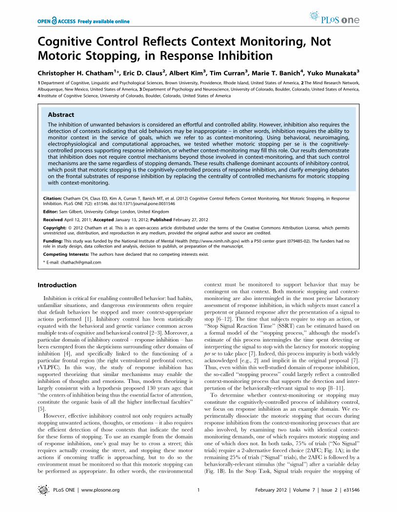
Event-related potentialsThe ERPs evoked by our tasks also reflected context-monitoring
demands rather than stopping demands. In particular, motoric
stopping accounts predict that a prefrontal ERP called the ‘‘Stop
P3’’ reflects stopping-specific processes [14] and should therefore
be enhanced in the Stop task. However, the so-called Stop P3 was
enhanced in the Double Go task, in direct contradiction to the
stopping account (Fig. 4A; t(35) = 2.92, p,.03; see also Figures S5
and S6), but consistent with our observations of increased transient
hemodynamics in the Double Go task relative to the Stop task (see
Univariate fMRI results, above).
These ERPs from the Stop and Double Go tasks were not two
distinct potentials masquerading as the same; individual differences
in this ERP were also highly correlated between tasks (Fig. 4B).
Critically, correlations between the ERPs elicited by each task were
disproportionately increased over prefrontal electrodes, relative to
occipital electrodes, following signal onset, when context-monitor-
ing is most required but stopping demands differ most (Fig. 4C;
F(1,98) = 12.59, p = .001).
PupillometryWe also found that context-monitoring, not motoric stopping,
explains the patterns of mental effort elicited during our tasks. We
measured pupil diameter, a psychophysiological index of mental
effort [15–16], following the onset of a signal (or the average signal
onset time in the case of No-Signal trials; Fig. 5). Averaging across all
time points, mental effort was less for stopping than for monitoring
for signals that fail to appear (StopSignal,StopNo-Signalt(85) = 7.00,
p,.001; StopSignal,Double GoNo-Signal t(85) = 2.07, p,.05). Mental
effort was also less for context monitoring and motoric stopping than
for context monitoring and an additional act of going (StopSignal,-
Double GoSignal t(85) = 13.67, p,.001). Finally, mental effort was
greater when monitoring for signals that would require a change to
the planned response than when monitoring for those that would not
(StopNo-Signal .Double GoNo-Signal t(85) = 10.25, p,.001), a result
which also rules out global reductions in effort during the Stop task
(e.g. from fatigue; see also Text S1). Thus, motoric stopping is not
itself associated with effort beyond that required for the processes
involved in other trial types, contrary to the idea that motoric
stopping of a response constitutes a particularly effortful component
of response inhibition. Instead, context-monitoring demands are
more central to mental effort, and this relationship is modulated by
the relevance of the monitored stimulus to the planned response.
Model-based decomposition of behavior andcorrelations with brain activity
Stopping is not associated with differential mental effort or
prefrontal recruitment, contrary to what might be predicted from
stopping-centric accounts of cognitive control. This pattern of
results could imply that motoric stopping is not a cognitively-
controlled process, given the widely-held assumption that con-
trolled, goal-directed processes recruit the prefrontal cortex and
require differential mental effort. Consistent with this idea, subjects
Figure 3. Multivariate pattern analysis reveals similar representations in the rVLPFC despite differing inhibitory demands. (A)rVLPFC was recruited in subject-specific but consistent ways regardless of stopping demands: individual differences in (Double) Go taskhemodynamic activity also differentiated subjects in the Stop task. (B). rVLPFC showed trial-type-specific recruitment that was consistent across tasks,contradicting stopping-specific accounts of rVLPFC function. ** p,.0001 *** p,.005.doi:10.1371/journal.pone.0031546.g003
Context Monitoring in Response Inhibition
PLoS ONE | www.plosone.org 4 February 2012 | Volume 7 | Issue 2 | e31546
appear to engage in reflexive stopping even on the Double Go task,
where such stopping runs contrary to instructed goals. Specifically,
although Double GoSignal trials require that subjects commit a
subset of the motor responses required on Double GoNo-Signal trials,
subjects were nonetheless slower to provide even their first response
to stimuli when they were followed by the signal than when they
appeared alone (Double GoSignal1st RT.Double GoNoSignal
Only RT;
t(148) = 9.59, p,.0005; Fig. 6A). To the extent that this behavioral
slowing in the Double Go task reflects some transient stopping,
it runs contrary to subjects’ goals in the Double Go task and
therefore might not be engaged in a controlled or goal-directed
manner.
On the other hand, the presence of goal-inconsistent slowing
during the Double Go task does not by itself refute the idea that
motoric stopping can be a controlled process in this task or in others.
Indeed, one alternative interpretation of this slowing is that it does in
fact reflect a controlled and goal-directed process: it may be an
attempt to stop or replace the motor plan required on Double GoNo-
Figure 4. Prefrontal event-related potentials do not strongly distinguish the tasks. A prefrontal positivity peaking around 300 ms, knownas the ‘‘Stop P3,’’ has been previously associated with stopping, but this component (darkened region of A) was significantly enhanced in the(Double) Go task. Individual differences in voltage were also highly correlated across tasks, indicating substantial overlap in the underlying corticalprocesses (B). Moreover, prefrontal correlations between the scalp voltage recorded across tasks were disproportionately increased following thepresentation of the signal, relative to the increase in occipital correlations observed at the same time (C). This difference indicates increased cross-tasksimilarity in prefrontal processing specifically at signal onset.doi:10.1371/journal.pone.0031546.g004
Figure 5. Patterns of mental effort assessed via pupillometry indicate that effort matches demands on context-monitoring, notstopping, and is modulated by the relevance of the infrequent stimulus to the planned response. In particular, stopping a response(StopSignal trials) was associated with more mental effort was required by monitoring for the appearance of stimuli that would demand stopping(StopNo-Signal trials) than by stopping itself (StopSignal trials) or by monitoring for the appearance of stimuli that would demand an additional act ofgoing (GoNo-Signal trials).doi:10.1371/journal.pone.0031546.g005
Context Monitoring in Response Inhibition
PLoS ONE | www.plosone.org 5 February 2012 | Volume 7 | Issue 2 | e31546

Signal trials (i.e. the motor plan for ‘‘respond once’’ is stopped or
replaced with the motor plan for ‘‘respond twice’’). We assessed this
possibility with a model-based decomposition of subjects’ behavior;
however, the results of this analysis argue against this possibility, and
further show that the efficiency of subjects’ context-monitoring,
rather than the efficiency of motoric stopping or motor plan replace-
ment, shares a closer relationship with SSRT.
To assess the alternative accounts, we developed a formal model
of context-monitoring and stopping by building on the classic race
model of the Stop task [7] (see also Text S1 and Figure S7A)
in order to precisely estimate the duration of motoric slowing
experienced by subjects in the Double Go task, as well as exactly
which trials underwent such slowing (Figure S7B). The race model
of the Stop task posits that responses undergo inhibition when
a stopping process, triggered by the onset of the Stop signal,
completes before the ‘‘going’’ processes triggered by the onset of the
2AFC stimulus. The race between stopping and going processes is
the model’s namesake, and is supported by the monotonically-
decreasing relationship of interstimulus interval (ISI) to successful
inhibition: larger ISIs give the ‘‘going’’ process an increasing
advantage in the race, and thus leads to less successful inhibition.
We observed a similar phenomenon in our Double Go task, such
that increasing ISIs led to less slowing of first responses; this effect
was visible at the group level (Figure S7C) but also even at the level
of individual subjects (Fig. 6B), who showed substantial variability
in the earliest ISI to yield zero observable slowing.
We utilized this behavioral variability to estimate individual
differences in Double Go task performance. First, we estimated the
Figure 6. Mixture model analyses separate slowed from unslowed trials in the Go task, and demonstrate this slowing is not thesource of the commonality across tasks. Response slowing was observed in the Double Go task (A), perhaps suggesting that stopping is notassociated with differential mental effort or prefrontal activity because it is an automatic consequence of detecting an infrequent stimulus. Critically,this slowing was dependent on ISI; indeed, large individual differences were observed in the shortest ISI to yield zero slowing (B contains data fromfour representative subjects). A subtraction of reaction times on (Double) GoNo-Signal trials from those with a corresponding percent rank on (Double)GoSignal trials reveals a pronounced positive skew to these equipercentile residuals (C), indicating that some proportion of reaction times on GoSignal
trials are disproportionately delayed. Trials undergoing this slowing were identified as those more likely to come from a distribution not centered onzero, as determined through a two-component mixture model (see overlaid lines on histogram in C). This procedure adequately separated theslowed and unslowed distributions, as revealed by zero significant difference between GoSignal trials categorized as unslowed and theircorresponding reaction times in the GoNo-Signal distribution, but a large difference between GoSignal trials categorized as slowed and theircorresponding reaction times in the GoNo-Signal distribution (D). From this we estimated two individual differences: how long subjects are slowed(duration of slowing; DoS) and the time at which signals are detected (time of signal detection; TOSD). Only TOSD positively correlated with SSRT,whereas DoS showed a slight negative correlation, indicating that the slowing experienced by subjects in the Double Go task cannot be the source ofshared variance between the Stop and Double Go tasks (E). Brain-behavior correlations confirmed this conclusion: SSRT and TOSD, but not DoS,overlapped in their correlations with neural activity only in the rVLPFC (F).doi:10.1371/journal.pone.0031546.g006
Context Monitoring in Response Inhibition
PLoS ONE | www.plosone.org 6 February 2012 | Volume 7 | Issue 2 | e31546

probability that each trial belonged to either the ‘‘slowed’’ or
‘‘unslowed’’ distributions of reaction times. This categorization
was accomplished by fitting a mixture model to the difference
between reaction times of Double GoSignal1st RT and Double
GoNoSignalOnly RT trials of corresponding percent rank. To the
extent these reaction times come from the same (i.e., unslowed)
distribution, these equipercentile residuals should be centered on
zero; however, there was pronounced positive skew (Fig. 6C),
indicating that a substantial proportion of trials did undergo slow-
ing. We considered as ‘‘slowed’’ those trials that were marginally
less likely to come from a Gaussian distribution centered on zero,
relative to an alternative distribution with a positive mean (see
overlaid curves on Fig. 6C, and Text S1). This method clearly
separated ‘‘slowed’’ from ‘‘unslowed’’ trials on the basis of the first
RT on Double GoSignal trials: ‘‘unslowed’’ trials showed approx-
imately zero slowing relative to corresponding trials within the
No Signal distribution, whereas ‘‘slowed’’ trials were significantly
longer than corresponding trials within the No Signal distribution
(Fig. 6D).
Next, we estimated for each subject the amount of time that
must elapse after signal presentation until responses are catego-
rized as ‘‘slowed’’ (yielding the time of signal detection [TOSD],
our measure of context-monitoring), and the difference between
that subjects’ ‘‘slowed’’ and ‘‘unslowed’’ reaction times (yielding
the duration of slowing [DoS], our measure of stopping from the
Double Go task). If motoric stopping (or, equivalently, motor plan
replacement) is controlled, and initiated in this controlled fashion
in the Double Go task, then the process of motoric stopping or
motor plan replacement should should cease (as estimated by DoS,
in the Double Go task) in proportion to how quickly competing
motor plans can be stopped, as assessed by SSRT in the Stop task.
That is, the ‘‘controlled motoric stopping’’ and ‘‘controlled motor
plan replacement’’ accounts both predict that DoS and SSRT
should be positively correlated.
However, DoS and SSRT were not positively correlated –
they instead showed a weak negative correlation (Pearson R =
2.188, p = .048; Fig. 6E), in direct contradiction to the prediction
motivated by these alternative accounts. SSRT was instead
positively correlated only with TOSD – i.e., the efficiency with
which signals could be detected (Fig. 6E; R = .418, p,.0005) – as
predicted by accounts which posit that context-monitoring
underlies the commonalities of the Double Go and Stop Signal
tasks. This positive relationship persisted when controlling for DoS
(R = .410, p,.0005), indicating that the overlapping variance in
TOSD and SSRT does not reflect motoric stopping or motor plan
replacement. Strikingly, this relationship of context-monitoring to
SSRT was also regionally-specific: SSRT and TOSD overlapped
in their relationship to hemodynamics only within the rVLPFC
(Fig. 6F).
A second, independent assessment of the origin of the observed
commonalities across our tasks is also enabled by our formal
model. Specifically, the model identifies exactly which trials
undergo motoric stopping/slowing within the Double Go task, and
thus permits these trials to be excluded from analysis. To the
extent that similar hemodynamic, electroencephalographic, and
pupillometric patterns are observed when these ‘‘slowed’’ trials are
excluded, it would suggest that the commonalities across our tasks
do not reflect a motoric stopping process common to these tasks.
Consistent with the claim that a common and cognitively-
controlled process of context-monitoring – and not a common
process of motoric stopping – underlies the commonalities of our
tasks, a complete re-analysis of the data without such ‘‘slowed’’
trials replicated all of our primary results: the increased transient
hemodynamic response in the rVLPFC during the Double Go
task, the prominent sustained hemodynamic activity observed in
that task, the multivariate hemodynamic commonalities across
tasks, the increased Stop P3 response in the Double Go task, the
strong correlations of scalp voltage across tasks as well as the
selective increase in those correlations over frontal electrodes
following signal onset, and yields qualitatively similar patterns of
mental effort (see Text S1 and Table S4). This analysis further
substantiates our conclusion that context-monitoring, not motoric
stopping, reflects the cognitively-controlled component of this
canonical response inhibition task.
Discussion
By matching our tasks on all characteristics except motoric
stopping demands [6,8–12,19–21], we find that monitoring
context for behaviorally-relevant signals, not stopping, is the more
effortful, controlled, and prefrontally-based process engaged
during a canonical test of response inhibition. All of these results
replicated even when utilizing only the trials that were categorized
as ‘‘unslowed’’ from the Double Go task, indicating that the
slowing in that task was not the origin of the hemodynamic,
electroencephalographic, and pupillometric commonalities. This
conclusion is consistent with recent evidence that the behavioral
slowing expressed in context-monitoring tasks is not related to
hemodynamics in rVLPFC, nor to that in any portion of lateral
prefrontal cortex [8]. In contrast, SSRT was instead more closely
related to our measure of context monitoring, TOSD. Although
these two measures are calculated in mathematically analogous
ways, these calculations are nonetheless performed on tasks with
quite dissimilar demands on inhibitory control. This correlation is
surprising given that response inhibition tasks can fail to correlate
even with superficially-varying versions of themselves [22].
It nonetheless remains possible that the prefrontal cortex
could subserve some form of motoric stopping, or motor plan
replacement, or that motoric stopping could in other contexts or
tasks be cognitively controlled. Our results indicate only that there
is no need to assume that motoric stopping occurs in a cognitively
controlled fashion within the canonical task of response inhibition,
the Stop task. Instead, many of the phenomena from this task –
including both transient and sustained hemodynamics, multivar-
iate patterns in those hemodynamics, event-related potentials,
mental effort as quantified through pupillometry, and the primary
behavioral measure from this task – seem to primarily reflect this
task’s demands on context monitoring processes. More broadly,
our conclusions are also in line with those drawn on the basis of
comparisons of the same Double Go task we used above [11] and
alternative context monitoring tasks [8–10,23–24] with the Stop
Signal task, as described below.
Relation to Recent WorkOur study addresses the evolving debate on the functional
specialization of the rVLPFC [6,8,10,18–21,25] in three ways: by
developing a formal model, by distinguishing subprocesses within
these tasks that may have led to otherwise unresolved discrepan-
cies across previous findings, and by testing a question of different
scope: whether by any major criteria, motoric stopping could be
considered a specific cognitive control mechanism utilized during
response inhibition. Previous formal models did not separately
account for motoric stopping and monitoring, or even explicitly
distinguish between them [7]. Previous empirical attempts to
dissociate monitoring and motoric stopping yielded conflicting
results: less, more, or equivalent recruitment of either the same or
separable subregions of rVLPFC [8–11]. Finally, previous
neuroimaging work has focused largely on transient prefrontal
Context Monitoring in Response Inhibition
PLoS ONE | www.plosone.org 7 February 2012 | Volume 7 | Issue 2 | e31546
hemodynamics [8–9,11,19–21] (but see [10] for an exception). By
investigating not only transient but also the sustained and effortful
components to inhibitory control, their goal-directedness, and the
extent to which they drive individual differences in behavior,
event-related potentials, and multivariate hemodynamics, we
demonstrate the importance of context-monitoring as a mecha-
nism of cognitive control.
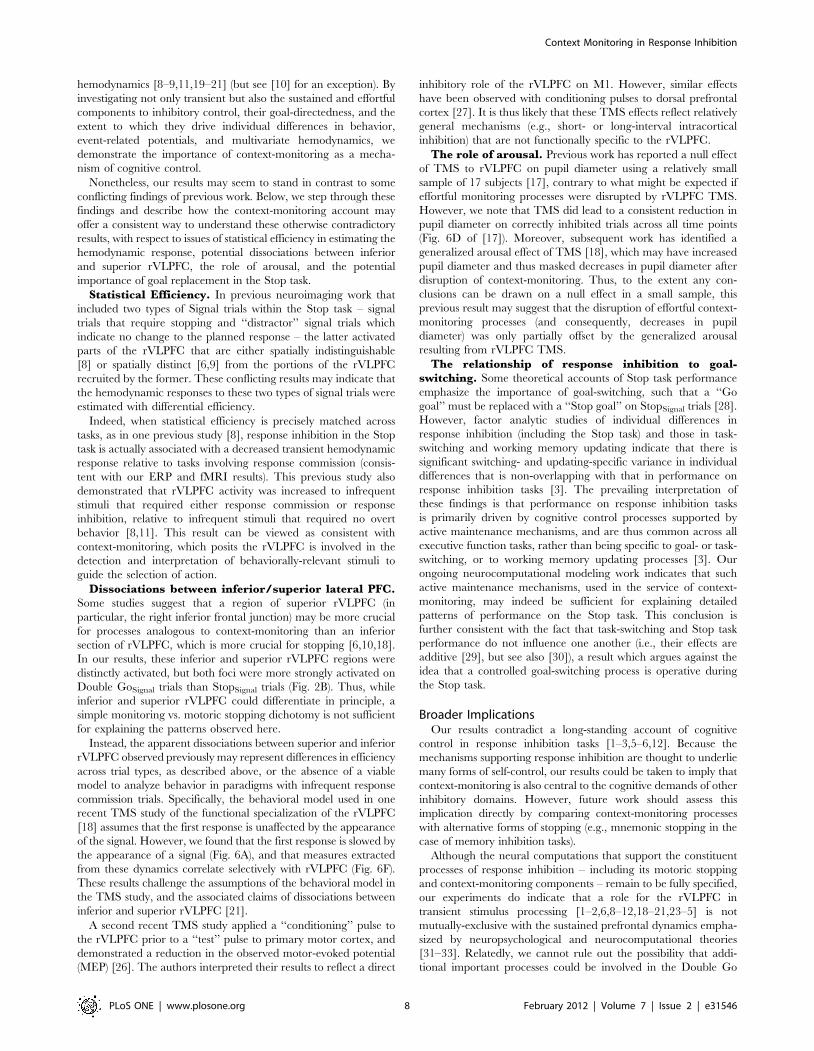
Nonetheless, our results may seem to stand in contrast to some
conflicting findings of previous work. Below, we step through these
findings and describe how the context-monitoring account may
offer a consistent way to understand these otherwise contradictory
results, with respect to issues of statistical efficiency in estimating the
hemodynamic response, potential dissociations between inferior
and superior rVLPFC, the role of arousal, and the potential
importance of goal replacement in the Stop task.
Statistical Efficiency. In previous neuroimaging work that
included two types of Signal trials within the Stop task – signal
trials that require stopping and ‘‘distractor’’ signal trials which
indicate no change to the planned response – the latter activated
parts of the rVLPFC that are either spatially indistinguishable
[8] or spatially distinct [6,9] from the portions of the rVLPFC
recruited by the former. These conflicting results may indicate that
the hemodynamic responses to these two types of signal trials were
estimated with differential efficiency.
Indeed, when statistical efficiency is precisely matched across
tasks, as in one previous study [8], response inhibition in the Stop
task is actually associated with a decreased transient hemodynamic
response relative to tasks involving response commission (consis-
tent with our ERP and fMRI results). This previous study also
demonstrated that rVLPFC activity was increased to infrequent
stimuli that required either response commission or response
inhibition, relative to infrequent stimuli that required no overt
behavior [8,11]. This result can be viewed as consistent with
context-monitoring, which posits the rVLPFC is involved in the
detection and interpretation of behaviorally-relevant stimuli to
guide the selection of action.
Dissociations between inferior/superior lateral PFC.Some studies suggest that a region of superior rVLPFC (in
particular, the right inferior frontal junction) may be more crucial
for processes analogous to context-monitoring than an inferior
section of rVLPFC, which is more crucial for stopping [6,10,18].
In our results, these inferior and superior rVLPFC regions were
distinctly activated, but both foci were more strongly activated on
Double GoSignal trials than StopSignal trials (Fig. 2B). Thus, while
inferior and superior rVLPFC could differentiate in principle, a
simple monitoring vs. motoric stopping dichotomy is not sufficient
for explaining the patterns observed here.
Instead, the apparent dissociations between superior and inferior
rVLPFC observed previously may represent differences in efficiency
across trial types, as described above, or the absence of a viable
model to analyze behavior in paradigms with infrequent response
commission trials. Specifically, the behavioral model used in one
recent TMS study of the functional specialization of the rVLPFC
[18] assumes that the first response is unaffected by the appearance
of the signal. However, we found that the first response is slowed by
the appearance of a signal (Fig. 6A), and that measures extracted
from these dynamics correlate selectively with rVLPFC (Fig. 6F).
These results challenge the assumptions of the behavioral model in
the TMS study, and the associated claims of dissociations between
inferior and superior rVLPFC [21].
A second recent TMS study applied a ‘‘conditioning’’ pulse to
the rVLPFC prior to a ‘‘test’’ pulse to primary motor cortex, and
demonstrated a reduction in the observed motor-evoked potential
(MEP) [26]. The authors interpreted their results to reflect a direct
inhibitory role of the rVLPFC on M1. However, similar effects
have been observed with conditioning pulses to dorsal prefrontal
cortex [27]. It is thus likely that these TMS effects reflect relatively
general mechanisms (e.g., short- or long-interval intracortical
inhibition) that are not functionally specific to the rVLPFC.
The role of arousal. Previous work has reported a null effect
of TMS to rVLPFC on pupil diameter using a relatively small
sample of 17 subjects [17], contrary to what might be expected if
effortful monitoring processes were disrupted by rVLPFC TMS.
However, we note that TMS did lead to a consistent reduction in
pupil diameter on correctly inhibited trials across all time points
(Fig. 6D of [17]). Moreover, subsequent work has identified a
generalized arousal effect of TMS [18], which may have increased
pupil diameter and thus masked decreases in pupil diameter after
disruption of context-monitoring. Thus, to the extent any con-
clusions can be drawn on a null effect in a small sample, this
previous result may suggest that the disruption of effortful context-
monitoring processes (and consequently, decreases in pupil
diameter) was only partially offset by the generalized arousal
resulting from rVLPFC TMS.
The relationship of response inhibition to goal-switching. Some theoretical accounts of Stop task performance
emphasize the importance of goal-switching, such that a ‘‘Go
goal’’ must be replaced with a ‘‘Stop goal’’ on StopSignal trials [28].
However, factor analytic studies of individual differences in
response inhibition (including the Stop task) and those in task-
switching and working memory updating indicate that there is
significant switching- and updating-specific variance in individual
differences that is non-overlapping with that in performance on
response inhibition tasks [3]. The prevailing interpretation of
these findings is that performance on response inhibition tasks
is primarily driven by cognitive control processes supported by
active maintenance mechanisms, and are thus common across all
executive function tasks, rather than being specific to goal- or task-
switching, or to working memory updating processes [3]. Our
ongoing neurocomputational modeling work indicates that such
active maintenance mechanisms, used in the service of context-
monitoring, may indeed be sufficient for explaining detailed
patterns of performance on the Stop task. This conclusion is
further consistent with the fact that task-switching and Stop task
performance do not influence one another (i.e., their effects are
additive [29], but see also [30]), a result which argues against the
idea that a controlled goal-switching process is operative during
the Stop task.
Broader ImplicationsOur results contradict a long-standing account of cognitive
control in response inhibition tasks [1–3,5–6,12]. Because the
mechanisms supporting response inhibition are thought to underlie
many forms of self-control, our results could be taken to imply that
context-monitoring is also central to the cognitive demands of other
inhibitory domains. However, future work should assess this
implication directly by comparing context-monitoring processes
with alternative forms of stopping (e.g., mnemonic stopping in the
case of memory inhibition tasks).
Although the neural computations that support the constituent
processes of response inhibition – including its motoric stopping
and context-monitoring components – remain to be fully specified,
our experiments do indicate that a role for the rVLPFC in
transient stimulus processing [1–2,6,8–12,18–21,23–5] is not
mutually-exclusive with the sustained prefrontal dynamics empha-
sized by neuropsychological and neurocomputational theories
[31–33]. Relatedly, we cannot rule out the possibility that addi-
tional important processes could be involved in the Double Go
Context Monitoring in Response Inhibition
PLoS ONE | www.plosone.org 8 February 2012 | Volume 7 | Issue 2 | e31546

task, such as goal-switching (though see discussion above) or other
as-yet unspecified control operations. We claim only that such
assumptions are unnecessary to explain our results, nor those from
other recent demonstrations of similarities between the Stop task
and closely matched (yet non-inhibitory) tasks that involve only
single responses [8–9] or responses across multiple effectors [18].
Nonetheless, even at its current level of detail, the context-
monitoring account of rVLPFC function does accord with
numerous emerging taxonomies of prefrontal organization. Ac-
cording to one recent taxonomy [33], ventral areas of the prefrontal
cortex are particularly important for contextual processing of
stimulus significance (broadly speaking, ‘‘what’’ processing) whereas
more dorsal areas may be particularly important for the processing
contextually-appropriate responses to those stimuli (‘‘how’’ process-
ing’’). The putative localization of context-monitoring to rVLPFC is
fully compatible with this framework. To the extent that response-
related ‘‘stopping’’ processes have a dedicated prefrontal substrate,
this taxonomy predicts that such processes should localize to areas
dorsal to the rVLPFC, some of which project to the STN with
similar or greater density [34]. However, our results do not suggest
that dorsal areas are differentially associated with stopping,
consistent with the wider literature [2,6–10], and further supporting
our conclusion that motoric stopping does not have a dedicated
lateral prefrontal substrate within the Stop task. Thus, while motoric
stopping may instead be subserved by a collection of heterogenous
mechanisms, including the subthalamic nucleus and primary motor
cortex, the rVLPFC’s interaction with these circuits appears to
derive more from its role in ‘‘what’’ processing (and perhaps with
associated temporoparietal connectivity), rather than from a role
that is more integrated with response demands (whether inhibitory
or not) [33].
This context-monitoring role of rVLPFC may also be
understood as arising from the proximity of rVLPFC to the
anterior insula, which appears to monitor interoceptive informa-
tion [35], in some cases proactively [36]. The anterior insula also
shows greater hemodynamic responses to demands on action
selection than to demands on motoric stopping, and is thought
to be tightly integrated with the rVLPFC [37]. Thus, a basic
mechanism in anterior insula for monitoring the internal sig-
nificance of upcoming stimuli may have been evolutionarily
adapted for use in monitoring the goal-relevance of stimuli for
action selection in the nearby rVLPFC. These representations may
even be tightly integrated, such that context monitoring can be
effectively recruited when it counts most – under conditions of
threat or pain. Indeed, target detection is improved when the
targets are predictive of pain, an effect that is associated with
greater activity in both rVLPFC and anterior insula [37].
The context-monitoring account is also compatible with recent
revisions to a classical taxonomy of the effects of prefrontal insult,
in which the inhibitory deficits arising from right lateral prefrontal
damage are now explained as monitoring deficits instead [31]. The
match between our findings and those motivating this taxonomic
revision may indicate the need to rethink a broad range of putative
inhibitory deficits. For example, focal rVLPFC damage can lead to
poor target detection, such that even when the location of an
upcoming target is cued before trial onset, this location is not
effectively monitored following the onset of any stimulus [38].
While this deficit might reflect problems with inhibiting locations
in space, our results suggest this patient’s focal rVLPFC damage
may have yielded a deficit in monitoring contextually-appropriate
locations in the service of target detection and action selection.
In addition to the significance of our result for understanding
neural insult, our result may also impact the treatment of patho-
logical impulse control deficits (e.g. as in substance abuse or
attention-deficit hyperactivity disorder [ADHD]). Specifically, our
result suggests that pathological impulse control deficits might not
reflect a failure to stop in particular, but rather the more effortful
and prefrontal processes involved in context-monitoring. For
example, ADHD may be associated with a monitoring deficit
in which many stimuli, regardless of their behavioral-relevance,
are thought to warrant attention. This prediction is supported
by the finding that ADHD is more strongly associated with
increased reaction time variability, as might result from a context-
monitoring deficit, than with deficits in tasks that require stopping
[39]. Relatedly, the resistance of response inhibition to improve-
ment via training [40] may reflect that monitoring context for
contingent action selection, not the act of stopping, is the
controlled process to be targeted for effective intervention.
Materials and Methods
Ethics StatementAll research was approved by the institutional review board at
the University of Colorado, and written informed consent was
obtained from all participants.
ParticipantsFor experiment 1, 86 subjects (mean age 19.11 years; SD = 1.17
years; 32 males) were recruited using the University of Colorado
undergraduate research pool and successfully completed the
Double Go and Stop Tasks. 2 subjects failed eyetracker calibration
and were excluded from pupillometric analyses. For experiment 2,
45 subjects (mean age 19.86 years; SD = 2.21; 23 males) were
recruited using the University of Colorado undergraduate research
pool and successfully completed the Double Go and Stop Tasks. 7
of these subjects were excluded from ERP analysis for artifacts
caused by excessive blinking (.60% of trials). For experiment 3,
19 subjects (mean age 23.3: SD = 4.4; 10 males) were recruited
from the local community and successfully completed the Double
Go and Stop Tasks. One subject was excluded from fMRI analyses
due to motion artifact.
Behavioral TaskAll subjects in all experiments completed the Double Go Task
prior to completing the Stop Task. This fixed task order was
adopted for reasons described in Text S1 – in particular, the use of
a fixed task order is ideal for the investigation of individual
differences [3], which was a central goal of the study reported
here. Nonetheless, appropriate precautions were taken to prevent
the contamination of experimental effects with cognitive phenom-
ena that might arise from the fixed task order (e.g., the use of
within-task baselines are used for all pupillometry, ERPs, and
fMRI analyses, so as to control for the relatively general effects of
phenomena like fatigue).
In all respects the Double Go and Stop Task were identical
within any given experiment (e.g., the precise interstimulus and
intersignal intervals, the presence of ‘‘null’’ trials, etc), with the
following exception: subjects are naturally aware of when they fail
to successfully stop a response, but seem unaware of their relative
speed on trials with the infrequent stimulus. To avoid any possible
mismatch across the two tasks owing to this difference, we
provided explicit feedback on all signal trials. Specifically, in the
Double Go Task, the signal turned red if subjects were slower than
their average running RT (experiments 2 & 3); in experiment 1
this was presented as sham feedback. (Double Go task trials with
categorically incorrect responses – such as a failure to respond
twice on Signal trials, or anything but a single correct response on
No Signal trials – were extremely rare and excluded from all
Context Monitoring in Response Inhibition
PLoS ONE | www.plosone.org 9 February 2012 | Volume 7 | Issue 2 | e31546

analysis). Similarly, in the Stop Task, the signal turned red if
subjects failed to successfully stop their response on that trial (in all
experiments). Additional cross-experiment differences in our tasks
suggest the generality of our results across minor variations in
experimental procedure (see Figure S1 & Table S1).
Statistical Analysis of fMRIData were acquired with a 3T GE Signal whole-body MRI
scanner at the University of Colorado Health Sciences Center,
using T2-weighted echo-planar imaging (EPI) (TR = 2000 ms,
TE = 32 ms, flip angle = 70u). Additional acquisition details are
available in Text S1.
Image pre-processing and analyses were conducted with FSL
(FMRIB’s Software Library). The first six volumes of each run
were discarded to allow the MR signal to reach steady state, the
remaining images in each participant’s time series were motion
corrected using MCFLIRT, and non-brain voxels were removed
using a brain extraction algorithm (BET). The data series was
spatially smoothed with a 3D Gaussian kernel (FWHM = 5 mm),
intensity normalized for all volumes, and high-pass filtered
(s = 50 sec).
After statistical analysis of each time series (details of the
regression model are available in Text S1), statistical maps were
normalized into the MNI-152 stereotaxic space using FLIRT
(FMRIB’s Linear Image Registration Tool). Parameter estimates
(PE) were transformed into a common stereotaxic space using the
above-mentioned three-step registration prior to the group
analyses with FLAME (FMRIB’s Local Analysis of Mixed Effects).
Z-statistic images were thresholded using clusters with z .2.58 as
well as a whole-brain corrected cluster significance threshold of
p,.05 using the theory of Gaussian Random Fields.
ROIs for Brodmann areas were anatomically defined using the
Talairach labeled atlas (see Figure S2), and mean percent signal
change was extracted using FSL’s featquery tool. The subthalamic
nucleus was anatomically defined using a 10 mm3 region centered
on the MNI coordinates previously used in the Stop Task to
interrogate BOLD in the STN (10,215,25) [41]. The TPJ was
anatomically defined using a 30 mm3 region centered on the MNI
coordinates (254, 252, 30) previously observed in a target
detection task [42].
Pattern classification analyses were conducted on the beta-
weights resulting from the above fMRI analysis pipeline, with
four minor exceptions. First, the BOLD data were not spatially
smoothed; second, the PEs were not statistically thresholded; third;
the PEs were z-transformed across all voxels within a given ROI for
each subject, to ensure that the classifiers were forced to operate
on the basis of distributed patterns of activation instead of overall
magnitudes. Finally, voxels with z-values falling outside of +/2 4.5
were windsorized. Classifiers were implemented as neural net-
works in Emergent [43]; separate networks were then trained,
using Hebbian and Contrastive Hebbian learning, for each ROI
(and therefore differed in terms of the number of input units), and
for identifying which individuals generated the data vs. what trial
type the data was estimated from (and therefore differed in terms
of the number of output units) but all other aspects of the network
architecture were the same. See Text S1 for full details on classifier
implementation.
Statistical Analysis of ERPs. During the Double Go and
Stop Tasks scalp voltages were recorded with a 128-channel
geodesic sensor net [44]. Amplified analog voltages (0.1- to 100.0-
Hz bandpass) were digitized at 250 Hz. Individual sensors were
adjusted until impedances were less than 50 k. The EEG was
digitally low-pass filtered at 40 Hz. Trials were discarded from
analyses if they contained incorrect responses, eye movements (eye
channel amplitudes over 70 V), or more than 20% of channels
were bad (average amplitude over 100 V or transit amplitude over
50 V). Individual bad channels were replaced on a trial-by-trial
basis with a spherical spline algorithm. EEG was measured with
respect to a vertex reference (Cz), but an average-reference trans-
formation was used to minimize the effects of reference-site activity
and accurately estimate the scalp topography of the measured
electrical fields. The average reference was corrected for the polar
average reference effect [45]. ERPs were obtained by stimulus-
locked averaging of the EEG recorded in each condition. ERPs
were baseline-corrected with respect to a 200-ms prestimulus
recording interval. These baselines were calculated separately for
each task, thereby controlling for nonspecific effects like fatigue.
Where montages are used, the occipital montage was centered
on Oz (including Oz, O1, O2, and the contiguous set of electrodes
76, 70, 74 and 82) and the frontal montage was centered on Fz
(including Fz and the contiguous set of electrodes 4, 5, 10, 12, 16
18 and 19). For scalp-wide voltage correlations (Fig. 4B) we
calculated Pearson’s R across tasks at every time point as the
variance shared between the subjects x electrode matrix across
tasks. Thus, this correlation reflects changes in voltage that covary
across tasks in the same subjects at the same electrode sites. For
montage-based voltage correlations (main text Fig. 4C) we
calculated Pearson correlations separately for the frontal and
occipital montages both before and after signal onset.
Statistical Analysis of Pupillometry. Pupil diameter was
recorded continuously during the Double Go and Stop Tasks
via a Tobii X50 infrared eyetracker calibrated to each subject.
Sampling at 50 Hz was synchronized to fixation onset, and pupil
diameter was calculated as the average diameter of successfully-
tracked eyes for each sample. Baseline measurements of pupil
diameter were calculated as the average diameter during the
200 ms preceding the onset of each signal (or the corresponding
time period for no signal trials); this value was subtracted from
the averaged samples recorded following the onset of the signal
(or the average signal onset for no signal trials). Baseline periods
were calculated independently for the Stop and Double Go t
asks, providing a within-task baseline to control for nonspecific
cognitive effects like fatigue. These normalized, averaged pupil
diameter samples were then smoothed using a box-car filter with
width of 60 ms.
Statistical Analysis of Behavior – Double Go Task. In
the Double Go Task, all RTs falling below 150 ms or above
750 ms were excluded from analysis, as well as those on No Signal
trials falling outside of 3.5 standard deviations of the iteratively-
calculated mean for each subject. RTs were only analyzed on
correct trials (i.e., trials in which two responses of the correct type
were provided on Signal trials, and where one and only one
response of the correct type was provided on No Signal trials).
Individual differences were extracted from the Double Go task
using a mixture model-based adaptation of the classic race model of
the Stop task (see also Text S1). Specifically, to classify individual
trials as slowed or unslowed, we first decomposed the distribution of
equipercentile residuals into two underlying distributions: a Gaussian
distribution with a mean of zero (corresponding to unslowed first
RTs), and a Gamma distribution (corresponding to the slowed first
RTs). The two free parameters to the Gamma and the one free
parameter to the Gaussian were fit in a fixed-effects analysis using
maximum likelihood estimation via with the Nelder-Meade simplex
algorithm [46–47]. The maximum likelihood fit is illustrated as
overlaid lines on the residual histogram (Fig. 6C), which was
relatively stable across multiple optimizations with different starting
parameters and yielded a better overall fit (see Table S1) than a single
Gaussian in terms of the Bayesian Information Criterion (BIC),
Context Monitoring in Response Inhibition
PLoS ONE | www.plosone.org 10 February 2012 | Volume 7 | Issue 2 | e31546

calculated as:
BIC~{2:XN
n~1
ln(XD
d~1
Wd LdRTn)zDp:ln(N)
Where N is the total number of observations, D is the total number of
distributions fit, Dp is the total number of free parameters used in
fitting those distributions, Wd is the weight of the dth distribution, and
Ld(RTn) is the likelihood of the nth RT given the best fit parameters for
the dth distribution (m and s for Gaussian and k and H for Gamma).
We next categorized individual trials as slowed or unslowed
using the likelihood of observing each RT under either of the two
fitted distributions. RTs were categorized as slowed if there
was even weak evidence in favor of the RT belonging to that
distribution (as quantified by a difference in BIC of $2.35);
otherwise RTs were categorized as unslowed. Other standards of
evidence lead to similar results as those presented here, but do not
as cleanly separate the slowed and unslowed trials (c.f. Fig. 6D).
To calculate TOSD, we subtracted the signal delay from the nth
percentile of no signal trial RTs, where n corresponds to the
proportion of RTs classified as unslowed at that signal delay. This
approach is conceptually identical to that used to calculate SSRT in
the race model, in which the signal delay is subtracted from the nth
percentile of No Signal RTs, where n corresponds to the proportion
of unsuccessful stop trials at that signal delay. TOSD was calculated
for each subject as the median of these estimates across all signal
delays. This estimate was unreasonably high for subjects for whom no
RTs had been classified as slowed (n = 34 out of 150), so in those cases
we used the minimum estimate of TOSD across all signal delays.
We then calculated the duration of slowing as the average
difference between RTs classified as slowed and RTs of corre-
sponding percent rank in the no signal RT distribution; subjects for
whom no RTs had been classified as slowed were excluded from all
analyses involving duration of slowing. The resulting estimates of
TOSD and duration of slowing can be found in Table S2.
Statistical Analysis of Behavior – Stop Task. In keeping
with the recommendations based on Monte Carlo simulations
[48], we estimated SSRT as the nth percentile of the No Signal RT
distribution minus the signal delay, where n is the proportion of
errors observed at each signal delay. This estimate was averaged
across the signal delays yielding 15% to 85% accuracy for each
subject to generate the recommended dependent measure for Stop
Tasks with fixed interstimulus intervals (SSRTAV). Data from the
Stop Task confirmed assumptions of the race model: RTs were
faster on Signal trials than on No Signal trials (t(145) = 11.31,
p,.0005) and accuracy was a monotonically decreasing function
of interstimulus interval (100 vs 150: t(145) = 13.52, p,.0005; 150
vs 250: t(145) = 17.20, p,.0005; 250 vs 300: t(145) = 7.14,
p,.0005). These and other behavioral indices of performance
across tasks are also reported in Table S3.
Supporting Information
Figure S1 Stimuli used in the three experiments. (A)
Experiment 1 included null trials consisting only of a fixation ring,
constituting 33% of the total number of trials. Of the remaining
trials, 75% were No-Signal trials – i.e., 2AFC trials in which either
a left-pointing or right-pointing arrow was presented. 25% were
Signal trials, in which a white box followed the onset of the 2AFC
stimulus. (B) Experiments 2 & 3 used this slightly different set of
stimuli, in which the arrows were replaced with left- or right-
pointing triangles, and the number of illuminated pixels was
matched between the triangles and squares.
(TIF)
Figure S2 rVLPFC ROIs were used in the univariate andmultivariate fMRI analyses. Subregions of the rVLPFC
include Brodmann Areas 44 (blue), 45 (red), and 47 (green).
(TIF)
Figure S3 MVPA methods. (A). For classifying subjects, neural
networks received inputs consisting of 1 unit per voxel in a given
ROI, where the activity of those units corresponds to the z-
transformed and trimmed parameter estimates from the unsmoothed
BOLD data. This input layer projects to a hidden ‘‘Scalar Val’’ layer,
which transforms each input unit’s activity into a distributed pattern
across 30 dedicated units. Finally, this hidden layer is fully connected
with an output layer consisting of 18 units, one corresponding directly
to each of our subjects. (B). For classifying trial types, we used the
same architecture as in A except that only 2 output units were used,
corresponding directly to each of the trial type contrasts. In addition,
separate networks were trained for each subject.
(TIF)
Figure S4 Tasks can be discriminated in all ROIs,including V1. Although tasks were best classified on the basis of
the Signal . Null contrast (white bars), this is unlikely to reflect
stopping-specific processes, since activity patterns in V1 allowed
the best classification on this contrast. Indeed, V1 showed the best
classification of tasks across all ROIs, when averaging across
contrasts. Because our tasks were collected in separate runs, this
good classification performance is likely to reflect run-specific
variance, rather than task-specific variance. This conclusion is
further supported by above-chance discrimination of tasks on the
basis of nuisance trials, during which both stimuli and responses
were precisely matched across tasks/runs.
(TIF)
Figure S5 Scalp topographies of the P3. The typical pattern
of ‘‘P3 anteriorization’’ in tasks that demand stopping was reversed
in our tasks, such that Double GoSignal trials elicited a larger P3
than the StopSignal trials at the site where the Stop P3 is typically
maximal (A). In contrast, the opposite was true of more posterior
electrodes (B & C), indicating that anteriorization effects cannot
not be taken to index explicit motoric stopping demands.
(TIF)
Figure S6 The group-average scalp distribution of ERPselicited by StopSignal and Double GoSignal trials weremarkedly similar, consistent with the strong relation-ship of these ERPs at the level of individual differences.In particular, the anteriorization of the P3 ERP elicited by Double
GoSignal trials, relative to that elicited by StopSignal trials, is visible
in the highlighted portion of each figure. Each contour represents
a change of .79 mV; red is positive.
(TIF)
Figure S7 Schematic illustration of the process modelsof our tasks. (A). The race model is used to analyze behavior in
the Stop task, such that the amount of warning necessary to stop
(Stop Signal Reaction Time, or SSRT) can be extracted as the nth
percentile of the StopNo-Signal distribution, where n corresponds to
the percent of unsuccessfully stopped responses at a particular
signal delay. (B) A conceptually similar model is used to analyze
behavior in the Double Go Task, but allows the extraction of two
underlying parameters. The duration of slowing can be estimated
Context Monitoring in Response Inhibition
PLoS ONE | www.plosone.org 11 February 2012 | Volume 7 | Issue 2 | e31546

as the difference between slowed 1st responses on Double
GoSignaltrials and responses of the same percent rank on Double
GoNo-Signal trials. The time of signal detection can be estimated as
the amount of time that must elapse following a signal before
responses are slowed. (C) The process model of the Double Go
Task predicts that slowing should be larger when signals are
presented earlier; this prediction was confirmed.
(TIF)
Table S1 Differences between Experiments 1–3.(DOCX)
Table S2 Mixture model estimates.(DOCX)
Table S3 Descriptive statistics for model-based analy-ses across Experiments.(DOCX)
Table S4 Statistical results from all primary analysesboth with and without those trials designated by themixture model as ‘‘slowed’’ in the Go task.
(DOCX)
Text S1 Additional methods, results, and discussion.
(DOC)
Acknowledgments
We thank the DEFD Research Group and the Badre & Frank labs.
Author Contributions
Conceived and designed the experiments: CHC EDC AK TC MTB YM.
Performed the experiments: CHC. Analyzed the data: CHC ED. Wrote
the paper: CHC EDC AK TC MTB YM.
References
1. Aron AR (2007) The neural basis of inhibition in cognitive control.Neuroscientist 13: 214–28.
2. Aron AR, Poldrack RA (2005) The cognitive neuroscience of response
inhibition: Relevance for genetic research in ADHD. Biological Psychiatry,57: 1285–92.
3. Friedman NP, Miyake A, Young SE, Defries JC, Corley RP, et al. (2008)
Individual differences in executive functions are almost entirely genetic in origin.Journal of Experimental Psychology: General 137: 201–25.
4. MacLeod CM, Dodd MD, Sheard ED, Wilson DE, Bibi U (2003) In Opposition
to Inhibition. In: BH. Ross, ed. The Psychology of Learning and Motivation.San Diego: Academic Press. pp 163–214.
5. Ferrier D (1876) The functions of the brain. London: Elder. 353 p.
6. Chikazoe J, Jimura K, Asari T, Yamashita K, Morimoto H, et al. (2009)Functional dissociation in right inferior frontal cortex during performance of
Go/No-Go Task. Cerebral Cortex 19: 146–52.
7. Logan GD, Cowan WB (1984) On the ability to inhibit thought and action: Atheory of an act of control. Psychological Review 91: 295–327.
8. Hampshire A, Chamberlain SR, Monti MM, Duncan J, Owen AM (2010) The
role of the right inferior frontal gyrus: inhibition and attentional control.Neuroimage 50: 1313–9.
9. Sharp DJ, Bonelle V, De Boissezon X, Beckman CF, James SG, et al. (2010)Distinct frontal systems for response inhibition, attentional capture, and error
processing. Proc Natl Acad Sci U S A 107: 6106–11.
10. Cai W, Leung HC (2011) Rule-Guided Executive Control of ResponseInhibition: Functional Topography of the Inferior Frontal Cortex. PLoS One
6(6): e20840. doi:10.1371/journal.pone.0020840.
11. Dodds CM, Morein-Zamir S, Robbins TW (2011) Dissociating Inhibition,Attention, and Response Control in the Frontoparietal Network Using
Functional Magnetic Resonance Imaging. Cerebral Cortex 21: 1155–65.
12. Aron AR (2010) From Reactive to proactive and Selective Control: Developing aRcicher Model for Stopping Inappropriate Responses. Biological Psychiatry(69):
55–68.
13. Jahfari S, Stinear CM, Claffey M, Verbruggen F, Aron AR (2010) Respondingwith restraint: what are the neurocognitive mechanisms? Journal of Cognitive
Neuroscience 22: 1479–92.
14. Smith JL, Johnstone SJ, Barry RJ (2008) Movement-related potentials in theGo/NoGo task: the P3 reflects both cognitive and motor inhibition. Clinical
Neurophysiology 119: 704–14.
15. Kahneman D, Beatty J (1966) Pupillary changes in two memory tasks. Science154: 1583–5.
16. Beatty J, Lucero-Wagoner B (2000) The Pupillary System. In: Caccioppo J,
Tassinary LG, Berntson G, eds. The Handbook of Psychophysiology. Hillsdale:Cambridge University Press. pp 142–62.
17. Chambers CD, Bellgrove MA, Stokes MG, Henderson TR, Garavan H, et al.(2006) Executive ‘‘brake failure’’ following deactivation of human frontal lobe.
Journal of Cognitive Neuroscience 18: 444–55.
18. Verbruggen F, Aron AR, Stevens MA, Chambers CD (2010) Theta burststimulation dissociates attention and action updating in human inferior frontal
cortex. Proc Natl Acad Sci U S A 107: 13966–71.
19. Chikazoe J, Konishi S, Asari T, Jimura K, Miyashita Y (2007) Activation of rightinferior frontal gyrus during response inhibition across response modalities.
Journal of Cognitive Neuroscience 19: 69–80.
20. Konishi S, Nakajima K, Uchida I, Sekihara K, Miyashita Y (1998) No-godominant brain activity in human inferior prefrontal cortex revealed by
functional magnetic resonance imaging. European Journal of Neuroscience 10:1209–1213.
21. Rubia K, Smith AB, Brammer MJ, Taylor E (2003) Right inferior prefrontal
cortex mediates response inhibition while mesial prefrontal cortex is responsiblefor error detection. Neuroimage 20: 351–8.
22. Shilling VM, Chetwynd A, Rabbitt PM (2002) Individual inconsistency across
measures of inhibition: an investigation of the construct validity of inhibition in
older adults. Neuropsychologia 40: 605–19.
23. Corbetta M, Patel G, Shulman GL (2008) The reorienting system of the human
brain: from environment to theory of mind. Neuron 58: 306–324.
24. Corbetta M, Shulman GL (2002) Control of goal-directed and stimulus-driven
attention in the brain. Nature Reviews Neurosciences 3: 201–215.
25. Barch DM, Braver TS, Nystrom LE, Forman SD, Noll DC, et al. (1997)
Dissociating working memory from task difficulty in human prefrontal cortex.
Neuropsychologia 35: 1373–1380.
26. Neubert FX, Mars RB, Buch ER, Olivier E, Rushworth MF (2010) Cortical and
subcortical interactions during action reprogramming and their related white
matter pathways. Proc Natl Acad Sci U S A 107: 13240–5.
27. Civardi C, Cantello R, Asselman P, Rothwell JC (2001) Transcranial magnetic
stimulation can be used to test connections to primary motor areas from frontal
and medial cortex in humans. Neuroimage 14: 1444–1453.
28. Verbruggen V, Logan G (2009) Automatic and Controlled Response Inhibition:
Associative Learning in the Go/No-Go and Stop-Signal Paradigms. Journal of
Experimental Psychology 137: 649–672.
29. Verbruggen F, Liefooghe B, Szmalec A, Vandierendonck A (2005) Inhibiting
Responses When Switching: Does it Matter? Experimental Psychology 52:
125–130.
30. Aron AR, Monsell S, Sahakian BJ, Robbins TW (2004) A componential analysis
of task–switching deficits associated with lesions of left and right frontal cortex.
Brain 127: 1561–1573.
31. Stuss DT, Alexander MP (2007) Is there a dysexective syndrome? Philos
Trans R Soc Lond B Biol Sci 362: 901–915.
32. Miller EK, Cohen JD (2001) An integrative theory of prefrontal cortex function.
Ann Rev Neurosci 24: 167–202.
33. O’Reilly RC (2010) The What and How of prefrontal cortical organization.
Trends in Neurosciences 33: 355–361.
34. Nambu A, Tokuno H, Inase M, Takada M (1997) Corticosubthalamic input
zones from forelimb representations of the dorsal and ventral divisions of the
premotor cortex in the macaque monkey: comparison with the input zones from
the primary motor cortex and the supplementary motor area. Neurosci Lett 239:
13–16.
35. Craig AB (2009) How do you feel – now? The anterior insula and human
awareness. Nature Reviews Neuroscience 10: 59–70.
36. Lovero KL, Simmons AN, Aron JL, Paulus MP (2009) Anterior insular cortex
anticipates impending stimulus significance. Neuroimage 45: 976–83.
37. Lim SL, Padmala S, Pessoa L (2009) Segregating the significant from the
mundane on a moment-to-moment basis via direct and indirect amygdala
contributions. Proc Natl Acad Sci U S A 106: 16841–16846.
38. Michael GA, Garcia S, Fernandez D, Sellal F, Boucart M (2006) The ventral
premotor cortex (vPM) and resistance to interference. Behavioral Neuroscience
120: 447–62.
39. Castellanos FX, Sonuga-Barke EJS, Milham MP, Tannock R (2006)
Characterizing cognition in ADHD: beyond executive dysfunction. Trends
Cog Sci 10: 117–123.
40. Thorell LB, Lindqvist S, Nutley SB, Bohlin G, Klingberg T (2009) Training and
transfer effects of executive functions in preschool children. Dev Sci 12:
106–113.
41. Aron AR, Poldrack RA (2006) Cortical and subcortical contributions to stop
signal response inhibition: role of the subthalamic nucleus. J Neurosci 26:
2424–2433.
42. Corbetta M, Kincade JM, Ollinger JM, McAvoy MP, Shulman GL (2000)
Voluntary orienting is dissociated from target detection in human posterior
parietal cortex. Nature Neuroscience 3: 292–7.
Context Monitoring in Response Inhibition
PLoS ONE | www.plosone.org 12 February 2012 | Volume 7 | Issue 2 | e31546

43. Aisa B, Mingus B, O’Reilly RC (2008) The emergent neural modeling system.
Neural Networks 21: 1146–1152.44. Tucker DM (1993) Spatial sampling of head electrical fields: The geodesic sensor
net. Electroencephalography & Clinical Neurophysiology 87: 154–16.
45. Junghoefer M, Elber T, Tucker DM, Braun C (1999) The polar averagereference effect: A bias in estimating the head surface integral in EEG recording.
Clinical Neurophysiology 110: 1149–1155.
46. Nelder JA, Mead RA (1965) Simplex method for function minimization. Comp J
7: 308–313.
47. Ratcliff R, Starns JJ (2009) Modeling confidence and response time in
recognition memory. Psychological Review 116: 59–83.
48. Band GP, van der Molen MW, Logan GD (2003) Horse-race model simulations
of the stop-signal procedure. Acta Psychologica 112: 105–42.
Context Monitoring in Response Inhibition
PLoS ONE | www.plosone.org 13 February 2012 | Volume 7 | Issue 2 | e31546