coexpression of neurotrophins their nervous - pnas.org filethe neurotrophins (1-12). members of the...
TRANSCRIPT

Proc. Natl. Acad. Sci. USAVol. 90, pp. 6711-6715, July 1993Neurobiology
Coexpression of neurotrophins and their receptors in neurons ofthe central nervous system
(in situ hybridizatIon/braIn-derived neurotrophic factor/brain damage/hippocampus/epilepsy)
ZAAL KOKAIA*t, JOHAN BENGZON*, MADIS METSIS*, MERAB KoKAIA*, HAKAN PERSSONf,AND OLLE LINDVALL**Restorative Neurology Unit, Department of Neurology, University Hospital, S-221 85 Lund, Sweden; and tDepartment of Medical Chemistry, Laboratory ofMolecular Neurobiology, Karolinska Institute, S-104 01 Stockholm, Sweden
Communicated by Jan G. Waldenstrom, April 14, 1993 (receivedfor review February 3, 1993)
ABSTRACT Nerve growth factor (NGF) and brain-derived neurotrophic factor (BDNF) are neuronal survivalmolecules which utilize the Trk family of tyrosine kinasereceptors. Using double-label in situ hybridization, we dem-onstrate that mRNAs for BDNF and its high-affinity receptorTrkB are coexpressed in hippocampal and cortical neurons.Also, a large number of neurons in these areas coexpress NGFand BDNF mRNAs. Epileptic seizures lead to increased levelsof both BDNF/TrkB and NGF/BDNF mRNAs in double-labeled ceils. Our results show that individual neurons of thecentral nervous system can coexpress neurotrophins and theirreceptors and produce two neurotrophic factors. These factorscould support neuronal survival after brain insults, not only viaretrograde transport but also through autocrine mechanisms.
Neurotrophic factors are necessary for the normal develop-ment and maintenance of the peripheral and central nervoussystems. Nerve growth factor (NGF), brain-derived neuro-trophic factor (BDNF), neurotrophin 3 (NT-3), and neuro-trophin 4 (NT-4), also named neurotrophin 5, belong to afamily of structurally related neurotrophic molecules, calledthe neurotrophins (1-12). Members of the Trk family oftyrosine kinase receptors-TrkA, TrkB, and TrkC, havebeen shown to encode essential components ofthe functionalhigh-affinity receptors for NGF, BDNF, NT-3, and NT-4 (10,12-21). NGF mediates its effect viaTrkA (14-16), and BDNFand NT-4 via TrkB (10, 12, 13, 17-19), whereas NT-3interacts mainly with the TrkC receptor but also binds toTrkA and TrkB receptors (21).The neurotrophins are all expressed in neurons of the
central nervous system, with the highest mRNA levels in thehippocampus (22, 23). NGF is the prototype of a target-derived neurotrophic molecule and promotes the survival ofbasal forebrain cholinergic neurons (24, 25). NGF is pro-duced by hippocampal and cortical neurons, taken up incholinergic nerve terminals, and transported retrogradely tothe cell bodies (24, 25). Recently BDNF and NT-3 werereported to display distinct patterns of retrograde axonaltransport in central neurons (26). In the brain, TrkA mRNAexpression appears to be restricted to NGF-responsive cells(27, 28), whereas cells expressing TrkB and TrkC mRNAs arewidely distributed (21, 28-30). Epileptic seizures markedlyincrease levels of mRNA and protein for NGF and BDNF(31-38), reduce NT-3 mRNA expression (34, 39), and lead toelevated levels of TrkB mRNA and protein (29). Similarchanges are observed after cerebral ischemia and insulin-induced hypoglycemic coma in the absence ofseizure activity(40). These findings have raised the possibility that theneurotrophins act locally and protect against neuronal ne-
The publication costs of this article were defrayed in part by page chargepayment. This article must therefore be hereby marked "advertisement"in accordance with 18 U.S.C. §1734 solely to indicate this fact.
crosis after brain insults characterized by excessive gluta-mate release. Such a role would require that neurotrophinsand their receptors are localized in the same or neighboringcells-i.e., that they function through autocrine or paracrinemechanisms. In fact, autocrine or paracrine stimulation ofcholinergic neurons by NGF has been postulated to occur inthe embryonic hippocampus (41) and in the adult striatum(41) and basal forebrain (42). In this study we show, usingdouble-label hybridization histochemistry, that mRNAs forBDNF and TrkB and for BDNF and NGF are coexpressed inneurons in the normal brain and that seizure activity inducesincreased expression ofboth mRNA species in the same cell.
MATERIALS AND METHODSAdult male Sprague-Dawley rats were either subjected torecurrent seizures induced by electrical kindling stimulationsin the hippocampus or injected with kainic acid (10 mg/kg,i.p.). For kindling, stainless steel electrodes (o.d., 0.25 mm)were implanted bilaterally in the ventral hippocampus byusing a Kopf stereotaxic frame (43). One week later, animalswere given 40 stimulations on the left side (400-,A, 10-Hz,1-ms square-wave pulses for 10 s with 5-min intervals) (44).Electroencephalographic activity was continuously moni-tored and behavioral seizures were scored. Control rats wereconnected to the stimulating-recording device or injectedwith saline. Two and 3 hr after the last kindling stimulationand the injection of kainic acid or vehicle, respectively,animals were decapitated and the brains were immediatelyfrozen on dry ice and analyzed by in situ hybridizationhistochemistry.
Cryostat sections (14 um) through the hippocampus werefirst hybridized to a 35S-labeled NGF or TrkB cRNA probeand then to a digoxygenin-11-dUTP-labeled BDNF cRNAprobe, which had been prepared as follows. A 456-bp PCRfragment including primers and restriction site and encom-passing nucleotides 1135-1515 in the rat trkB sequence (45)and a 762-bp fragment of the 3' exon of the rat NGF gene (46)encompassing nucleotides 334-1095 were subcloned in plas-mid pBluescript KS (Stratagene). The plasmid was linearizedand the cloned DNA was transcribed in vitro by using T3(antisense) or T7 (sense) RNA polymerase in the presence of[a-[35S]thio]UTP (Amersham, >1000 Ci/mmol; 1 Ci = 37GBq). A 243-bp partial cDNA for rat BDNF (47) encodingamino acids 75-155 in prepro-BDNF was subcloned in pB-SKS, linearized, and transcribed in vitro with T3 (antisense)orT7 (sense)RNA polymerases and labeled with digoxygeninDNA-labeling mixture (Boehringer Mannheim), containingdigoxygenin-11-dUTP. Some sections were hybridized to aBDNF cRNA probe labeled with [a-[35S]thio]UTP.
Abbreviations: NGF, nerve growth factor; BDNF, brain-derivedneurotrophic factor; NT-3, neurotrophin-3.tTo whom reprint requests should be addressed.
6711
6712 Neurobiology: Kokaia et al.
.........A. l...
*CA1! t
..15' ... .:s
If.
P
A4~~~~. \,.= v
A::
-A.
::::::.V
A^
Sw...
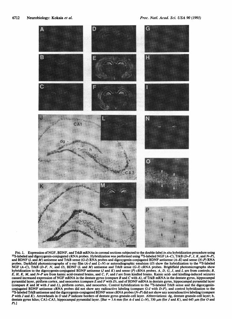
FiG. 1. Expression ofNGF, BDNF, and TrkB mRNAs in coronal sections subjected to the double-label in situ hybridization procedure using35S-labeled and digoxygenin-conjugated cRNA probes. Hybridization was performed using 35S-labeled NGF (A-C), TrkB (D-F, J, K, and N-P),and BDNF (L and M) antisense and TrkB sense (G-1) RNA probes and digoxygenin-conjugated BDNF antisense (A-K) and sense (N-P) RNAprobes. Darkfield photomicrographs of x-ray film (A-I and L-N) or autoradiographic emulsion (0) show the hybridization to the 35S-labeledNGF (A-C), TrkB (D-F, N, and 0), BDNF (L and M) antisense and TrkB sense (G-l) cRNA probes. Brightfield photomicrographs showhybridization to the digoxygenin-conjugated BDNF antisense (J and K) and sense (P) cRNA probes. A, D, G, J, and L are from controls; B,E, H, K, M, and N-P are from kainic acid-treated brains, and C, F, and I are from kindled brains. Kainic acid- and kindling-induced seizurescaused increased expression ofNGF mRNA in the dentate gyrus (compare B and C with A), ofTrkB mRNA in the dentate gyrus, hippocampalpyramidal layer, piriform cortex, and neocortex (compare E and F with D), and ofBDNF mRNA in dentate gyrus, hippocampal pyramidal layer(compare K and M with J and L), piriform cortex, and neocortex. Control hybridization to the 35S-labeled TrkB sense and the digoxygenin-conjugated BDNF antisense cRNA probes did not show any radioactive labeling (compare G-I with D-F), and control hybridization to the35S-labeled TrkB antisense and the digoxygenin-conjugated BDNF sense cRNA probes (N-P) did not show any nonradioactive labeling (compareP with J and K). Arrowheads in 0 and P indicate borders of dentate gyrus granule-cell layer. Abbreviations: dg, dentate granule-cell layer; h,dentate gyrus hilus; CA1-CA3, hippocampal pyramidal layer. [Bar = 3.6 mm (for A-I and L-N), 330 ,um (for J and K), and 645 ,um (for 0 andP).]
Proc. Natl. Acad. Sci. USA 90 (1993)
~...?t

Proc. Natl. Acad. Sci. USA 90 (1993) 6713
In the first step, the sections were thawed onto poly(L-lysine)-coated slides (50 ug/ml). After fixation with 3%formaldehyde in phosphate-buffered saline (PBS) for 5 min,sections were rinsed once with PBS and twice with deionizedand autoclaved water and were placed for 15 min in 0.2 MHCI. Sections were then rinsed twice with PBS and immersedin 0.25% acetic anhydride in 0.1 M triethanolamine (pH 8.0)for 20 min. After two washes in PBS, sections were dehy-drated and air-dried, and 80 ul of hybridization buffer [50%(vol/vol) deionized formamide/0.33 M NaCl/20 mMTris-HCl, pH 7.5/1 mM EDTA/0.1 M dithiothreitol/10%(wt/vol) dextran sulfate/Sx Denhardt's solution with yeasttRNA at 0.5 mg/ml and synthetic poly(A) RNA at 0.1 mg/ml]containing 35S-labeled cRNA probe at 2.5 x 10⁶ cpm/ml wasapplied per section. After hybridization overnight at 42°C,slides were washed four times (15 min each) in 1 x SSC(standard saline citrate) at 55°C, cooled to room temperature,briefly dipped in deionized and autoclaved water, and air-dried.
In the second step, 100 ,ul of hybridization buffer contain-ing 50 ng of digoxygenin-11-dUTP-labeled BDNF cRNAprobe was applied per section. After hybridization overnightat 37°C, slides were washed twice in 1 x SSC at 48°C, placedfor 30 min at 37°C in 0.5 M NaCl/10 mM Tris-HCl, pH 8.0/1mM EDTA with RNase A at 10 ,ug/ml, and washed twice in1x SSC at 48°C and in 0.5 x SSC and 0.1 x SSC at 60°C. Slideswere cooled to room temperature, rinsed twice with buffer 1(100mM Tris-HCl, pH 7.5/150mM NaCl, incubated with 5%normal sheep serum in buffer 1 for 1 hr (150 IlI per section),and then incubated for 5 hr at room temperature with 1%normal sheep serum, 0.3% Triton X-100, and alkaline-phosphatase-conjugated anti-digoxygenin serum (BoehringerMannheim; 1:500) in buffer 1. Sections were washed twice for10 min in buffer 1 and for 5 min in buffer 3 (100 mM Tris HCl,pH 9.5/100 mM NaCl/50 mM MgCl2). They were thenincubated overnight at room temperature in the dark in buffer3 containing nitroblue tetrazolium salt (0.34 mg/ml) and5-bromo-4-chloro-3-indolyl phosphate toluidinium salt (0.18mg/ml) (Boehringer Mannheim). The sections were rinsedtwice in buffer 3 and immersed in buffer 4 (10mM Tris HCl/1mM EDTA, pH 8.0) for 6-8 hr and then in 1 x SSC overnightat room temperature. On the next day the slides were brieflyrinsed in distilled water and air-dried, and sections wereapposed to ,3-max x-ray film (Amersham). When films hadbeen developed, photos of the distribution of digoxygenin-positive cells were taken under the microscope and thesections were then dipped in Ilford K5 emulsion and exposedfor 8 weeks.
RESULTSThe specificity of the hybridization reaction was inferredfrom the following observations. (i) In situ hybridization ofsections with a digoxygenin-labeled BDNF sense probe afterhybridization to a 35S-labeled TrkB antisense probe gave riseto radioactive labeling of TrkB mRNA (Fig. 1 N and 0) butdid not result in any specific labeling of BDNF mRNA (Fig.1P). Conversely, hybridization to a 35S-labeled TrkB senseprobe and subsequent hybridization to a digoxygenin-labeledBDNF antisense probe showed no specific TrkB mRNAsignal (Fig. 1 G-1). (ii) Identical regional distribution ofNGFand TrkB mRNAs, as assessed with radiolabeled probes, wasseen before and after hybridization to the digoxygenin-labeled BDNF cRNA probe (Fig. 1 A-F). Similarly, nonra-dioactive hybridization to the BDNF cRNA probe (Fig. 1 Jand K) was not changed by hybridization to 35S-labeled NGFor TrkB cRNA probe, and regional expression of BDNFmRNA was similar with both 35S- and digoxygenin-labeledprobes (Fig. 1 J-M). (iii) The distribution, density, andmRNA levels of cells which hybridized to the various probesin the present double-labeling procedure in intact rats and
after kindling and kainic acid (Fig. 1 A-F, L, and M) wereidentical to what has been described previously with singlelabeling techniques (22, 23, 28, 29, 35, 48-50).
In the control brains, BDNF and NGF mRNAs werecolocalized in scattered neurons in the dentate gyrus granule-cell layer and hilus (Fig. 2A), in the pyramidal layer of thehippocampal CA1-CA3 regions, and in piriform cortex (Fig.2C). All NGF mRNA-positive cells also expressed BDNFmRNA, whereas >95% of BDNF mRNA-positive cells inthese regions were not labeled for NGF mRNA. No clearlydouble-labeled neurons were observed in the neocortex.Kainic acid-induced and kindled seizures markedly increasedNGF mRNA expression in the dentate gyrus and piriformcortex and BDNF mRNA levels in dentate gyrus, hippocam-pal CA1-CA3 regions, piriform cortex, amygdala, and neo-cortex (Fig. 1 A-C and J-M). Following these treatments,>95% of BDNF mRNA-containing neurons in the dentategyrus (Fig. 2B) and piriform cortex (Fig. 2D) exhibited NGFmRNA labeling, and the levels of both mRNAs withindouble-labeled cells were markedly higher than in controls.Also after seizures, all NGF mRNA-containing cells in den-tate gyrus and piriform cortex expressed BDNF mRNA,while all labeled neurons in neocortex and amygdala con-tained BDNF mRNA alone.The control brains showed coexistence ofBDNF and TrkB
mRNAs in many regions. Virtually all BDNF mRNA-expressing neurons in the dentate granule-cell layer and hilus(Fig. 3A), pyramidal layer ofhippocampal CA1-CA3 regions,and piriform cortex (Fig. 3E) were also labeled for TrkBmRNA. A few TrkB mRNA-positive profiles, possibly glialcells, were present outside the principal neuronal layers inthese regions. Also in neocortex (Fig. 3C) and amygdaloidcomplex, all BDNF mRNA-containing cells hybridized to theTrkB mRNA probe. Cells expressing only TrkB mRNA were
#5 t >if~@
A *. .i vo.4. -
'''-io'7 B -
.v Ks
Vs *,Ot
.4% .,N, ..
D
. *,W
FIG. 2. Brightfield photomicrographs showing hybridization tothe 35S-labeled NGF and digoxygenin-conjugated BDNF cRNAprobes in the dentate gyrus (A and B) and piriform cortex (C and D).(A) A large hilar neuron coexpresses NGF and BDNF mRNA.Granule cells contain BDNF mRNA but only scattered neurons alsoexpress NGF mRNA. (B) Kainic acid markedly increases BDNF andNGF mRNA levels in granule cells, most of which now coexpressboth mRNA species. Increased expression of BDNF mRNA in adouble-labeled large hilar neuron is also observed. (C and D)Examples of cells coexpressing NGF and BDNF mRNAs in control(C) and, with higher levels of both mRNA species, after kainic acid(D). Abbreviations: dg, dentate granule-cell layer; h, hilar neuron.(Bar = 20 Am.)
Neurobiology: Kokaia et al.
Proc. Natl. Acad. Sci. USA 90 (1993)
A
ICr.
B
D iw**.w *
s.. . . ,<,.,.,
F., t *v .
_W. '. --
FiG. 3. Brightfield photomicrographs showing hybridization tothe 35S-labeled TrkB and digoxygenin-conJugated BDNF cRNAprobes in the dentate gyrus hilus (A and B), the neocortex (C and D),and the piriform cortex (E and F). Examples of cells coexpressingBDNF and TrkB mRNAs are seen in controls (A, C, and E), and withhigher levels of both mRNA species, following kainic acid (B and F)or kindling (D). A cell in Fexpressing TrkB mRNA alone is indicatedby arrowheads. (Bar = 16 pm.)
scarce (Fig. 3F) and in neocortex, for example, representedonly about 3% of the labeled cells. Following kainic acid orkindling, the intensity of labeling for both BDNF and TrkBmRNAs within double-labeled neurons had increased mark-edly in the dentate granule-cell layer and hilus (Figs. 3B and4B), hippocampal CA1-CA3 regions, piriform cortex (Figs.3F and 4A), and neocortex (Fig. 3D).
DISCUSSIONOur data demonstrate that NGF and BDNF mRNAs can besynthesized by an individual neuron and that seizure activityleads to increased expression of both mRNA species in thesame cell. The two neurotrophins produced by the same cell
could have separate functional roles. NGF synthesized in ahippocampal or cortical neuron might act as a target-derivedtrophic factor for cholinergic neurons (24, 25), whereasBDNF in the same cell could provide local trophic supportvia an autocrine mechanism. The proposed similarities in thethree-dimensional structure of the neurotrophins (51) mayalso suggest that NGF and BDNF could form heterodimericcomplexes as a consequence ofcoexpression in the same cell.Receptor activation by such complexes may lead to hithertounknown biological activities of the neurotrophins.
Supporting a role forNGF as a target-derived neurotrophicfactor, mRNA for its high-affinity receptor, TrkA, has beenfound in basal forebrain cholinergic cell bodies but not in theirhippocampal and cortical projection areas (27, 28). In con-trast, the widespread coexpression of BDNF and TrkBmRNAs demonstrated here provides evidence that BDNFacts via an autocrine mechanism. Throughout the forebrainall BDNF mRNA-containing cells hybridized also to theTrkB probe. The TrkB probe used in this study detectsmessages encoding both truncated receptors lacking thetyrosine kinase domain and full-length TrkB receptors. Vir-tually all dentate granule cells and pyramidal neurons of thepiriform cortex contain TrkB mRNA coding for the func-tional receptor (29). Since we found that the overall majorityof neurons in these areas hybridized to both the BDNF andTrkB mRNA probes, it seems highly likely that BDNFmRNA is also coexpressed with mRNA for the functionalTrkB receptor. The small portion of cells which hybridized tothe TrkB mRNA probe alone often had a glial distribution,being localized in areas largely devoid of neurons.
If BDNF acts via an autocrine mechanism it is probablysecreted and activates the TrkB receptor at the surface of theBDNF-synthesizing cell itself. A local effect ofBDNF in thehippocampus is supported by data showing rapid induction ofthe c-fos gene in cultures of embryonic hippocampal neuronsafter addition ofBDNF (52). Increased expression of BDNFand TrkB mRNAs in vivo occurs already after brief periods(1-2 min) of epileptic seizures, cerebral ischemia and hypo-glycemic coma (34, 35, 40). If prolonged, these insults lead toneuronal necrosis in, for example, cortex and hippocampus(53). Our finding ofa widespread increase ofBDNF and TrkBmRNAs within the same neuron after kainic acid-induced andkindled seizures indicates that the rapid induction of BDNF
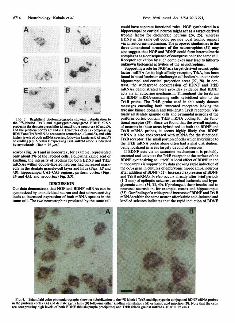
FIG. 4. Brightfield color photomicrographs showing hybridization to the 35S-labeled TrkB and digoxygenin-conjugated BDNF cRNA probesin the piriform cortex (A) and dentate gyrus hilus (B) following either kindling stimulations (A) or kainic acid injection (B). Note that the cellsare coexpressing high levels of both BDNF (bluish/purple precipitate) and TrkB (black grains) mRNAs. (Bar = 19 gm.)
6714 Neurobiology: Kokaia et al.
.. ;p0. .. v
.0

Proc. Natl. Acad. Sci. USA 90 (1993) 6715
mRNA expression after brain insults acts to protect theBDNF-synthesizing neuron itself. Elevated levels ofboth thefull-length and the truncated TrkB receptor may amplify theprotective effect of BDNF, the truncated receptor by buff-ering the concentration of the ligand (i.e., maintaining a highlocal concentration ofBDNF), and the full-length receptor byincreasing the biological response to BDNF.Growth factor stimulation via autocrine mechanisms has
been implicated in cell transformation and tumor growth (54).Autocrine stimulation may also occur in normal cells-forexample, by platelet-derived growth factor (PDGF) in dorsalroot ganglia and Schwann cells (55) and by BDNF and NT-3in developing sympathetic and dorsal root ganglia (56, 57). Inthe brain, synthesis of a trophic factor and its receptor by thesame neuron, as shown here for BDNF and TrkB, has notbeen demonstrated previously, although the overlappingdistribution of cells expressing a neurotrophin and a neuro-trophin receptor, respectively, has suggested a local mode ofaction in the hippocampus (29, 41). Both PDGF and PDGFreceptors have been detected in central neurons (58-60),though not reported in the same cell, which suggests thatautocrine regulation of neuronal function may be a moregeneral principle for trophic factor action in the centralnervous system. Multiple trophic factors may be necessaryfor survival of the synthesizing neuron and its afferentneuronal inputs both under physiological conditions andfollowing brain insults.
We thank A. Bjorklund, K. Campbell, A. Dagerlind, and A.Lippoldt for valuable discussions. This work was supported by grantsfrom the Swedish Medical Research Council (14X-8666), the RoyalPhysiographic Society, the Swedish Society for Medical Research, theRoyal Swedish Academy of Sciences, the Swedish Natural ScienceResearch Council, the Bank of Sweden Tercentenary Fund, theEuropean Science Foundation, the Medical Faculty, University ofLund, and the Elsa and Thorsten Segerfalk Foundation.
1. Barde, Y.-A., Edgar, D. & Thoenen, H. (1982) EMBO J. 1, 549-553.2. Levi-Montalcini, R. (1987) Science 237, 1154-1162.3. Leibrock, J., Lottspeich, F., Hohn, A., Hofer, M., Hengerer, B.,
Masiakowski, P., Thoenen, H. & Barde, Y.-A. (1989) Nature (London)341, 149-152.
4. Ernfors, P., Ib6llez, C. F., Ebendal, T., Olson, L. & Persson, H. (1990)Proc. Natl. Acad. Sci. USA 87, 5454-5458.
5. Hohn, A., Leibrock, J., Bailey, K. & Barde, Y.-A. (1990) Nature(London) 344, 339-341.
6. Jones, K. R. & Reichardt, L. F. (1990) Proc. Natl. Acad. Sci. USA 87,8060-8064.
7. Kaisho, Y., Yoshimura, K. & Nakahama, K. (1990) FEBS Lett. 266,187-191.
8. Maisonpierre, P. C., Belluscio, L., Squinto, S., Ip, N. Y., Furth, M. E.,Lindsay, R. M. & Yancopoulos, G. D. (1990) Science 247, 1446-1451.
9. Rosenthal, A., Goeddel, D. V., Nguyen, T., Lewis, M., Shih, A.,Laramee, G. R., Nikolics, K. & Winslow, J. W. (1990) Neuron 4,767-773.
10. Berkemeier, L. R., Winslow, J. W., Kaplan, D. R., Nikolics, K., Goed-del, D. V. & Rosenthal, A. (1991) Neuron 7, 1031-1041.
11. Hallb6ok, F., IMbfiez, C. F. & Persson, H. (1991) Neuron 6, 845-858.12. Ip, N., IMbiez, C., Nye, S., McClain, J., Jones, P., Gies, D., Beliuscio,
L., LeBeau, M., Espinosa, R., Squinto, S., Persson, H. & Yancopoulos,G. (1992) Proc. Natl. Acad. Sci. USA 89, 3060-3064.
13. Klein, R., Lamballe, F., Bryant, S. & Barbacid, M. (1992) Neuron 8,947-956.
14. Hempstead, B., Kaplan, D. R., Martin-Zanca, D., Panda, L. F. & Chao,M. V. (1991) Nature (London) 350, 678-683.
15. Kaplan, D. R., Martin-Zanca, D. & Parda, L. F. (1991) Nature (Lon-don) 350, 158-160.
16. Klein, R., Jing, S., Nanduri, V., O'Rourke, E. & Barbacid, M. (1991) Cell65, 189-197.
17. Klein, R., Nanduri, V., Jing, S., Lamballe, F., Tapely, P., Bryant, S.,Cordon-Cardo, C., Jones, K. R., Reichart, L. F. & Barbacid, M. (1991)Cell 66, 395-403.
18. Soppet, D., Escandon, E., Maragos, J., Middlemas, D. S., Reid, S. W.,Blair, J., Burton, L. E., Stanton, B. R., Kaplan, D. R., Hunter, T.,Nikolics, K. & Parada, L. F. (1991) Cell 65, 895-903.
19. Squinto, S. P., Stitt, T. N., Aldrich, T. H., Davis, S., Bianco, S. M.,Radziejewski, C., Glass, D. J., Masiakowski, P., Furth, M. E., Valen-zuela, D. M., DiStefano, P. S. & Yancopoulos, G. D. (1991) Cell 65,885-893.
20. Kaplan, D. R., Hempstead, B., Martin-Zanca, D., Chao, M. V. &Panda, L. F. (1991) Science 252, 554-558.
21. Lamballe, F., Klein, R. & Barbacid, M. (1991) CeUl 66, 967-979.22. Ernfors, P., Wetmore, C., Olson, L. & Persson, H. (1990) Neuron 5,
511-526.23. Phillips, H. S., Hains, J. M., Laramee, G. R., Rosenthal, A. & Winslow,
J. W. (1990) Science 250, 290-294.24. Barde, Y.-A. (1989) Neuron 2, 1525-1534.25. Dreyfus, C. F., Bend, P., Martinez, H. J., Rubin, S. J. & Black, I. B.
(1989) Exp. Neurol. 104, 181-185.26. DiStefano, P. S., Friedman, B., Radziejewski, C., Alexander, C., Bo-
land, P., Schik, C. M., Lindsay, R. M. & Wiegand, S. J. (1992) Neuron8, 983-993.
27. Holtzman, D. M., Li, Y., Panda, L. F., Kinsman, S., Chen, C.-K.,Valleta, J. S., Zhou, J., Long, J. B. & Mobley, W. C. (1992) Neuron 9,465-478.
28. Merlio, J.-P., Ernfors, P., Jaber, M. & Persson, H. (1992) Neuroscience51, 513-532.
29. Merlio, J.-P., Ernfors, P., Kokaia, Z., Bengzon, J., Kokaia, M., Smith,M.-L., Siesjo, B. K., Middlemas, D. S., Hunter, T., Lindvall, 0. &Persson, H. (1993) Neuron 10, 151-164.
30. Klein, R., Conway, D., Parada, L. F. & Barbacid, M. (1990) Cell 61,647-656.
31. Zafra, F., Hengerer, B., Leibrock, J., Thoenen, H. & Lindholm, D.(1990) EMBO J. 9, 3545-3550.
32. Ballarin, M., Ernfors, P., Lindefors, N. & Persson, H. (1991) Exp.Neurol. 114, 35-43.
33. Gall, C., Murray, K. & Isackson, P. J. (1991) Brain Res. 9, 113-123.34. Bengzon, J., Kokaia, Z., Ernfors, P., Kokaia, M., Leanza, G., Nilsson,
O., Persson, H. & Lindvall, 0. (1993) Neuroscience 53, 433-446.35. Ernfors, P., Bengzon, J., Kokaia, Z., Persson, H. & Lindvall, 0. (1991)
Neuron 7, 165-176.36. Gall, C. & Isackson, P. J. (1989) Science 24, 758-761.37. Isackson, P. J., Huntsman, M. M., Murray, K. D. & Gall, C. M. (1991)
Neuron 6, 937-948.38. Bengzon, J., Soderstrom, S., Kokaia, Z., Kokaia, M., Ernfors, P.,
Persson, H., Ebendal, T. & Lindvall, 0. (1992) Brain Res. 587, 338-342.39. Rocamora, N., Palacios, J. M. & Mengold, G. (1992) Brain Res. 13,
27-33.40. Lindvall, O., Ernfors, P., Bengzon, J., Kokaia, Z., Smith, M.-L., Siesjo,
B. K. & Persson, H. (1992) Proc. Natl. Acad. Sci. USA 89, 648-652.41. Lu, B., Buck, C. R., Dreyfus, C. F. & Black, I. B. (1989) Exp. Neurol.
104, 191-199.42. Lauterborn, J. C., Isackson, P. J. & Gall, C. M. (1991)J. Comp. Neurol.
306, 439-446.43. Kokaia, M., Bengzon, J., Kalen, P. & Lindvall, 0. (1989) Brain Res. 491,
398-402.44. Lothman, E. W., Hatlelid, J. M., Zorumski, C. F., Conry, J. A., Moon,
P. F. & Perlin, J. B. (1985) Brain Res. 360, 83-91.45. Middlemas, D. S., Lindberg, R. A. & Hunter, T. (1991) Mol. Cell. Biol.
11, 143-153.46. Whittemore, S. R., Friedman, P. L., Larhammar, D., Persson, H.,
Gonzalez-Carvajal, M. & Holets, V. R. (1988) J. Neurosci. Res. 20,403-410.
47. Timmusk, T., Palm, K., Metsis, M., Reintam, T., Paalme, V., Saarma,M. & Persson, H. (1993) Neuron 10, 475-489.
48. Ayer-LeLievre, C., Olson, L., Ebendal, T., Seiger, A. & Persson, H.(1988) Science 240, 1339-1341.
49. Hofer, M., Paglusi, S. R., Hohn, A., Leibrock, J. & Barde, Y.-A. (1990)EMBO J. 9, 2459-2464.
50. Wetmore, C., Cao, Y., Petersson, R. F. & Olson, L. (1991) Proc. Natl.Acad. Sci. USA 88, 9843-9847.
51. McDonald, N. Q., Lappato, R., Murray-Rust, J., Gunning, J.,Wlodawer, A. & Blundel, T. L. (1991) Nature (London) 354, 411-414.
52. Collazo, D., Takahashi, H. & McKay, R. D. G. (1992) Neuron 9,643-656.
53. Auer, R. N. & Siesjo, B. K. (1988) Ann. Neurol. 24, 699-707.54. Heldin, C.-H. & Westermark, B. (1989) Eur. J. Biochem. 184, 487-496.55. Eccleston, P. A., Funa, K. & Heldin, C.-H. (1993) Dev. Biol. 155,
459-470.56. Enfors, P. & Persson, H. (1991) Eur. J. Neurosci. 3, 953-961.57. Schecterson, L. C. & Bothwell, M. (1992) Neuron 9, 449-463.58. Sasahara, M., Fries, J. W. U., Raines, E. W., Gown, A. M., Westrum,
L. E., Frosch, M. P., Bonthron, D. T., Ross, R. & Collins, T. (1991) Cell64, 217-227.
59. Smits, A., Kato, M., Westermark, B., Nister, M. & Heldin, C.-H. (1991)Proc. Natl. Acad. Sci. USA 88, 8159-8163.
60. Yeh, H.-Y., Ruit, K. G., Wang, X.-Y., Parks, W. C., Snider, W. D. &Deuel, T. F. (1991) Cell 64, 209-216.
Neurobiology: Kokaia et al.