cochlear implant (3)
TRANSCRIPT
Presented to Bombay Hospital
By Chandra veer singh, 15th 2014
Cochlear Implant Technology
•A cochlear implant is a small, complex electronic device to restore some hearing in profoundly deaf or severely hard-of hearing people when organ of corti is not developed or destroyed by injury to such an extent that no hearing can be obtained by Hearing Aids. • In CI bypass damaged hair cells. Convert the acoustic input signal into electrical impulses to stimulate the auditory nerve fibers in the cochlea.• The resulting electrical sound information is sent through the auditory system to the brain for interpretation.

Introduction
• There is an orderly development of auditory responses in infants between 4 and 16 months
• A normal hearing alert infant will respond in a predictable manner in accordance to mental age
• The type of responses obtained are age specific depending on maturation of the infant

Age 20th week to at birth
Human cochlea has normal adult function after 20th week of gestation
Foetus can hear a mother’s voice, but sound lacks tone because of attenuated high frequencies (querleu, Renard, and Crepin, 1981)
At birth, infant have heard sounds for four months - fluid-borne, but still true hearing.
At birth, infant can discriminate a mother’s voice and show a preference to that voice

Neonatal Hearing
• There is an orderly development of auditory responses in infants between 4 and 16 months
• A normal hearing alert infant will respond in a predictable manner in accordance to mental age
• The type of responses obtained are age specific depending on maturation of the infant

Neuroplasticity
The brain’s availability and malleability to grow, develop and alter its structure as a
function of external stimulation(C. Flexer)

Cochlear Implants provide auditory stimulation to children who have hearing loss
Cochlear Implants take advantage of the neuroplasticity of the infant brain to “hard wire” efficient auditory brain pathways

Absence of Sound
Neural deficits occur
Brain reorganizes itself to receive input from other senses, primarily vision
“Cross-modal” reorganization – reduces auditory neural capacity

The Window is Small
Neuroplasticity is greatest in the first 3 ½ years of life…
The goal of early auditory intervention is to maintain a child’s ability to learn through a developmental model
“Developmental synchrony”

Anatomy of the Ear
• Outer ear
• Middle ear
• Inner ear - cochleaNormal Inner Ear Damaged Ear
Inner
Hair Cells
Outer

Age of implantation 1) Age at implantation is decreasing from 2 to 1year ,
2) children in oral education programs obtain more benefit from a cochlear implant than children in total communication programs, 3) Children who undergo implantation before 2 years of age show greater benefit than children who undergo implantation between 2 and 3 years of age,4) More younger children are using oral communication than older children, 5) More children with good auditory skills before implantation and more residual hearing are undergoing implantation.
6) Cochlear implant in elderly increase self confidence and improved quality of life.

Anatomy (lack of calcification, is there an auditory nerve, malformed / no cochlea)
Perhaps one ear accepts electrical stimulation better than other Leave ear with better hearing, implant worse ear, then if not successful can revert back to aiding that ear.
Implant better ear (opposite argument) - It has already benefited from hearing aid, will more readily acclimate to implant
Facial nerve too close to cochlea-may pick other ear
If no difference may want it on right- as speech and hearing centers of brain on left
Want on right- later for later when driving. Can hear people in the car

What is a Cochlear Implant ?
•Surgically implanted device.• Electrode Array and a Receiver-Stimulator
•But it works only if used with:• External components :Speech Processor, microphone, transmitter
•To work, it needs:• Programming with a computer
•To work even more optimally: • Rehabilitation sessions necessary

Type of cochlear implants
• Single vs Multiple channels
• Monopolar vs Bipolar
• Speech processing strategies– Spectral pea(nucleus)– Continuous interleaved sampling (nucleus, clarion)
– Advanced combined encoder(nucleus)– Simultaneous analog strategy (clarion)

The speech processor uses a bank of bandpass filters (or a Fourier analyzer) to analyze the signal before passing it
along to the array of electrodes

External components: Speech Processor, Headpiece & Battery

What does it look l ike?
Illustration from Advanced Bionics

Indication for CI in Adults• Bilateral severe to profound SNHL 70 db
HINT sentence score <20% -50% in quiet
• Limited benefit from appropriate hearing aids i.e. poor speech recognition
• Telephone use is difficult, limited or impossible
• Patient relies heavily on speech reading or note writing to understand speech
• Patient is distressed by the inability to communicate efficiently on a daily basis
• No medical contraindications
• No anatomic contraindications

Indication for CI in Children• Severe to profound sensorineural hearing loss in both ears
(>90db in better ear)• Lack of benefit from hearing aids and therapy• No medical contraindications• High motivation and expectations for child and family• Placement in aural educational program that emphasizes
auditory skills
• 12 months and up; may be indicated earlier for special cases
• No anatomic contraindications

Contraindications• Incomplete hearing loss
• Neurofibromatosis II, mental retardation, psychosis, organic brain dysfunction,
• Active middle ear disease
• CT finding of cochlear agenesis(Michel deformity) small IAC ( 8th cn atresia)
• Labyrinthitis ossificans
• Advanced otosclerosis
• H/o CWD mastoidectomy
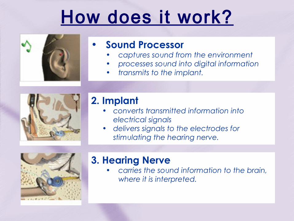
How does it work?• Sound Processor
• captures sound from the environment • processes sound into digital information • transmits to the implant.
2. Implant • converts transmitted information into
electrical signals • delivers signals to the electrodes for
stimulating the hearing nerve.
3. Hearing Nerve • carries the sound information to the brain,
where it is interpreted.

Evaluation Process
• Audiological• Medical• Psychological & Social Worker (children)• Auditory-Verbal Therapy (children)• Speech Language Pathology (for adults,
when required)

Audiological Assessment
• Diagnostic testing:– Audiogram >50 db– Tympanometry & Acoustic Reflexes– Auditory Brainstem Response– Otoacoustic Emissions
• Speech perception tests with appropriate amplification <60
• Hearing in noise test (HINT) <60 % in quiet • Consonant nucleus consonant word testing
(CNC) <30 %

Medical Assessment• Otologic history & examination
• General medical health
• Pneumococcal meningitis vaccine: Pneumovax
• Diagnostic Imaging• Computed Tomography (CAT)• Magnetic Resonance Imaging(MRI)*
• Electronystagmography ENG(Adult only)Electronystagmography (ENG) is a diagnostic test to record involuntary movements of the eye
caused by a condition known as nystagmus. It can also be used to diagnose the cause of vertigo, dizziness or balance dysfunction by testing the vestibular system.

Diagnostic Assessment
CT MRIMorphology of cochlea and semicircular canals ++ +++
Potency of cochlear duct + ++
Status of cochlear nerve - +++
Anatomy of facial nerve and fallopian canal ++ +
Defect of the modiolar + +++
Defect of cribiform area +++ ++
Enlarged vestibular aqueduct ++ +++
Enlarged cochlear aqueduct +++ +
Presence of round or oval window +++ -
CNS abnormalities +++

1. Primary means of determining suitability for cochlear implantation.2. The patients to undergo implantation were postlingually deafened adults with no hearing and who received no benefit from conventional amplification. Many or all aspects of spoken language had developed before the onset of their deafness. There was no likelihood that their hearing could worsen with cochlear implantation. Knowledge gained from these patients and with improved technology, candidacy criteria have broadened to include prelingually deafened children and patients with some minimal residual hearing.
Audiologic Assessment

3. Patients who become deaf at or after age 5 are classified as postlingually deafened.4. Once access to auditory input and feedback is lost, rapid deterioration of speech intelligibility often occurs. Implantation soon after the onset of deafness potentially can ameliorate this rapid deterioration.5. Cochlear implantation may be less successful in postlingually deafened patients if there is a long delay between the onset of deafness and implantation.6. A postlingual onset of deafness is an infrequent occurrence in the pediatric population

Psychological Assessment
• 1. Performed to identify subjects who have organic brain dysfunction,mental retardation, undetected psychosis, or unrealistic expectations.
• 2. Valuable information related to the family dynamics and other factors in the patient’s
milieu that may affect implant acceptance and performance are assessed.

Method of hearing assessment:in infants and children
• Neonatal screening procedures• Arousal test: high frequency narrow band noise stimuli
3time for 2 sec >aroused twice
• Auditory response cradle: trunk, limb movement ,head jerk and respiration seen on auditory stimuli
• Otoacoustic emissions:+nt in healthy outer hair cell– Absent in damaged outer hair cell
– transient evoked emission are absent in ear where hearing loss>30 dB
» Evoked by click
– Distortion product emissions are absent in ear where hearing loss>50 dB
» 2 tone are simultaneously present to cochlea to produce distorsion

ABR• screening test and definitive hearing assessment
– Elicit brainstem responses to auditory stimulation of clicks 40nHL or less is criteria of passing in screening test
– Find hearing threshold in infant ,1st at higher stimulus then lowered till wave V is just identifiable and repeatable.
– Identifiable waveform are present 10-20 dB above behavioral threshold.
– done under sedation
– Find integrity of central auditory pathway (EE COLI)
– In normal child 7 waves are produced in 1stten millisecond
– 1st 3rd 5th waves are stable n used in measurement.
– Waves are studied for absolute latency , inter-wave latency and amplitude.

Behaviour observation audiometry
– Moro’s reflex-sudden movement of limb and extension of head in response to sound of 90dB
– Cochleopalpebral reflex : blink to loud sound
– Cessation reflex:• starts crying in response to sound of 90 dB • Or stop activity

Distraction techniques
– Used in 6- 7 month child
– Child turn his head to locate source of sound-when assistant distracts child attention.
– High frequency sound – 8 kH rattle– Low frequency sound – hum– Narrow band sound- whispered sound

Conditioning techniques
• Play audiometry-– used in child 2-5 years
– each time after hear a sound signal child perform an act
• Speech audiometry:– Child is asked to repeat the names of object
– Voice can be gradually lowered
– Hearing level and speech discrimination can be tested
• visual reinforcement audiometry• Child is trained to look for an auditory stimulus by turning
his head
Objective test
– ABR– Otoacoustic emission– Impedance audiometry:
• stapedius ms contracts in response to sound of 70-100 dB and this reflex can be recorded.
• Middle ear disorder,retrocochlear hearing loss profound HL-absence of acoustic reflex
• Absent of acoustic reflex +(n)tympanometry=SNHL• Absent of acoustic reflex +(abn)tympanometry=CHL

Surgery• Pre operative procedure Intravenous antibiotics should be given at least 20 minutes before the incision is made. Antibiotics should be continued for the first 24 hours postoperatively and then discontinued
•Requires general anesthesia•Duration ~ 3-4 hours

Behind-The-Ear/Device Marking Template
The Behind-the-Ear/Device Marking Template is used to allow adequate clearance between the pinna, the speech processor and the head piece.
From Advanced Bionics

Device Coil Gauge
After placing the Device Coil Gauge, an outline is drawn around the template.
From Advanced Bionics

Incision Line
The surgeon will determine the length of the incision line.
A conventional postauricular-scalp incision approximately 5.0-6.0cm in length for children may be used. The incision may be longer in adults if the scalp needs to be thinned.incision-
U shaped incisionCshaped incisionJ shaped incision
From Advanced Bionics

Incision and Skin Flap
• Flap can be elevated either as a single layer or in two layers.If two layers are separately elevated, the superficial layer should be elevated first and then deep tissues, which include the periosteum of the mastoid, temporalis fascia, and temporalis muscle, should be left intact. •The periosteum of the mastoid should be elevated as an anteriorly based Palva flap, which can be sutured back into position at the end of the case to protect the electrode array in the mastoid cavity. • The Palva flap should be as large as possible and, will cover the take off point of the electrodes for the Nucleus and Advanced Bionics devices

• Stimulator/receiver should be placed 2.5 cm posterior to the posterior border of the external auditory canal. surgical drill is used to create a defect in the skull contoured to exactly fit the implanted device exactly.• The skull of small children, between 1 and 2 years of age, may be only 2 and 3 mm in thickness. For these children, the implant often rests on exposed dura. • Some surgeons seek to leave an "island" of bone in the center of the area of exposed dura, whereas other removing all the bone from the dura.
The Well

Recess Marking Template
The Recess Marking Template is used to determine the location of the recess bed and channel for the electrode lead.
From Advanced Bionics

• Incision and skin flap
• Mastoidectomy-not sauceried
• Exposing incus, LSC ,facial reces
• Posterior tympanotomy done through facial recess
• The facial recess is a triangular area bound by (1) the fossa incudis superiorly, (2) the chorda tympani nerve laterally and anteriorly (3) facial nerve medially and posteriorly
• Round window niche, incudostapedial joint ,pyramid exposed
• Cochleostomy made and widen (0.8-2mm)
• implant-receiver well/recess bed and electrode lead channel are drilled .
• Fixing of implant into bed after electrode fixed
• Neutral electrode placed under temporalis ms
• Insertion of cochlear electrode into cochlea
• Withdraw of stilete to hug modiolar.
• Palva flap closed
• Skin closer

Mastoidectomy
The mastoidectomy cavity should not be saucerized. The edges should be left as acute. These edges will help retain the electrode leads within the confines of the mastoid cavity.
After mastoidectomy is complete, LSC,incus is identified then facial recess is identified and widely opened. The most inferior portion of the facial recess is of greatest importance for visualization of the round window niche.

Some bone medial to the facial nerve must be removed to see even the anterior boundary of the round window niche.
Almost all anomalous facial nerves are displaced anteriorly and medially. Just distal to the oval window, they turn directly into the hypotympanum and run just inferior to or directly over the round window area

Mastoidectomy-Facial Recess Approach
After completion of a mastoidectomy-facial recess approach, the implant-receiver well/recess bed and electrode lead channel are drilled.
Suture tie-down holes to stabilize the implant are placed.
A standard cochleostomy is used.
From Advanced Bionics

Cochleostomy
•Once the facial recess has been widely opened, the round windowniche can be clearly seen. It is often useful to remove the anterior lip of the round window niche so that the anterior attachment of the round window membrane itself can be visualized. • Make the cochleostomy inferior to the inferior attachment of the round window membrane to avoid the "hook" of the cochlea. This allows a straighter, more direct insertion of the electrode array into scala tympani. "pure" RW insertion avoids the trauma and bone dust associated with a classic promontory cochleostomy. Insertion through the RW ensures entrance into scala tympani.•The size of the cochleostomy will be of 2 mm or more. Most currently available devices can be easily inserted through a cochleostomy of between 0.8 and 1.2 mm in diameter.

Electrode Insertion
The insertion tool is used to insert the electrode array in the usual fashion.
The Insertion Tube is placed just inside the cochlea toward the basal turn of the scala tympani, with the insertion tube slot directed toward the modiolar (or inner) wall.
From Advanced Bionics

Closure
The skin incision is closed in layers.
From Advanced Bionics

Electrode Array Placement Within the Cochlea
Illustration courtesy of Cochlear Corporation

Audiological testing in the OR
• Impedance check on all electrodes
• Neural response testing to help estimate required levels

X-Ray to confirm posit ion
of internal device
Courtesy of Cochlear Corporation

INITIAL FITTING OF COCHLEAR IMPLANT
• 1. External processor and transmitter fit approximately 1 month after surgery.
• 2. Magnet strength for transmitter determined.
• 3. Electrical threshold and comfort levels determined.
• 4. Map created

After the surgery•Initial stimulation: 4-6 weeks post surgery
•Adjustments made regularly based on feedback from patients, parents, therapists and educators
•Rehabilitation to meet specific patient needs
•Regular follow-up appointments

• Facial nerve injury –intra operative
• Chorda tympani nerve injury- intra operative
• Post operative bleeding and hematoma
• Infection –open wound and tt with antibiotic
• Wound dehiscence-flap necrosis
• Early Device failure-out of box failure
• CSF leak-penetration of dura -presence of modiolar defect
• Labyrinthisis
• Meningitis
• Balance Disturbance-Dizziness - vertigo
Post operative complication

Post operative complication-late
1 Extrusion or exposure of device -used pericranial flap to fully cover device
2 Displacement- d/t injiry- do CT scan
3 late Device failure-d/t trauma –replacement of external components-no improvement-CTscan-no explanation-electrical integrity check-soft failure-replace device
4 Otitis Media infection – tt with antibiotics
5 Meningitis- d/t Pneumococcal infection-pneumococcal vaccination

Intracochlear ossification
• Ossification at round window common in post meningitic patients
• In these patients, a cochleostomy is developed anterior to round window. New bone is drilled. If an open scala tympani is entered, a full insertion is performed.

• Less frequently, the scala tympani is completely obliterated by bone. - drill open the basal turn and create a tunnel approximately 6 mm in depth and partially inert a straight electrode. This allows implantation of 10 to 12 active electrodes which has proven satisfactory.
• Specially designed split electrodes have been developed One branch of the electrode array is placed into the tunnel described earlier and the second active electrode is inserted into a second cochleostomy developed just anterior to the oval window.

Cerebrospinal fluid leak
• Eliminated by drilling shallow well for implant package not exposing dura and eliminating control holes for tie-down sutures.
• CSF gushers have occurred in children with a Mondini deformity and major inner ear malformations and patients with the large vestibular aqueduct syndrome.

• The flow of CSF has been controlled by entry into the cochlea through a small fenestra, allowing the CSF reservoir to drain off, insertion of the electrode into the cochleostomy, and tight packing of the electrode with fascia At the cochleostomy site.
• It is postulated that the source of the leak is through the lateral end of the internal auditory canal.
• The eustachian tube is occluded with tissue and fibrin glue is placed in the middle ear.

Posit ive psychological & social benefits
• Decline in:-Loneliness-Depression-Social isolation
• Increase in:– Self-esteem– Independence– Social integration– Vocational prospect

Negative psychological & social impacts
• Concerns about the maintenance and/or malfunctioning of the Cochlear Implant
• Difficulty in background noise
• Unreasonable expectations of aural-only benefit on the part of the implant user or their family and friends

Potential Benefits1. Better speech understanding compared to a
hearing aid2. Awareness and responsiveness to environmental
sounds3. Less dependence on family members for day to
day living4. Reconnection with the world of sound5. Facilitation of communication with family and loved
ones6. Ability to talk on the phone 7. Better appreciation of music