cochise regional hospital radiology department policies...
TRANSCRIPT

Reviewed: 07/2014 Revised: 07/2014
Cochise Regional Hospital Radiology Department Policies & Procedures

Reviewed: 07/2014 Revised: 07/2014
ADMINISTRATION OF ORAL CONTRAST MEDIA FOR OUTPATIENT EXAMS PURPOSE: To provide a guideline for outpatient procedures which require the administration fo an oral contrast media.
PROCEDURE:
1. All patients having a computed tomography exam which requires oral contrast media as an outpatient, will be advised to arrive one hour before their scheduled procedure.
2. A registered radiologic technologist will obtain the patients medical history; to include any medication allergies.
3. The technologist will dispense the oral contrast media to the patient with appropriate drinking instructions.
4. The exam will be performed after the oral contrast media has been ingested and the technologist feels it has fully coated the bowl.
Reviewed: 07/2014 Revised: 07/2014
DIAGNOSTIC IMAGING STAT OUTPATIENT PROCEDURES AFTERHOURS
PURPOSE: To establish guidelines for after-hours STAT outpatient Diagnostic Imaging procedures.
AFTER HOURS IMAGING FOR COMPUTED TOMOGRAPHY, NUCLEAR MEDICINE, AND ULTRASOUND PROCEDURES
Diagnostic Imaging after-hours is considered Monday- Friday 4:30pm- 6pm.
Note. After 6pm on weekdays or on holidays or weekends patients with a STAT order for any of the above mentioned modalities should be directed to the Emergency Dept.
• All STAT after-hours outpatients must be registered and the order must be placed into Cerner RadNet.
• The technologist will notify the on-call radiologist of the patients name, procedure/exam ordered, reason for exam, name and contact number of ordering physician. If the on-call radiologist cannot be reached, the technologist will try to contact another staff radiologist.
• NO STAT procedure/exam will be started without prior approval by the on-call or staff radiologist(s).
• Upon completion of the procedure/exam the technologist will notify the on-call radiologist and place the patient in the old radiology waiting area. The on-call radiologist will communicate results with the ordering physician and inform the technologist if the patient needs to be admitted to the Emergency Department or can go home. Note: The radiologist or technologist will directly communicate with the patient regarding results. Tech aides or students will be prohibited from communicating results to any patient
• STAT PROCEDURES FROM URGENT CARE FACILITIES • Patients requiring STAT imaging from Urgent Care Facilities after 6pm on weekdays or
on weekends or holidays should be admitted to the Emergency Dept. per Policy# DI127 DI Prioritization Afterhours.

Reviewed: 07/2014 Revised: 07/2014
Anatomical Site Check
POLICY:
To insure the correct anatomical site is radiographed or the correct side (left or right) is
correctly identified before any interventional imaging procedure is performed.
PROCEDURE:
It is the policy of the Department of Radiology and Diagnostic Imaging that all patients undergoing any imaging procedure are to have the correct site identified before the exam begins in order to insure patient safety.
To achieve the above, the following safety measures will be followed:
1. The technologist will check for the correct patient by two means, i.e., the patient’s name and date of birth.
2. The technologist will confirm the spelling of the name and confirm the date of birth. 3. The technologist will check the written order to verify “left” or “right”. 4. The technologist will ask the patient on which side they are having the procedure
performed and to point to the specific area. 5. The technologist will place a “spot-marker” on the all extremities where the patient is
having pain.

Reviewed: 07/2014 Revised: 07/2014
ARIZONA STATUES FOR RADIOLOGY SERVICES POLICY All rules and regulation will be followed and adhered to in compliance with Arizona Department of Health Services Statute R9-10-219 as related to Radiology and Diagnostic Imaging Services.

Reviewed: 07/2014 Revised: 07/2014
CARE OF CRITICALLY ILL PATIENT
POLICY: To establish guidelines for the care of the critically ill patient in the Department of Diagnostic Imaging.
PROCEDURE: • A Registered Nurse must accompany the patient to and from the Department of Radiology
as well as remaining during the entire x-ray procedures.
• The x-ray room must be prepared to accommodate the patient in case of emergency (oxygen, crash cart, suction, etc.).
• Expediency of the exam is emphasized. The technologist shall utilize the radiographic and auxiliary equipment to its maximum potential and shall always be alert to the patient's condition.
• When radiography is required for a patient in the room or at bedside, the technologist will always report to the nurse in charge on the ward, station or floor.
• The technologist will check the patient ID and verify the patient name and date of birth.
• The technologist should remember the directions and cautions the charge nurse communicated concerning the patient, and make any necessary adjustments to accommodate the patient's special needs and/or condition.
• When it is necessary to change a patient's position, the rules of body mechanics shall be observed, to safely and comfortably lift and move the patient.
• After completion of radiographic procedure, the technologist shall make the patient comfortable and advise the charge nurse of the completion of the examination.
Policy All rules and regulation will be followed and adhered to in compliance with Arizona Department of Health Services Statute R9-10-219 as related to Radiology and Diagnostic Imaging Services.

Reviewed: 07/2014 Revised: 07/2014
CARE OF PATIENTS FROM ANOTHER HEALTH CARE FACILITY
POLICY: To insure appropriate care of patients arriving at Cochise Regional Hospital (CRH) from another health care facility for the purposes of radiological examinations or procedures.
PROCEDURE:
It is the policy of the Diagnostic Imaging Department that all patients being transferred from another health care facility for the purposes of radiological examinations or procedures will be transferred via ambulance. The patient will be registered in client services and received into our Diagnostic Imaging Department. The ambulance transport team will remain with the patient at all times.
Between the hours of 0700 and 1530
1. The Radiology Registered Nurse (RN) will accept the patient if the patient is stable and able to undergo the radiological procedures as ordered. The Radiology RN will be responsible for periodic assessment of the patient while the patient remains in the Diagnostic Imaging Department.
2. In the event that the RN is obligated to assist with patient care in other previously scheduled radiological procedures, the Nursing Supervisor will be notified for further nursing assistance.
3. In the event that the patient’s status deteriorates, the patient will be taken to the Emergency Department to be treated as an acute Emergency Department admission.
Between the hours of 1530 and 0700 or on Weekends
1. The Nursing Supervisor will be notified of the request for Diagnostic Imaging on a patient from another health care facility. The Nursing Supervisor will locate and assign an RN staff to accept this patient upon arrival. This nurse will follow steps number 1, 2, and 3 as written above.
2. The transferring facility will provide the patient’s medical chart to SVRHC upon transfer to our facility to aide in appropriate and safe patient care.
3. Patients may not be transferred from another facility via his/her own personal vehicle. Appropriate transportation must be arranged by the transferring facility.

Reviewed: 07/2014 Revised: 07/2014
CONTRAST REACTION – EMERGENCY REACTION MAIN CAMPUS POLICY: To establish the necessary actions to be taken during a contrast/emergency reaction.
PROCEDURE: 1. A small emergency tray of medication is kept in the CT room in the Diagnostic Imaging
Department. This tray contains antihistamines, adrenaline, steroids and other drugs to counteract adverse reactions to contrast media. Whenever these drugs are administered, the patient’s chart must be logged accordingly and signed by the radiologist or the attending physician.
2. Contrast allergy will be noted on Contrast Form and Power Chart.
Contrast Reaction Treatment
Mild • Vomiting Observe / Monitor • Nausea Oxygen • Localized hives/itching Benadryl (PO)
Moderate • Facial swelling Observe / Monitor • Generalized hives/itching Oxygen • Early Laryngeal Edema • Respiratory Involvement Epinephrine (SQ /Epi Pen)
* Wheezing Benadryl PO * Shortness of Breath Call 911
Severe Call Code Blue - 88 • Laryngeal Edema - call 911 • Pulmonary edema - Oxygen • Unconsciousness - • Shock - Epinephrine (SQ Epi Pen) • Respiratory or Cardiac Arrest
Necessary actions to be taken during an emergency contrast reaction:
1. Contact Radiologist or Radiology Nurse. 2. Start CPR if necessary.
Reviewed: 07/2014 Revised: 07/2014
CONTRAST REACTION – EMERGENCY REACTION EAST CAMPUS
POLICY:
To establish the necessary actions to be taken during a contrast/emergency reaction.
PROCEDURE:
1. A small emergency tray of medication is kept in the diagnostic x-ray room in the Diagnostic Imaging Department. This tray contains antihistamines, adrenaline, steroids and other drugs to counteract adverse reactions to contrast media. Whenever these drugs are administered, the patient’s chart must be logged accordingly and signed by the radiologist or the attending physician.
2. The film jacket and the patient flash card shall be marked accordingly to indicate that the patient had an allergic reaction to the contrast used in that examination.
Necessary actions to be taken during an emergency contrast reaction:
1. Remove patient and call 911. 2. Start CPR if necessary.

Reviewed: 07/2014 Revised: 07/2014
PROCEDURE FOR CT THORAX WITH I.V. CONTRAST FOR PULMONARY EMBOLISM
Policy:
To assure that all CT Thorax with I.V. contrast exams are performed with the proper size I.V. catheter, all images are optimal, and to reduce extravasations.
PROCEDURE:
• All CT Thorax with I.V. contrast for Pulmonary Embolism require at least a 20 gauge I.V. catheter in the Antecubital Vein or above. Note: If a 20 gauge I.V. catheter can not be placed in the Antecubital Vein or above, the ordering department must be notified. The exam will not be performed if the patient does not have a 20 gauge I.V. catheter or greater in the Antecubital Vein or above.
• The I.V. access must be flushed with regular saline to assure patency before and after any I.V. contrast injection.
• A rate of at least 3.5ml per second will be used for ALL Pulmonary Embolism studies.

Reviewed: 07/2014 Revised: 07/2014
DEPARTMENTAL SAFETY
Policy :
To assure safety of all employees and patients.
PROCEDURE:
• The Diagnostic Imaging Department Manager is responsible for maintaining safety standards, developing and refining safety rules, and supervising and training personnel in departmental standards.
• The Diagnostic Imaging Department Manager is responsible for notifying the Safety Officer in case of any safety hazard.
• All Diagnostic Imaging Department employees shall report defective equipment, unsafe conditions, acts or safety hazards to the Manager.
• Smoking or the consumption of alcoholic beverages will not be allowed at any time, while on duty.
• Proper body mechanics and lifting techniques will be observed at all times. • Electrical cords will be clear of traffic areas. Electrical extension cords will not be used
without written approval from the Physical Plant. Physical Plant personnel will inspect all personal electrical appliances before use. All electrical machines with heat producing elements must be turned off or unplugged when not in use.
• Only authorized personnel will be allowed to operate diagnostic imaging equipment. • Faulty equipment will be reported to the Physical Plant or the vendor, per policy. • Equipment and furniture must be arranged to allow adequate passage and access to exits at
all times. • The employee who discovers a spill will clean up minor spills, such as water. This is to be
done immediately. Environmental Services will clean up major spills. • The Physical Plant will be notified immediately of improper illumination and/or ventilation. • Scissors, knives, pins, razor blades and other sharp instruments must be stored and used
safely. Use of sharp spindles is prohibited. • File drawers and cabinet doors will be closed when not in use. • Employee clothing will be in accordance with hospital policy. • Only authorized personnel shall be allowed in exam rooms. • Transport or technologist who calls for patient will check the ID band on the patient's wrist
to verify correct patient identity. • Outpatients will be asked date of birth and/or to give full name and spelling of name. • A “Radiology Hand-off Communication Form” must be completed by the patient’s nurse
BEFORE transporting to the Diagnostic Imaging Department. • Employees will be aware of location of fire extinguishers and fire exits. Employees will be
educated in evacuation of area during a fire code.

Reviewed: 07/2014 Revised: 07/2014
DIAGNOSTIC IMAGING SERVICE Policy:
To insure proper storage and safety of diagnostic imaging agents.
Procedure:
1. The Diagnostic Imaging Department shall properly store the agents (i.e., protect from light) and keep them secured.
2. When needed, the Diagnostic Imaging Department will order the necessary medications from the pharmacy on the appropriate order form.
3. Stock will be rotated by Material Services.
4. Managed inventory is performed by Material Services.

Reviewed: 07/2014 Revised: 07/2014
DIAGNOSTIC IMAGING – CALL BACK
POLICY:
To establish policy for call-back and completion of imaging orders in the Diagnostic Imaging
Department.
The following Diagnostic Imaging services will be available 24 hours/7days per week:
CT Ultrasound
X-Ray Echosonography
CT and X-ray staff is available in-house 24 hours/7days per week.
Ultrasound, Nuclear Medicine and Echosonography is available in-house during business hours Monday through Friday from 7am – 5pm.
Diagnostic Imaging modalities that are not staffed 24 hours/7 days per week except Nuclear Medicine are on call after business hours weekly and Friday evening through opening of business hours on Monday, and on holidays.
Diagnostic Imaging staff will be notified by the ordering department of all STAT imaging orders during on-call hours. Nuclear Medicine is on-on call only on Saturdays from 8am to 4pm. Diagnostic Imaging staff will be notified by the ordering department of all STAT imaging orders during on-call hours. The Diagnostic Imaging on-call staff will call each patient care unit during weekend call shifts to inquire if any ROUTINE procedures have been ordered. Routine imaging procedures will be started within 24 hours.
PROCEDURE:
STAT procedures will be started within 30 minutes or as soon as physically possible. ROUTINE procedures will be started within 24 hours.
Diagnostic Imaging staff must be notified of all STAT orders pending. Upon arrival to the hospital, the Diagnostic Imaging staff will punch in as “call-back” on the time clock and begin imaging the STAT patient requests. During the weekend upon completion of any STAT request, the Technologist will review the on-line worklist for any pending ROUTINE procedures. Routine procedures will be imaged and the technologist will check with every department for pending orders before leaving or clocking out.
The Technologist is not to clock in at separate intervals for each patient. Call-back pay will be accrued according to the Call-back policy number PAY105.

Reviewed: 07/2014 Revised: 07/2014
DIAGNOSTIC IMAGING PRIORITIZATION AFTERHOURS
POLICY: To establish guidelines for prioritization and after-hours Diagnostic Imaging procedures for the Inpatient and Emergency Department settings.
The following Diagnostic Imaging services will be available 24 hours/7days per week: CT X-Ray Ultrasound
PROCEDURE: IMAGING PRIORITIZATION
Diagnostic Imaging procedures may be ordered STAT or ROUTINE.
STAT procedures will be started within 30 minutes or As Soon As Physically Possible. ROUTINE procedures will be started within 24 hours.
STAT PROCEDURE
STAT procedures are ordered ONLY when a physician deems that a patient may suffer loss of life, limb or body function. Patients requiring STAT imaging from Urgent Care facilities should be admitted to the Emergency Department.
If possible, the ordering physician will review STAT plain films after-hours. A radiologist (radiologist on-call or nighthawk) is available for consult of STAT plain films if requested.
The radiologist, nighthawk services, will respond to STAT orders for CT and Ultrasound according to the Medical Staff By-Laws. If the ordering physician would like verbal results, contact information from the ordering physician must be written on the orders for the radiologist to call results. For example, “Please call me at 417-XXXX with results”. The technologist will place this information in the comment section of Cerner.
ROUTINE PROCEDURE
ROUTINE procedures are ordered when the physician deems the imaging could be performed within the next 24 hours. The procedure will be started within 24 hours of receipt of the order.
ROUTINE procedures with a specific time requested by the ordering physician will be started by the specific time requested, unless other STAT orders are preceding it. The test will then be completed at its earliest convenience. If the test cannot be completed within one hour of the requested time, the radiology technician will contact the ordering physician/charge nurse on the unit to follow-up and give the anticipated competed time.

Reviewed: 07/2014 Revised: 07/2014
Procedures ordered by physician offices or Urgent Care facilities for same day service will be completed within our normal business hours of 0800 to 1630. The physician should write “TODAY” on the order. If orders cannot be completed on this day due to triage of patients, the radiology department will notify the physician/patient to make arrangements for the procedure to be completed the next business day.
If the ordering physician would like verbal results, contact information from the ordering physician must be written on the orders for the radiologist to call results. Otherwise, results will be faxed to the ordering physician every two hours between 0700 to 1900 hours.
SPECIAL PROCEDURES:
Special procedures that require the assistance of the radiologist and/or nurse are the following. These procedures are performed during normal business hours of 0800 to1630 (except holidays):
• Image-guided biopsies • US guided biopsies or paracenthesis • Fluoroscopy
Esophagus Barium Enema UGI Portacath Patency Dubb Hoff Advancement from the Stomach J-Tube Patency Fistulogram SBF
If a referring physician feels an order for a special procedure is necessary after-hours or on weekends or holidays, the referring physician must call the radiologist on-call for arrangements. Otherwise, the order will be complete as a ROUTINE procedure on the next business day.
The radiologist must be called for approval of placement of drainage tubes. This includes during normal business hours. AFTER HOURS IMAGING
Diagnostic Imaging technical staff is available in-house 24 hours a day / 7 days per week for CT and X-ray imaging. Ultrasound staff is on-call during weekends, holidays, and after-hours for STAT inpatient and Emergency Department patients only. Nuclear Medicine is available Saturday and Sundays and Holidays 8am – 4pm.
After-hours for interpretation of diagnostic imaging is considered when our radiologist(s) is not on-call, currently 5pm-5am M-F, weekends and Holidays During this time, Nighthawk radiology services will be utilized for all STAT CT and Ultrasound Imaging.

Reviewed: 07/2014 Revised: 07/2014
• On-call Diagnostic Imaging technical staff must be notified when a STAT order is written on an inpatient or ED patient. The Diagnostic Imaging Staff will respond according to the above prioritization procedure.
• If a physician order requests a timed procedure to be completed by a certain time, the order with the specific timeframe must be inputted into Project ISIS and relayed to the appropriate on-call technician. The Diagnostic imaging technician will then ensure that the procedure is completed by this time.
• For all procedures, the referring department must ensure that the patient is prepped and in a gown, pregnancy consent form completed (if needed), and ready for each procedure before the on-call diagnostic imaging technician arrives. No patient should be treated at anytime without proper identification bands.
• If the diagnostic imaging technician is called in to do a STAT procedure, the technologist will review the on-line work list for ROUTINE orders. After completing the STAT procedures, the technician will complete any pending ROUTINE procedures before leaving the hospital, if needing to be completed within the 24-hour timeline.
If the diagnostic imaging technician is NOT called in to do a STAT procedure, the technologist will call the weekend tech to determine if there are any ROUTINE orders that need to be completed within 24 hours. If so, the technician will complete any of these pending ROUTINE procedures.

Reviewed: 07/2014 Revised: 07/2014
EAST CAMPUS IMAGING ESCORTING PATIENTS THROUGH RADIOLOGY
POLICY: To ensure customers are properly escorted through Radiology. PROCEDURE:
• After a patient is checked in at the registration or greeter station, the greeter or expeditor will take the patient back to the waiting room.
• The greeter or expeditor will gather the patient medical record from the registration desk, greet the patient, and escort the patient to the dressing and/or waiting room.
o If the patient needs to undress, the greeter or expeditor will instruct the patient on what needs to be removed for the procedure, give the patient a clean gown, and direct the patient to a curtained dressing area
o The patient will be instructed to place their clothing in a locker but to take their valuables with them.
o The patient will then be instructed to the designated waiting room.
• After escorting the patient to the dressing/waiting room, the greeter or expeditor will drop the patient chart off to its designated modality, and notify the technician of the patient arrival.
• The tech will then pick the patient up from the waiting room.
o The tech will ask for 2-patient identifiers before beginning any exam with a patient.
• Upon completion of the exam, the technician will escort the patient to its designated waiting and/or dressing room.

Reviewed: 07/2014 Revised: 07/2014
EAST CAMPUS IMAGING REGISTRATION POLICY: To ensure timely registration of Radiology patients. PROCEDURE:
• Walk-In patients that need to register for East Campus (EC) diagnostic imaging services will be registered using the current hospital standards as outlined in the training manual. o The clerk will collect the signed copy of the COA, copy of referral and/or authorization,
the original order, face sheet and one sheet of labels for the patient. o Pending scheduled appointments will be kept in a locked file drawer until the day of
the patient visit. o Upon completion of the exam, Diagnostic Imaging will route to HIM via courier.
• Fast tracked patients (already scheduled outpatient patients) are sent directly to EC Diagnostic Imaging Center; their paperwork is already complete and sent to the EC the day before the procedure. They are admitted by the front desk concierge.
• A copy of the patient’s insurance cards, picture ID, script and referral and/or authorization will be made and sent to EC diagnostic imaging along with paperwork.
• A properly completed Physician’s order should be obtained prior to sending the patient to the EC Diagnostic Imaging Center. When needed, the clerk will contact the physician’s office and request a corrected order to be faxed to registration.
• Patient Registration staff will assure that if an authorization or referral is required, there is one on file prior to sending the patient to the EC Diagnostic Imaging Center for the scheduled procedure. When unable to get the appropriate script, authorization, referral or Physician’s signature in a timely manner, inform the EC Diagnostic Imaging Center for further instructions.
• All patients sent to the EC Diagnostic Imaging Center must be properly banded with their pertinent information. Armbands are to be verified and initialed by the patient or patient’s representative.

Reviewed: 07/2014 Revised: 07/2014
EAST CAMPUS IMAGING CENTER SCOPE OF SERVICES
POLICY:
To outline the scope of care at Cochise Regional Hospital.
SERVICES OFFERED:
• X-Ray • CT • Ultrasound • Mammography • Stereotactic • Dexascan • MRI
PROCEDURE:
HOURS OF OPERATION:
• Scheduled outpatient imaging services will be offered and performed Monday through Friday from 8:00 AM to 5:00 PM, excluding holidays.
SCHEDULING:
• All outpatient imaging requests are scheduled through the Scheduling Department at CRH by calling 520-364-7931 ext 5733 or 5734. X-ray walk-ins will be scheduled at the outpatient imaging center.
• All services requiring the administration of contrast need to be scheduled in designated slots to coordinate with the work schedule and on-site availability of physician.
PATIENT REQUESTS:
All outpatient requests must have a written order from a licensed physician or practitioner. The Scheduling Department will obtain pre-authorization, if necessary, and provide patient with information and instructions pertaining to their appointment and exam.
Reviewed: 07/2014 Revised: 07/2014
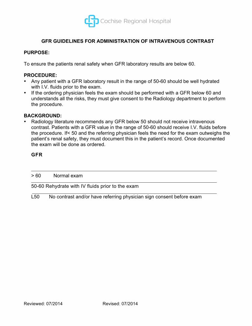
GFR GUIDELINES FOR ADMINISTRATION OF INTRAVENOUS CONTRAST
PURPOSE: To ensure the patients renal safety when GFR laboratory results are below 60.
PROCEDURE: • Any patient with a GFR laboratory result in the range of 50-60 should be well hydrated
with I.V. fluids prior to the exam. • If the ordering physician feels the exam should be performed with a GFR below 60 and
understands all the risks, they must give consent to the Radiology department to perform the procedure.
BACKGROUND: • Radiology literature recommends any GFR below 50 should not receive intravenous
contrast. Patients with a GFR value in the range of 50-60 should receive I.V. fluids before the procedure. If< 50 and the referring physician feels the need for the exam outweighs the patient’s renal safety, they must document this in the patient’s record. Once documented the exam will be done as ordered.
GFR
> 60 Normal exam
50-60 Rehydrate with IV fluids prior to the exam
L50 No contrast and/or have referring physician sign consent before exam

Reviewed: 07/2014 Revised: 07/2014
HANDLING OF RADIOACTIVE MATERIAL
PURPOSE
To assure safe and effective handling of radioactive materials delivered to Sierra Vista
Regional Health Center.
PROCEDURE
All radioactive material will be delivered directly to the Material Services Department.
• Material Services will inspect the package for any damage and will keep a log which must include the following:
1. Name of the person who delivered and received the package. 2. Time the package was received by Material Services. 3. The condition the package was in upon delivery to Material Services. Note: If
the package is damaged in any form or if the packaging seal has been broken, the Nuclear Medicine Technologist will inspect the box and determine that no radioactive material has leaked.
4. Any suspicion of damage will be reported to the Radiation Safety Officer.. 5. The package must not be opened by anyone who is not trained in
the procedure for handling radioactive materials.
• Material Services will deliver the shipment to the Nuclear Medicine Department upon receipt. No deliveries will be accepted after normal working hours.
• The Nuclear Medicine Department will keep a “Radioactive Material-Package Receipt Log” and a “Radioactive Seeds – Inventory/Utilization Log”.
• The radioactive material receipt information is documented on a “Radioactive Material Inventory Form”
• After opening the package, the contents are checked for agreement with the packing slip and the integrity of the radioactive material is checked. Any suspicion of damage to package is reported to the Radiation Safety Officer and to the manufacturer. Any suspicion of contamination is evaluated by performing a wipe test and assay by the Nuclear Medicine technologist.
• The empty package will be checked for radiation levels, the radioactive materials labels are removed and the package either kept for return or trashed.
INFECTION CONTROL GUIDELINES

Reviewed: 07/2014 Revised: 07/2014
POLICY:
To ensure consistency with the implementation of infection control guidelines within the Diagnostic Imaging department.
PROCEDURE:
I. Personnel
A. Employee health guidelines will be followed by all employees of the Radiology and Diagnostic Imaging department.
II. General infection control practices
A. Careful hand hygiene must be practiced as outlined in the hospital infection control manual. Hand hygiene must be performed after patient contact, contact with contaminated items, or contact with mucous membranes.
B. Standard precautions will be followed for all patients. Body substances from all patients are to be considered potentially infectious.
C. Isolation precautions will be observed as appropriate. Specific precautions and indications for isolation can be found in the hospital infection control manual.
D. Linen is to be changed between each patient. Clean linen is stored in a closed cupboard. Soiled linen is disposed of in dirty linen hampers within the department. Soiled linen is collected by EVS.
E. Disposable items are for single patient use only and discarded after use. F. Sterile patient care items will be kept in closed cupboards. All supplies will be
checked for outdates periodically and prior to patient use for damage to outer package.
G. During sterile procedures, only personnel involved in the procedure are permitted in the room.
H. Exam tables/Buckys/patient contact surfaces in all imaging areas (MRI, Mammography, Stereotactic, Dexa, Ultrasound, X-ray, CT, and Nuclear Medicine) will be cleaned between each patient with an Infection Control Committee approved disinfectant.
I. Instruments/sterile trays are returned to Sterile Processing for decontamination and sterilization. Items must be transported in a closed bag/container, which is labeled as biohazard.
J. A schedule for routine cleaning of all portable equipment must be maintained and cleaning must be documented. In addition, portable equipment must be cleaned prior to entering a surgical suite and upon leaving an isolation room.
K. Laboratory specimens should be collected in a careful manner. Prior to transport, tubes or slides must be placed in a plastic bag and sealed. The bag must be labeled as biohazard.
L. Sterile technique must be observed when starting IV lines or inserting urinary catheters.

Reviewed: 07/2014 Revised: 07/2014
M. Care must be taken when handling contaminated sharps. Used syringes must be disposed of in an appropriate puncture resistant biohazard container.
N. Injectable fluids must be checked for expiration date and any sign of degradation (cloudiness or particulates) prior to use.
O. Sonographic probes that will have contact with mucous membranes should be covered with a latex barrier, if possible. Probes must be cleaned and high-level disinfected after use.
P. Mammography / Stereotactic Biopsy see Policy # MAM120.
Reviewed: 07/2014 Revised: 07/2014
INTRAVENOUS ACCESS/INFUSION FOR THE EAST CAMPUS
POLICY:
To ensure safe and appropriate initiation of intravenous access for administration of contrast media for prescribed diagnostic imaging procedures.
PROCEDURE:
1. Prior to initiation of intravenous access:
a. The diagnostic imaging technologist will be familiar with the Infection Control Policy and departmental and hospital policies regarding management of contrast reaction.
b. The diagnostic imaging technologist will have attended and successfully completed the Cardiopulmonary Resuscitation (CPR) course and CPR certification will be current.
2. The diagnostic imaging technologist will ensure the following:
a. An order for the procedure is completed. b. Adequate clinical information is obtained (disease history, medications, and laboratory
results) as needed for specified procedure. If there is a history of renal disease or diabetes, the attending radiologist should be notified.
c. If there is a GFR level less than 60, the attending radiologist should be notified (refer to policy DI133 and MRI013).
d. The patient’s identity is verified prior to initiation of intravenous access. This is done verbally as well as by visually examining the patient identification bracelet.
e. The patient has given informed consent prior to the invasive procedure, and a ‘Consent for IV Contrast’ form has been signed by the patient.
f. Any known allergies or significant history is reported to the radiologist prior to the procedure.
g. The type, strength, and volume of contrast agent are documented.
3. Appropriate aseptic technique will be employed to minimize the risk of cross-infection, to include the following:
a. Thorough hand washing technique. b. Proper use of alcohol wipes. c. Use of disposable gloves. d. Safe disposal of needle and injecting equipment following injection. e. If an existing intravenous access exists, the site will be flushed with normal saline
to ensure patency prior to injection with contrast. f. The technologist will seek the assistance of the diagnostic imaging nurse following two
failed venipuncture attempts or prior to attempts if the technician is not confident to proceed with venipuncture on a particular patient.
Reviewed: 07/2014 Revised: 07/2014
g. If an adverse reaction occurs, the covering physician will be summoned immediately. Should the reaction be deemed immediately life- threatening, 911 will be dialed.
h. All adverse reaction will be entered in Quantros.

Reviewed: 07/2014 Revised: 07/2014
INTRAVENOUS ACCESS/INFUSION
POLICY: To ensure safe and appropriate initiation of intravenous access for administration of contrast media for prescribed diagnostic imaging procedures.
PROCEDURE: 1. For inpatients who have existing intravenous infusions, it is the responsibility of the
ordering department nurse to determine: a. If the infusion may be stopped and the site converted to a saline lock, or b. If the infusion is to continue and another access placed.
2. The above information must be communicated to the technologist prior to the diagnostic imaging procedure. The diagnostic imaging technologist is not authorized to stop or interrupt an infusion in order to inject contrast into the same intravenous access as the continuous infusion.
3. Prior to initiation of intravenous access: a. The diagnostic imaging technologist will be familiar with the Infection Control Policy
and departmental and hospital policies regarding management of contrast reaction. b. The diagnostic imaging technologist will have attended and successfully completed the
Cardiopulmonary Resuscitation (CPR) course and CPR certification will be current.
4. The diagnostic imaging technologist will ensure the following: a. An order for the procedure is completed. b. Adequate clinical information is obtained (disease history, medications, and
laboratory results) as needed for specified procedure. If there is a history of renal disease or diabetes, the attending radiologist should be notified.
c. If there is a GFR level less than 60, the attending radiologist should be notified. d. The patient’s identity is verified prior to initiation of intravenous access. This is
done verbally as well as by visually examining the patient identification bracelet. e. The patient has given informed consent prior to the invasive procedure, and
a ‘Consent for IV Contrast’ form has been signed by the patient. f. Any known allergies or significant history is reported to the radiologist prior to
the procedure. g. The type, strength, and volume of contrast agent are documented. h. The radiologist, diagnostic imaging nurse, or other emergency medical response
team members, as well as emergency code cart are readily available.
5. Appropriate aseptic technique will be employed to minimize the risk of cross-infection, to include the following: a. Thorough hand washing technique. b. Proper use of alcohol wipes.

Reviewed: 07/2014 Revised: 07/2014
c. Use of disposable gloves. d. Safe disposal of needle and injecting equipment following injection. e. If an existing intravenous access exists, the site will be flushed with normal
saline to ensure patency prior and after to injection with contrast. f. The technologist will seek the assistance of the diagnostic imaging nurse
following two failed venipuncture attempts or prior to attempts if the technician is not confident to proceed with venipuncture on a particular patient.
g. If an adverse reaction occurs, the radiologist and diagnostic imaging nurse will be summoned immediately. Should the reaction be deemed immediately life- threatening, the cardiac arrest team will be alerted without delay using the overhead page, by dialing 88 and speaking into phone receiver: “CODE BLUE to...(CT, MRI, etc.)”

Reviewed: 07/2014 Revised: 07/2014
LEAD APRON QUALITY ASSURANCE
PURPOSE:
To maintain quality assurance of lead aprons used to reduce exposure to
radiation.
POLICY: Lead aprons will be inspected annually under fluoroscopy.
PROCEDURE: Annual inspection of lead aprons under fluoroscopy by a radiological technologist.
• Gather lead aprons. • Annually fluoroscope aprons to visualize holes or cracks in the lead. • Document lead apron number and status of apron on appropriate form. • Document disposal of aprons that show evidence of cracks or radiation permeation
in the body of the apron. • Notify manager of aprons that must be discarded so that a replacement apron
may be ordered. • A record of all discarded lead aprons and the reason for the discard will be kept on
file. • Radiological technologist is responsible to evaluate quality of lead aprons in
Diagnostic Imaging, Operating Room, Cath Lab, Special Procedures and Ambulatory Surgery.
• Department manager will be notified of any defects in lead aprons. • Department managers will be responsible for reordering new aprons as defective
aprons are destroyed. • Department managers will be responsible for notifying Diagnostic Imaging
Manager of new aprons acquired. • Newly acquired aprons will be tagged with a number, inspected and added to the
list for subsequent annual inspections. • Findings of the lead apron inspection report will be logged by the Radiology
Administrative Assistant and verified for accuracy by the Radiologic technologist who scanned the aprons.
. Personal lead aprons will not be used for any purpose in the facility.

Reviewed: 07/2014 Revised: 07/2014
LINEN USAGE AT THE EAST CAMPUS
POLICY:
To establish a policy and procedure for cost efficient linen usage in the East Campus Imaging Department.
PROCEDURE:
1. Linens will be stored in the following locations:
a. Ultrasound Room 1 b. Mammography Exam Room c. X-ray Room 1 d. CT Exam Room
2. Linens will be stored in a linen cabinet.
3. Section leaders maintain an inventory of linens in the section for which they are responsible.
4. One sheet and one pillowcase are acceptable for each patient, as well as a blanket, as necessary.
5. One or two gowns may be used for each patient, depending on need. If a gown is soiled, it will be replaced with a clean one.
6. White washcloths and towels are for patient use.
7. Chux will be utilized in place of Geri pads, and then discarded in appropriate container after use.
8. Yellow protective wear will be available in all exam rooms.

Reviewed: 07/2014 Revised: 07/2014
LINEN USAGE AT THE MAIN CAMPUS
PURPOSE: To establish a policy and procedure for cost efficient linen usage in the Diagnostic Imaging Department.
PROCEDURE: 1. Linens will be stored in the following locations:
a. Ultrasound Room 1 b. Nuclear Medicine room c. X-ray Room 1 d. CT Exam Room
2. Linens will be stored in a linen cabinet. 3. Section leaders maintain an inventory of linens in the section for which
they are responsible. 4. One sheet and one pillowcase are acceptable for each patient, as well as a
blanket, as necessary. 5. One or two gowns may be used for each patient, depending on need. If a
gown is soiled, it will be replaced with a clean one. 6. White washcloths and towels are for patient use. 7. Chux will be utilized in place of Geri pads, and then discarded in appropriate
container after use. 8. Yellow protective wear will be available in all exam rooms. 9. A blue mesh clean linen reject bag hangs in X-ray Room 2. Clean linen that is
torn, frayed, or otherwise no longer fit for use is to be placed in this reject linen bag for recycling.

Reviewed: 07/2014 Revised: 07/2014
LOST AND FOUND
PURPOSE:
To establish guidelines for the Diagnostic Imaging Department in compliance with Sierra
Vista Regional Health Center’s Security Management Plan.
PROCEDURE:
1. Any items found in patient exam or other areas are to be secured in the following manner:
a. A Lost and Found Form is to be completed and attached to the item. b. The item will be placed in the Lost and Found Box prominently displayed
in the Client Services Office. c. A valuable item will be given directly to a Client Services clerk who will lock it
in the safe.
2. Do not keep a lost item and attempt to contact the patient or patient’s family.
3. The Security Department will be responsible for logging in all items placed in the box in Client Services, and following up to return the item to the correct individual.
4. If a patient or customer reports a lost item to an employee of the Diagnostic Imaging Department, the employee should contact the Security Department and make them aware of the situation.
5. If a Security Officer is not available, a phone message or email should be made to the Security Department. Included in the message should be the name and telephone number of the person who has lost an item, the item missing, and your name and extension number.
6. If someone coming to claim a lost item approaches an employee, the employee should contact the Security Officer on duty at that time.
Items will be retained for a period of 30 days. If left unclaimed after 30 days, items will be turned over to the Salvation Army and St. Vincent De Paul support centers on an alternating basis.

Reviewed: 07/2014 Revised: 07/2014
OCCUPATIONAL EXPOSURE MONITORING
POLICY It is the policy of Sierra Vista Regional Health Center (SVRHC)to monitor personnel working with or around radiation emitting sources or devices and who are likely to receive 10% of the annual radiation dose limits identified in by the Arizona Radiation Regulatory Agency (ARRA).
PURPOSE The purpose of this policy is to establish guidelines to ensure personnel exposures to radiation are maintained as low as reasonably achievable (ALARA) and meet the SVRHC ALARA goals.
AUTHORITY AND RESPONSIBILITY Office of Radiation Safety is responsible for:
1. Providing radiation monitoring devices as requested by personnel. 2. Ensure appropriate personal monitoring equipment is provided for the type or
radiation to be monitored. 3. Providing instructions to personnel on how to wear personal monitoring
equipment. 4. Reviewing personnel monitoring reports. 5. Investigating causes for employee exposures which exceed the ALARA
investigational limits or have abnormally high exposure quarterly readings.
Employees are responsible for: 1. Wearing the personal monitoring equipment (dosimeter) assigned while
working in areas where radiation emitting sources or devices are used and/or stored.
2. Making sure that the dosimeter does not leave SVRHC property at any time except when being sent out for development and reading.
3. Making sure that the dosimeter for a particular wear period is exchanged for a dosimeter for the new wear period by the return due date.
4. Informing the Radiation Safety Officer, in writing, if they want to declare their pregnancy.
5. Using appropriate ALARA principles (time, distance and shielding) when required or applicable to maintain individual exposure to within ALARA levels.
MONITORING REQUIREMENTS • All persons whose work is associated with radiation that could result in exposure
above 10% of the above limits must wear radiation monitoring badges (5% for persons under 18 years of age). * Whole body badges and extremity badges are issued for a

Reviewed: 07/2014 Revised: 07/2014
three-month wear cycle and are used to monitor exposure from high-energy beta, gamma-ray, and neutron sources.
• Ring badges and whole body badges are required for workers using I-131, TC-99m,
• I-125 and Xe-133. • Workers that use 10 mCi or more of P-32 or other high-energy beta emitters at
a time or use more than 1 mCi of a gamma-ray source are required to wear a whole-body dosimeter and ring badge.
• Employees whose work is associated with radiation from X-ray producing equipment and are likely to receive exposure in excess of 10% of the annual dose limits must wear radiation monitoring badges (dosimeters).
• A declared pregnant women must be monitored if she is likely to receive during the entire pregnancy, from radiation sources external to the body, a deep dose equivalent in excess of 1 mSv (0.1 rem) or is likely to receive a committed effective dose equivalent in excess of 0.5 mSv (0.05 rem). Pregnant employees have the option to voluntarily declare their pregnancy, in writing, to the Radiation Safety Officer. Declaration of the pregnancy allows the radiation exposure to the fetus to be closely monitored and allow for additional precautions, if needed. If you should have any questions, please contact the Office of Radiation Safety.
• Exposure Limits - Quarterly
1. Total Effective Dose Equivalent (TEDE) [Exposure to the Whole Body]: 1,250 mRem
2. Shallow Dose Equivalent (SDE) [Exposure to the Skin or any Extremity]: 1,875 mRem
3. Minor Dose Limits [Less than 18 years old]: 10% of Adult Doses listed in Items 1 – 3 above
4. Declared Pregnant Worker [Dose Equivalent to an Embryo/Fetus]: 500 mRem during the gestation period
REQUESTING OR CANCELING RADIATION MONITORING BADGES
1. To initiate monitoring service for exposure to radiation an individual must complete all information on the radiation monitoring request sheet. This will ensure the proper monitoring device(s) is issued to the individual and will assist in determining if the individual has any previous exposure history. The individual shall submit the request sheet to their manager for signature. The completed request sheet shall be submitted to the Office of Radiation Safety in Nuclear Medicine.
2. The Office of Radiation Safety will issue the monitoring device(s) to the individual as noted on the request sheet.
3. Radiation monitoring badges must be ordered and discontinued by the Office of Radiation Safety several weeks in advance. The manager must submit

Reviewed: 07/2014 Revised: 07/2014
request sheets in our office by the 15th of the month to ensure that a permanent badge is started or canceled effective the first of the following month.
LOCATION OF INDIVIDUAL MONITORING DEVICE
The radiation monitoring device shall be worn in the appropriate location on the whole body or extremity as follows:
• The whole body monitoring device shall be worn at the unshielded location of the whole body likely to receive the highest exposure. Note: When a protective apron is worn, the location of the monitoring device is typically at the neck (collar). The whole body means, for purposes of external exposure, head, trunk (including male gonads), arms above the elbow and legs above the knee.
• The extremity monitoring device shall be worn on the extremity likely to receive the highest exposure and shall be oriented on the appropriate finger (label inward toward palm) to measure the highest dose to the extremity being monitored. The extremity badge must be protected from contamination; therefore, it must be worn under gloves when you are working with unsealed radioactive material.
• The monitoring device to monitor the dose to an embryo/fetus of a declared pregnant woman shall be located at the waist under any protective apron being worn by the woman.
• Radiation monitoring badge should remain in a secure area and should not be taken home after normal work hours.
Please Note: Radiation monitoring badges are to be worn only by the individual to whom they are assigned to.
EXCHANGE AND PROCESSING OF MONITORING DEVICE
• The manager is responsible to ensure every monitored individual in their section shall exchange their radiation monitoring device quarterly for the new wear period monitor by the 1st day of the month of the current (new) badge wear period.
• The manager is responsible to collect the old badges and return them to the Office of Radiation Safety by the 10th of each
• The Office of Radiation Safety will collect and ship the monitors to the outside vendor for processing.
• The vendor provides exposure reports to the Office of Radiation Safety and a copy is provided to the Principal Investigator or Department.
• The exposure reports are reviewed by the Radiation Safety Officer (RSO) or designee.
Reviewed: 07/2014 Revised: 07/2014
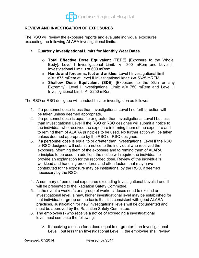
REVIEW AND INVESTIGATION OF EXPOSURES
The RSO will review the exposure reports and evaluate individual exposures exceeding the following ALARA investigational limits:
• Quarterly Investigational Limits for Monthly Wear Dates
o Total Effective Dose Equivalent (TEDE) [Exposure to the Whole Body]: Level I Investigational Limit: >/= 300 mRem and Level II Investigational Limit: >/= 600 mRem
o Hands and forearms, feet and ankles: Level I Investigational limit >/= 1875 mRem at Level II Investigational knee >/= 5625 mREM
o Shallow Dose Equivalent (SDE) [Exposure to the Skin or any Extremity]: Level I Investigational Limit: >/= 750 mRem and Level II Investigational Limit >/= 2250 mRem
The RSO or RSO designee will conduct his/her investigation as follows:
1. If a personnel dose is less than Investigational Level I no further action will be taken unless deemed appropriate.
2. If a personnel dose is equal to or greater than Investigational Level I but less than Investigational Level II the RSO or RSO designee will submit a notice to the individual who received the exposure informing them of the exposure and to remind them of ALARA principles to be used. No further action will be taken unless deemed appropriate by the RSO or RSO designee.
3. If a personnel dose is equal to or greater than Investigational Level II the RSO or RSO designee will submit a notice to the individual who received the exposure informing them of the exposure and to remind them of ALARA principles to be used. In addition, the notice will require the individual to provide an explanation for the recorded dose. Review of the individual’s workload and handling procedures and often factors that may have contributed to the exposure may be institutional by the RSO, if deemed necessary by the RSO.
4. A summary of personnel exposures exceeding Investigational Levels I and II will be presented to the Radiation Safety Committee.
5. In the event a worker’s or a group of workers’ doses need to exceed an investigational level, a new, higher investigational level may be established for that individual or group on the basis that it is consistent with good ALARA practices. Justification for new investigational levels will be documented and must be approved by the Radiation Safety Committee.
6. The employee(s) who receive a notice of exceeding a investigational level must complete the following:
o If receiving a notice for a dose equal to or greater than Investigational Level I but less than Investigational Level II, the employee shall review

Reviewed: 07/2014 Revised: 07/2014
their procedural technique for possible reduction of exposure and apply the basic rules of time, distance and shielding to keep their exposure ALARA.
o If receiving a notice for a dose greater than Investigational Level II, the employee shall be consulted with their supervisor and the RSO. In addition, the employee shall review their procedural technique for possible reduction of exposure and apply the basic rules of time, distance and shielding to keep their exposure ALARA.
o If an employee exceeds annual allowable limit identified by ARRA before the end of the calendar year, the employee may be reassigned to a different position to minimize future radiation exposure.
7. The RSO or RSO designee will determine if any other actions should be implemented to assure adequate protection in the future.
Reviewed: 07/2014 Revised: 07/2014
ORDERING EXAMS FOR OUTPATIENTS
POLICY:
To ensure that only appropriate exams are performed.
PROCEDURE:
1. Exams shall be performed only upon the order of a person who is lawfully authorized to diagnose, treat and prescribe.
2. All requests for exams should contain the reasons for the examination. The requesting medical staff member is responsible for providing this information.
3. For outpatients, a physician’s prescription should be provided.

Reviewed: 07/2014 Revised: 07/2014
ORDERING EXAMS
POLICY:
To ensure that only appropriate exams are performed.
PROCEDURE:
1. Exams shall be performed only upon the order of a person who is lawfully authorized to diagnose, treat and prescribe.
2. All requests for exams should contain the reasons for the examination. The requesting medical staff member is responsible for providing this information.
3. In the case of inpatients, the requisition or order for examination should be provided in compliance with the hospital’s established procedure.
4. In the case of outpatients, a physician’s prescription should be provided. 5. All requisitions on inpatients shall be verified against the physician’s orders on
the patient’s chart or prescription. Any contraindication requires an immediate call to the referring physician for clarification of the order.
6. Once the order or prescription is confirmed, check the patient’s ID bracelet or otherwise establish the patient’s identity to make sure the correct patient is being scanned. Always verify the patient identity twice, by name and date of birth.
Reviewed: 07/2014 Revised: 07/2014
PATIENT ASSESMENT
POLICY:
Patient assessment is made with the interdisciplinary approach of the physician, nursing and the Diagnostic Imaging Department technologist to provide the most relevant information to allow for the optimum radiological exam and results.
PROCEDURE:
It is the policy that the assessment of patients undergoing diagnostic imaging procedures
takes place in the following manner:
1. A history of the patient’s condition will be reviewed prior to the test being performed.
2. A written order will be reviewed by the radiologist and technologist. 3. The patient will be questioned about his/her condition by the technologist or
radiology nurse and the information documented on the requisition for the radiologist to review.
4. Questionnaires will be given to patients to fill out when pertinent or for safety measure. (MRI, Mammography, Invasive or Special procedures).
5. Verbal communication between the ordering physician and the radiologist is encouraged.
6. Technologists and nursing will assess the patient during the procedure being performed.
7. If the patient condition changes, it will be reported to the radiologist or ordering physician immediately.
8. All actions necessary for response to an adverse reaction will be documented by staff and reported in Quantros.
9. The radiology nurse will be available for pre and post monitoring when necessary.

Reviewed: 07/2014 Revised: 07/2014
PATIENT CARE GUIDELINES FOR DIAGNOSTIC IMAGING STAFF
PURPOSE:
To have a keen sense of all our patient’s feelings and needs, and to be perceived by all others (both internal and external) as a knowledgeable, understandable, helpful and caring resource. To make all patients feel special.
PROCEDURE:
1. Professionalism and appearance – to look and conduct oneself in a manner perceived as positive by all others, both internal and external. To create a work environment that projects an image of excellence.
a. Dress code adhered to. b. Managing emotions and stress at all times in all situations. c. Proper knowledge, use and care of equipment in all areas of assigned work. d. Clean, safe and organized work area. e. Accurate record keeping.
2. Knowledge and expertise – to be perceived as knowledgeable and up-to-date in the field of radiological technology and all the services offered by the department.
a. Possess knowledge of all the services offered with the ability to guide and describe each modality to doctors, nursing, interdepartmental staff, clinic personnel, lay people, etc.
b. Have equipment knowledge and annual proficiencies reviewed and documented. c. Maintain professional certifications as mandated by ARRT, ACR, JCAHO,
and the Arizona State Board of Nursing. d. Keep up-to-date in the field of radiology and areas of expertise, by attending
seminars, in-services, and organizational/professional meetings. e. Network with staff at other hospitals and/or clinics. f. Adhere to radiation protection and safety guidelines at all times in all situations.
Follow ALARA (to keep all radiation exposure as low as reasonably achievable). Shield all patients, especially those of childbearing age. Follow all standards set forth by the NCRP (National Council on Radiation Protection and Measurements).
g. Adhere to JCAHO standards.
3. Communication and projection – Communicate positively with all internal and external customers and project through communication, professionalism, knowledge and high standards.

Reviewed: 07/2014 Revised: 07/2014
a. Be sensitive to people of different cultural and religious backgrounds. They may view illness and treatment methods differently.
b. Patient and their families, many of whom may not speak or understand English, need to know that the hospital staff is acting in their best interest. (Please ask for assistance in communication when needed. There are staff members who speak different languages who may be of assistance.)
c. Always use appropriate and effective delivery and tonal quality.
4. Geriatrics and pediatrics – Caring for the adolescent or geriatric patient can present unique challenges for the technologist or nurse. Each age group has particular anxieties and concerns. It is up to the technologist or nurse to provide an understanding, supportive, and compassionate environment. All staff members who assess, treat, or care for these patients should be able to understand, adjust and meet their special needs.
a. Geriatrics
i. Address each client appropriately and professionally at his or her level. (i.e. Adult) “Hello, Mr. Smith. My name is Jane and I will be performing your CT exam today.”
ii. Never ignore your patient, even though you may think they do not hear or understand. Address them appropriately and explain what you are going to do before you do it.
iii. Never call an elderly patient “sweetie, honey, or dear”; use their respectful title or name.
iv. Never treat an elderly patient like a child. v. Never leave a patient unattended. Always put up the side rails on carts.
Always check to make sure the brake is set on the cart or wheelchair for patient safety.
b. Pediatrics
i. Address each client appropriate and professionally at his or her level. (i.e. Child) “Hi, Jimmy. My name is Jane and I am going to take a picture of your chest today with a special camera that can see inside of you. Maybe we will be able to see why you have been coughing so hard.”
ii. Do not confuse children by using technical terms. Talk to them on their level, and look at them directly when speaking to them.
iii. Praise them for holding still and cooperating with you. iv. Demonstrate what you are going to do before you do it. v. Always shield children, and document such on the requisition. vi. Let the parents know what you are going to do. If the mother is not pregnant,
you may ask her to help with the child (be sure to give her a lead apron to wear, and note on the requisition that the mother stated she was not pregnant and was given a lead apron for radiation protection). Children are

Reviewed: 07/2014 Revised: 07/2014
more comfortable with their parents nearby in strange surroundings and situations.
vii. Never leave children unattended.
5. Customer focus – Understand and service customers’ needs and wants to meet their expectations.
a. Know what your customer wants. b. Be a key link to the patient care effort. c. Market your department and educate others of your services. d. Think in terms of service excellence and CQI.
6. Standards – Set and adhere to high work standards that are noticed and regarded as positive by all others.
a. Follow the organizational values, vision and mission statement. b. Produce high quality radiographs at all times. c. Have ownership and accountability of work. d. Have pride in work and the department. e. Set high levels of performance. f. Be flexible to continue to meet the demands of the healthcare field of
today and tomorrow. g. To follow the RT and RN Code of Ethics at all times.

Reviewed: 07/2014 Revised: 07/2014
PATIENT SHIELDING
PURPOSE:
To insure patient safety during radiographic procedures/examination.
PROCEDURE:
It is the policy that appropriate measures will be taken to protect patients from unnecessary direct and scatter radiation through the following measures:
• All females of childbearing age will be shielded with a lead apron. • The technologist will ensure that all children being radiographed have proper
gonadal shielding and that proper collimation of the x-ray machine be utilized to expose only the area or anatomy of interest.
• All expectant females will be properly shielded and the x-ray collimated to the area or anatomy of interest only. Orders should be carefully considered against the risks.
• Expectant females MUST NOT be allowed to hold or immobilize children for radiographs and they WILL NOT be allowed in the x-ray area during exposures.

Reviewed: 07/2014 Revised: 07/2014
PROCEDURE FOR RADNET DOWNTIME
POLICY: To establish guidelines for downtime of the Radnet computer system.
PROCEDURE:
Scheduled Radnet Downtime
1. Client Services will send a copy of the Patient Information Sheet (PIS) to the Diagnostic Imaging Department. The Diagnostic Imaging Department will use NCR form XR-101 to record orders for patients. The white copy goes to the ordering department. The yellow copy is kept by the scheduling/reception staff to be used to record information into the Radnet system after Radnet is back online.
2. The Diagnostic Imaging Department will manually forward the results of the study to the requesting departments.
3. There will be no transcription of reports in Powerchart during downtime. Once Radnet returns to normal function, it will be the responsibility of the Radiology Technologist to enter the orders for procedures completed during the downtime.
4. During downtime the Radiologist will dictate on tape recorders, which will be retrieved and typed by the transcriptionist. Report migration to Powerchart once the system is functional will be the responsibility on the Transcription Department.

Reviewed: 07/2014 Revised: 07/2014
RADIATION PHYSICST SERVICES
POLICY: To ensure radiation safety procedures are followed.
PROCEDURE:
The services of a Radiation Physicist should be available for the Diagnostic Imaging
Department for the following:
- Consultation
- Periodic safety checks
- Supervision of radiation safety procedures
- Participation in educational programs
- Consultant to the Hospital Radiation Safety Committee
- Review of QC Program
- Review of MQSA for mammography
- Annual equipment calibration

Reviewed: 07/2014 Revised: 07/2014
RADIOLOGIC EXAMINATIONS IN PREGNANT PATIENTS
PURPOSE
To assure that all reasonable steps are taken to protect an unborn child during radiological exams.
PROCEDURE
• All female patients will be asked if they may be pregnant prior to the examination. .
• A written informed consent is required in the event that a radiological exam must be performed on a pregnant patient.
• The pregnant patient will be shielded and technique adjusted to be as low as possible without compromised diagnostic quality.
• For exams of the abdominal and pelvic area:
- The radiologist is to be made aware of the scheduled procedure.
- The radiologist will contact the referring physician to discuss possible alternatives or modifications of the exam to minimize exposure to the fetus/embryo.
- Due to emergency, if informed consent cannot be obtained, the radiologist will document in the medical record the reason for the exam and steps taken to minimize risks to the embryo/fetus.
• In utero irradiation:
- The radiologist will notify the physicist for retrospective estimate of fetal dose.
- A report will be filed with the Radiation Safety Officer by the next working day. The radiologist will inform the referring physician regarding the patient's exposure and will counsel the patient on the radiation risks.
• Nuclear Medicine:
If the patient is, or thinks that she may be pregnant, the radiologist in consultation with the
referring physician will determine if the benefits of the exam outweigh the risks.
• A written, informed consent will be obtained by the radiologist. • Magnetic Resonance Imaging:

Reviewed: 07/2014 Revised: 07/2014
An MRI is not to be done during the first trimester of pregnancy unless it is a medical
emergency.
Should the radiologist, in consultation with the referring physician, feel that the test should be
performed, a written informed consent will be obtained. (REFER TO 4237-8)

Reviewed: 07/2014 Revised: 07/2014
RADIOLOGY EXAM ROOM CLOSURE PROCEDURE
POLICY:
To establish the procedure to follow when requesting a room closure.
PROCEDURE:
The Radiology leads are allowed to request the closing only with Manager/Director approval.
The request has to be done by e-mail; verbal orders will not be accepted.
1. The lead will e-mail request to the Manager/Director requesting to close the room, with a carbon copy to the Scheduling Coordinator.
2. The Manager/Director will approve/not approve the request via email and cc the Scheduling Coordinator
3. Once the Scheduling Supervisor completes the request, she will “reply to all” on the original e-mail to let everybody know that it has been completed.

Reviewed: 07/2014 Revised: 07/2014
RADIOLOGY INTERPRETATION AND FOLLOW UP
POLICY:
To provide follow-up care of all patients who have had standard radiographs done while in the Emergency Department.
PROCEDURE:
1. Under normal operating circumstances, all preliminary and final readings will be completed by the Radiologist.
2. Results will post in Cerner during normal business hours. After hours, Virtual Radiologic will fax results for STAT exams for the following modalities: CT and Ultrasound.
3. The Radiologists will do their best to interpret exams in a timely manner during normal business hours. If the Emergency Department Physician feels a preliminary read is needed after business hours, they will conduct a “wet read” and a notation will be made in Cerner Radiology Desktop documenting their preliminary findings.
4. The radiologist will complete the final read the following morning on all X-ray exams completed after normal business hours. Should a major discrepancy arise between the readings, the Radiologist will discuss the results with the Emergency Room Physician. Non-critical discrepancies will be reported to the ED Nurse Manager or charge Nurse via printed documentation.

Reviewed: 07/2014 Revised: 07/2014
THE READING OF FILMS TAKEN AT THE CRH EAST CAMPUS
POLICY:
To establish a policy for the reading of films taken at the CRH East Campus Imaging Center.
PROCEDURE:
1. Films taken at the CRH East Campus Imaging Center with the mini C-arm for the surgeon’s/physician’s use during surgery or for documentation of the surgical procedure will not be sent for reading to the radiologist. These films are not for diagnostic purposes.
2. The radiologist will read only those films from the CRH Outpatient Imaging Center which meet the following criteria:
a. Films required for immediate diagnosis or further care of the patient. b. The physician has submitted a written order for such exams. c. The exam has been ordered in the Diagnostic Imaging Department
computer application. d. A radiological technologist is called to take appropriate films following
proper positioning standards and diagnostic techniques.

Reviewed: 07/2014 Revised: 07/2014
RADIOLOGY SERVICES FOR THE AMBULATORY SURGERY CENTER
POLICY:
To define Radiology support at the Ambulatory Surgery Center.
PROCEDURE:
1. A portable x-ray machine, two full sizes C-arm, and a mini C-arm are available for use at the ASC.
2. A Technologist is required to operate the full size C-arm and Portable x-ray machine. 3. Attending surgeon will be responsible for the use of the mini C-arm. 4. The Surgical Department will be responsible for the maintenance and record
keeping of the mini C-arm. The Radiology Department is available for direction if needed.
Portable film requests:
5. When surgeon/anesthesiologist determines a portable film is necessary, the supervisor/charge will notify the Radiology Department at the East Campus.
6. A radiology technologist will be sent to the ASC to perform the exam. 7. Requested film is taken and cassette is taken to the East Campus Center by
radiology technologist. 8. Preliminary reading of the film will be done by the ordering physician. 9. Radiologist will call ASC with results if requested by the ordering physician.
- ALL Fluoroscopy time must be recorded and logged.

Reviewed: 07/2014 Revised: 07/2014
SCANNER QUALITY CONTROL FOR CT
POLICY:
To ensure that all imaging is of the highest possible quality.
PROCEDURE:
1. Quality Control test procedures are to be performed to meet all manufacturer recommendations.
2. Systems that have software-driven mandatory system checks may have all checks completed without additional (written) records.
3. Documentation of QC Daily Requirements shall be retained and shall be available for use by operators and engineering service personnel.
4. All scanner problems should be reported to Biomed for proper resolution. Remedial repair items should be communicated to the service engineer by use of communications logbook for correction during routine scheduled service.
5. Radiographic equipment will be operated only by personnel meeting all State of Arizona and federal licensing requirements.
6. All radiographic equipment will be annually calibrated by the physicist.
Reviewed: 07/2014 Revised: 07/2014
RADIOLOGY SCHEDULING DATA ENTRY
POLICY:
To establish and administer a uniform and consistent policy and procedure concerning Radiology Scheduling which will help monitor accurate data entry of orders and improve customer service.
PROCEDURE:
The Diagnostic Imaging Department will monitor data entry for accurate scheduling from the original order. All Radiology Scheduling staff who is directly involved in the data entry or review of orders will be held accountable to the following:
1. The correct entry of the ordering provider. 2. The correct entry of the exam/procedure according to the physician script/order. 3. The correct entry of billing information (i.e. financial number, encounter). 4. The correct location of the exam scheduled (i.e. Main or East Campus). 5. The correct patient service type (SDM, CL, SDS, RBX). 6. The appointment is entered into appointment book.
Occurrences of errors as listed above shall result in disciplinary action as follows:
1. 3 occurrences in a 6 month period: verbal warning 2. 4 occurrences in a 6 month period: documented verbal warning 3. 5 occurrences in a 6 month period: written warning 4. 6 occurrences in a 6 month period: second written warning, further
disciplinary action, not to exclude suspension or termination.

Reviewed: 07/2014 Revised: 07/2014
SCHEDULING PROCEDURES AT THE EAST CAMPUS
POLICY:
To insure efficient workflow, scheduling, and tracking of patients and exams.
PROCEDURE:
1. Diagnostic radiologic procedures shall be performed upon receipt of the written request of a physician.
2. All requests for radiologic procedures shall contain the reasons for the examination. A space is provided on the x-ray request for this information. It is the responsibility of the person filling out the request to provide this information and to initial said request. The Diagnostic Imaging Department personnel will process outpatient requests.
3. All outpatient radiologic procedures will be scheduled through the Radiology Scheduling Department, at which time specific instructions for any special preparation will be given the patient if he/she has not received it from the ordering physician.
4. Only physicians with proper privileges, and who have been authorized to do so by the medical staff may authenticate reports. These reports are part of the medical record and are filed accordingly. Inpatient reports are filed in medical records along with Emergency Department patient reports. Outpatient reports are sent to the requesting physician. Reports are archived in the RIS computer program.

Reviewed: 07/2014 Revised: 07/2014
SCHEDULING PROCEDURES AT THE MAIN CAMPUS
POLICY:
To insure efficient workflow, scheduling, and tracking of patients and exams.
PROCEDURE:
1. Diagnostic radiologic procedures shall be performed upon receipt of the written request of a physician, except for mammography or DEXA exams.
2. All requests for radiologic procedures shall contain the procedure, diagnosis, and/or reasons for the examination and doctor’s signature.
3. All inpatient diagnostic radiologic procedures will be performed as soon as possible upon receiving the request, with the exception of those which require special preparation such as diagnostic gastrointestinal work. Preparation for routine fluoroscopic procedures is available at the nurses’ stations as well as in the Diagnostic Imaging Department. These preparations will be followed routinely unless otherwise specified by either the referring physician or the radiologist. In cases where several procedures are ordered simultaneously, preferential treatment will be given those patients who have undergone special preparation for a specific examination.
4. All outpatient radiologic procedures will be scheduled through the Radiology Scheduling Department, at which time specific instructions for any special preparation will be given the patient if he/she has not received it from the ordering physician.
5. Only physicians with proper privileges, and who have been authorized to do so by the medical staff may authenticate reports. These reports are part of the medical record and are filed accordingly. Inpatient reports are filed in medical records along with Emergency Department patient reports. Outpatient reports are sent to the requesting physician. Reports are archived in the RIS computer program.

Reviewed: 07/2014 Revised: 07/2014
SCOPE OF SERVICE
POLICY:
To outline the scope of service for the Diagnostic Imaging Department.
PROCEDURE:
This department is under the direct supervision of a Radiologist, certified by the American Board of Radiology and having a current license from the State of Arizona to practice medicine.
A technologist registered by the American Registry of Radiologic Technologists and certified by the State of Arizona is available 24 hours per day and will assist the radiologist(s) in acquiring needed images on a referred patient.
Radiographs, commonly called x-rays, must be ordered by an attending physician, and are taken by a certified Radiologic Technologist. Following processing of the radiographs, the radiologists dictate their interpretation.
Radiographic images are permanently stored in the Picture Archiving and communication System (PACS).
The goal of the Department of Radiology will be to ensure that all patients treated will receive high quality care in the most expedient and professional manner possible.
Although services include CT scanning, diagnostic ultrasound and magnetic resonance imaging (MRI), nuclear medicine, and mammography, x-ray procedures still constitute the majority of the daily procedural load. Services related or concomitant to imaging include quality assurance monitoring and evaluation, quality control (including protecting patients and staff from harmful radiation), image interpretation, dictation, transcription, patient billing, marketing, equipment purchasing and continuing education.
Technologists or other non-physician personnel do not perform interventional studies or diagnostic fluoroscopy without a radiologist present. Portable x-ray equipment allows radiographs to be obtained in surgery, as well as medical/surgical and intensive care units.
Radiologists are consultants, responsible for advising referring physicians on which imaging procedures to do and in which sequence. In addition, when emergency physicians request films and interpret them, staff radiologists are responsible for the confirming or amending of the emergency physician’s initial interpretations.
All personnel within the department are under the direction of the Diagnostic Imaging Department Director.

Reviewed: 07/2014 Revised: 07/2014
IMAGING TECHNOLOGIST CERTIFICATION AND DUTIES
Policy :
All technologists in the Diagnostic Imaging (DI) department will be categorized and work in the modalities to which they are registered. They must be able to perform all required procedures with expertise and proficiency, following established protocols and guidelines on patients of all ages and conditions.
Description of Modalities
Diagnostic Radiology and Special Procedures: Able to perform ED, Fluoroscopy, surgery and portable procedures. May include myelograms, arthrograms, venograms, hysterosalpingogram, vertebroplasty, and other special procedures as required. Mammography: Able to perform screening, diagnostic, magnification, needle localizations, specimen and advanced special views as needed. May be asked to do general diagnostic radiographic procedures if necessary.
MRI: Able to do all diagnostic procedures and rotate through diagnostic radiology as schedule demands.
CT: Able to do all diagnostic procedures and rotate through diagnostic radiology as schedule demands.
Ultrasound: Able to perform all diagnostic studies (Vascular, OB-GYN, Abdominal, small parts). Advanced training in Echosonography may be required.
Nuclear Medicine: Able to perform all diagnostic procedures and may be asked to do general diagnostic radiographic procedures if necessary.

Reviewed: 07/2014 Revised: 07/2014
IMAGING TECHNOLOGIST RESTRICTIONS
POLICY:
To insure appropriate patient care is provided by the delivery of results of procedures through the proper channels to avoid misdiagnosis and/or treatment.
PROCEDURE:
It is the policy of the Diagnostic Imaging Department that all technologists working in the department will not work beyond their scope of practice.
1. Technologists will not perform any diagnostic procedure without the written order of a physician.
2. All technologists work under the supervision of the radiologists. 3. Technologists will not make a diagnosis based on any radiograph or image. 4. Technologists will not operate any equipment without having been trained to
operate it safely and effectively. 5. Technologists will not report results to any patient; this shall be done by the
physician or the radiologist. 6. Technologists will not be allowed to work without a valid and current
license by the ARRT and the MRTBE. 7. Technologists will not perform breast palpations except to position the
breast for radiographic purposes.

Reviewed: 07/2014 Revised: 07/2014
VOLUNTEER TESTING FOR NEW CLINICAL APPLICATION PROCEDURES
PURPOSE:
It is occasionally necessary for SVRHC staff to test new technologies, FDA approved technologies, and/or new equipment applications prior to its’ full implementation. In order to accomplish this, and to assure the safe and proper testing of such new technologies and equipment and for quality control purposes it is necessary to enlist the assistance of human subject volunteers. All volunteers will be requested to sing the SVRHC Volunteer Acknowledgement of Non-Liability, Consent and Release. The contents and implications of this Acknowledgement will be fully and completely explained to the volunteer by an appropriate SVRHC staff member.
PROCEDURE FOR VOLUNTEERS:
1. Upon receipt of approval of the request for volunteers, the appropriate department Director or Manager will send an SVRHC email to “ALL” advising staff members fo the need for volunteers to assist in the Application competency process. The email will provide information on the procedure to be performed and instruct any interested staff member to make contact with the Department Manager in order to determine suitability and to be screened by the radiologist for he desired procedure. Note: 1) the first ten (10) volunteers, who meet criteria, will be accepted for the procedure and 2) Exams on staff volunteers will ONLY be performed on “off-hours” and not during scheduled work shifts, nor will compensation be offered for time volunteered.
2. All volunteers will have a script written by a staff radiologist for the procedure/exam deemed necessary during the applications training period. The volunteer will be screened and the procedure and associated risks will be explained.
3. Images will only be used to establish protocols and imaging parameters/sequences and will NOT be used for ANY clinical evaluations.
4. Images will not be archiving in PACS and will not remain part of the volunteer’s medical record.
5. All volunteers will be given post procedure discharge instructions. 6. In that these procedures are conducted for testing on a new “Application” they
will be performed on a “NO CHARGE” basis.
PROCEDURE FOR VOLUNTEERS WITH A SCRIPT FROM A PRIMARY CARE PHYSICIAN
1. All volunteers must have a valid script from a physician.

Reviewed: 07/2014 Revised: 07/2014
2. Upon receipt of the script/exam order, all volunteers will be required to complete SVRHC’s patient registration process and must complete and sign Form AD-100E Conditions of Admission and/or Treatment.
3. On all APPLICATIONS TESTING PATIENTS, Client Services staff will be required to select the institutional account #01987191 to register the volunteers and will document SVRHC as the account guarantor.
4. The volunteer will be screened and the procedure and associated risks will be explained and Form NU-216 Consent to Operate or Procedure and/or Anesthetics and/or Blood or Blood Products and Form XR-136 Patient information, and if necessary, Guidelines an Risks for contrast Material must be completed and signed by the volunteer.
5. Patient identifiers will be labeled on all test and/or images. Images will be archived under the patient’s name in PACS and remain part of their medical record.
6. Volunteers will be given post procedure discharge instructions and a copy of Form OR-107 Same Day Surgery/Procedure Discharge Instructions.
7. To validate quality, radiologists or other medical professionals may view the procedure results. A report will be generated with the exam findings and will become part of the volunteer’s medical record.
8. In that these procedures are conducted for testing on a new “Application” they will be performed on a “NO CHARGE” basis. All account charges will be adjusted off via a general ledger PAT code established by the Finance Department.