coarctation of the aorta interrupted aortic arch
TRANSCRIPT
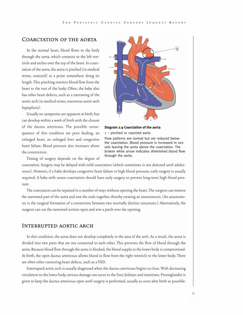
Coarctation of the aorta
In the normal heart, blood flows to the body
through the aorta, which connects to the left ven-
tricle and arches over the top of the heart. In coarc-
tation of the aorta, the aorta is pinched (in medical
terms, coarcted) at a point somewhere along its
length. This pinching restricts blood flow from the
heart to the rest of the body. Often, the baby also
has other heart defects, such as a narrowing of the
aortic arch (in medical terms, transverse aortic arch
hypoplasia).
Usually no symptoms are apparent at birth, but
can develop within a week of birth with the closure
of the ductus arteriosus. The possible conse-
quences of this condition are poor feeding, an
enlarged heart, an enlarged liver and congestive
heart failure. Blood pressure also increases above
the constriction.
Timing of surgery depends on the degree of
coarctation. Surgery may be delayed with mild coarctation (which sometimes is not detected until adoles-
cence). However, if a baby develops congestive heart failure or high blood pressure, early surgery is usually
required. A baby with severe coarctation should have early surgery to prevent long-term high blood pres-
sure.
The coarctation can be repaired in a number of ways without opening the heart. The surgeon can remove
the narrowed part of the aorta and sew the ends together, thereby creating an anastomosis. (An anastomo-
sis is the surgical formation of a connection between two normally distinct structures.) Alternatively, the
surgeon can cut the narrowed section open and sew a patch over the opening.
Interrupted aortic arch
In this condition, the aorta does not develop completely in the area of the arch. As a result, the aorta is
divided into two parts that are not connected to each other. This prevents the flow of blood through the
aorta. Because blood flow through the aorta is blocked, the blood supply to the lower body is compromised.
At birth, the open ductus arteriosus allows blood to flow from the right ventricle to the lower body. There
are often other coexisting heart defects, such as a VSD.
Interrupted aortic arch is usually diagnosed when the ductus arteriosus begins to close. With decreasing
circulation to the lower body, serious damage can occur to the liver, kidneys and intestines. Prostaglandin is
given to keep the ductus arteriosus open until surgery is performed, usually as soon after birth as possible.
T H E P E D I A T R I C C A R D I A C S U R G E R Y I N Q U E S T R E P O R T
33
Diagram 2.9 Coarctation of the aorta
1 – pinched or coarcted aorta
Flow patterns are normal but are reduced belowthe coarctation. Blood pressure is increased in ves-sels leaving the aorta above the coarctation. Thebroken white arrow indicates diminished blood flowthrough the aorta.

C H A P T E R 2 ■ P E D I A T R I C C A R D I A C I S S U E S
34
Diagram 2.11 Pulmonary stenosis (valvular)
1 – narrowed pulmonary valve
Blood flow patterns are normal but blood flowthrough the pulmonary artery is reduced as indicated by the broken white arrows.
Diagram 2.10 Interrupted aortic arch
1 – interruption of aortic arch
2 – descending aorta connected to pulmonary artery by large patent ductus arteriosus
3 – ventricular septal defect
Flow patterns are normal to the upper body.However, there is no flow of oxygenated bloodto the lower body unless there exist, as in thisdrawing, shunts such as a ventricular septaldefect that allows oxygenated blood into thepulmonary artery, and a patent ductus arteriosus that allows the partially oxygenatedblood to travel from the pulmonary artery tothe descending aorta (as indicated by the broken white arrow).
Diagram 2.12 Aortic stenosis (valvular)
1 – narrowed aortic valve
Flow patterns are normal but blood flow to theaorta is reduced as indicated by the broken whitearrows.
Most often a one-stage repair is undertaken, patching the arch or connecting the two ends of the aortic
arch together, closing any VSD, and tying off and dividing the ductus arteriosus into two parts.
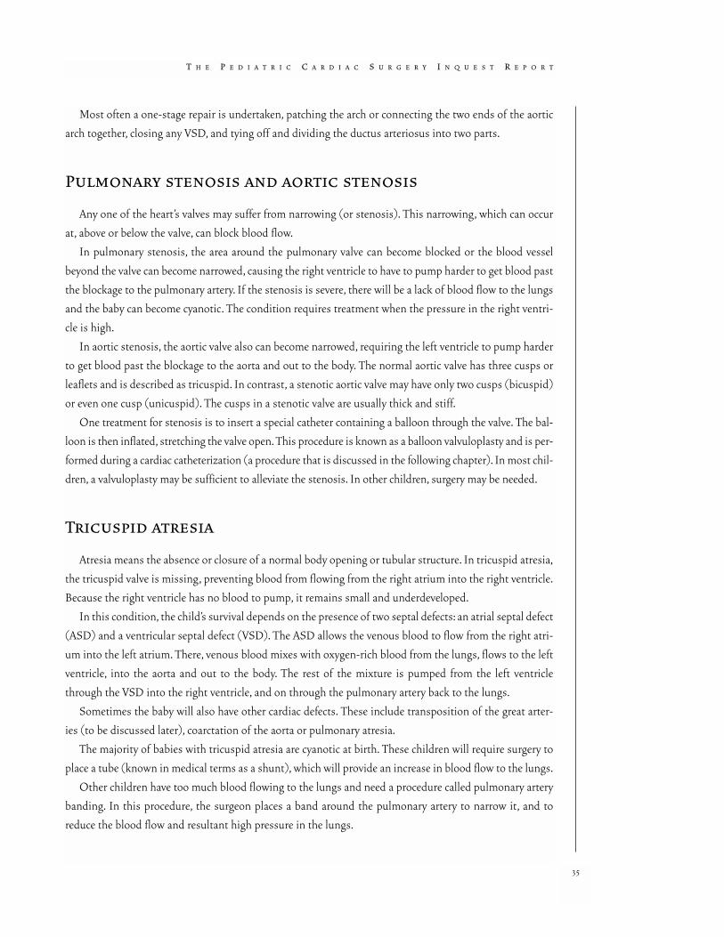
Pulmonary stenosis and aortic stenosis
Any one of the heart’s valves may suffer from narrowing (or stenosis). This narrowing, which can occur
at, above or below the valve, can block blood flow.
In pulmonary stenosis, the area around the pulmonary valve can become blocked or the blood vessel
beyond the valve can become narrowed, causing the right ventricle to have to pump harder to get blood past
the blockage to the pulmonary artery. If the stenosis is severe, there will be a lack of blood flow to the lungs
and the baby can become cyanotic. The condition requires treatment when the pressure in the right ventri-
cle is high.
In aortic stenosis, the aortic valve also can become narrowed, requiring the left ventricle to pump harder
to get blood past the blockage to the aorta and out to the body. The normal aortic valve has three cusps or
leaflets and is described as tricuspid. In contrast, a stenotic aortic valve may have only two cusps (bicuspid)
or even one cusp (unicuspid). The cusps in a stenotic valve are usually thick and stiff.
One treatment for stenosis is to insert a special catheter containing a balloon through the valve. The bal-
loon is then inflated, stretching the valve open. This procedure is known as a balloon valvuloplasty and is per-
formed during a cardiac catheterization (a procedure that is discussed in the following chapter). In most chil-
dren, a valvuloplasty may be sufficient to alleviate the stenosis. In other children, surgery may be needed.
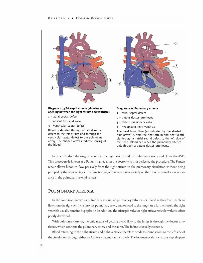
Tricuspid atresia
Atresia means the absence or closure of a normal body opening or tubular structure. In tricuspid atresia,
the tricuspid valve is missing, preventing blood from flowing from the right atrium into the right ventricle.
Because the right ventricle has no blood to pump, it remains small and underdeveloped.
In this condition, the child’s survival depends on the presence of two septal defects: an atrial septal defect
(ASD) and a ventricular septal defect (VSD). The ASD allows the venous blood to flow from the right atri-
um into the left atrium. There, venous blood mixes with oxygen-rich blood from the lungs, flows to the left
ventricle, into the aorta and out to the body. The rest of the mixture is pumped from the left ventricle
through the VSD into the right ventricle, and on through the pulmonary artery back to the lungs.
Sometimes the baby will also have other cardiac defects. These include transposition of the great arter-
ies (to be discussed later), coarctation of the aorta or pulmonary atresia.
The majority of babies with tricuspid atresia are cyanotic at birth. These children will require surgery to
place a tube (known in medical terms as a shunt), which will provide an increase in blood flow to the lungs.
Other children have too much blood flowing to the lungs and need a procedure called pulmonary artery
banding. In this procedure, the surgeon places a band around the pulmonary artery to narrow it, and to
reduce the blood flow and resultant high pressure in the lungs.
T H E P E D I A T R I C C A R D I A C S U R G E R Y I N Q U E S T R E P O R T
35
In other children the surgeon connects the right atrium and the pulmonary artery and closes the ASD.
This procedure is known as a Fontan, named after the doctor who first perfected the procedure. The Fontan
repair allows blood to flow passively from the right atrium to the pulmonary circulation without being
pumped by the right ventricle. The functioning of this repair relies totally on the preservation of a low resist-
ance in the pulmonary arterial vessels.
Pulmonary atresia
In the condition known as pulmonary atresia, no pulmonary valve exists. Blood is therefore unable to
flow from the right ventricle into the pulmonary artery and onward to the lungs. As a further result, the right
ventricle usually remains hypoplastic. In addition, the tricuspid valve or right atrioventricular valve is often
poorly developed.
With pulmonary atresia, the only means of getting blood flow to the lungs is through the ductus arte-
riosus, which connects the pulmonary artery and the aorta. The infant is usually cyanotic.
Blood returning to the right atrium and right ventricle therefore needs to shunt across to the left side of
the circulation, through either an ASD or a patent foramen ovale. The foramen ovale is a natural septal open-
C H A P T E R 2 ■ P E D I A T R I C C A R D I A C I S S U E S
36
Diagram 2.13 Tricuspid atresia (showing no opening between the right atrium and ventricle)
1 – atrial septal defect
2 – absent tricuspid valve
3 – ventricular septal defect
Blood is shunted through an atrial septaldefect to the left atrium and through the ventricular septal defect to the pulmonaryartery. The shaded arrows indicate mixing ofthe blood.
Diagram 2.14 Pulmonary atresia
1 – atrial septal defect
2 – patent ductus arteriosus
3 – absent pulmonary valve
4 – hypoplastic right ventricle
Abnormal blood flow (as indicated by the shadedblue arrow) is from the right atrium and right ventri-cle through an atrial septal defect to the left side ofthe heart. Blood can reach the pulmonary arteriesonly through a patent ductus arteriosus.
ing in the fetal heart that allows blood to flow between the atria. In most newborns this opening closes at
birth. Failure of the foramen ovale to close results in a type of atrial septal defect, known as a patent fora-
men ovale (PFO). Where there is an ASD or a PFO, blood leaves the right atrium and mixes with the oxy-
gen-rich blood in the left atrium. This blood is then pumped by the left ventricle into the aorta and out to
the body.
A further problem arises when the ductus arteriosus closes, causing severe cyanosis. The infant is then
usually given prostaglandin to keep the ductus arteriosus open. As a palliative measure, a surgeon may cre-
ate a shunt between the pulmonary artery and the aorta to increase blood flow to the lungs. If the right ven-
tricle is too small to be effective as a pump, a Fontan procedure is performed later, connecting the right atri-
um directly to the pulmonary artery. The ASD or PFO is also closed to relieve the cyanosis.
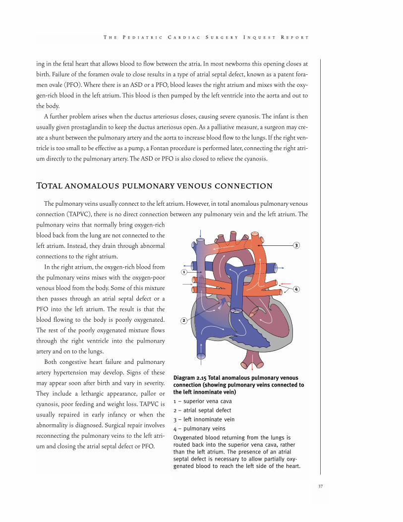
Total anomalous pulmonary venous connection
The pulmonary veins usually connect to the left atrium. However, in total anomalous pulmonary venous
connection (TAPVC), there is no direct connection between any pulmonary vein and the left atrium. The
pulmonary veins that normally bring oxygen-rich
blood back from the lung are not connected to the
left atrium. Instead, they drain through abnormal
connections to the right atrium.
In the right atrium, the oxygen-rich blood from
the pulmonary veins mixes with the oxygen-poor
venous blood from the body. Some of this mixture
then passes through an atrial septal defect or a
PFO into the left atrium. The result is that the
blood flowing to the body is poorly oxygenated.
The rest of the poorly oxygenated mixture flows
through the right ventricle into the pulmonary
artery and on to the lungs.
Both congestive heart failure and pulmonary
artery hypertension may develop. Signs of these
may appear soon after birth and vary in severity.
They include a lethargic appearance, pallor or
cyanosis, poor feeding and weight loss. TAPVC is
usually repaired in early infancy or when the
abnormality is diagnosed. Surgical repair involves
reconnecting the pulmonary veins to the left atri-
um and closing the atrial septal defect or PFO.
T H E P E D I A T R I C C A R D I A C S U R G E R Y I N Q U E S T R E P O R T
37
Diagram 2.15 Total anomalous pulmonary venousconnection (showing pulmonary veins connected tothe left innominate vein)
1 – superior vena cava
2 – atrial septal defect
3 – left innominate vein
4 – pulmonary veins
Oxygenated blood returning from the lungs is routed back into the superior vena cava, ratherthan the left atrium. The presence of an atrial septal defect is necessary to allow partially oxy-genated blood to reach the left side of the heart.
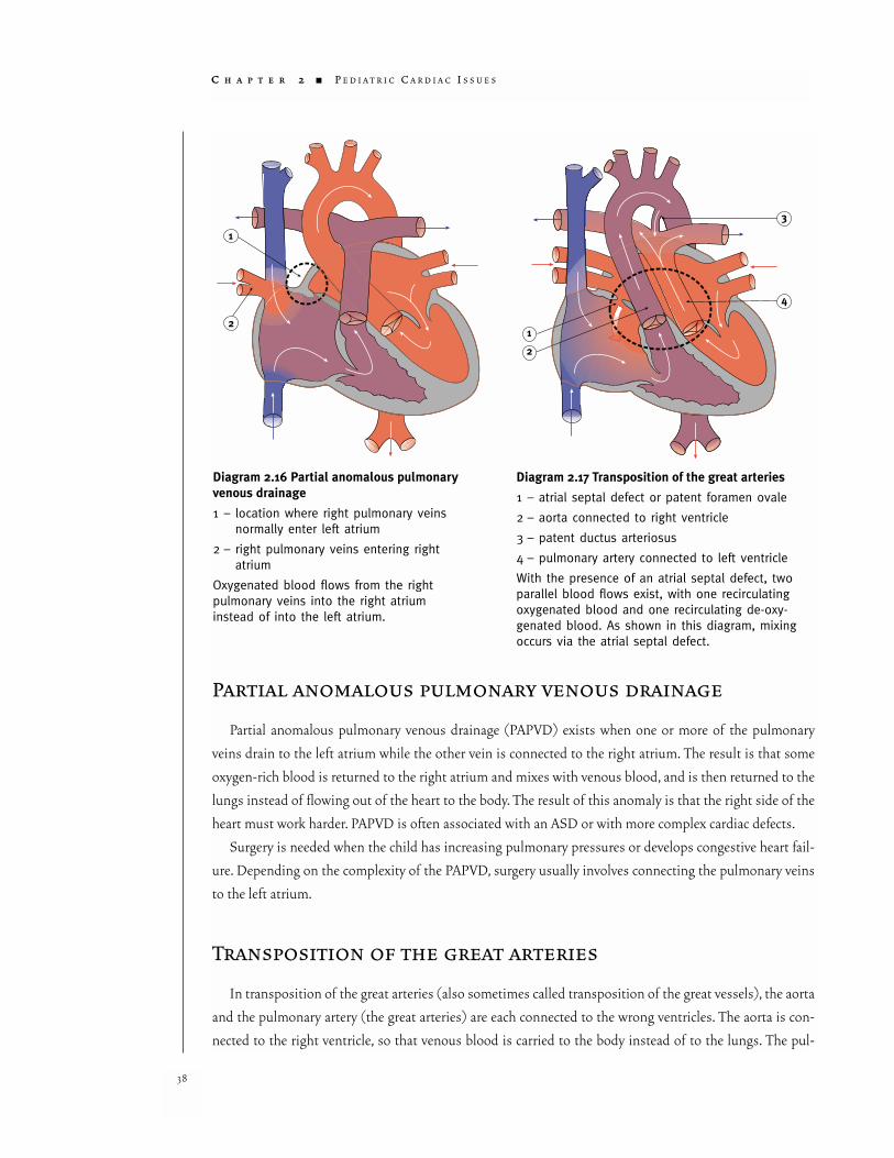
Partial anomalous pulmonary venous drainage
Partial anomalous pulmonary venous drainage (PAPVD) exists when one or more of the pulmonary
veins drain to the left atrium while the other vein is connected to the right atrium. The result is that some
oxygen-rich blood is returned to the right atrium and mixes with venous blood, and is then returned to the
lungs instead of flowing out of the heart to the body. The result of this anomaly is that the right side of the
heart must work harder. PAPVD is often associated with an ASD or with more complex cardiac defects.
Surgery is needed when the child has increasing pulmonary pressures or develops congestive heart fail-
ure. Depending on the complexity of the PAPVD, surgery usually involves connecting the pulmonary veins
to the left atrium.
Transposition of the great arteries
In transposition of the great arteries (also sometimes called transposition of the great vessels), the aorta
and the pulmonary artery (the great arteries) are each connected to the wrong ventricles. The aorta is con-
nected to the right ventricle, so that venous blood is carried to the body instead of to the lungs. The pul-
C H A P T E R 2 ■ P E D I A T R I C C A R D I A C I S S U E S
38
Diagram 2.16 Partial anomalous pulmonaryvenous drainage
1 – location where right pulmonary veinsnormally enter left atrium
2 – right pulmonary veins entering right atrium
Oxygenated blood flows from the right pulmonary veins into the right atrium instead of into the left atrium.
Diagram 2.17 Transposition of the great arteries
1 – atrial septal defect or patent foramen ovale
2 – aorta connected to right ventricle
3 – patent ductus arteriosus
4 – pulmonary artery connected to left ventricle
With the presence of an atrial septal defect, twoparallel blood flows exist, with one recirculatingoxygenated blood and one recirculating de-oxy-genated blood. As shown in this diagram, mixingoccurs via the atrial septal defect.

monary artery is attached to the left ventricle, so that oxygen-rich blood is carried back to the lungs instead
of to the body.
Newborns with transposition survive only if they have one or more connections that let oxygen-rich
blood reach the body. These connections may be in the form of a hole between the two ventricles, a patent
foramen ovale or a patent ductus arteriosus. Most babies born with transposition are extremely blue soon
after birth because their bodies are not receiving enough oxygenated blood.
In order to immediately improve the body’s oxygen supply, prostaglandin is given to keep the ductus
arteriosus open. It is also possible to enlarge the patent foramen ovale by inflating a small balloon during a
heart catheter procedure. This is known as a balloon atrial septostomy and allows more mixing between the
right and left sides of the circulation.
A common surgical procedure for treating transposition is known as an arterial switch. In this operation,
the great arteries are disconnected from the ventricles to which they are attached at birth and are recon-
nected to the appropriate ventricles. The pulmonary artery is connected to the right ventricle, while the aorta
is connected to the left ventricle. The coronary arteries are also re-implanted. Depending on the condition
of the baby, this procedure may be done in the first few weeks after birth. The repair is more complicated if
there is also a large VSD or other defects. An arterial switch operation is considered a high-risk procedure.
Double outlet right ventricle
Double outlet right ventricle (DORV) is a condition in which both the pulmonary artery and the aorta
connect to the right ventricle. DORVs are usually
accompanied by one of a number of ventricular
septal defects. The three types of VSDs most like-
ly to occur with DORV are subaortic VSD (the
defect is located beneath the aortic valve), subpul-
monary VSD (the defect is located beneath the
pulmonary valve) and doubly committed VSD
(the defect is located within the septal band and
immediately beneath the leaflets of the aortic and
pulmonary valves). Sometimes the baby will have
other associated cardiac defects.
Symptoms tend to occur early in life, often
within days of birth. The two major symptoms are
congestive heart failure and cyanosis. In some
cases, cyanosis can become severe because of
reduced pulmonary blood flow.
T H E P E D I A T R I C C A R D I A C S U R G E R Y I N Q U E S T R E P O R T
39
Diagram 2.18 Double outlet right ventricle
1 – overriding aorta
2 – ventricular septal defect
De-oxygenated blood enters the aorta from the rightventricle and is returned to the body.
The goal of the surgical treatment of DORV is a complete anatomic repair. The left ventricle is connect-
ed to the aorta, the right ventricle is connected to the pulmonary artery and the VSD is closed. Usually a
complete repair is undertaken at as early an age as possible.
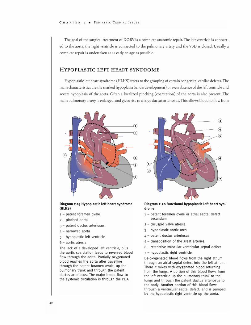
Hypoplastic left heart syndrome
Hypoplastic left heart syndrome (HLHS) refers to the grouping of certain congenital cardiac defects. The
main characteristics are the marked hypoplasia (underdevelopment) or even absence of the left ventricle and
severe hypoplasia of the aorta. Often a localized pinching (coarctation) of the aorta is also present. The
main pulmonary artery is enlarged, and gives rise to a large ductus arteriosus. This allows blood to flow from
C H A P T E R 2 ■ P E D I A T R I C C A R D I A C I S S U E S
40
Diagram 2.19 Hypoplastic left heart syndrome(HLHS)
1 – patent foramen ovale
2 – pinched aorta
3 – patent ductus arteriosus
4 – narrowed aorta
5 – hypoplastic left ventricle
6 – aortic atresia
The lack of a developed left ventricle, plusthe aortic coarctation leads to reversed bloodflow through the aorta. Partially oxygenatedblood reaches the aorta after travellingthrough the patent foramen ovale, up thepulmonary trunk and through the patent ductus arteriosus. The major blood flow tothe systemic circulation is through the PDA.
Diagram 2.20 Functional hypoplastic left heart syn-drome
1 – patent foramen ovale or atrial septal defectsecundum
2 – tricuspid valve atresia
3 – hypoplastic aortic arch
4 – patent ductus arteriosus
5 – transposition of the great arteries
6 – restrictive muscular ventricular septal defect
7 – hypoplastic right ventricle
De-oxygenated blood flows from the right atriumthrough an atrial septal defect into the left atrium.There it mixes with oxygenated blood returningfrom the lungs. A portion of this blood flows fromthe left ventricle up the pulmonary trunk to thelungs and through the patent ductus arteriosus tothe body. Another portion of this blood flowsthrough a ventricular septal defect, and is pumpedby the hypoplastic right ventricle up the aorta.

the right ventricle into the aorta and out to the body. Other characteristics of HLHS often include a combi-
nation of aortic and mitral stenosis or aortic and mitral atresia.
HLHS is also used to describe cases in which the right ventricle is hypoplastic and the child also suffers
from transposition of the great arteries. In such cases the hypoplastic right heart pumps into the same artery
that the left ventricle normally pumps blood through, and therefore the circulation is the same as in HLHS.
This is sometimes referred to as functional HLHS.
HLHS is a severe form of congenital heart disease. Without surgical intervention, HLHS is fatal. Infants
are often diagnosed within 24 to 48 hours of birth. Symptoms appear when the ductus arteriosus begins to
close.
There are three options for treating these children: supportive care until death occurs, staged recon-
struction of the heart, or a heart transplant.
Supportive careBefore surgical treatment was developed, at
least 90 per cent of infants with this condition
died by the age of one month. Even today, some
infants are not candidates for surgical therapy. For
these infants, supportive care is the only option.
Exactly what this care entails will depend on the
condition of the infant, but would likely include
assistance with breathing, the provision of fluids
and management of pain.
Reconstruction of the heartStaged reconstruction is the treatment of
choice for hypoplastic left heart syndrome.
Reconstruction takes place in three stages.
The first stage, known as a Norwood operation,
is undertaken as soon as possible after birth. This
stage calls for a homograft1 to be used to enlarge
the rudimentary aortic arch and then join it to the
pulmonary trunk. This is known as an anastomo-
sis. The pulmonary trunk will have been discon-
nected from the left and right pulmonary arteries.
The newly created blood vessel functions as the
T H E P E D I A T R I C C A R D I A C S U R G E R Y I N Q U E S T R E P O R T
41
Diagram 2.21 Stage 1 of hypoplastic left heart syndrome reconstruction (Norwood)
1 – Blalock-Taussig shunt (temporary)
2 – atrial septum removed
3 – patch where pulmonary trunk is disconnectedfrom left and right pulmonary artery
4 – aorta and pulmonary trunk anastomosedtogether and the aorta made larger
Blood flows through the anastomosed aorta andpulmonary trunk to the aortic arch. A shunt con-nects the aorta to the pulmonary arteries, providingthe lungs with blood to oxygenate.
1 A homograft is a graft of tissue between animals of the same species. In the case of humans, a homograft comes from a tissue donor. A homograft is in con-trast to an autograft, which is a graft of tissue transferred from another part of the patient’s body. With pulmonary homografts, the pulmonary artery isremoved from a tissue donor shortly after death. The pulmonary homograft is prepared and stored indefinitely at –70 degrees Celsius in liquid nitrogenuntil needed, when it is thawed and prepared under sterile conditions.)
patient’s aorta. The right ventricle then becomes a common ventricle, pumping blood through the aorta to
the rest of the body.
Blood flow to the lungs is provided by means of a modified Blalock-Taussig shunt joining the innomi-
nate artery to the pulmonary artery. This increases the flow of oxygen-enriched blood that will be sent to the
body. The Norwood operation also involves making a hole in the wall between the left and right atria. In
medical terms this is known as an atrial septectomy.
After the Norwood operation, children require medical management with various drugs. Some drugs,
such as digoxin, are needed to improve the strength of the heart’s contractions, and other drugs, such as
diuretics, are required to remove excess fluid from the body.
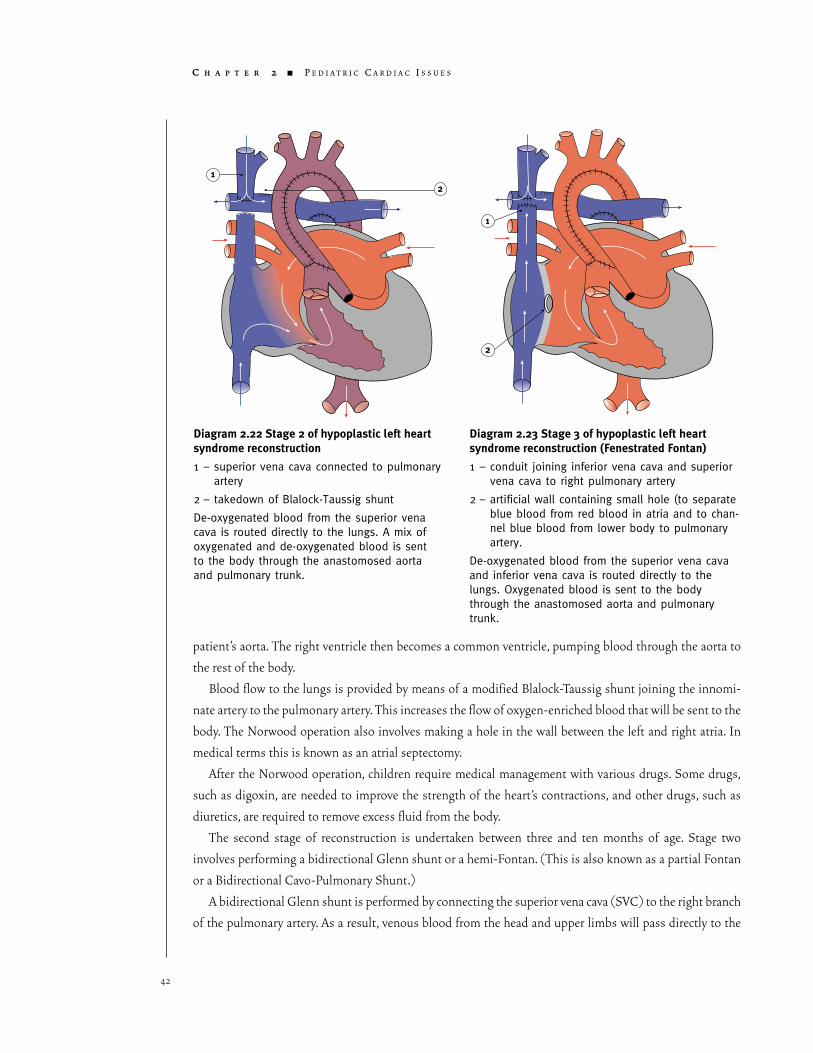
The second stage of reconstruction is undertaken between three and ten months of age. Stage two
involves performing a bidirectional Glenn shunt or a hemi-Fontan. (This is also known as a partial Fontan
or a Bidirectional Cavo-Pulmonary Shunt.)
A bidirectional Glenn shunt is performed by connecting the superior vena cava (SVC) to the right branch
of the pulmonary artery. As a result, venous blood from the head and upper limbs will pass directly to the
C H A P T E R 2 ■ P E D I A T R I C C A R D I A C I S S U E S
42
Diagram 2.22 Stage 2 of hypoplastic left heartsyndrome reconstruction
1 – superior vena cava connected to pulmonaryartery
2 – takedown of Blalock-Taussig shunt
De-oxygenated blood from the superior venacava is routed directly to the lungs. A mix ofoxygenated and de-oxygenated blood is sentto the body through the anastomosed aortaand pulmonary trunk.
Diagram 2.23 Stage 3 of hypoplastic left heart syndrome reconstruction (Fenestrated Fontan)
1 – conduit joining inferior vena cava and superiorvena cava to right pulmonary artery
2 – artificial wall containing small hole (to separateblue blood from red blood in atria and to chan-nel blue blood from lower body to pulmonaryartery.
De-oxygenated blood from the superior vena cavaand inferior vena cava is routed directly to thelungs. Oxygenated blood is sent to the bodythrough the anastomosed aorta and pulmonarytrunk.

lungs, bypassing the right ventricle. However, the venous blood from the lower half of the body will contin-
ue to return to the heart.
While avoiding the risk of failure of a complete Fontan operation, the partial Fontan helps to relieve symp-
toms. The procedure decreases the volume of blood delivered to the single ventricle, thus reducing the
amount of work that the ventricle must perform. Post-operatively, oxygen saturation is improved.
In addition, because the bi-directional Glenn is a low-pressure shunt, it does not carry the risk of caus-
ing thickening and hardening of the blood vessels of the lungs. (This is a normal response of the lung’s
blood vessels to high pressures.) This operation creates a more favorable setting in which to complete a
Fontan reconstruction at one and a half to two years of age.
At 18 to 24 months of age, if the patient does well after the Glenn shunt and pulmonary resistance
remains low, the third stage of reconstruction is possible. This involves the completion of the Fontan pro-
cedure (as described in the earlier discussion of tricuspid atresia).
With the Fontan operation, the venous blood from the lower part of the body is also diverted to the
lungs, thus creating the ‘complete’ Fontan circulation. However, if the child is not well or the pulmonary
resistance is considered too high, then no further surgical treatment is possible.
TransplantationHeart transplantation is carried out using the same basic techniques as for other transplants. In the case
of pediatric heart transplants, the greatest problem is that infant donor hearts are in short supply. Thus
transplantation is a treatment for only a limited number of babies with hypoplastic left heart syndrome. In
addition, recipients can survive only with the assistance of heavy doses of expensive medication to prevent
rejection of the transplanted tissue. These drugs also have significant side-effects, such as the risk of infec-
tion and lymphatic cancer, and must be taken by recipients for the rest of their lives. For those reasons, heart
transplants are not considered the standard treatment for children suffering from HLHS.
SUMMARY
The defects mentioned in this chapter are not an exhaustive list of each and every defect or lesion that
pediatric cardiac surgical programs are called upon to deal with. There are other defects not listed here that
would be seen by professionals involved in the care of children with heart problems.
Nor is this chapter to be taken as being an authoritative statement of what the defects involve and how
best they are treated. For such information, readers should refer to authoritative medical literature and texts.
In addition, it is also important to note that frequently there are individual variations in each of these
lesions. No two cases are alike.
This chapter is intended only to acquaint the reader with those defects that will receive the most atten-
tion in this Inquiry. As one reads through the following chapters, reference back to the information con-
tained in this chapter (as well as in the Glossary and in Chapter 3) may be helpful.
T H E P E D I A T R I C C A R D I A C S U R G E R Y I N Q U E S T R E P O R T
43

RISK AND PEDIATRIC
CARDIAC SURGERY
There is always risk involved in pediatric cardiac surgery. Various attempts have been made to estimate
the level of risk associated with surgically treating each of the known pediatric congenital heart lesions.
During the course of 1994, parents in Winnipeg were given estimates by the doctors involved as to how
many patients were likely to survive a particular procedure. At the time, there was no single book or list of
percentages that contained a definitive estimation of the risk for each procedure, although information was
available from pediatric cardiac surgeons and cardiologists working in other centres. Risk depends on many
factors, including the specifics of the patient’s condition, the skills and experience of the team, available
technology and the overall growth of medical knowledge. One variable that recent research has focused
attention on is the relationship between the numbers of cases a hospital deals with and results as measured
by mortality.
Two academic articles presented to this Inquest examined this question. The articles were “In-Hospital
Mortality for Surgical Repair of Congenital Heart Defects: Preliminary Observations of Variation by
Hospital Caseload,” by Kathy J. Jenkins MD MPH, Jane W. Newburger MD MPH, James E. Lock MD, Roger
B. Davis ScD, Gerald A. Coffman MSc and Lisa I. Iezzoni MD MSc, published in the journal Pediatrics
(Volume 95, Number 3 (March 1995), pages 323–330) and “Pediatric Cardiac Surgery: The Effect of
Hospital and Surgeon Volume on In-Hospital Mortality,” by Edward L. Hannan PhD, Michael Racz MS,
Rae-Ellen Kavey MD, Jan M. Quaegebeur MD PhD and Roberta Williams MD, published in the journal
Pediatrics (Volume 101, Number 6 (June 1998), pages 963–969).
Both articles came to similar conclusions. The Jenkins study made the following comment:
In our study population, the risk of dying in-hospital was much less for children who underwentsurgical correction of a congenital heart defect at institutions with the highest volume of suchpatients, in comparison with lower volume centres. Outcomes at the highest volume institutionsappeared clearly different than those for other institutions; below 300 cases annually, no consistenttrends in mortality by annual case volume were observed. (Jenkins et al, pages 327–328)
The Hannan study commented:
Findings of the study were that annual hospital volume and annual surgeon volume were both sig-nificantly related to inpatient mortality rates, even after controlling for patient age and several clini-cal risk factors in addition to procedure complexity. The maximal differentiation in mortality ratesbetween high and low-volume providers was 100 procedures annually for hospital and 75 proce-dures annually for surgeons. (Hannan et al, page 968)
This issue of the relationship between the number of procedures carried out in a pediatric cardiac sur-
gery program and the mortality rate is one that will be returned to later in this report.
C H A P T E R 2 ■ P E D I A T R I C C A R D I A C I S S U E S
44