co-expression of cd133r/cd44r in human colon cancer and ... et... · rosa divella,2 elvira foglia...
TRANSCRIPT

Co-Expression of CD133R/CD44R
in Human Colon Cancer andLiver MetastasisANTONIA BELLIZZI,1 SINTO SEBASTIAN,1 PASQUALE CEGLIA,1 MATTEO CENTONZE,1
ROSA DIVELLA,2 ELVIRA FOGLIA MANZILLO,3 AMALIA AZZARITI,4 NICOLA SILVESTRIS,5
SEVERINO MONTEMURRO,6 COSIMO CALIANDRO,6 RAFFAELE DE LUCA,6
GIUSEPPE CICERO,7 SERGIO RIZZO,7 ANTONIO RUSSO,7* MICHELE QUARANTA,2
GIOVANNI SIMONE,3 AND ANGELO PARADISO1
1Invasion and Metastatization Laboratory, Department of Experimental Oncology, National Cancer Centre ‘‘Giovanni Paolo II’’,
Bari, Italy2Clinical Pathology Laboratory, Department of Experimental National Cancer Centre ‘‘Giovanni Paolo II’’, Bari, Italy3Department of Pathology, National Cancer Centre ‘‘Giovanni Paolo II’’, Bari, Italy4Clinical and Preclinical Pharmacology Unit, Department of Experimental National Cancer Centre ‘‘Giovanni Paolo II’’, Bari, Italy5Medical and Experimental Oncology Unit, National Cancer Centre ‘‘Giovanni Paolo II’’, Bari, Italy6Department of Surgical Oncology, National Cancer Centre ‘‘Giovanni Paolo II’’, Bari, Italy7Section of Medical Oncology, Department of Surgical and Oncological Sciences, University of Palermo, Palermo, Italy
Although relatively good therapeutic results are achieved in non-advanced cancer, the prognosis of the advanced colon cancer still remainspoor, dependent on local or distant recurrence of the disease.Oneof the factors responsible for recurrence is supposed to be cancer stemcells (CSCs) or tumor-initiating cells, which are a population of cancer cells with ability to perpetuate themselves through self-renewaland to generate differentiated cells, thought to be responsible for tumor recurrence. This study globally approach the possible role oftissue-derived stemcells in the initiation of colon cancer and itsmetastatic process in the liver. Fresh surgical specimens fromcolon cancer,non-tumor tissue and liver metastasis were obtained directly from the operating room, examined, and immediately processed. CSCswere selected under serum-free conditions and characterized by CD44 and CD133 expression levels. CD133þ/CD44þ cell populationswere then investigated in paraffin-embedded tissues and circulating tumor cells isolated from peripheral blood of the same group of coloncancer patients. Our data demonstrate that metastatic properties of cell populations from blood and liver metastasis, differently fromprimitive tumors, seem to be strictly related to the phenotype CD133 positive and CD44 positive.J. Cell. Physiol. 228: 408–415, 2013. � 2012 Wiley Periodicals, Inc.
Colon cancer is the second leading cause of cancer-relateddeath. The majority of these deaths are due to metastasis, withthe liver easily the most common site of deposit (LeGolvan andResnick, 2010).
Combining surgery and chemotherapy in the treatment ofpatients with colon hepatic metastases is increasingly becomingthe standard of care. However, controversy remains regardingthe juxtapositioning of chemotherapy and surgery, the durationof chemotherapy, and particularly, the use of preoperativechemotherapy in the treatment of patients with initiallyresectable metastases (Nordlinger et al., 2010).
Cancermetastasis has been explained by at least twomodels:a progression model and an initiating model. In the former,metastatic capacity is acquired during cancer progression in asubpopulation of cells through sequential genetic mutationsor epigenetic alterations (Gray, 2010), in genes associatedwith proteolysis of local extracellular matrix attachments(Nagashima et al., 1997), adhesive alterations (Furger et al.,2001), angiogenesis (Weber, 2008), viable vasculardissemination, distant embolization, and survival in a newenvironment (Folkman, 1990; Hynes, 2003; Bird et al., 2006).However, not all of these genetic alterations occur duringthe process of liver metastasis (Gray, 2010). In the initiationmodel, cells with metastatic potential are determined by earlymutational events in a progenitor cell, named cancer stem cell
(CSCs) or tumor-initiating cells (TIC; Clarke and Fuller, 2006;Polyak and Hahn, 2006; Odoux et al., 2008), even if few datasupport the hypothesis of its role in the colon metastaticprocess in humans (Horst et al., 2009b; Puglisi et al., 2009;Ju et al., 2011).
CD44 andCD133 have already been validated as informativemarkers of stem cells in both primary tumors and xenografts(O’Brien et al., 2007; Ricci-Vitiani et al., 2007). CD133,originally known as AC133, is a glycoprotein also known inhumans and rodents as Prominin 1 (PROM1; Kawamoto et al.,
Contract grant sponsor: Italian Ministry of Health, ‘‘ProgrammaIntegrato Oncologia (PIO) 2007’’.
*Correspondence to: Antonio Russo, Section of Medical Oncology,Department of Surgical and Oncological Sciences, Universita diPalermo, Via del Vespro 127, 90127 Palermo, Italy.E-mail: [email protected]
Manuscript Received: 19 March 2012Manuscript Accepted: 18 June 2012
Accepted manuscript online in Wiley Online Library(wileyonlinelibrary.com): 27 June 2012.DOI: 10.1002/jcp.24145
ORIGINAL RESEARCH ARTICLE 408J o u r n a l o fJ o u r n a l o f
CellularPhysiologyCellularPhysiology
� 2 0 1 2 W I L E Y P E R I O D I C A L S , I N C .

2010). It is the founding member of pentaspan transmembraneglycoproteins (5-transmembrane, 5-TM), whichwas specificallyregarded as the colon CSC-surface markers. CD133 has beenused to identify and isolate cancer initiating cells from humancolon cancer and it has been demonstrated that CD133þ cellsare able to maintain themselves as well as differentiate andre-establish tumor heterogeneity upon serial transplantation invivo (O’Brien et al., 2007; Ricci-Vitiani et al., 2007). CD44 is acell surface glycoprotein involved in cell–cell interactions, celladhesion, and migration. It was identified as the cell-surfacemarkers of breast cancer and also recently observed to berelated to the distant metastasis of colon cancer (Huang et al.,2012).
Currently, there are some studies addressing theCD133 andCD44 co-expression of CSC antigens in colon cancer patients(Horst et al., 2009a; Salnikov et al., 2009). CD44/CD133co-expression was significantly higher in colon cancers withearly liver metastases compared to those without early livermetastases and co-expressionwas also associatedwith poorestprognosis (Huang et al., 2012), suggesting that CD133 andCD44 proteins co-expression in colon cancer may be apotential biomarker for early liver metastases. Indeed, Chenet al. (2011) demonstrated that FACS sorted CD133þ/CD44þ
HCT116 cells are undifferentiated, endowed with extensiveself-renewal and epithelial lineage differentiation capacity invitro, more invasive in vitro and responsible solely for livermetastasis in vivo. However, CD133 and CD44 expressionstatus and its relation to liver metastasis is still controversial.
A recent study (Hou et al., 2011) showed the critical role ofCD133þ/CD44þ tumor cells in hematogenous metastasis ofliver cancers, underlying that CD133 is responsible for tumorgrowth and CD44 is important for invasion; two importantfactors in tumor metastasis. Their data also suggest thatCD133þ cells act as TIC-like populations, however were notnecessarily highly invasive, and that CD44 was a marker ofan invasive, but not tumorigenic, sub-population. In additionto CD133, CD44 has a critical role in tumor metastasis.Moreover, based on studies of HCC cell lines, CD133þ andCD44þ were proposed to be markers of TICs in liver cancers(Zhu et al., 2010). Therefore, CD133 and CD44 could beregarded asmarkers ofmetastasis-facilitating pathways throughwhich liver tumor metastasis is jointly promoted.
Therefore, starting from these observations, in this study wehave generated stem cell-enriched human colonospherecultures from fresh samples deriving from the same group ofcolon cancer and liver metastasis specimens under serum-freeculture conditions, comparing the sphere forming potentialof the clones isolated from the primary culture comingfrom both compartments and the differences observed inthe corresponding non-tumor counterpart, adjacent to theneoplastic lesion. We have established long-term culturespheres from the three compartments, analyzing theexpression of CD44 and CD133.
Indeed, we have examined colon cancer specimens andcirculating tumor cells (CTC) to investigate the CD133 andCD44 expression and localization.
Materials and MethodsPatients
Biological tissues for laboratory assays were obtained from21 patients with a first diagnosis of primary colon cancer with(n¼ 11) or without (n¼ 10) liver metastasis. All patients wereconsecutively treated with primary surgery at the GastrointestinalSurgery Unit of NCC Bari, Italy. Before undergoing routinesurgery, all patients signed an informed consent authorizing theInstitute to use their removed biological tissues for researchpurposes. Routine staging procedures were adopted for diseasestaging according to UICC criteria (Greene and Sobin, 2002). Just
after surgical removal of the colon and, if any, of liver metastasis,tissues were macroscopically analyzed by the pathologist whoperformed a sampling from the primary tumor, from adjacentnormal mucosa (located not less than 10 cm from the primary site),and from liver metastasis. H&E staining confirmed the diagnosis ofcolon cancer in all primary and metastatic sites as well the absenceof cancer cells in normal mucosa. About 100mg of tissue withoutfat and necrosis was placed in sterile tubes with culture mediumon ice. Patients with liver metastasis were categorized as themetastatic group (M1); those without liver metastases as the non-metastatic group (M0).
Isolation and expansion of colon stem cell cultures
After sampling, biological samples were processed as describedpreviously (Cammareri et al., 2008). The obtained tissue fragmentswere washed extensively with PBS and were mechanicallydissociated using scalpels and vigorous trituration to yield smallfragments (<1mm3) and single cells. Enzymatic digestion wasperformed using collagenase/hyaluronidase (StemcellTechnologies, Vancouver, Canada) in DMEM/F12 containing 5mMHepes (Sigma, St. Louis, MO) for 2 h at 378C. The suspension wasthen filtered through a 100-mm pore size to separate the tissuefragments from the single cells. The single cell suspension wascultured in advanced DMEM/F12 (Life Technologies, Monza, Italy)supplemented with 0.6% BSA (Sigma), 0.6% glucose (Sigma), 2mML-glutamine (Euroclone, SPA, Pero, Milano), 9.6mg/ml putrescin(Sigma), 6.3 ng/ml progesterone (Sigma), 5.2 ng/ml sodium selenite(Sigma), 25mg/ml insulin (Sigma), 100mg/ml apotransferrin (Sigma),5mM Hepes (Sigma), 10ml antibiotic–antimycotic (LifeTechnologies, Monza, Italy), and 4mg/ml Heparin (Sigma). Growthfactors [20 ng/ml EGF (Peprotech, London, UK) and 10 ng/ml b-FGF (Peprotech)] were added to the cell culture medium freshlyeach week. All cell culture was carried out in non-tissue culturetreated petri at 378C in a 5% CO2 humidified incubator.
The viable cells were counted using a light microscope bytrypan blue dye exclusion test at day 7. Surviving immature tumorcells slowly proliferate, and grow as non-adherent spheres. Theformation of such spheres, containing about 50 cells, takes 2months. Sphere formation was scored as follows: (�) no sphereobserved; (þ) two spheres per field.
Cytospin cell preparation and immunocytochemical staining ofCD44 and CD133
Cells were centrifuged at 800 rpm for 5min, washed with PBS,and re-suspended in PBS. The cells were attached to coatedmicroscope slides (Bio Optica, Milano, Italy) in a Cyto-Tekcentrifuge (Sakura Finetek, Alphen Aan Den Rijn, Netherlands) at2,000 rpm for 10min and dried overnight on a slide warmer at378C. They were then fixed with acetone and stained. Primaryantibodies specific for CD133 (Abcam, Cambridge, UK), CD44(DakoCytomation, Glostrup, Denmark) were used, withprimary antibody binding detected using corresponding DakoEnVision System-HRP Labelled Polymer secondary antibodies(DakoCytomation) and AEC Substrate Chromogen(DakoCytomation). The cells were counterstained with Mayer’shematoxylin (DakoCytomation). After incubation, cells werewashed with PBS, and incubated with biotinylated link for 30min,peroxidase-labeled streptavidin for 30min and 3-amino-9-ethylcarbazole substrate-chromogen (Labelled Streptavidin-Biotin2 System-Horseradish Peroxidase; DakoCytomation) for15min in the dark. After PBS washing, the slides werecounterstained with hematoxylin and mounted with aqueousmounting medium (DakoCytomation). For negative controls, theprimary antibody was omitted and replaced by PBS.
Photomicrographs of cytospin preparations were acquiredunder bright field illumination with a Leica DMLB opticalmicroscope (Leica, Cambridge, UK) and analyzed with LeicaIM1000 software.
JOURNAL OF CELLULAR PHYSIOLOGY
C S C s I N P R I M A R Y A N D M E T A S T A T I C S I T E 409

Immunohistochemical staining of CD44 and CD133
Double immunostaining with antibodies for detection of CD44 andCD133 was performed on paraffin-embedded tissue sections andcytospins, using primary antibodies specific for CD133 (Abcam),CD44 (DakoCytomation), and Alexa 488 goat anti-rabbit andthe Alexa 568 goat anti-mouse immunoglobulin G1 secondaryantibodies conjugate (Invitrogen). Tissue sections were thenmounted with aqueous mounting medium (DakoCytomation). Fornegative controls, the primary antibody was omitted and replacedby PBS. Images were obtained on a BX40 microscope (Olympus,Tokyo, Japan) with a SenSys 1401E Photometrics CCD camera.
Immunohistochemical evaluation
The scoring was performed twice by two persons in a blindedfashion. CD133 and CD44 staining was detected mainly in themembrane andwas evaluated as the percentage of immunoreactivecells with respect to the total analyzed. The result was the mean of15–20 random field at 40� magnification.
Collection of blood samples
Fifteen milliliters of peripheral blood were collected from eachparticipant in a vacutainer system with lithium-heparin beforesurgery. A written consent should be obtained from all patientsprior to enrolment in the study, and the Ethical Committee of theNCI approved the protocol which was in accord with the ethicalguidelines of the 1975 Declaration of Helsinki Whole. A controlgroup found to be healthy from laboratory data was enrolledamong donors (N¼ 30). Their median age was 50 years (range:40–70 years).
Magnetic labeling
Fifty milliliters of anti-coagulated bloodwere centrifugedwith 400gfor 35min. Buffy coat were collected into 50-ml conical tubes. Formagnetic labeling, the cells were first permeabilized using CellPermSolution (40ml) from the Carcinoma Cell Enrichment Kit (MiltenyiBiotec, Bergisch Gladbach, Germany) and incubated for 5minat room temperature. Fixation was done using 5ml of Cell FixSolution for 30min at room temperature. The cells were thenwashed twice in dilution buffer. Two hundred microliters of FcRBlocking Reagent (Miltenyi Biotec) was used for the preventionof unspecific binding. Two hundred microliters of anti-cytokeratin
(7/8) Micro Bead was used for magnetic labeling. The magneticlabeling was performed at room temperature for 45min. Finally,the cells werewashedonce in cell stain solution and resuspended in1ml of dilution buffer for magnetic cell separation.
Magnetic separation of cytokeratin 7/8-positive tumor cells
For magnetic separation, the labeled cells were passed throughMSþ (Miltenyi Biotec) separation columns that had beenequilibratedwith dilution buffer. The negative cellswerewashedoffthe column with 3ml of dilution buffer. The retained cells wereeluted from the column outside the magnetic field by pipetting 1mlof dilution buffer onto the column and the cellular suspension wasfiltered through a mesh with 30-mm diameter holes.
Immuncytochemical labeling and microscopic analysis
Cells were magnetically separated as described above, and theenriched cell fraction was spun down on slides using acytocentrifuge. Slides were air-dried overnight at roomtemperature. Fast Red TR/Naphthol AS-MX Substrate Solutionwas added to cell spots for 15min in humidity chamber at 258C.Slides were then stained with dilute Meyer’s hemalum solution 1:2in 100mM of Tris–HCl, pH 8.2, washed twice for 1min in doubledistilled water, and coverslipped using glycerin plus PBS (1:1).
Results
Tumor tissues of 21 patients with primary colon cancer,togetherwithmatched adjacent normal tissues, were collected;11 patients had also liver metastasis. Clinical pathologicalcharacteristics and biological specimens of each patient aresummarized in Table 1. Fresh tissues (Fig. 1A) were processedand after dissection, viable cells were obtained from 17 out of18 tumor tissues, 11 out of 18 normal mucosa, and 11 out of 11livermetastasis (Table 2).We evaluated the growth potential ofthese primary cells under serum-free conditions. Firstgeneration of colon spheres (Fig. 1B)were successfully culturedin 11 out of 17 samples from tumors, in 9 out of 11 frommetastasis and in 6 out of 11 from adjacent normal tissues(Table 2). To assess whether the ability to form spheres wasmaintained during the time of culture, cells were expanded andafter 5months (Table 2) 5 out of 17 samples from tumor tissues,6 out of 11 from liver metastasis, and 2 out of 11 from non-
TABLE 1. Summary of patient population, tumor sample information, and phenotype characteristics
Sample Age/sexTumorstage Grade
Tumor Liver metastasis Normal tumor adjacent tissue
Vitality Spheres CSCs CD133 CD44 Vitality Spheres CSCs CD133 CD44 Vitality Sphere SCs CD133 CD44
1 66/M T2N0M0 2 þ þ � � þ þ þ � � �2 70/M T2N0M0 2 þ � � � þ þ þ � � �3 64/M T3N0M0 2 þ � � � þ �4 81/F T3N2M0 3 þ � � � þ þ þ þ � þ5 68/M T3N0M0 3 þ � � � � þ � � � �6 68/M T4N2M0 3 þ � � � � þ � � � �7 62/M T2N0M0 2 þ þ � � � þ þ � � þ8 42/M T3N1M0 3 þ þ � þ þ þ � � � þ9 82/M T4N2M0 3 þ þ þ þ � �10 70/M T3N1M0 3 þ þ þ þ � þ � � � þ11 63/F T4N1M1 3 þ þ þ þ þ þ þ þ þ þ þ þ þ � þ12 52/M T3N2M1 2 þ þ þ þ þ þ þ þ þ þ �13 87/F T4N2M1 2 þ þ þ þ þ þ þ þ þ þ �14 73/M T3N2M1 3 � þ þ þ þ þ �15 67/M T4N2M1 2 þ þ � þ þ þ þ þ þ þ þ þ � � þ16 72/M T3N2M1 3 þ þ � þ þ þ þ � þ þ �17 61/F T3N2M1 2 þ þ � þ þ þ � � þ þ �18 63/F T4N0M1 3 þ � � þ þ þ þ � þ þ þ � � � �19 66/F T4N2M1 3 þ þ � � �20 64/F T3N1M1 2 þ � � þ þ21 74/M T3N1M1 3 þ þ þ þ þ
F, female; M, male.
JOURNAL OF CELLULAR PHYSIOLOGY
410 B E L L I Z Z I E T A L .

tumor tissues were able to form spheroids of second and thirdgeneration (Fig. 1C).
The presence of CD44þ cells and CD133þ cells wasevaluated at two isolation steps (Table 2): on the cellsuspensions obtained 1 week after sample processing,representative of naıve samples (Fig. 2A), and in clones obtainedafter 5 months of cell selection (Fig. 3). CD44 was detectable in13 out of 17 samples from primary tumors, in 6 out of 11 fromnormal adjacent mucosa, and in 10 out of 11 from metastasis.All non-tumor specimens were CD133�, whereas 11 out of
17 tumor and 10 out of 11 metastasis samples were CD133þ.CD133þ cells were significantly higher in the tumorcompartment than in the metastatic compartment, and higherin liver metastasis than in non-tumor tissue (21.6� 1.8 vs.5.4� 1.09 vs. 0.56� 0.3). The CD44þ cells were significantlyhigher in the liver metastasis than tumor and non-tumorcompartment (12.6� 1.19 vs. 10.7� 1.37 vs. 9.18� 2.2). Then,we confirmedCD44 andCD133 expression levels in the clonesobtained after 5 months of cells selection (Fig. 3) and wefound that 100% of the CSCs isolated from the non-tumor
Fig. 1. A:Freshtissuesofprimarycoloncancer(T),matchedadjacentnormaltissues(NT),andlivermetastasis (MET).B:Phasecontrastphotooffirst and (C) third generation colon spheres growth under serum-free conditions (40T).
TABLE 2. Summary of patients phenotype and functional characteristics
NT T Liver metastasis
Number 18 18 11Naive sample
Vitality 11/18 (61%) 17/18 (94.4%) 11/11 (100%)Expression markers/patients
CD133 0/11 (0%) 11/17 (64.7%) 10/11 (90.9%)CD44 6/11 (54.5%) 13/17(76.5%) 10/11 (90.9%)
Markers expression mean%CD133 positive cells 0.56� 0.3 21.6� 1.8 5.4� 1.09%CD44 positive cells 9.18� 2.2 10.7� 1.37 12.6� 1.19
Sphere production (20 days) 6/11 (54.5%) 11/17 (64.7%) 9/11 (81.8%)SCs isolated after 5 months cultured 2/11(18.2%) 5/17 (29.4%) 6/11 (54.5%)
SCs phenotypeCD133þ/CD44þ 0 3/5 (60%) 6/6 (100%)CD133þ/CD44� 0 2/5 (40%) 0CD133�/CD44þ 2/2 (100%) 0 0CD133�/CD44� 0 0 0
P-e sampleCD133þ/CD44þ cells/patients 0/18 (0%) 9/18 (50%) 10/11 (90.9)% CD133þ/CD44þ cells 1.3� 0.2 13.8� 1.1 8.5� 0.7
P-e, paraffin-embedded; SCs, stem cells.
JOURNAL OF CELLULAR PHYSIOLOGY
C S C s I N P R I M A R Y A N D M E T A S T A T I C S I T E 411

compartments were CD133�/CD44þ; CSCs isolated from thetumor compartmentwereCD133þ/CD44þ in 60%of cases andCD133þ/CD44� in 40%; CSCs from metastasis were allCD44þ/CD133þ.
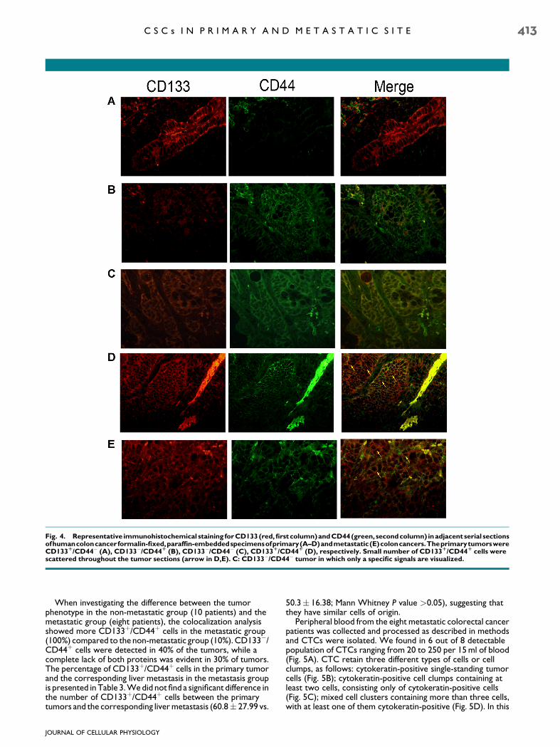
We also performed immunofluorescence analysis todetermine the proportion of CD133þ/CD44þ putativetumorigenic cells within each tumor, scanning for the presenceof the FITC and TRITC staining in the same cell (Fig. 4).Immunohistochemical staining for CD133 (red, first column)and CD44 (green, second column) showed membranelocalization of the two proteins in epithelial tumor cells
localized at the base of the crypt (Fig. 4B–D), or in tumor cellsscattered throughout the tumor sections (Fig. 4A). Smallnumber of CD133þ/CD44þ cells were observed only inprimary tumors of patients who develop liver metastasis(Fig. 4D).Metastasis consists of cancer cells showingmembraneCD133/CD44 accumulation (Fig. 4E).
We evaluated the difference in the presence of CD44þ/CD133þ cells and found that are significantly higher in thetumor compartment than in the metastatic once and higher inliver metastasis than in non-tumor tissue (13.8� 1.1 vs.8.5� 0.7 vs. 1.3� 0.2).
Fig. 2. CD44 and CD133 expression levels in tumor cells clusters (20T). CD44 and CD133 expression were evaluated. A,B: One week aftersamples processing in tumor cells clusters.
Fig. 3. Representative CD44 and CD133 staining in colon spheres growth for 5 months under serum-free conditions.
JOURNAL OF CELLULAR PHYSIOLOGY
412 B E L L I Z Z I E T A L .
When investigating the difference between the tumorphenotype in the non-metastatic group (10 patients) and themetastatic group (eight patients), the colocalization analysisshowed more CD133þ/CD44þ cells in the metastatic group(100%) compared to the non-metastatic group (10%). CD133�/CD44þ cells were detected in 40% of the tumors, while acomplete lack of both proteins was evident in 30% of tumors.The percentage of CD133þ/CD44þ cells in the primary tumorand the corresponding liver metastasis in the metastasis groupis presented in Table 3.Wedid not find a significant difference inthe number of CD133þ/CD44þ cells between the primarytumors and the corresponding livermetastasis (60.8� 27.99 vs.
50.3� 16.38; Mann Whitney P value >0.05), suggesting thatthey have similar cells of origin.
Peripheral blood from the eight metastatic colorectal cancerpatients was collected and processed as described in methodsand CTCs were isolated. We found in 6 out of 8 detectablepopulation of CTCs ranging from 20 to 250 per 15ml of blood(Fig. 5A). CTC retain three different types of cells or cellclumps, as follows: cytokeratin-positive single-standing tumorcells (Fig. 5B); cytokeratin-positive cell clumps containing atleast two cells, consisting only of cytokeratin-positive cells(Fig. 5C); mixed cell clusters containing more than three cells,with at least one of them cytokeratin-positive (Fig. 5D). In this
Fig. 4. Representative immunohistochemical staining forCD133(red,firstcolumn)andCD44(green, secondcolumn) inadjacent serial sectionsofhumancoloncancerformalin-fixed,paraffin-embeddedspecimensofprimary(A–D)andmetastatic(E)coloncancers.TheprimarytumorswereCD133R/CD44� (A), CD133�/CD44R (B), CD133�/CD44� (C), CD133R/CD44R (D), respectively. Small number of CD133R/CD44R cells werescattered throughout the tumor sections (arrow in D,E). C: CD133�/CD44� tumor in which only a specific signals are visualized.
JOURNAL OF CELLULAR PHYSIOLOGY
C S C s I N P R I M A R Y A N D M E T A S T A T I C S I T E 413
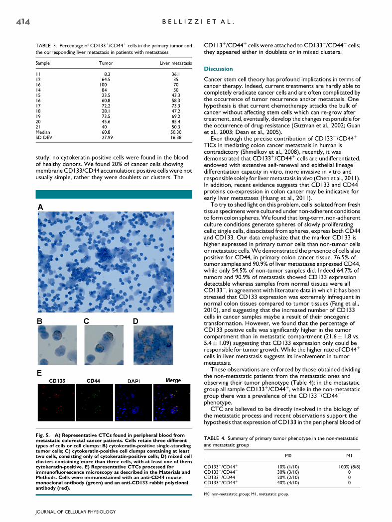
study, no cytokeratin-positive cells were found in the bloodof healthy donors. We found 20% of cancer cells showingmembrane CD133/CD44 accumulation; positive cells were notusually simple, rather they were doublets or clusters. The
CD113þ/CD44þ cells were attached to CD133�/CD44� cells;they appeared either in doublets or in mixed clusters.
Discussion
Cancer stem cell theory has profound implications in terms ofcancer therapy. Indeed, current treatments are hardly able tocompletely eradicate cancer cells and are often complicated bythe occurrence of tumor recurrence and/or metastasis. Onehypothesis is that current chemotherapy attacks the bulk ofcancer without affecting stem cells which can re-grow aftertreatment, and, eventually, develop the changes responsible forthe occurrence of drug-resistance (Guzman et al., 2002; Guanet al., 2003; Dean et al., 2005).
Even though the precise contribution of CD133þ/CD44þ
TICs in mediating colon cancer metastasis in human iscontradictory (Shmelkov et al., 2008), recently, it wasdemonstrated that CD133þ/CD44þ cells are undifferentiated,endowed with extensive self-renewal and epithelial lineagedifferentiation capacity in vitro, more invasive in vitro andresponsible solely for livermetastasis in vivo (Chen et al., 2011).In addition, recent evidence suggests that CD133 and CD44proteins co-expression in colon cancer may be indicative forearly liver metastases (Huang et al., 2011).
To try to shed light on this problem, cells isolated from freshtissue specimenswere cultured under non-adherent conditionsto form colon spheres.We found that long-term, non-adherentculture conditions generate spheres of slowly proliferatingcells; single cells, dissociated from spheres, express both CD44and CD133. Our data emphasize that the marker CD133 ishigher expressed in primary tumor cells than non-tumor cellsor metastatic cells.We demonstrated the presence of cells alsopositive for CD44, in primary colon cancer tissue. 76.5% oftumor samples and 90.9% of liver metastases expressed CD44,while only 54.5% of non-tumor samples did. Indeed 64.7% oftumors and 90.9% of metastasis showed CD133 expressiondetectable whereas samples from normal tissues were allCD133�, in agreement with literature data in which it has beenstressed that CD133 expression was extremely infrequent innormal colon tissues compared to tumor tissues (Fang et al.,2010), and suggesting that the increased number of CD133cells in cancer samples maybe a result of their oncogenictransformation. However, we found that the percentage ofCD133 positive cells was significantly higher in the tumorcompartment than in metastatic compartment (21.6� 1.8 vs.5.4� 1.09) suggesting that CD133 expression only could beresponsible for tumor growth.While the higher rate of CD44þ
cells in liver metastasis suggests its involvement in tumormetastasis.
These observations are enforced by those obtained dividingthe non-metastatic patients from the metastatic ones andobserving their tumor phenotype (Table 4): in the metastaticgroup all sample CD133þ/CD44þ, while in the non-metastaticgroup there was a prevalence of the CD133þ/CD44�
phenotype.CTC are believed to be directly involved in the biology of
the metastatic process and recent observations support thehypothesis that expression of CD133 in the peripheral blood of
TABLE 3. Percentage of CD133þ/CD44þ cells in the primary tumor and
the corresponding liver metastasis in patients with metastases
Sample Tumor Liver metastasis
11 8.3 36.112 64.5 3516 100 7014 84 5015 23.5 43.316 60.8 58.317 72.2 73.318 28.1 47.219 73.5 69.220 45.6 85.421 40 50.3Median 60.8 50.30SD DEV 27.99 16.38
Fig. 5. A) Representative CTCs found in peripheral blood frommetastatic colorectal cancer patients. Cells retain three differenttypes of cells or cell clumps: B) cytokeratin-positive single-standingtumor cells; C) cytokeratin-positive cell clumps containing at leasttwo cells, consisting only of cytokeratin-positive cells; D) mixed cellclusters containing more than three cells, with at least one of themcytokeratin-positive. E) Representative CTCs processed forimmunofluorescence microscopy as described in the Materials andMethods. Cells were immunostained with an anti-CD44 mousemonoclonal antibody (green) and an anti-CD133 rabbit polyclonalantibody (red).
TABLE 4. Summary of primary tumor phenotype in the non-metastatic
and metastatic group
M0 M1
CD133þ/CD44þ 10% (1/10) 100% (8/8)CD133�/CD44� 30% (3/10) 0CD133þ/CD44� 20% (2/10) 0CD133�/CD44þ 40% (4/10) 0
M0, non-metastatic group; M1, metastatic group.
JOURNAL OF CELLULAR PHYSIOLOGY
414 B E L L I Z Z I E T A L .

patients affected with CRC might identify high-risk patients bydetecting putative circulating CSCs that might be responsiblefor disease progression after apparently radical surgery (Pilatiet al., 2012). We found detectable population of CTCs in 75%of metastatic colon cancer patients: 20% of CTCs showedmembrane CD133�/CD44þaccumulation; positive cells werenot usually simple, rather they were doublets or clusters. TheCD113þ/CD44þ cells were attached to CD133�/CD44� cells;they appeared either in doublets or in mixed clusters. Ouranalysis supports the premises that CTCs represent a samplingof the phenotypic cell types present in the primary andmetastatic tumor deposits, and the possibility that CTCsconsist of a combination of cells with malignant potential, stemcell characteristics and actively migrating cells that may go on toform liver metastatic foci.
Collectively, these results suggest thatCD133þ colon cancercells might play an important role in both primary tumors aswell as in metastatic lesions but metastasis of the liver seems tobe strictly related to the phenotype CD133þ and CD44þ. Ourdata underline the importance of CD133þ/CD44þ CSCs inliver metastasis thus warranting further studies on the role(s)of this subset of cells in the liver metastatic process.
Acknowledgments
This study was partially supported by the Italian Ministry ofHealth, ‘‘Programma Integrato Oncologia (PIO) 2007.’’
Literature Cited
Bird NC, Mangnall D, Majeed AW. 2006. Biology of colorectal liver metastases: A review.J Surg Oncol 94:68–80.
Cammareri P, Lombardo Y, FrancipaneMG, Bonventre S, TodaroM, Stassi G. 2008. Isolationand culture of colon cancer stem cells. Methods Cell Biol 86:311–324.
Chen KL, Pan F, Jiang H, et al. 2011. Highly enriched CD133(þ)CD44(þ) stem-like cells withCD133(þ)CD44(high) metastatic subset in HCT116 colon cancer cells. Clin ExpMetastasis 28:751–763.
Clarke MF, Fuller M. 2006. Stem cells and cancer: Two faces of eve. Cell 124:1111–1115.Dean M, Fojo T, Bates S. 2005. Tumour stem cells and drug resistance. Nat Rev Cancer5:275–284.
FangDD,KimYJ, LeeCN, et al. 2010. ExpansionofCD133(þ) colon cancer cultures retainingstem cell properties to enable cancer stem cell target discovery. Br J Cancer 102:1265–1275.
Folkman J. 1990.What is the evidence that tumors are angiogenesis dependent? JNatl CancerInst 82:4–6.
Furger KA, Menon RK, Tuck AB, Bramwell VH, Chambers AF. 2001. The functional andclinical roles of osteopontin in cancer and metastasis. Curr Mol Med 1:621–632.
Gray J. 2010. Cancer: Genomics of metastasis. Nature 464:989–990.Greene FL, Sobin LH. 2002. The TNM system: Our language for cancer care. J Surg Oncol80:119–120.
Guan Y, Gerhard B, Hogge DE. 2003. Detection, isolation, and stimulation of quiescentprimitive leukemic progenitor cells from patients with acute myeloid leukemia (AML).Blood 101:3142–3149.
Guzman ML, Swiderski CF, Howard DS, et al. 2002. Preferential induction of apoptosis forprimary human leukemic stem cells. Proc Natl Acad Sci USA 99:16220–16225.
Horst D, Kriegl L, Engel J, Kirchner T, Jung A. 2009a. Prognostic significance of the cancerstem cell markers CD133, CD44, and CD166 in colorectal cancer. Cancer Invest 27:844–850.
Horst D, Scheel SK, Liebmann S, Neumann J, Maatz S, Kirchner T, Jung A. 2009b. The cancerstem cell marker CD133 has high prognostic impact but unknown functional relevance forthe metastasis of human colon cancer. J Pathol 219:427–434.
Hou Y, Zou Q, Ge R, Shen F, Wang Y. 2011. The critical role of CD133(þ)CD44(þ/high)tumor cells in hematogenous metastasis of liver cancers. Cell Res 22:259–272.
Huang X, Sheng Y, Guan M. 2012. Co-expression of stem cell genes CD133 and CD44 incolorectal cancers with early liver metastasis. Surg Oncol 21:103–107.
Hynes RO. 2003. Metastatic potential: Genetic predisposition of the primary tumor or rare,metastatic variants-or both? Cell 113:821–823.
Ju HX, An B, Okamoto Y, Shinjo K, et al. 2011. Distinct profiles of epigenetic evolutionbetween colorectal cancers with and without metastasis. Am J Pathol 178:1835–1846.
Kawamoto H, Yuasa T, Kubota Y, et al. 2010. Characteristics of CD133(þ) human coloncancer SW620 cells. Cell Transplant 19:857–864.
LeGolvan MP, Resnick M. 2010. Pathobiology of colon cancer hepatic metastases with anemphasis on prognostic factors. J Surg Oncol 102:898–908.
Nagashima Y, Hasegawa S, Koshikawa N, et al. 1997. Expression of matrilysin in vascularendothelial cells adjacent to matrilysin-producing tumors. Int J Cancer 72:441–445.
Nordlinger B, Vauthey JN, PostonG, Benoist S, Rougier P, VanCutsem E. 2010. The timing ofchemotherapy and surgery for the treatment of colon liver metastases. Clin ColorectalCancer 9:212–218.
O’Brien CA, Pollett A, Gallinger S, Dick JE. 2007. A human colon cancer cell capable ofinitiating tumour growth in immunodeficient mice. Nature 445:106–110.
Odoux C, Fohrer H, Hoppo T, et al. 2008. A stochastic model for cancer stem cell origin inmetastatic colon cancer. Cancer Res 68:6932–6941.
Pilati P, Mocellin S, Bertazza L,Galdi F, BriaravaM,MammanoE, Tessari E, ZavagnoG,Nitti D.2012. Prognostic value of putative circulating cancer stem cells in patients undergoinghepatic resection for colorectal liver metastasis Ann Surg Oncol 19:402–408.
Polyak K, Hahn WC. 2006. Roots and stems: Stem cells in cancer. Nat Med 12:296–300.Puglisi MA, SgambatoA, SaulnierN, et al. 2009. Isolation and characterizationofCD133þ cellpopulationwithin human primary andmetastatic colon cancer. Eur RevMed Pharmacol Sci13:55–62.
Ricci-Vitiani L, Lombardi DG, Pilozzi E, et al. 2007. Identification and expansion of humancolon-cancer-initiating cells. Nature 445:111–115.
Salnikov AV, Kusumawidjaja G, Rausch V, et al. 2009. Cancer stem cell marker expression inhepatocellular carcinoma and liver metastases is not sufficient as single prognosticparameter. Cancer Lett 275:185–193.
Shmelkov SV, Butler JM, Hooper AT, et al. 2008. CD133 expression is not restricted to stemcells, and both CD133þ and CD133�metastatic colon cancer cells initiate tumors. J ClinInvest 118:2111–2120.
Weber GF. 2008. Molecular mechanisms of metastasis. Cancer Lett 270:181–190.Zhu Z, Hao X, Yan M, Yao M, Ge C, Gu J, Li J. 2010. Cancer stem/progenitor cells are highlyenriched in CD133þ CD44þ population in hepatocellular carcinoma. Int J Cancer126:2067–2078.
JOURNAL OF CELLULAR PHYSIOLOGY
C S C s I N P R I M A R Y A N D M E T A S T A T I C S I T E 415