cmv diagnosis and management in children - home … findings neonate congenital, intrapartum and...
TRANSCRIPT

CMV Diagnosis and Management in
children Melissa Lawler
Paediatric Infectious Disease
UKZN

Herpesviridae
Betaherpesvirinae
Human cytomegalovirus
double-stranded DNA

HCMV Pathophysiology Site of inoculation in healthy host mucosal surface in the upper respiratory or genital tract
Sources of infection: oropharyngeal secretions, urine,cervical and vaginal secretions, semen, breast milk,tears, feces and blood
Viraemia – dissemination
Shedding after 4 to 6 weeks continues for months to years
Latency
Disease = Reactivation/primary infection in disabled immune system

CMV Seroepidemiology

Clinical findings NORMAL HOST
Early childhood – saliva in family or day care settings
Adulthood- sexually and via saliva ,urine, blood transfusions or transplanted organs
Mononucleosis (about 8% of infectious mononucleosis cases)
Rare complications include pneumonia, hepatitis and CNS disease
In children <7 years of age, CMV infection may result in severe liver or respiratory disease
Recurrent infection is rare in the normal host
Clinical findings NEONATE
Congenital, intrapartum and postnatal routes of infection
Postnatal Cervical secretions during vaginal delivery Ingestion of CMV-infected breast milk Term - rarely result in significant symptoms or
sequelae Low-birth weight premature infants Worsening respiratory status, neutropenia, or
sepsis Long-term sequelae independent from
prematurity unclear

Impact of HIV on congenital CMV HIV-infected women CMV seropositive More frequent CMV recurrences
Increased risk for congenital CMV infection in neonates
HIV-infected - 3-fold higher risk for symptomatic congenital CMV infection

CMV may act as a cofactor for HIV disease progression
Risk for infant mortality is increased in HIV-CMV-coinfected infants
Accelerated progression of CNS disease in survivors
Resource-limited settings - high rate of coinfections in pregnant women with HIV-1
? influences the transplacental transmissibility of CMV


Clinical clues IUGR
Hydrops
generalized petechiae, purpura
Thrombocytopenia
Jaundice
Hepatosplenomegaly
Pneumonitis
Microcephaly,periventricular calcifications, seizures
Chorioretinitis
sensorineural hearing loss
Bone abnormalities, abnormal dentition, and hypocalcified enamel

Children with HIV Associated with T cell activation in HIV infected children
Often other opportunistic infection
CMV pneumonia Increased mortality and treatment failure in HIV-
infected infants Viraemia peaks around 3–4 months of age Interstitial pneumonitis

CMV GI disease Colitis – stool occult blood or frank bloody
CMV retinitis Children- relatively rare (developed world) Necrotic rapidly progressing retinitis with brushfire
retinitis Children- strabismus or failure to fix and follow
objects may be important clues to the diagnosis
Diagnostic methods for CMV Serology
Antigenaemia
PCR
Cytology/Histology
Culture
Immunohistochemistry

Diagnostic methods for CMV Serology
Antigenaemia
PCR
Cytology/Histology

Serology CMV IgG - Past infection
CMV IgM - Acute or recent infection
ELISAs are the most widely used and are based on crude viral preparations
Lack specificity for primary infection false-positive results
can persist for months after primary infection
reactivated CMV infections
Inaccurate in immunocompromised
Pregnant women

Serology
IgG avidity assays distinguish primary from non-primary CMV infection
Avidity increases over time reflecting maturation of the immune response
Reported as the avidity index

Antigenemia Detect the viral pp65 antigen
Structural late protein expressed in blood leukocytes during the early phase of the CMV replication cycle
Immunoflurescence assay for the CMV
Limited to detection of the virus in leukocytes
Qualitative result and quantitative
Correlating closely with viraemia
and clinical disease severity in
immunosupressed populations

Disadvantages Labor intensive with low throughput Not amenable to automation Subjective bias Have to be immediately processed
(within 6 hours)
Neutropenic patients- false-negative results
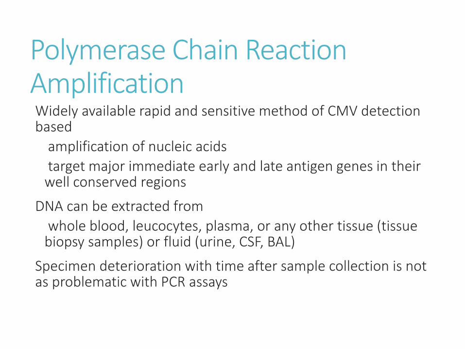
Polymerase Chain Reaction Amplification Widely available rapid and sensitive method of CMV detection based
amplification of nucleic acids
target major immediate early and late antigen genes in their well conserved regions
DNA can be extracted from
whole blood, leucocytes, plasma, or any other tissue (tissue biopsy samples) or fluid (urine, CSF, BAL)
Specimen deterioration with time after sample collection is not as problematic with PCR assays

Qualitative or quantitative
Threshold of the qualitative method calibrated to prevent over-detection
Quantitative PCR (Real-Time PCR)
more expensive compared to the antigaenemia assay
rapid and can be automated

Cytology/ Histology Characteristics intranuclear inclusions in specimens
Saliva, milk, cervical and tracheal secretions, and in touch preparations from biopsy or necropsy tissues
Hallmark of CMV infection "owl's eye"
Papanicolaou or hematoxylin-eosin stains
Clusters of small intracytoplasmic inclusions may also be seen
Sensitivity of the standard cytologic
techniques is low relative to virus isolation
Irrespective of the type of specimen

Diagnosing Congenital CMV Antibody titers maternal CMV IgG crosses the placenta neonates mount weak IgM responses
Viral detection in body fluids PCR, culture, or antigen testing (pp65 antigen) first 3 weeks of life
>3 weeks – Congenital vs postnatally acquired infection
Saliva and urine - newborns shed high levels of the virus
Saliva samples more easily obtained As reliable as urine samples in diagnosing CMV


Neuroimaging assessment Cerebral ultrasound, CT, and MRI for suspected or proven congenital infection
Before 19 weeks post menstrual age Lissencephaly with a thin cortex, cerebellar hypoplasia,
ventriculomegaly, periventricular calcification and delayed myelination
18-24 weeks Migrational abnormalities
CNS lesions may include delayed myelination, dysmyelination and white matter disease
All cases, calcification is a common finding


Ophthalmologic and audiology assessment Opthalmology chorioretinitis, optic atrophy, and cortical visual
impairment Strabmismus is also a common long-term
ophthalmologic complication
Audiological assessment SNHL may be absent at birth progressive in nature frequent evaluations are required throughout
childhood

Diagnosis in HIV CMV viraemia Usually present in end-organ disease Low CD4 cell counts in the absence of end-organ
disease Negative serum or plasma PCR assay also does not
rule out CMV end-organ disease Not recommended for diagnosis of CMV poor PPV
CMV retinitis 70% in the blood Rest diagnosed by clinical criteria plus response to
therapy CMV DNA detected in the vitreous in ~80% of cases


Pneumonitis Diagnosis difficult
Consistent clinical and radiological findings
Multiple CMV inclusion bodies associated with inflammation in lung tissue or cytology
Absence of any other pathogens
Isolation of CMV from isolates including BAL does not prove that the child has CMV pneumonia
Co-infection with both PJP and CMV is common

Lung biopsy gold standard
pp65 - sensitivity and specificity 73% and 50%
VL
Hsiao et al
Significantly higher CMV viral load in infants with probable CMV pneumonia
No cut-off identified
Whole blood HCMV viral load above 4.1 log copies/ml is useful in clinical practice
HIV infected with probable HCMV pneumonia
Ganciclovir treatment

Management of Congenital Infections Who???
Central nervous system (CNS) involvement, including SNHL
Considered in infants with serious end-organ disease
First month of life
What????
Ganciclovir
Valganciclovir
Management of congenital cytomegalovirus infection: an evidence-based approachRS Gandhi et al Acta Pædiatrica 2010 99, pp. 509–515

Ganciclovir Synthetic acyclic nucleoside analogue
Safe and well-tolerated in newborns
No sustained effect on CMV shedding
May be long-term neurodevelopmental benefit for some infants with congenital CMV
6 weeks of intravenous ganciclovir therapy is recommended in the management of babies with symptomatic congenital CMV disease involving the CNS
6 mg/kg/dose IV 12 hourly

Monitor for toxicity
Full blood counts
serum electrolytes and renal function
Neutropenia
H- GCS factor therapy can be administered
Dosage adjustments made, when treating infants with impaired renal function
Realistic expectations-will not reverse established CNS injury

Valganciclovir Oral prodrug of ganciclovir
Neonates who can take enterally
Very well absorbed following oral administration
Rapidly metabolized following oral dosing into ganciclovir
Studies in neonates have demonstrated stable drug levels following oral administration
Dose – 16mg/kg/dose 12 hourly po
6 weeks versus 6 months of valganciclovir performed by the collaborative antiviral study Group

Retinitis Systemic therapy
Disease part of systemic infection
FDA – Ganciclovir,Foscarnet and Cidofovir
Higher induction dose for 2-3 weeks
Maintenance to prevent relapse
Stop once CD4 > 100cells/uL for 3-6 months
Antivitreal therapy
Ganciclovir/Foscarnet
Sight threatening disease
Induction – 2-3 x weekly
Implant – no longer available
Long – term suppression of CMV retinitis – HAART

Panel on Opportunistic Infections in HIV-Infected Adults and Adolescents. Guidelines for the prevention and treatment of opportunistic infections in HIV-infected adults and adolescents: CDC and IDSA