cluster analysis and its applications to gene expression dataroded/schering.pdf · 1 cluster...
TRANSCRIPT

1
Cluster Analysis and its Applications to Gene Expression Data R. SHARAN1
R. ELKON2
R. SHAMIR1
1School of Computer Science, Tel-Aviv University, Tel-Aviv 69978, Israel.
{roded,rshamir}@post.tau.ac.il. 2 The David and Inez laboratory for genetic research, Department of Human Genetics and Molecular Medicine, Sackler School of Medicine, Tel-Aviv University, Tel-Aviv 69978, Israel.

2
1. Introduction
Technologies for generating high-density arrays of cDNAs and oligonucleotides
are developing rapidly, and changing the landscape of biological and biomedical
research. They enable, for the first time, a global, simultaneous view on the
transcription levels of many thousands of genes, when the cell undergoes specific
processes and in certain conditions. For several organisms, the sequences of all
genes are available, and thus, transcript levels of the complete gene collection can
already be monitored today. The potential of such technologies is tremendous:
Monitoring gene expression levels in different developmental stages, tissue types,
clinical conditions and different organisms can help understanding gene function
and gene networks, assist in the diagnosis of disease conditions and reveal the
effects of medical treatments. Undoubtedly, other applications will emerge in
coming years.
A word on terminology: gene expression data is usually represented by a matrix,
with rows corresponding to genes, and columns corresponding to conditions,
experiments or time points. The content of the matrix is the expression levels of
each gene under each condition. Those levels may be absolute, relative or
otherwise normalized. Each column contains the results obtained from a single
array in a particular condition, and is called the profile of that condition. Each row
vector is the expression pattern of a particular gene across all the conditions.
More formal definitions will be given in the sequel.
What is Clustering: A key initial step in the analysis of gene expression data is
the detection of groups of genes that exhibit similar expression patterns. This
translates to the algorithmic problem of clustering. A clustering problem consists
of elements and a characteristic vector for each element. A measure of similarity
is defined between pairs of such vectors. (In gene expression, elements are usually
genes and the vector of each gene is its expression pattern; similarity can be
measured in various ways that are problem dependent, for example, by the
correlation coefficient between vectors.) The goal is to partition the elements into
subsets, which are called clusters, so that two criteria are satisfied: Homogeneity -
elements in the same cluster are highly similar to each other; and separation -
elements from different clusters have low similarity to each other.

3
The Array Technology: There are currently two main technologies that generate
large-scale gene expression data. Both are based on performing a large number of
hybridizations in parallel in a single experiment on high-density arrays (a.k.a.
“DNA chips”), between probes and targets. cDNA microarrays (Schena et al.
1996, Schena 1996, Marshall and Hodgson 1998, Ramsay 1998) contain large sets
of cDNA sequences, each several hundred bases long, immobilized on a solid
substrate. In an array experiment, many gene-specific cDNAs are spotted on a
single matrix. The matrix is then simultaneously probed with cDNA
representations of total RNA pools from test and reference cells, tagged with
distinct fluorescent dyes, allowing one to determine the relative amount of
transcript present in the pool by the relative intensities of the fluorescent signals
generated at each spot. In oligonucleotide microarrays (Fodor et al. 1993,
Lipshutz et al. 2000, Harrigton et al. 2000), each gene is represented on the array
by a set of 15-20 oligonucleotide probes, typically 25 bases long, designed to
hybridize to different regions of the same RNA. This use of multiple detectors
greatly improves reproducibility and accuracy of RNA quantification, and reduces
the rate of false-positives and miscalls.
Outline of this Chapter: In this chapter we shall describe applications of
clustering to gene expression data. We shall outline some of the popular
algorithms in this field, and focus mainly on the CLICK algorithm developed by
Sharan and Shamir (2000). In clustering of genes, we shall present results of
CLICK in comparison to the other algorithms. Some of these results were
previously reported in (Sharan and Shamir 2000,2001). The latter also contains a
broader exposition of other clustering methods. In addition, we shall present
results of using CLICK to find common regulatory motifs in upstream regions of
clustered genes, and to classify tissues based on their expression profiles. The
results demonstrate the utility of clustering in general, and CLICK in particular, in
a wide variety of applications to gene expression analysis.
2. Algorithms
In this section we first give basic mathematical background on clustering. We
then describe three clustering algorithms used for gene expression analysis. We

4
end the section with a discussion on how to measure the quality of a suggested
clustering solution.
2.1 Mathematical Background
Let N={e1,…,en} be a set of n elements, and let C={C1,…,Cl} be a partition of N
into disjoint subsets. Each subset is called a cluster, and C is called a clustering
solution, or simply a clustering. Two elements ei and ej are called mates with
respect to C if they are members of the same cluster in C. In the gene expression
context, the elements are the genes and we often assume that there exists some
correct partition of the genes into “true” clusters. Alternatively, the elements are
the conditions or tissues, that are assumed to belong to one of several categories,
e.g., tumor or normal tissues. When C is the true clustering of N, elements that
belong to the same true cluster are simply called mates.
The input data for a clustering problem is typically given in one of two forms: (I)
Fingerprint data - each element is associated with a real-valued vector, called its
fingerprint, or pattern, which contains p measurements on the element, e.g.,
expression levels of an mRNA at p different conditions (cf. Eisen and Brown
1999). (II) Similarity data - pairwise similarity values between elements. These
values can be computed from fingerprint data, e.g., by correlation between
vectors. Alternatively, the data can represent pairwise dissimilarity, e.g., by
computing distances. Fingerprints contain more information than similarity
values, but the latter are completely generic and can be used to represent the input
to clustering in any application. (Note that there is also a practical storage
consideration regarding the presentation: The fingerprint matrix is of order n×p
while the similarity matrix is of order n×n. In gene expression applications often
n>>p. In contrast, in tissue classification applications often n<<p.)
The goal in a clustering problem is to partition the set of elements into
homogeneous and well-separated clusters. That is, we require that elements from
the same cluster will be highly similar to each other, while elements from
different clusters will have low similarity to each other. This “meta-formulation”
does not define a specific optimization problem, since homogeneity and
separation can be mathematically formulated in various ways, leading to a variety
of optimization problems. Note also, that even when homogeneity and separation

5
are precisely defined, those are two opposing objectives: The better the
homogeneity – the poorer the separation, and vice versa.
Clustering problems and algorithms are often represented in graph-theoretic terms
and we shell use this representation here. We therefore include some basic
definitions on graphs. We refer the readers to Golumbic (1980) and Even (1979)
for more background and terminology on graphs.
Let G=(V,E) be a weighted graph. We denote the vertex set V of G also by V(G).
For a subset R of V, the subgraph induced by R, is obtained from G by deleting all
vertices not in R and the edges incident on them. The weight of a vertex v is the
sum of the weights of the edges incident on v. A cut C in G is a subset of its
edges, whose removal disconnects G. The weight of C is the sum of the weights of
its edges. A minimum weight cut is a cut in G with minimum weight.
For a set of elements K⊆N, we define the fingerprint or centroid of K to be the
mean vector of the fingerprints of the members of K. For two fingerprints x and y
we denote their similarity by S(x,y) and their dissimilarity by d(x,y). A similarity
graph is a weighted graph in which vertices correspond to elements and edge
weights are derived from the similarity values between the corresponding
elements. Hence, the similarity graph is just an equivalent representation of the
similarity matrix.
An alternative formulation of the clustering problem is hierarchical: Rather than
asking for a single partition of the elements, one seeks an iterated partition: A
dendogram is a rooted weighted tree, with leaves corresponding to elements. The
customary (perhaps counter-intuitive) way is to present the tree with the root at
the top and the leaves at the bottom. Each edge defines the cluster of elements
contained in the subtree below that edge. The edge's weight (or length) reflects the
dissimilarity between that cluster and the remaining elements. In this formulation
the clustering solution is the dendogram, and each non-singleton cluster,
corresponding to a rooted subtree, is split into subclusters. The determination of
disjoint clusters is left to the judgment of the user. Typically, one tends to
consider as a genuine cluster the set of elements of a subtree just below a
connecting edge of high weight.
Irrespective of the representation of the clustering problem input, judicious
preprocessing of the raw data is essential to meaningful clustering. This
preprocessing is application dependent and must be chosen in view of the

6
expression technology used and the biological questions asked. The goal of the
preprocessing is to normalize the data and calculate the pairwise element
(dis)similarity, if applicable. Common procedures for normalizing fingerprint data
include transforming each fingerprint to have mean zero and variance one, a fixed
norm or a fixed maximum entry. Statistically based methods for data
normalization have also been developed recently (cf. Kerr et al. 2000).
Several algorithmic techniques were previously used for clustering gene
expression data, including hierarchical clustering (Eisen et al. 1998), self
organizing maps (Tamayo et al. 1999), and graph theoretic approaches (Sharan
and Shamir 2000). We describe these approaches in the sequel. For other
approaches to clustering expression patterns, see (Ben-Dor et al. 1999, Hartuv and
Shamir 2000, Milosavljevic et al. 1995, Alon et al. 1999, Getz et al. 2000, Heyer
et al. 1999). For more extensive reviews and more information and background on
clustering, see (Hartigan 1975, Everitt 1993, Mirkin 1996, Hansen and Jaumard
1997, Shamir and Sharan 2001).
2.2 Hierarchical Clustering
Hierarchical clustering solutions are typically represented by a dendogram.
Algorithms for generating such solutions often work either in a top-down manner,
by repeatedly partitioning the set of elements, or in a bottom-up fashion. We shall
describe here the latter approach. Such agglomerative hierarchical clustering
algorithms are among the oldest and most popular clustering methods (Cormack
1971). They proceed from an initial partition into singleton clusters by successive
merging of clusters until all elements belong to the same cluster. Each merging
step corresponds to joining two clusters. The general scheme due to Lance and
Williams (1967) follows. It is assumed that D=(d(i,j)) is the input dissimilarity
matrix.
The agglomerative hierarchical clustering scheme:
1. Find a minimal entry d(i,j) in D, and merge clusters i and j.
2. Modify D by deleting rows and columns i,j and adding a new row and
column i∪j, with their dissimilarities defined by:
d(k,i∪j)=d(i∪j,k)=αid(k,i)+αjd(k,j)+γ|d(k,i)-d(k,j)|
3. If there is more than one cluster, then go to Step 1.

7
Common variants of this scheme, obtained for appropriate choices of the α-s and
γ parameters, are the following:
Single-linkage: d(k, i∪j)=min{d(k,i), d(k,j)}.
Complete-linkage: d(k, i∪j)=max{d(k,i), d(k,j)}.
Average-linkage: d(k, i∪j)= (ni⋅d(k,i) + nj⋅d(k,j))/(ni+nj), where ni denotes the
number of elements in cluster i.
Eisen et al. (1998) developed a clustering software package based on average-
linkage hierarchical clustering. The clustering program is called Cluster, and the
accompanying visualization program is called TreeView. Both programs are
available at http://rana.stanford.edu/software/. The gene similarity metric used is a
form of correlation coefficient. The algorithm iteratively merges elements whose
similarity value is the highest, as explained above. The output of the algorithm is a
dendogram and an ordered fingerprint matrix. The rows in the matrix are
reordered based on the dendogram, so that groups of genes with similar
expression patterns are adjacent.
2.3 Self Organizing Maps
The self organizing map (SOM) was developed by Kohonen (1997) as a method
for fitting a number of ordered discrete reference vectors to the distribution of
vectorial input samples. The method assumes that the number of clusters is
known. Those clusters are organized as a set of nodes in a hypothetical “elastic
network”, with a simple neighborhood structure on the nodes, e.g., a two-
dimensional k×l grid. Each node n in the grid is associated with a reference vector.
In the process of running the algorithm, the input vectors direct the movement of
the reference vectors towards the denser areas of the input vectors space, so that
an organization of the input vectors over the network emerges. In the following
we describe the SOM algorithm in the Euclidean space.
The SOM process is iterative. Denote by fi(n) the position of the reference vector
of node n at the i-th iteration. The initial positioning f1 is random. The algorithm
iteratively selects a random data point p, identifies the node np whose reference

8
vector fi(np) is closest to p, and updates the position of all reference vectors
towards p according to a predefined learning function l(·):
)]()[,,()()(1 nfpinnlnfnf ipii −+=+
The amount of position adjustment determined by l(n,np,i) decreases as the
distance between n and np (in the grid) and the iteration number grow. The
intuition for this learning process is that the reference vectors that are close
enough to p will be pulled towards it, and the stiffness of the grid structure will
propagate some of impact to neighboring nodes. For much more on self
organizing maps the reader is referred to Kohonen (1997).
Tamayo et al. (1999) devised a gene expression clustering software, GeneCluster,
which uses the SOM algorithm. The software is available at
http://waldo.wi.mit.edu/MPR/. In their implementation they incorporated a
neighborhood learning function, which is zero for nodes distant from np and
equals 0.02T /(T+100i) for nodes close to np, where T is the maximum number of
iterations. GeneCluster accepts an input file of expression levels together with a
two dimensional grid geometry for the nodes. The number of grid points is the
prescribed number of clusters. The resulting clusters are visualized by presenting
for each cluster its average expression pattern along with error bars displaying the
standard deviation at each condition. Clusters are presented in their grid order, as
clusters of close nodes tend to be similar.
Another implementation of SOM for clustering gene expression profiles was
developed by Toronen et al. (1999).
2.4 CLICK
The CLICK (CLuster Identification via Connectivity Kernels) algorithm (Sharan
and Shamir 2000) employs a graph theoretic approach to clustering. The software
is available at http://www.math.tau.ac.il/~rshamir/click.html. The algorithm first
preprocesses the input data and forms a weighted similarity graph. It then
recursively partitions the current set of elements into two subsets. Before a
partition, the algorithm tests if the subgraph induced by the current subset of
elements is a kernel of a cluster (the definition of a kernel is given below). If this
is the case, the subgraph is not partitioned further. Otherwise, a minimum weight
cut is computed in the subgraph, and the current set of elements is split into the

9
two subsets separated by that cut. The output is a list of kernels which serve as a
basis for the eventual clusters, and a set of singletons, i.e., single vertices to be
handled later. This scheme is detailed below. It is assumed that procedure
MinWeightCut(G) computes a minimum weight cut of G and returns a partition of
G into two subgraphs H and K according to this cut.
Form-Kernels(G):
If V(G)={v} then move v to the singleton set.
Else if G is a kernel then output V(G).
Else
(H,K) ← MinWeightCut(G).
Form-Kernels(H).
Form-Kernels(K).
CLICK builds on a statistical model, which gives probabilistic meaning to edge
weights in the similarity graph and to the stopping criterion. The key probabilistic
assumption of CLICK is that pairwise similarity values between elements are
normally distributed: Similarity values between mates are normally distributed
with mean µT and variance σT2, and similarity values between non-mates are
normally distributed with mean µF and variance σF2, where µT>µF. This situation
often holds on real data, and can be asymptotically justified under certain
assumptions (Sharan and Shamir 2000).
The algorithm uses the values of these distribution parameters as well as the
probability pmates that two randomly chosen elements are mates. These parameters
can be computed directly from a known solution on a subset of the elements
(such a solution is often available, e.g., in oligofingerprinting experiments
(Poustka et al. 1999)). Alternatively, the parameters can be estimated using the
EM algorithm, assuming the above probabilistic model (see, e.g., Mirkin 1996,
Sec. 3.2.7).
Let S=(Sij) be the input similarity matrix. Form a weighted similarity graph G in
which the weight wij of the edge (i,j) reflects the probability that i and j are mates,
and is derived from the normal density function and Bayes Theorem:

10
2
2
2
2
2
)(
2
)()1(
ln)| mates-non are ,Pr(
)| mates are ,Pr(ln
T
Tij
F
Fij
Tmates
Fmates
ij
ijij
SSp
pSji
Sjiw
σ
µ
σ
µ
σ
σ −
+
−
+
−
==
The current subgraph is determined to be a kernel if the value of a minimum cut in
it is positive. This is the case if and only if for every cut C in the current
subgraph, the probability that it contains only edges between mates exceeds the
probability that C contains only edges between non-mates.
The actual implementation omits from the graph all edges with values below some
predefined non-negative threshold, computes the minimum cut in that simplified
graph using the Hao-Orlin algorithm (1994), and corrects the solution value for
the missing edges. In order to reduce running time on very big instances, a
screening heuristic removes low weight vertices from large components. The
removed vertices are marked as singletons and handled at a later stage.
CLICK first produces kernels which form the basis of the eventual clusters.
Subsequent processing includes singleton adoption, recursive clustering process
on the set of remaining singletons, and an iterative merging step. The singletons
adoption step is based on computing similarities between singletons' and clusters'
fingerprints. The merging step iteratively merges two kernels whose fingerprint
similarity is the highest, provided that this similarity exceeds a predefined
threshold. Visualization of the clusters shows average expression patterns and
standard error bars for each cluster.
2.5 Assessment of solutions
A key question in the design and analysis of clustering techniques is how to
evaluate solutions. We present in this section figures of merit for measuring the
quality of a clustering solution. Different measures are applicable in different
situations, depending on whether a (partial) true solution is known or not, and
whether the input is fingerprint or merely similarity data. We describe below
some of the applicable measures in case the true solution is unknown. For other
possible figures of merit we refer the reader to (Everitt 1993, Hansen and Jaumard
1997, Yeung et al. 2000).
When the true solution is not known, we evaluate the quality of a suggested
solution by computing two figures of merit that measure its homogeneity and
separation. For fingerprint data, homogeneity is evaluated by the average
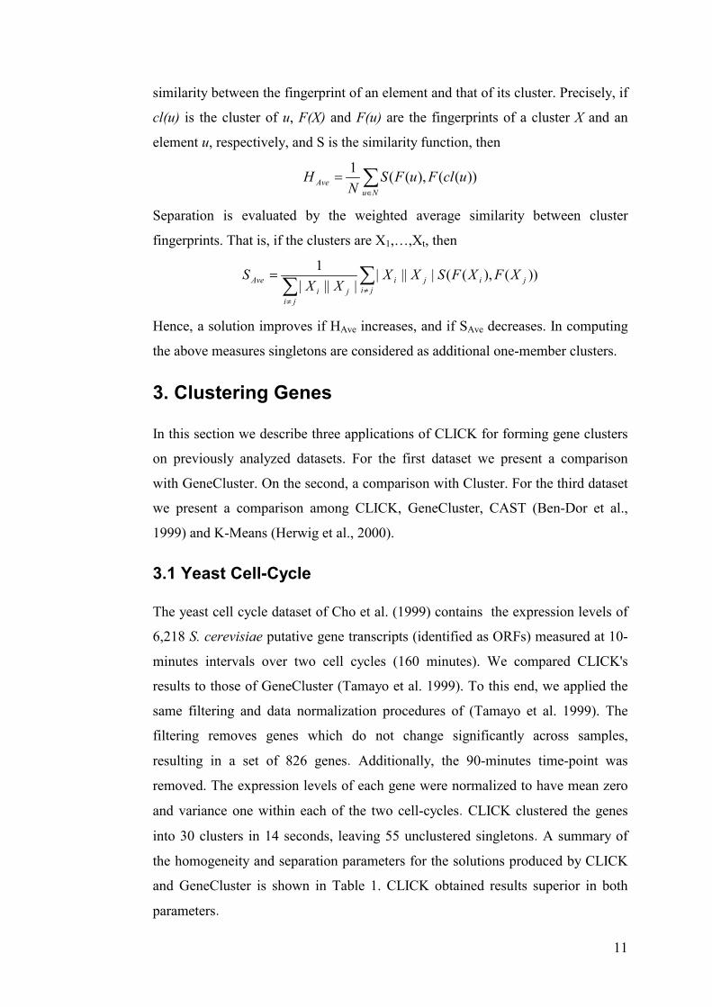
11
similarity between the fingerprint of an element and that of its cluster. Precisely, if
cl(u) is the cluster of u, F(X) and F(u) are the fingerprints of a cluster X and an
element u, respectively, and S is the similarity function, then
∑∈
=
NuAve uclFuFS
NH ))((),((1
Separation is evaluated by the weighted average similarity between cluster
fingerprints. That is, if the clusters are X1,…,Xt, then
∑∑ ≠
≠
=
jijiji
jiji
Ave XFXFSXXXX
S ))(),((||||||||
1
Hence, a solution improves if HAve increases, and if SAve decreases. In computing
the above measures singletons are considered as additional one-member clusters.
3. Clustering Genes
In this section we describe three applications of CLICK for forming gene clusters
on previously analyzed datasets. For the first dataset we present a comparison
with GeneCluster. On the second, a comparison with Cluster. For the third dataset
we present a comparison among CLICK, GeneCluster, CAST (Ben-Dor et al.,
1999) and K-Means (Herwig et al., 2000).
3.1 Yeast Cell-Cycle
The yeast cell cycle dataset of Cho et al. (1999) contains the expression levels of
6,218 S. cerevisiae putative gene transcripts (identified as ORFs) measured at 10-
minutes intervals over two cell cycles (160 minutes). We compared CLICK's
results to those of GeneCluster (Tamayo et al. 1999). To this end, we applied the
same filtering and data normalization procedures of (Tamayo et al. 1999). The
filtering removes genes which do not change significantly across samples,
resulting in a set of 826 genes. Additionally, the 90-minutes time-point was
removed. The expression levels of each gene were normalized to have mean zero
and variance one within each of the two cell-cycles. CLICK clustered the genes
into 30 clusters in 14 seconds, leaving 55 unclustered singletons. A summary of
the homogeneity and separation parameters for the solutions produced by CLICK
and GeneCluster is shown in Table 1. CLICK obtained results superior in both
parameters.

12
Table1
Two putative true cluster are the sets of late G1-peaking genes and M-peaking
genes, reported in (Cho et al. 1999). Out of the late G1-peaking genes that passed
the filtering, CLICK placed 91% in a single cluster. In contrast, Tamayo et al.
(1999) report that in their solution 87% of these genes were contained in three
clusters. Out of the M-peaking genes that passed the filtering, CLICK placed 95%
in a single cluster, while in the solution of (Tamayo et al. 1999) 92.5% of the
genes are again partitioned among three different clusters.
3.2 Serum Response
The dataset of Iyer et al. (1999) contains the expression levels of 8,613 human
genes obtained as follows: Human fibroblasts were deprived of serum for 48
hours and then stimulated by addition of serum. Expression levels of genes were
measured at 12 time-points after the stimulation. An additional data-point was
obtained from a separate unsynchronized sample. A subset of 517 genes whose
expression levels changed substantially across samples was analyzed by the
hierarchical clustering method of Eisen et al. (1998). The data was normalized by
dividing each entry by the expression level at time zero, and taking a logarithm of
the result. For ease of manipulation, we also transformed each fingerprint to have
a fixed norm. The similarity function used was dot-product, giving values
identical to those used by Eisen et al. (1998). CLICK clustered the genes into 10
clusters in 32 seconds, leaving 26 singletons. These clusters are shown in Figure
1. Table 2 presents a comparison between the clustering quality of CLICK and the
hierarchical clustering of Eisen et al. (1998) on this dataset. (The determination of
clusters from the dendogram was done manually by Eisen et al.) Again, CLICK
performs better in both parameters.
Table 2

13
3.3 Yeast Cell-Cycle Revisited
Here we describe a ‘blind’ comparison among several clustering algorithms,
including CLICK, CAST, GeneCluster and K-Means, on a yeast cell-cycle dataset
containing the gene expression levels of yeast ORFs over 79 conditions. The
dataset is available at http://cellcycle-www.stanford.edu. This comparison was
originally reported in (Shamir and Sharan 2001). The original dataset (Spellman
et al. 1998) contains samples from yeast cultures synchronized by four
independent methods: α factor arrest (samples taken every 7 minutes for 119
minutes); arrest of a cdc15 temperature sensitive mutant (samples taken every 10
minutes for 290 minutes); arrest of a cdc28 temperature sensitive mutant (this part
of the data is from (Cho et al. 1999), as described in section 3.1), and elutriation
(samples taken every 30 minutes for 6.5 hours). It also contains separate
experiments in which G1 cyclin Cln3p or B-type cyclin Clb2p were induced.
Spellman et al. identified in this data 800 genes that are cell-cycle regulated
(Spellman et al. 1998). The dataset that we used contains the expression levels of
698 out of those 800 genes, which have no missing entries, over the 72 conditions
that cover the α factor, cdc28, cdc15, and elutriation experiments. As in (Tamayo
et al. 1999), the 90 minutes datapoint was omitted from the cdc15 experiment.
Each row of the 698×72 matrix was normalized to have mean 0 and variance 1.
(Note that by normalizing the variance different gene amplitudes are
deemphasized and periodicity is made more prominent).
Based on the analysis conducted by Spellman et al. we expect to find in the data
five main clusters: G1-peaking genes, S-peaking genes, G2-peaking genes, M-
peaking genes, and M/G1-peaking genes. Each of these clusters was shown to
contain biologically meaningful sub-clusters. The dataset was clustered using four
methods: K-Means (Herwig et al. 2000), GeneCluster (Tamayo et al. 1999),
CAST (Ben-Dor et al. 1999), and CLICK (Sharan and Shamir 2000). The
similarity measure used was Pearson correlation coefficient. The authors of each
of the programs were given the dataset and asked to provide a clustering solution.
The identity of the dataset was not described and genes were permuted in an
attempt to disguise the data source. (Admittedly, the test was not really blind,
since someone familiar with the gene expression literature could have identified
the nature of the data.) The authors were told in advance that the criteria of

14
average homogeneity and average separation would be used to evaluate the
quality of the solutions.
Table 3 summarizes the solutions produced by each program, and their
homogeneity and separation parameters. The so-called 'True' clustering, reported
in Spellman et al. (1998) was obtained manually by comparing the expression
patterns and the literature and is not very accurate (M. Eisen, private
communication). The solution produced by CLICK contains 67 unclustered
singletons.
Table 3
4. Identifying regulatory motifs
Global gene expression data enables the delineation of genetic regulatory
networks via direct or indirect approaches. In the direct approach, the effects of
activation or repression of a specific transcription factor (TF) on gene expression
are monitored. This way, genes located downstream from p53 (Zhao et al. 2000),
BRCA1 (Harkin et al. 1999) and C-myc (Coller et al. 2000) in their respective
pathways, were identified.
The indirect approach for the delineation of regulatory networks relies on the
hypothesis that genes exhibiting similar expression patterns across a large panel of
biological conditions share common regulatory elements in their promoter
regions. In other words, co-expression is correlated with co-regulation (Tavazoie
et al. 1999, Zhang 1999, Brazma and Vilo 2000). The regulatory elements in the
promoter region represent the “switches” that respond to signals from various
cellular signaling pathways. The response can be either as part of the normal
developmental program of the organism, or in response to external perturbations,
stresses and alterations in physiological conditions. The binding of transcription
factors to their binding sites in the promoter region enhances (or represses) the
transcription initiation complex recruitment and assembly on the basal promoter
in the proximity of the transcription start site, thereby influencing transcription
initiation.

15
The approach that aims to detect cis-regulatory TF’s binding sites from co-
expression comprises two steps: (I) Cluster analysis aimed at the identification of
clusters of genes sharing similar expression patterns. (II) Sequence analysis,
which searches for sequence patterns that are over-represented in upstream
regions of members of the same cluster. The derivation of regulatory networks
through the identification of common cis-regulatory elements shared by co-
regulated genes was successfully demonstrated in yeast (Spellman et al. 1998,
Cho et al. 1998, Jelinsky et al. 2000) and arabidopsis thaliana (Maleck et al.
2000).
In order to test the utility of CLICK for motif identification, we have analyzed the
dataset published recently by Jelinsky et al. (2000). In that experiment, gene
expression levels of all 6,200 ORFs of the yeast Saccharomyces Cerevisiae were
measured in order to study the cellular response to DNA damage. In total, gene
expression profiles in 26 biological conditions were measured, including
treatments with various DNA damaging agents at several time points and doses.
2,610 genes that changed by a factor of 3 or more in at least one condition were
subjected to cluster analysis. The clustering reported in (Jelinsky et al. 2000)
consists of 18 clusters, obtained by GeneCluster. In comparison, CLICK
identified 33 clusters with more than 10 members.
Once the clusters are identified, the motif finding algorithm is applied to promoter
regions of the genes in each cluster. In this study, the search was performed on the
500 bases upstream to the ORFs’ translation start sites. The analysis was
performed using the AlignACE package (http://atlas.med.harvard.edu/, Roth et al.
1998, Hughes et al. 2000) as done in (Jelinsky et al. 2000). (As the motif finding
software was recently modified, in addition to its application to CLICK’s
clustering, we also reapplied AlignACE to the clustering reported in (Jelinsky et
al. 2000).) AlignACE employs Gibbs sampling for detecting over-represented
motifs in a target set of sequences. It utilizes the mononucleotide frequencies as
genomic background. To focus on regulatory motifs that potentially form the
mechanistic basis for the observed co-expression, as well as to suppress false-
negatives, only motifs that exceed two score thresholds are reported. The first is
an alignment score, which gauges the statistical significance of the identified
motif over the genomic background. The second is a specificity score, which

16
gauges how specific is the identified motif to promoters of genes in the cluster,
relative to promoter regions of other genes in the genome (Hughes et al. 2000).
In total, 26 significant motifs were identified in CLICK’s clusters, and 30 such
motifs were identified in GeneCluster’s clusters. The identified motifs were
matched against the SCPD database of experimentally verified yeast’s TF binding
sites (http://cgsigma.cshl.org/jian/, Zhu and Zhang 1999). Table 4A summarizes
the number of motifs identified in each clustering as well as the number of motifs
that had a match in the SCPD DB. For both clustering methods, more than 60% of
the motifs had a verified TF binding site match, a fact that indicates the utility of
this approach. In addition, motifs with no match to known binding site were
detected as well. Each of these motifs forms a hypothesis that should be subjected
to further biological research.
Of the 26 motifs identified in CLICK’s clusters, 17 were common with
GeneCluster’s motifs. Common motifs were identified using CompareAce (part of
the AlignAce package), which calculates a similarity coefficient for pairs of input
motifs. Motifs whose similarity exceeded a threshold of 0.7 were regarded as
common. Table 4B lists those common motifs. It is interesting to note that the
percent of verified common motifs is particularly high (more than 75%, see Table
4A). Hence, the four common, unidentified motifs are more likely to be true, as
they were obtained by two different methods.
The delineation of cis-regulatory motifs via analysis of promoters of co-regulated
genes was so far demonstrated in model organisms where the identification of
promoter regions is straightforward. Most TF binding sites are located within a
few hundred bases upstream from the transcription start site. As the 5’-
untranslated regions (UTRs) are very short (100-200b), the predicted translation
start sites are very proximate to the transcription start sites (most of which are
unmapped). Therefore, sequence analysis aimed at detecting the regulatory
elements, can be conducted on sequences in the upstream vicinity of the easy-to-
determine translation start sites. In contrast, the identification of promoters in
mammals is much more difficult, as the 5’-UTR can be very long (up to several
kilobases). However, several full length cDNA projects are being conducted,
which will substantially increase the number of genes for which transcription start
sites are mapped (Werner 2001). A recently published research indeed
demonstrated that this approach for deciphering regulatory mechanism can be

17
successfully implemented in mammals, provided that promoters of co-regulated
genes are mapped (Livesey et al. 2000).
Table 4
5. Tissue classification
One of the most promising applications of gene expression analysis is the
classification of tissue types according to their gene expression profiles (Golub et
al. 1999). The power of gene expression analysis is directed at two important
problems in this context: Cancer classification and drug discovery. Several recent
studies (Alon et al.1999, Golub et al. 1999, Alizadeh et al. 2000) demonstrated
that gene expression data can be used in distinguishing between similar cancer
types, whose distinction is hard otherwise, thereby allowing more accurate
diagnosis and treatment. Drug assessment is aided by expression profiling before,
during and after treatment: The profiles pinpoint drug responsive genes, and
indicate treatment outcome (Clarke et al. 1999).
Here we focus on application of gene expression analysis, and cluster analysis in
particular, to cancer classification. In cancer related gene expression studies, the
data consist of expression levels of thousands of genes in several tissues. The
tissues originate from two or more known classes, e.g., normal and tumor. The
analysis aims at studying the typical expression profile of each class and
predicting the classification of new unlabeled tissues. Classification methods
employ supervised learning techniques, i.e., the known classification of the
tissues is used to guide the algorithm in building a classifier. These include
support vector machines (Ben-Dor et al. 2000, Furey et al. 2000), boosting,
clustering (Ben-Dor et al. 2000), discriminant analysis (Xiong et al. 2000) and
weighted correlation (Golub et al. 1999). Classification can be aided by first
filtering the dataset from genes which are irrelevant to the required distinction.
Several methods have been suggested to choose subsets of informative genes, on

18
which improved classification accuracy can be attained (Ben-Dor et al. 2000,
Furey et al. 2000, Xiong et al. 2000, Dudoit et al. 2000).
Ben-Dor et al. (2000) have demonstrated the strength of clustering in
classification problems. Key to their method is combining the labeling (known
classification) information in the clustering process. Assume that we use a
clustering algorithm with at least one free parameter. Given an unlabeled tissue,
the clustering algorithm is applied repeatedly with different parameter values on
the set of all tissues (known and unknown). Each solution is scored by its level of
compatability with the labeling information, and the best solution is chosen. The
classification of the unlabeled tissue is then determined according to the content
of the cluster containing it, assigning it the most represented class in this cluster.
The compatibility score for a clustering solution used by Ben-Dor et al. (2001) is
simply the number of tissue pairs that are mates or non-mates in both the true
labeling and in the clustering solution. Singletons are considered as 1-member
clusters for this computation. The clustering algorithm used in that work was
CAST.
We have studied two classification datsets using CLICK. The first dataset of Alon
et al. (1999) contains 62 samples of colon epithelial cells. These samples were
collected from colon-cancer patients. They are divided into 40 ‘tumor’ samples
collected from tumors, and 22 ‘normal’ samples collected from normal colon
tissues of the same patients. Of the ~6000 genes represented in the experiment,
2000 genes were selected based on the confidence in the measured expression
levels. This data is available at http://www.sph.uth.tmc.edu/hgc. The second
dataset of Golub et al. (1999) contains 72 leukemia samples. These samples are
divided into 25 samples of acute myeloid leukemia (AML) and 47 samples of
acute lymphoblastic leukemia (ALL). Of the ~7000 genes represented in the
experiment, 3549 were chosen based on their variability in the dataset. The
complete dataset is available at http://www.genome.wi.mit.edu/MPR.
The application of CLICK to classify these datasets enumerates several
combinations of parameters for CLICK, and chooses the one which is most
compatible with the given labels. Compatibility was computed as in Ben-Dor et
al. (2000). A sample is not classified if either it is a singleton in the clustering
obtained, or no class has a majority in the cluster assigned to that sample.

19
In order to assess the performance of CLICK we employed the leave one out cross
validation (LOOCV) technique, as done in (Ben-Dor et al. 2000). According to
this technique, one trial is performed for each tissue in the dataset. In the i-th trial,
the algorithm tries to classify the i-th sample based on the known classifications
of the rest of the samples. The average classification accuracy is thus computed.
Table 5 presents a comparison between the classification based on CLICK and
that of CAST, as reported in Ben-Dor et al. (2000). The results are comparable,
with CAST performing slightly better on the colon dataset, and CLICK
performing slightly better on the leukemia dataset.
Table 5
Next, we tested CLICK’s utility in differentiating between two very similar types
of cancer. We concentrated on part of the leukemia dataset comprising of the 47
ALL samples only. For these samples an additional sub-classification into either
T-cell or B-cell, is provided. An application of CLICK to this dataset resulted in
an almost perfect classification (see Table 6).
Table 6
Finally we examined the influence of feature selection on the classification
accuracy. To this end, we sorted the genes in each dataset according to the ratio of
their between-sum-of-squares and within-sum-of-squares values, as suggested in
(Dudoit et al. 2000). This ratio is computed by the following formula:
∑∑
∑
= ∈
=
−
−
=
2,1
2,
2,1
2,
)(
)(
)()(
i ikig
kg
igigi
xx
xxn
gWSSgBSS

20
Here i denotes the class number, ni its size, k denotes the sample number, xg,i is the
average expression level of gene g at class i, xg is the average expression level of
gene g, and xgk is the expression level of gene g at sample k.
We chose the 50 genes with the highest value and performed the classification
procedure on the reduced dataset which contained the expression levels of these
50 genes only. The results of this analysis are shown in Table 6. It is evident that
in all cases performance was substantially improved on the reduced dataset.
Moreover, there were no unclassified tissues in the reduced datasets.
Acknowledgments
We thank Amir Ben-Dor, Pablo Tamayo and Ralf Herwig for providing us with
their clustering of the yeast cell-cycle dataset. The first author was supported by
an Eshkol fellowship from the ministry of science, Israel. The second author was
supported by a Joseph Sassoon fellowship. The third author was supported in part
by the Israel Science Foundation (grant number 565/99).
References
Alizadeh A.A. , M.B. Eisen, R. Davis, C. Ma, I. Lossos, A. Rosenwald, J. Boldrick, R. Warnke, R.
Levy, W. Wilson, M. Grever, J. Byrd, D. Botstein, P.O. Brown and L. M. Straudt (2000). Distinct
types of diffuse large B-cell lymphomas identified by gene expression profiling. Nature 403:503-
511.
Alon U., N. Barkai, D. A. Notterman, G. Gish, S. Ybarra, D. Mack, and A. J. Levine. (1999).
Broad patterns of gene expression revealed by clustering analysis of tumor and normal colon
tissues probed by oligonucleotide arrays. PNAS, 96:6745-6750.
Ben-Dor A., L. Bruhn, N. Friedman, I. Nachman, M. Schummer, Z. Yakhini (2000). Tissue
classification with gene expression profiles. Journal of Computational Biology 7(3/4):559-583.
Ben-Dor A., R. Shamir, and Z. Yakhini. (1999). Clustering gene expression patterns. Journal of
Computational Biology, 6(3/4):281-297.
Brazma A. and J. Vilo (2000). Gene expression data analysis. FEBS Letters 480:17-24.
Cho R.J., M.J. Campbell, E.A. Winzeler, L. Steinmetz, A. Conway, L. Wodica, T.G.Wolfsberg et
al (1998). A genome-wide transcriptional analysis of the mitotic cell cycle. Mol. Cell 2:65-73.
Clarke P.A., M. George, D. Cunningham, I. Swift, P. Workman (1999). Analysis of tumor gene
expression following chemotherapeutic treatment of patients with bowel cancer. In proc. Nature
Genetics Microarray Meeting 99, Scottsdale, Arizona, p. 39.
Coller H., C. Gradori, P. Tamayo, T. Colbert, E. Lander, R. Eisenman and T.R. Golub (2000).
Expression analysis with oligonucleotide reveals that C-Myc regulates genes involved in growth,
cell-cycle, signaling and adhesion. PNAS 97(7):3260-3265.

21
Cormack R.M.(1971). A review of classification (with discussion). J. Royal Statustical Society,
Series A, 134:321-367.
Dudoit S., J. Fridlyand, T.P. Speed (2000). Comparison of discrimination methods for the
classification of tumors using gene expression data. Technical report #576, Dept. of Statistics,
university of California, Berkeley.
Eisen M.B. and P. O. Brown (1999). DNA arrays for analysis of gene expression. In Methods in
Enzymology, Vol. 303, pages 179-205.
Eisen M.B., P. T. Spellman, P. O. Brown, and D. Botstein (1998). Cluster analysis and display of
genome-wide expression patterns. PNAS, 95:14863-14868.
Even S. (1979). Graph Algorithms. Computer Science Press, Rockville, Maryland.
Everitt B. (1993). Cluster analysis. Edward Arnold, London, third edition.
Fodor S.P., R. P. Rava, X. C. Huang, A. C. Pease, C. P. Holmes, and C. L. Adams (1993).
Multiplexed biochemical assays with biological chips. Nature 364:555-556.
Furey T.S., N. Cristianini, N. Duffy, D.W. Bendarski, M. Schummer, D. Haussler (2000). Support
vector machine classification and validation of cancer tissue samples using microarray expression
data. Bioinformatics 16:906-914.
Getz G., E. Levine, E. Domany, and M.Q. Zhang (2000). Super-paramagnetic clustering of yeast
gene expression profiles. Physica, A279:457.
Golub T., D. Slonim, P. Tamayo, C.M. Huard, J.M. Caasenbeek, H. Coller, M. Loh, J. Downing,
M. Caligiuri, C. Bloomfield, E. Lander (1999). Molecular classification of cancer: class discovery
and class prediction by gene expression monitoring. Science 286:531-537.
Golumbic M.C. (1980). Algorithmic Graph Theory and Perfect Graphs. Academic Press, New
York.
Hansen P. and B. Jaumard (1997). Cluster analysis and mathematical programming. Mathematical
Programming, 79:191-215.
Hao J. and J. Orlin (1994). A faster algorithm for finding the minimum cut in a directed graph.
Journal of Algorithms 17(3):424-446.
Harkin D.P., J. Bean, D. Miklos, Y. Song, V. Maheswaram, J. Oliver, D. Haber (1999). Induction
of GADD45 and JNK/SAPK-dependent apoptosis following inducible expression of BRCA1. Cell
97:575-586.
Harrington C.A., C. Rosenow, and J. Retief (2000). Monitoring gene expression using DNA
microarrays. Curr. Opin. Microbiol., 3(3):285-291.
Hartigan J.A. (1975). Clustering Algorithms. John Wiley and Sons.
Hartuv E. and R. Shamir (2000). A clustering algorithm based on graph connectivity. Information
Processing Letters, 76:175-181.
Herwig R., A.J. Poustka, C. Meuller, H. Lehrach, and J. O'Brien. Large-scale clustering of cDNA-
fingerprinting data (1999). Genome Research 9(11):1093-1105.
Heyer L.J., S. Kruglyak, and S. Yooseph (1999). Exploring expression data: identification and
analysis of coexpressed genes. Genome Research, 9(11):1106-1115.

22
Hughes J.D., P.E. Estep, S. Tavazoie and G.M. Church (2000). Computational identification of
cis-regulatory elements associated with groups of functionally related genes in Saccharomyces
cerevisiae. J. Mol. Biol. 296:1205-1214.
Iyer V.R., M.B. Eisen, D.T. Ross, G. Schuler, T. Moore, J.C.F. Lee, M. Trent, L.M. Staudt, J.
Hudson, M.S. Boguski, D. Lashkari, D. Shalon, D. Botstein, and P.O. Brown (1999). The
transcriptional program in the response of human fibroblast to serum. Science 283:83-87.
Jelinsky S.A., P. Estep, Q.M.Church and L.D.Samson (2000). Regulatory networks revealed by
transcriptional profiling of damaged Saccharomyces cerevisiae cells: Rpn4 links base excision
repair with proteasomes. MCB 20(21):8157-8167.
Kerr M.K., M. Martin, and G.A. Churchill (2000). Analysis of variance for gene expression
microarray data. Technical report, The Jackson Laboratory.
Kohonen T.(1997). Self-Organizing Maps. Springer, Berlin.
Lance G.N. and W.T. Williams (1967). A general theory of classification sorting strategies. 1.
hierarchical systems. The Computer Journal, 9:373-380.
Lipshutz R.J., S. P. A. Fodor, T. R. Gingeras, and D. J. Lockhart (2000). High density synthetic
oligonucleotide arrays. Nature Genetics Supplement, 21:20-24.
Livesey F.J., T. Furukawa, M.A. Steffen, G.M. Church and C.L. Cepko (2000). Microarray
analysis of the transcriptional network controlled by the photoreceptor homeobox gene Crx. Curr.
Biol. 10:301-310.
Maleck K., A. Levine, T. Eulgem, A. Morgan, J. Schmid, K. A. Lawton, J.L. Dangl and R.A.
Dietrich (2000). The transcriptome of Arabidopsis thaliana during systematic acquired resistance.
Nature Genetics 26:403-410.
Marshall A. and J. Hodgson (1998). DNA chips: an array of possibilities. Nat Biotechnol, 16:27-
31.
Milosavljevic A., Z. Strezoska, M. Zeremski, D. Grujic, T. Paunesku, and R. Crkvenjakov (1995).
Clone clustering by hybridization. Genomics, 27:83-89.
Mirkin B. (1996). Mathematical Classification and Clustering. Kluwer.
Poustka A.J., R. Herwig, A. Krause, S. Hennig, S. Meier-Ewert, and H. Lehrach (1999). Toward
the gene catalogue of sea urchin development: the construction and analysis of an unfertilized egg
cDNA library highly normalized by oligonucleotide fingerprinting. Genomics, 59:122-133.
Ramsay G.(1998). DNA chips: State-of-the art. Nat Biotechnol, 16:40-44.
Roth F.P., J.D. Hughes, P.W. Estep, and G.M. Church (1998). Finding DNA regulatory motifs
within unaligned noncoding sequences clustered by whole-genome mRNA quantitation. Nature
Biotech. 16:939-908.
Schena M.(1996). Genome analysis with gene expression microarrays. Bioessays, 18:427-431.
Schena M., D. Shalon, R. Heller, A. Chai, P. O. Brown, and R. W. Davis (1996). Parallel human
genome analysis: microarray-based expression monitoring of 1000 genes. PNAS 93:10614-9.
Sharan R. and R. Shamir (2000). CLICK: A clustering algorithm with applications to gene
expression analysis. In Proceedings of the Eighth International Conference on Intelligent Systems
for Molecular Biology (ISMB), pages 307-316.

23
Shamir R. and R. Sharan (2001). Algorithmic approaches to clustering gene expression data. In
Current topics in computational biology, T. Jiang, T. Smith, Y. Xu and M.Q. Zhang eds., MIT
Press.
Spellman P.T., G. Sherlock, M. Zhang, V.R. Iyer, K. Anders, M. Eisen, P.O. Brown, D. Botstein
and B. Futcher (1998). Comprehensive identification of cell cycle regulated gene of the yeast
Saccharomyces Cerevisia by microarray hybridization. Mol. Biol. Cell 9:3273-3297.
Tamayo P., D. Slonim, J. Mesirov, Q. Zhu, S. Kitareewan, E. Dmitrovsky, E. S. Lander, and T.R.
Golub (1999). Interpreting patterns of gene expression with self-organizing maps: Methods and
application to hematopoietic differentiation. PNAS, 96:2907-2912.
Tavazoie S., J. Hughes, M. Campbell, R. Cho and G. M. Church (1999). Systematic determination
of genetic network architecture. Nature Genetics 22:281-285.
Toronen P., M. Kolehmainen, G. Wong, and E. Castren (1999). Analysis of gene expression data
using self-organizing maps. FEBS Letters, 451:142-146.
Werner T. (2001). Target gene identification from expression array data by promoter analysis.
Biomolecular Engineering 17:87-94.
Xiong M., L. Jin, W. Li, E. Boerwinkle (2000). Computational methods for gene expression based
tumor classification. Biotechniques 29:1264-1270.
Yeung K.Y., D.R. Haynor, and W.L. Ruzzo (2001). Validating clustering for gene expression data.
Bioinformatics 17:309-318.
Zhang M.Q (1999). Large scale gene expression data analysis: a new challenge to computational
biologists. Genome Research 9:681-688.
Zhao R., K. Gish, Y. Yin, D. Notterman, W. Hoffman, E. Tom, D. Mak and A.J. Levine (2000).
Analysis of p53 regulated gene expression patterns using oligonucleotide arrays. Genes and Dev.
14:981-993.
Zhu J. and M.Q. Zhang (1999). SCPD: a promoter database of the yeast Saccharomyces cerevisiae.
Bioinformatics 15:607-611.

24
Table Legends
Table 1: A comparison between CLICK and GeneCluster on the yeast cell-cycle dataset of (Cho et
al. 1999).
Table 2: A Comparison between CLICK and the hierarchical clustering of Eisen et al. (1998) on
the dataset of response of human fibroblasts to serum of (Iyer et al. 1999).
Table 3: A summary of the clustering solutions and their figures of merit on the yeast cell-cycle
dataset of (Spellman et al. 1998).
Table 4: (A) Statistics on motifs identified in CLICK’s clusters (first column), GeneCluster’s
clusters (second column) and in both clusterings (third column). First row: total number. Second
row: number of motifs with a hit in the SCPD DB. (B) Motifs identified in both CLICK’s and
GeneCluster’s clusters. Left column: CLICK’s motif name. Each motif is denoted by two
numbers: the first is the cluster number and the second is the motif serial number in the cluster
(AlignACE is capable of finding multiple motifs in a target set of sequences by an iterative
masking procedure). The second row contains statistics on the prevalence of the motif in the
cluster. Second column: the motif’s consensus sequence. Third column: an experimentally verified
TF which matches the consensus sequence. (For TFs denoted by an ‘*’, there is one mismatch
between the TF binding site consensus and the identified motif consensus, otherwise, there is a
perfect match.) Forth column: The corresponding GeneCluster’s motifs (motifs with similarity
coefficient above 0.7 to CLICK’s consensus). Motifs found more than once are grouped together
in column 1.
Table 5: A comparison of classification quality between CLICK and CAST on the colon data of
(Alon et al. 1999) and the leukemia data of (Golub et al. 1999). For each dataset and clustering
algorithm the percent of correct classifications (in the LOOCV iterations), incorrect classifications
and unclassified elements are specified.
Table 6: A summary of the classifications obtained by CLICK on the colon data of (Alon et al.
1999), the whole leukemia dataset of (Golub et al. 1999), and part of the leukemia dataset which
contains ALL samples only. For each dataset classifications were performed with respect to the
total number of genes, and with respect to the 50 most informative genes. The percent of correct
classifications (in the LOOCV iterations), incorrect classifications and unclassified elements are
specified.

25
Figure Legends
Figure 1:CLICK's clustering of the fibroblasts serum response dataset of (Iyer et al., 1999). x axis:
1-12: synchronized time points. 13: unsynchronized point. y axis: normalized expression levels.
The solid line in each sub-figure plots the average pattern for that cluster. Error bars display the
measured standard deviation. The cluster size is printed above each plot.

26
Tables
Table 1:
Program #Clusters Homogeneity Separation
CLICK 30 0.8 -0.07
GeneCluster 30 0.74 -0.02
Table 2:
Program #Clusters Homogeneity Separation
CLICK 10 0.88 -0.34
Hierarchical 10 0.87 -0.13
Table 3:
Program #Clusters Homogeneity Separation
CLICK 6 0.66 -0.1
K-Means 49 0.63 0.09
GeneCluster 6 0.62 -0.07
CAST 5 0.6 -0.15
‘True’ 5 0.57 -0.13

27
Table 4:
4A.
CLICK GeneCluster Common
Found 26 30 17
Verified 17 19 13
4B.
CLICK Consensus Putative TF GeneCluster
Clk 1_2 16% (37/237)
GGTGGCAAAW UASPHR Som 14_2 30% (61/205)
Clk 2_1 63% (119/189)
RAAAAAAAAA PHO2;SWI5
Som 4_4 38% (52/136)
Clk 2_2 44% (83/189)
ATGTAYGGRTK RAP1 Som 1_2 52% (85/165)
Clk 2_3 30% (56/189) Clk 2_11 23% (44/189) Clk 9_3 87% (33/38)
RAAAAATTT AAAAAWTTT TGAAAAWTTTT
DAL82 Som 2_2 65% (48/74) Som 4_2 61% (83/136)
Clk 2_7 15% (29/189)
NSYAGGCNGNR RAP1*, BUF* Som 1_4 17% (28/165) Som 1_8 11% (18/165) Som 1_9 15% (25/165)
Clk 2_9 12% (23/189)
CYCNSCNRGNNGGA MCM1*
Som 1_5 15% (25/165)
Clk 3_3 21% (31/149)
YNCGGNSNNNSGGS RAP1*
Som 3_8 13% (23/175)
Clk 3_4 19% (28/149)
RRCCAATCAN ABF1,BAF1* Som 3_2 21% (36/175)
Clk 3_6 12% (18/149) Clk 3_7 8% (12/149)
GGCNGGGCRKC TSGGCGGCNNTT
URS1H Som 3_4 8% (14/175)
Clk 3_9 19% (28/149)
SGGNNNNNNNGGNNNGG
BUF * Som 3_6 16% (28/175)
Clk 2_8 15% (28/189)
SCNGCNNSCNGNNGSG
----- Som 1_6 17% (28/165)
Clk 3_11 13% (19/149)
MNNNGGGNNNRNNNRNGGGR
----- Som 6_7 25% (28/114)
Clk 3_21 9% (13/149)
NCCGNYGGNCCGR -----
Som 3_8 13% (23/175)
Clk 26_2 90% (9/10)
AGGGGCGGNG ----- Som 9_3 15% (23/150)

28
Table 5:
Dataset Method Correct Incorrect Unclassified
Colon CLICK 85.5 9.7 4.8
CAST 88.7 11.3 0.0
Leukemia CLICK 90.3 4.2 5.5
CAST 87.5 12.5 0.0
Table 6:
Dataset Size Correct Incorrect Unclassified
Colon 2000 85.5 9.7 4.8
50 90.3 9.7 0.0
Leukemia 3549 90.3 4.2 5.5
50 98.6 1.4 0.0
ALL 3549 95.8 2.1 2.1
50 100.0 0.0 0.0