clinically obscure venous malformation in the sub mandibular triangle: a rare presentation
TRANSCRIPT
Journal of Oral Biology and Craniofacial Research 2012 MayeAugustVolume 2, Number 2; pp. 119e122 Case Report
Clinically obscure venous malformation in the sub mandibular triangle:A rare presentation
Mamta Agrawala,*, Manish Kumarb, Vikrant Agrawalc
aReadColleg*CorreReceivCopyrihttp://d
ABSTRACT
Vascular lesions have been classified as hemangiomas or vascular malformations depending on the presence ofcellular proliferation. These lesions have been known to cause significant morbidity and even mortality in bothchildren and adults. Confusing nomenclature, relative rarity of these lesions, inappropriate diagnosis, lack of preciseimaging studies and deficiency of multidisciplinary approach are some of the factors which result in ineffectivetreatment of such patients. Here, we report a rare case of venous vascular malformation presenting as a mass in thesub mandibular region with no clinical stigmata which was diagnosed with the help of various imaging techniques andsubsequently treated by surgery.
Copyright © 2012, Craniofacial Research Foundation. All rights reserved.
Keywords: Venous vascular malformation, Vascular lesions, Vascular malformation in sub mandibular triangle
INTRODUCTION
Hemangiomas and vascular malformations are relativelycommon tumors of the face and neck region which repre-sent one of the largest groups among soft tissue tumorsand typically present with clinical stigmata as skin discolor-ation, dilated subcutaneous vessels and compressibilitysuggestive of the diagnosis. Vascular lesions were earliercategorized on the basis of the size of the channels andthe type of fluid in the lesions, according to which blood-containing lesions were called hemangiomas and lymph-containing lesions were referred to as lymphangiomas orcystic hygromas. Hemangiomas were further differentiatedon the basis of channel size into capillary hemangiomas,strawberry hemangiomas and cavernous hemangiomas.1
This classification system resulted in great confusion andin 1982 was replaced by a biologic classification systemby Mulliken and Glowacki2 which separates endothelialmalformations into two large groups, hemangiomas andvascular malformations, on the basis of their natural history,cellular turnover, and histology. Hemangiomas are benign,
er, Department of OMFS, Purvanchal Institute of Dental Sciences, GIDe, Mathura, cSenior Lecturer, Department of Radiology, BRD Medicasponding author. Fax: +91 0551 2202539, email: mamtaag02@yah
ed: 30.4.2012; Accepted: 19.5.2012ght � 2012, Craniofacial Research Foundation. All rights reserved.x.doi.org/10.1016/j.jobcr.2012.05.009
localized tumors of the blood vessels where as vascularmalformation result from anomalous development ofvascular plexuses.3 Hemangiomas are vascular neoplasmswith endothelial hyperplasia, these tend to be small orabsent at birth and often go unnoticed. Soon after birththey undergo proliferation and rapid growth which maylast for several months. This is followed by a stationaryperiod and then a period of involution. Conversely, vascularmalformations form secondary to an error of embryonicdevelopment with normal endothelial turnover; these arealways present at birth and grow commensurately withthe child, although the lesion may expand secondary totrauma, infection, hormonal changes, or embolic or surgicalintervention. They do not involute and remain presentthroughout the patient’s life. Vascular malformations aresubcategorized on the basis of their histological makeupas lymphatic, capillary, venous, arteriovenous, and mixedmalformations and according to hemodynamic featuresas slow flow (either capillary, venous, lymphatic, orcombined forms) or “fast-flow” (arteriovenous fistulas andarteriovenous malformation).4 Malformations with arterial
A, Gorakhpur 273001, bReader, Department of OMFS, K.D. Dentall College, Gorakhpur 273001, Uttar Pradesh, India.oo.co.in

120 Journal of Oral Biology and Craniofacial Research 2012 MayeAugust; Vol. 2, No. 2 Agrawal et al.
components are considered high-flow lesions and thosewithout arterial components are considered low-flowlesions.1
Here, we present a rare case of an obscure venousvascular malformation in the sub mandibular triangle,a low-flow lesion arising from the facial vein which withoutinvolving the gland was present in the fascial planesextending from the buccal to the lingual side of themandible.
CASE REPORT
A 25-year-old male presented with a swelling in the left submandibular region, he gave a history of a small swellingbeing present since childhood. Recently, the swelling hadincreased in size and was causing discomfort in neck move-ments. There was no change reported in the size of theswelling with meals or on straining and there was no historyof pain. He was otherwise asymptomatic.
Clinical examination revealed a large solitary, compress-ible swelling, palpable both on buccal and the lingual sideof the mandible with the overlying skin normal in color,texture, with a temperature similar to the surroundingskin, also the lesion was non pulsatile. No other mass orlymph node was palpable in the rest of the neck. Intraorally,a swelling was visible both on the lingual and buccal side ofthe left mandibular arch with bluish discoloration of theoverlying mucosa. Saliva flowed freely from the ipsilateralWharton’s duct and no calculi were palpable. Initial clinicaldiagnoses included a reactive nodal mass, sub mandibulargland neoplasm and branchial cleft cyst.
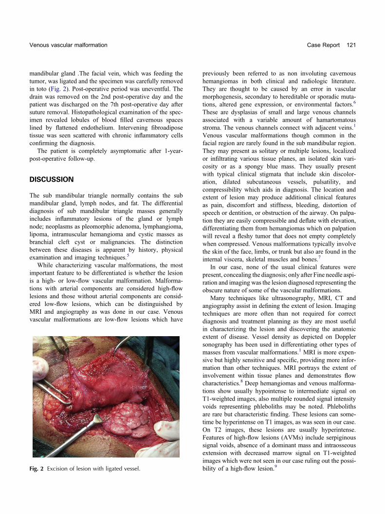
Fig. 1 (A) T1 W magnetic resonance image (sagittal section) showingbuccal and lingual side of the mandible. (B) T1 W MRI axial image shspace.
Fine needle aspiration revealed frank blood. ComputedTomography (CT) angiography and Magnetic resonanceimaging (MRI) was advised. CT of neck region showeda lobulated predominantly hypodense lesion in left side ofneck involving the superficial soft tissue and extendingalong, both the buccal and lingual aspect of mandible. Nodefinite bony involvement was seen. CT angiographyrevealed the feeders to be facial vessles.
MRI demonstrated a heterogeneous, well-defined mass,7 � 8 � 5 cm in size in sub mandibular triangle of neckon the left side. T1-weighted MRI images showed a large,lobulated, predominantly hyperintense space occupyinglesion. The lesion was seen extending toward the lingualside curving around the mandible, compressing the leftsub mandibular gland and extending anteriorly along thelingual margin of mandible toward the midline. The lesionwas abutting and compressing the myelohyoid muscle andmildly displacing the carotid vessels posteriorly. The under-lying bone showed no erosion. On the buccal side, thelesion was seen extending anteriorly along the body ofmandible, abutting the massater muscle with loss of fatplanes and no definite evidence of muscle invasion. Nosignal void was seen ruling out the possibility of high-flow lesion (Fig. 1A and B).
A diagnosis of venous vascular malformation of thefascial planes in the sub mandibular region was made onthe basis of history, clinical and radiological findings.Surgical excision of the lesion was planned as the lesionwas non infiltrative, well defined and confined to a singlefascial plane.
At surgery, a reddish blue colored lesion was seen lyingdeep to the superficial fascia and platysma above the sub
a lobulated mass in sub mandibular region extending along theowing the posterior extension of lesion to the parapharyngeal
Venous vascular malformation Case Report 121
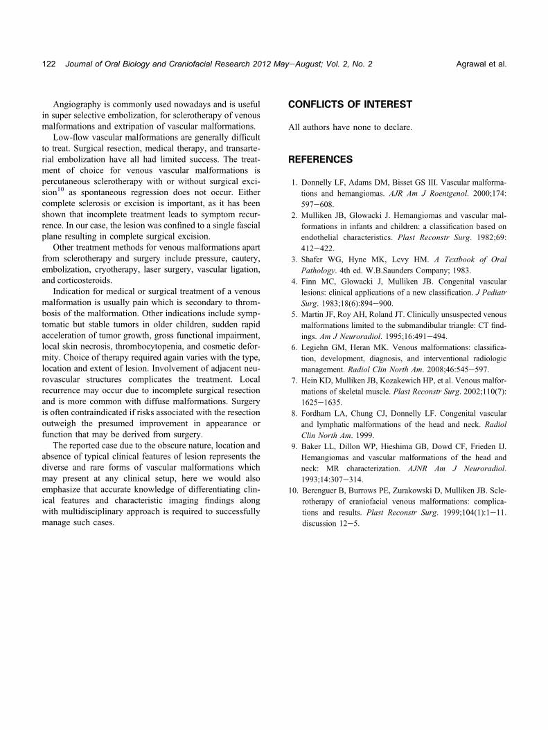
mandibular gland .The facial vein, which was feeding thetumor, was ligated and the specimen was carefully removedin toto (Fig. 2). Post-operative period was uneventful. Thedrain was removed on the 2nd post-operative day and thepatient was discharged on the 7th post-operative day aftersuture removal. Histopathological examination of the spec-imen revealed lobules of blood filled cavernous spaceslined by flattened endothelium. Intervening fibroadiposetissue was seen scattered with chronic inflammatory cellsconfirming the diagnosis.
The patient is completely asymptomatic after 1-year-post-operative follow-up.
DISCUSSION
The sub mandibular triangle normally contains the submandibular gland, lymph nodes, and fat. The differentialdiagnosis of sub mandibular triangle masses generallyincludes inflammatory lesions of the gland or lymphnode; neoplasms as pleomorphic adenoma, lymphangioma,lipoma, intramuscular hemangioma and cystic masses asbranchial cleft cyst or malignancies. The distinctionbetween these diseases is apparent by history, physicalexamination and imaging techniques.5
While characterizing vascular malformations, the mostimportant feature to be differentiated is whether the lesionis a high- or low-flow vascular malformation. Malforma-tions with arterial components are considered high-flowlesions and those without arterial components are consid-ered low-flow lesions, which can be distinguished byMRI and angiography as was done in our case. Venousvascular malformations are low-flow lesions which have
Fig. 2 Excision of lesion with ligated vessel.
previously been referred to as non involuting cavernoushemangiomas in both clinical and radiologic literature.They are thought to be caused by an error in vascularmorphogenesis, secondary to hereditable or sporadic muta-tions, altered gene expression, or environmental factors.6
These are dysplasias of small and large venous channelsassociated with a variable amount of hamartomatousstroma. The venous channels connect with adjacent veins.1
Venous vascular malformations though common in thefacial region are rarely found in the sub mandibular region.They may present as solitary or multiple lesions, localizedor infiltrating various tissue planes, an isolated skin vari-cosity or as a spongy blue mass. They usually presentwith typical clinical stigmata that include skin discolor-ation, dilated subcutaneous vessels, pulsatility, andcompressibility which aids in diagnosis. The location andextent of lesion may produce additional clinical featuresas pain, discomfort and stiffness, bleeding, distortion ofspeech or dentition, or obstruction of the airway. On palpa-tion they are easily compressible and deflate with elevation,differentiating them from hemangiomas which on palpationwill reveal a fleshy tumor that does not empty completelywhen compressed. Venous malformations typically involvethe skin of the face, limbs, or trunk but also are found in theinternal viscera, skeletal muscles and bones.7
In our case, none of the usual clinical features werepresent, concealing the diagnosis; only after Fine needle aspi-ration and imaging was the lesion diagnosed representing theobscure nature of some of the vascular malformations.
Many techniques like ultrasonography, MRI, CT andangiography assist in defining the extent of lesion. Imagingtechniques are more often than not required for correctdiagnosis and treatment planning as they are most usefulin characterizing the lesion and discovering the anatomicextent of disease. Vessel density as depicted on Dopplersonography has been used in differentiating other types ofmasses from vascular malformations.1 MRI is more expen-sive but highly sensitive and specific, providing more infor-mation than other techniques. MRI portrays the extent ofinvolvement within tissue planes and demonstrates flowcharacteristics.8 Deep hemangiomas and venous malforma-tions show usually hypointense to intermediate signal onT1-weighted images, also multiple rounded signal intensityvoids representing phleboliths may be noted. Phlebolithsare rare but characteristic finding. These lesions can some-time be hyperintense on T1 images, as was seen in our case.On T2 images, these lesions are usually hyperintense.Features of high-flow lesions (AVMs) include serpiginoussignal voids, absence of a dominant mass and intraosseousextension with decreased marrow signal on T1-weightedimages which were not seen in our case ruling out the possi-bility of a high-flow lesion.9
122 Journal of Oral Biology and Craniofacial Research 2012 MayeAugust; Vol. 2, No. 2 Agrawal et al.
Angiography is commonly used nowadays and is usefulin super selective embolization, for sclerotherapy of venousmalformations and extripation of vascular malformations.
Low-flow vascular malformations are generally difficultto treat. Surgical resection, medical therapy, and transarte-rial embolization have all had limited success. The treat-ment of choice for venous vascular malformations ispercutaneous sclerotherapy with or without surgical exci-sion10 as spontaneous regression does not occur. Eithercomplete sclerosis or excision is important, as it has beenshown that incomplete treatment leads to symptom recur-rence. In our case, the lesion was confined to a single fascialplane resulting in complete surgical excision.
Other treatment methods for venous malformations apartfrom sclerotherapy and surgery include pressure, cautery,embolization, cryotherapy, laser surgery, vascular ligation,and corticosteroids.
Indication for medical or surgical treatment of a venousmalformation is usually pain which is secondary to throm-bosis of the malformation. Other indications include symp-tomatic but stable tumors in older children, sudden rapidacceleration of tumor growth, gross functional impairment,local skin necrosis, thrombocytopenia, and cosmetic defor-mity. Choice of therapy required again varies with the type,location and extent of lesion. Involvement of adjacent neu-rovascular structures complicates the treatment. Localrecurrence may occur due to incomplete surgical resectionand is more common with diffuse malformations. Surgeryis often contraindicated if risks associated with the resectionoutweigh the presumed improvement in appearance orfunction that may be derived from surgery.
The reported case due to the obscure nature, location andabsence of typical clinical features of lesion represents thediverse and rare forms of vascular malformations whichmay present at any clinical setup, here we would alsoemphasize that accurate knowledge of differentiating clin-ical features and characteristic imaging findings alongwith multidisciplinary approach is required to successfullymanage such cases.
CONFLICTS OF INTEREST
All authors have none to declare.
REFERENCES
1. Donnelly LF, Adams DM, Bisset GS III. Vascular malforma-tions and hemangiomas. AJR Am J Roentgenol. 2000;174:597e608.
2. Mulliken JB, Glowacki J. Hemangiomas and vascular mal-formations in infants and children: a classification based onendothelial characteristics. Plast Reconstr Surg. 1982;69:412e422.
3. Shafer WG, Hyne MK, Lcvy HM. A Textbook of OralPathology. 4th ed. W.B.Saunders Company; 1983.
4. Finn MC, Glowacki J, Mulliken JB. Congenital vascularlesions: clinical applications of a new classification. J PediatrSurg. 1983;18(6):894e900.
5. Martin JF, Roy AH, Roland JT. Clinically unsuspected venousmalformations limited to the submandibular triangle: CT find-ings. Am J Neuroradiol. 1995;16:491e494.
6. Legiehn GM, Heran MK. Venous malformations: classifica-tion, development, diagnosis, and interventional radiologicmanagement. Radiol Clin North Am. 2008;46:545e597.
7. Hein KD, Mulliken JB, Kozakewich HP, et al. Venous malfor-mations of skeletal muscle. Plast Reconstr Surg. 2002;110(7):1625e1635.
8. Fordham LA, Chung CJ, Donnelly LF. Congenital vascularand lymphatic malformations of the head and neck. RadiolClin North Am. 1999.
9. Baker LL, Dillon WP, Hieshima GB, Dowd CF, Frieden IJ.Hemangiomas and vascular malformations of the head andneck: MR characterization. AJNR Am J Neuroradiol.1993;14:307e314.
10. Berenguer B, Burrows PE, Zurakowski D, Mulliken JB. Scle-rotherapy of craniofacial venous malformations: complica-tions and results. Plast Reconstr Surg. 1999;104(1):1e11.discussion 12e5.