clinical validity of longitudinal pre-ejectional
TRANSCRIPT

Circulation Journal Vol.71, December 2007
he early identification of viable myocardial extentafter reperfusion therapy in patients with myocar-dial infarction (MI) is of substantial clinical value,
in terms of patient management and predicting prognosis.1–6
Because myocardial reperfusion after a reversible ischemicinsult frequently results in a protracted contractile dysfunc-tion, which is often described as ‘myocardial stunning’,7,8
conventional wall motion assessment is limited in detectingviable myocardium with clinically acceptable confidence.Although dobutamine stress echocardiography, magneticresonance imaging (MRI) or nuclear myocardial perfusionimaging has been traditionally used to assess myocardial
viability, their applications are, in part, limited by their cost,availability, and variability.
Interestingly, 2 previous studies suggested that restinglongitudinal pre-ejectional velocity (PEVL) measured at theapical echocardiographic views by tissue Doppler imaging(TDI) is a simple and accurate parameter that allows theprediction of contractile recovery after percutaneous coro-nary intervention (PCI) in patients with occluded coronaryartery disease, implying the ability of PEVL to identify theextent of viable myocardium.9,10 However, because a largenumber of patients with coronary artery disease comprisethose with multi-vessel disease, not just a single-vessel dis-ease, and wide spectrum of the left ventricular (LV) systolicfunction, this potential association between PEVL andmyocardial viability requires confirmation before a generalapplication of this attractive index can be made to a cohortof patients with multi-vessel coronary artery disease. There-fore, the purpose of this study was to apply this intriguingnew TDI parameter to patients with recent MI who also hadsingle- and multi-vessel coronary artery disease, and toassess its reliability in determining the transmural extent of viable myocardium, using delayed-enhanced MRI as areference modality.
Circ J 2007; 71: 1904–1911
(Received May 21, 2007; revised manuscript received August 21,2007; accepted August 23, 2007)*Department of Internal Medicine, Cardiovascular Center, **Depart-ment of Diagnostic Radiology, †Seoul National University College ofMedicine, Seoul National University Hospital, Seoul, KoreaPart of this work was presented at the Annual Scientific Session of theAmerican Heart Association, Chicago, IL, USA, November 12–15,2006.Part of this work was supported by a grant from the Korean Instituteof Medicine, Seoul, Korea.Mailing address: Yong-Jin Kim, MD, Division of Cardiology, De-partment of Internal Medicine, Seoul National University College ofMedicine, 28 Yongon-dong, Chongno-gu, Seoul 110-744, Korea. E-mail: [email protected]
Clinical Validity of Longitudinal Pre-Ejectional Myocardial Velocity for Identifying the Transmural Extent
of Viable MyocardiumEarly After Reperfusion of an Infarct-
Related Coronary Artery
Hyung-Kwan Kim, MD*,†; Yong-Jin Kim, MD*,†; Sung-A Chang, MD*,†; Song-Yi Kim, MD*,†; Ho-Joon Jang, MD*,†; Whal Lee, MD**,†; Jin-Shik Park, MD*,†; Dae-Won Sohn, MD*,†;
Byung-Hee Oh, MD*,†; Young-Bae Park, MD*,†; Yun-Shik Choi, MD*,†
Background Positive longitudinal pre-ejectional velocity (+PEVL) was recently reported to be a reliable indexof myocardial recovery early after successful revascularization in myocardial infarction (MI); that is, it recognizesthe transmural extent of viable myocardium. The applicability of PEVL in the real-world clinical setting for identi-fying the transmural extent of viable myocardium in reperfused recent MI was assessed.Methods and Results Using tissue Doppler imaging, the resting basal and mid myocardial PEVLs were deter-mined within 3 days after revascularization in 41 consecutive patients with recent MI. Infarct thickness was semi-quantified using delayed gadolinium-enhanced magnetic resonance imaging (MRI) at baseline and at 6-monthfollow up to differentiate transmural from nontransmural MI. The proportion of segments showing the presenceof +PEVL was not significantly changed as infarct thickness increased (p=0.2), with 66.2% having +PEVL evenin segments involving >75% transmural infarction. Moreover, +PEVL was found in a large fraction of segmentswith akinesia (70.4%). Specificity and negative predictive value of +PEVL for assessing infarct nontransmuralitywere disappointingly low (32.0% and 26.9%, respectively). All of these results were not altered when the 6-monthfollow-up MRI was done.Conclusions +PEVL cannot be regarded as a reliable marker for predicting the transmural extent of viable myo-cardium in recent MI. (Circ J 2007; 71: 1904–1911)
Key Words: Magnetic resonance imaging; Myocardial infarction; Pre-ejectional velocity; Tissue Dopplerimaging
T

1905Clinical Validity of PEVL
Circulation Journal Vol.71, December 2007
MethodsStudy Population
Forty-one consecutive patients who had experienced afirst acute or subacute MI (age 55.6±11.6 years; range, 31–78 years) were prospectively recruited. All of them under-went successful PCI of an infarct-related coronary artery,and no significant coronary stenoses requiring an additionalrevascularization procedure remained after PCI in any pa-tient. Acute MI was defined as typical ischemic chest painlasting for at least 30min in combination with either anelevation of cardiac-specific troponins or an electrocardio-graphic change of ST-segment elevation within 24h fromsymptom onset and subacute MI, with a time gap betweenonset of symptoms and PCI performance from 24h to <2weeks. Patients with persistent severe heart failure (Killipclass III or IV and/or LV ejection fraction (LVEF) <20%),uncontrolled myocardial ischemia or ventricular tachycar-dia, infarct-related arteries unsuitable for PCI, unsuccessfulPCI, left bundle-branch block, atrial fibrillation, or morethan mild valvular regurgitation were excluded. Whether ornot patients enrolled in the present study had multi-vesselcoronary artery disease was not in the consideration of thepatient enrolment.
The study protocol was approved by the InstitutionalReview Board of the Seoul National University Hospital,and written informed consent was obtained from all partici-pants before study enrollment.
EchocardiographyWith the patient in a supine left lateral position, a com-
prehensive transthoracic echocardiography was performedwithin 72h after PCI, using a commercially available echo-cardiographic machine (Vivid 7, GE Medical System,Horten, Norway). Routine standard echocardiographic ex-aminations included LVEF by biplane modified Simpson’smethod and early (E) and late transmitral inflow velocities,and deceleration time of E velocity by pulsed-wave Dopplerwith the sample volume placed between the tips of mitralleaflets.
Three consecutive heart beats at the apical 4-, 2-, and 3-chamber images were digitally captured onto a magneto-optical disc in a cineloop format for later offline analysis ofTDI data, using a customized dedicated software package(EchoPac vers. 5.0.1 for PC, GE Medical System, Horten,Norway). Gain and filter settings were carefully adjustedfor better acquisition of myocardial tissue signals. Framerates were at least above 120 frames/second, which allowsfor a temporal resolution of <9ms, depending on the widthand depth of the images obtained. A sample volume of 5×5mm was placed at the center of the basal and mid myocar-dial segments as parallel as possible to the motional vectorof the myocardial segments being analyzed. All apical seg-ments were discarded from the analyses due to the difficultyin aligning the Doppler beam with the longitudinal motionvector.
Motion of the anterior mitral leaflet was tracked through-out the cardiac cycle by the color M-mode TDI technique todetermine cardiac time intervals, as described elsewhere.11
Longitudinal systolic (SL’) and early diastolic (EL’) myocar-dial tissue velocities, as well as longitudinal peak velocityduring the pre-ejectional period (PEVL), were obtainedusing a 16-segments model. When a biphasic PEVL patternwas encountered, the dominant one over the other, in termsof an absolute value, was chosen for subsequent analysis.
Average values for PEVL were taken from 3 consecutivecardiac cycles in each patient. In addition, as a categoricalterm, a positive PEVL value (+PEVL) was considered anindicator of myocardial viability, whereas a negative PEVL
value (–PEVL) was regarded as a nonviable marker, accord-ing to previous studies.12,13 Sensitivity, specificity, positiveand negative predictive values, and test accuracy of +PEVL
for the prediction of nontransmural MI were subsequentlyassessed in reference to the baseline and 6-month follow-upcardiac MRIs.
Measurement of Infarct Thickness by Delayed Hyperenhanced Cardiac MRI
Cardiac MRI (Sonata 1.5T, Siemens, Erlangen, Germany)was performed after PCI using a phased-array coil wrappedaround the chest. The acquisition time gap between echo-cardiography and cardiac MRI was fewer than 3 days. Afterlocalizing the heart, long axis and short axis cine imageswith a slice thickness of 8mm and a gap of 2mm were ob-tained throughout the entire LV using contiguous 2-dimen-sional steady-state precession sequences. After administer-ing an intravenous bolus of gadodiamide (0.1mmol/kg) at arate of 5ml/s using an infusion pump, delayed enhancementimaging was obtained with phase-sensitive inversion recov-ery sequence. Images were acquired during the patient’sshort breath-holding at end-expiration. Using ARGUS soft-ware (Siemens), infarct transmurality in the regions of in-terest was evaluated by an investigator blinded to the echo-cardiographic TDI results. For 31 patients (368 segments),cardiac MRI was repeated 6 months after the index MI andPCI by the same protocol. Quantification of the transmuralextent of delayed enhancement was determined on a 5-gradescale basis using a 16-segments model as follows: 0 repre-sents no hyperenhancement relative to the entire myocardialwall thickness; 1, 1–25% hyperenhancement; 2, 26–50%hyperenhancement; 3, 51–75% hyperenhancement; and 4,76–100% hyperenhancement.14 Regional wall motion scoreof an individual myocardium was also coded for eachmyocardium on a 3-grade scale basis: 1, normokinesia; 2,hypokinesia; and 3, akinesia. The analyses of cardiac MRIswere performed by one expert (WL), who was unaware ofpatient clinical and echocardiographic data.
Inter- and Intra-Observer Variabilities for PEVL
MeasurementsInter- and intra-observer variabilities for PEVL measure-
ments were determined by analyzing 60 segments in 5 ran-domly selected patients by 2 independent blinded observers,and these were further analyzed using least squares-fitlinear regression analysis with SEE.
Statistical AnalysisAll values are expressed as the mean±SD or as a percent-
age. Proportion of the presence of +PEVL was comparedusing linear-by-linear association test in reference to eachcategory of infarct thickness. Fisher’s exact test was em-ployed to evaluate differences in the failure rate of PEVL
measurements according to the coronary circulation char-acteristics; that is, anterior (mid inferoseptal, basal to midanterior, and basal to mid anteroseptal segments) or posterior(basal inferoseptal, basal to mid anterolateral, basal to midinferior, and basal to mid inferolateral segments) circulation.Test accuracy and the positive and negative predictive valuesof color-coded TDI-derived +PEVL for differentiating trans-mural from nontransmural infarction in a given myocar-

1906 KIM H-K et al.
Circulation Journal Vol.71, December 2007
dium were calculated using the following equations:
Test accuracy=(True positive+True negative)/Total num-ber of tests performed;
Positive predictive value = True positive/(True positive+False positive); and
Negative predictive value=True negative/(True negative+False negative).
All statistical analyses were performed using SPSS ver-sion 13.0 (SPSS Inc, Chicago, IL, USA), and a p value of<0.05 was considered statistically significant.
ResultsClinical characteristics and conventional echocardio-
graphic parameters are described in Table1. Overall, a totalof 464 myocardial segments were analyzed for SL’, EL’ andPEVL measurements. Among these, 17 myocardial segments(3.7%) were considered unfeasible for reliable assessmentsof PEVL, and SL’ and EL’ values were not procurable in 16myocardial segments (3.67%). The number of myocardialsegments subtended by anterior circulation from the leftanterior descending coronary artery was 192 (41.4%),whereas the number of those supplied by posterior circula-tion from the left circumflex coronary artery and/or right
Table 1 Clinical Characteristics and Conventional EchocardiographicParameters of the Study Patients
Study patients (n=41)
Clinical characteristics Demographics Age (years) 55.6±11.6 Male gender (%) 34 (82.9%) BMI (kg/m2) 24.2±2.8 Risk factors Diabetes mellitus 10 (24.4%) Hypertension 13 (31.7%) Smoking* 27 (65.9%) Dyslipidemia 8 (19.5%) Infarct-related artery LAD:LCX:RCA 22:4:15 STEMI:NSTEMI 32:9 Disease severity 1-:2-:3-vessel disease 21:13:7 Medical treatments Aspirin or clopidogrel 41 (100%) β-blocker 26 (63.4%) ACEI or ARB 36 (87.8%) Statin 37 (90.2%)
Conventional echocardiographic parameters LV systolic function LVESV (ml) 62.4±30.5 (range, 29.1–214.2) LVEDV (ml) 126.8±33.9 (range, 67.8–272.1) LVEF (%) 52.2±9.3 (range, 21.3–66.4) LV diastolic function E (m/s) 0.66±0.19 A (m/s) 0.63±0.18 E/A ratio 1.14±0.47 DT (m/s) 186.5±49.0
*‘Smoking’ means active smokers as well as those ex-smokers who stopped smoking <1 year before enrollment.BMI, body mass index; LAD, left anterior descending coronary artery; LCX, left circumflex coronary artery; RCA, right coronary artery; (N)STEMI, (Non) ST-elevation myocardial infarction; ACEI, angiotensin-converting enzyme inhibitors; ARB, angiotensin receptor blockers; LV, left ventricle; LVESV (LVEDV), left ventricular end-systolic (end-diastolic) volume; E, early mitral inflow velocity; A, late mitral inflow velocity; DT, deceleration time of mitral E velocity.
Tabl
e2
Rel
atio
nshi
p B
etw
een
SL’ o
r E
L’ a
nd th
e In
farc
t Thi
ckne
ss E
stim
ated
by
the
Bas
elin
e an
d 6-
Mon
th F
ollo
w-u
p C
ardi
ac M
RI
Bas
elin
e M
RI-
dete
rmin
ed in
farc
t thi
ckne
ss6-
Mon
th fo
llow
-up
MR
I-de
term
ined
infa
rct t
hick
ness
0%1–
25%
26–5
0%51
–75%
76–1
00%
Tota
lp
valu
e0%
1–25
%26
–50%
51–7
5%76
–100
%To
tal
p va
lue
S L’ (
m/s
)4.
3±1.
83.
3±2.
23.
2±1.
33.
9±1.
24.
2±1.
54.
2±1.
80.
14 5
.2±1
1.3
4.9±
1.7
4.0±
1.6
3.8±
1.5
3.9±
2.0
4.8±
9.5
0.88
EL’
(m/s
)–4
.6±2
.4
–4.5
±2.6
–3
.5±1
.7
–4.6
±3.0
–4
.8±2
.1
–4.6
±2.4
0.
21–4
.6±2
.5
–5.7
±2.2
–4
.7±2
.5
–4.4
±2.3
–4
.1±2
.5
–4.6
±2.5
0.
36
SL’,
long
itudi
nal s
ysto
lic m
yoca
rdia
l vel
ocity
; E
L’, e
arly
dia
stol
ic m
yoca
rdia
l vel
ocity
; M
RI,
mgn
etic
res
onan
ce im
agin
g.

1907Clinical Validity of PEVL
Circulation Journal Vol.71, December 2007
coronary artery was 272 (58.6%). In terms of the failure ofPEVL measurements, no significant differences were ob-served between segments from anterior and posterior circu-lation (9 segments (4.7%) vs 8 segments (2.9%), p=0.33).
Relation Between SL’ or EL’ Values and Cardiac MRI-Determined Infarct Thickness
As expected, SL’ failed to differentiate transmural fromnontransmural infarction (4.2±1.8m/s vs 4.1±1.1m/s withthe baseline MRI as a reference standard, p=0.76; 3.8±1.7m/s vs 5.0±10.4m/s with the 6-month follow-up MRI asa reference standard, p=0.36). Moreover, SL’ values stratifiedby the baseline MRI-based infarct thickness (0%, 1–25%,26–50%, 51–75%, and 76–100% hyperenhancement) were4.3±1.8 m/s, 3.3±2.2 m/s, 3.2±1.3 m/s, 3.9±1.2 m/s, and4.2±1.5 m/s, respectively (p=0.14). The correspondingvalues in relation to the 6-month follow-up MRI were5.2±11.2 m/s, 4.9±1.7 m/s, 4.0±1.6 m/s, 3.8±1.5 m/s, and3.9±2.0 m/s, respectively (p=0.88) (Table 2), confirmingthat SL’ is unhelpful for estimating the magnitude of infarctthickness. Indeed, all SL’ values feasible for analysis dis-played positive values regardless of the infarct transmurality.
With the baseline cardiac MRI as a reference, EL’, simi-larly to SL’, was unsuccessful for differentiating transmuralfrom nontransmural MI (–4.3±2.4m/s vs –4.7±2.5m/s; p=0.23), indicative of its limited power for predicting myo-cardial viability in the setting of recent MI. This was alsotrue for the relation between EL’ and infarct thickness asdetermined by both the baseline and the 6-month follow-upcardiac MRIs (Table 2). All EL’ values feasible for analysisdisplayed negative values irrespective of infarct thickness.
For further supporting evidence, we included only 114segments with regional wall motion abnormalities on thebaseline cardiac MRI, among which 5 segments were dis-carded because of failure to obtain a PEVL value. For thissubgroup analyses, again, both SL’ and EL’ did not showany differences between segments with transmural andthose with nontransmural infarction (3.9±1.8m/s vs 3.4±1.7m/s, p=0.14 for SL’ and –4.5±2.3m/s vs –4.4±1.7m/s,p=0.84 for EL’), again upholding the concept that TDI-derived SL’ and EL’ are unhelpful for differentiating infarcttransmurality.
Relation Between PEVL and Cardiac MRI-Determined Infarct Thickness
In Reference to the Baseline Cardiac MRI as a ReferenceModality No statistically significant relationship wasfound between PEVL (treated as a continuous variable) andthe different categories of MRI-determined infarct thick-ness (p=0.18). In addition, the proportion of myocardialsegments with +PEVL in each category failed to show anymeaningful trend as MRI-determined infarct thicknessincreased (p=0.2) (Table3). Among 323 segments with de-layed enhancement involving <25% of the myocardial wall,241 segments (74.6%) clearly showed +PEVL, whereas 47of 71 segments (66.2%) with >75% extent of infarctiondemonstrated +PEVL. Likewise, based on the subgroupanalyses using 109 segments with regional wall motionabnormalities on the baseline cardiac MRI, there was nosignificant relationship between PEVL (as a continuousvariable) and MRI-determined infarct thickness (p=0.11).The proportion of segments showing +PEVL was not sig-nificantly different throughout the categories stratifiedaccording to infarct thickness (p=0.35).
In Reference to the 6-Month Follow-up Cardiac MRI as Tabl
e3
Com
pari
son
of th
e P
ropo
rtio
n of
Seg
men
ts W
ith
+P
EV
L A
ccor
ding
to C
ardi
ac M
RI-
Det
erm
ined
Inf
arct
Thi
ckne
ss
Bas
elin
e M
RI-
dete
rmin
ed in
farc
t thi
ckne
ss6-
Mon
th fo
llow
-up
MR
I-de
term
ined
infa
rct t
hick
ness
0%1–
25%
26–5
0%51
–75%
76–1
00%
Tota
lp
valu
e0%
1–25
%26
–50%
51–7
5%76
–100
%To
tal
p va
lue
+P
EV
L23
65
1921
4732
819
514
2025
1727
1
% w
ithin
infa
rct t
hick
ness
74.7
%71
.4%
79.2
%72
.4%
66.2
%78
.0%
93.3
%74
.1%
78.1
%56
.7%
–PE
VL
80
2 5
824
119
0.2
55
1 7
713
83
0.15
% w
ithin
infa
rct t
hick
ness
25.3
%28
.6%
20.8
%27
.6%
33.8
%22
.0%
6.7
%25
.9%
21.9
%43
.3%
Tota
l31
67
2429
7144
731
915
2732
3035
4
PE
VL,
long
itudi
nal p
re-e
ject
iona
l vel
ocity
. Oth
er a
bbre
viat
ion
see
in T
able
2.

1908 KIM H-K et al.
Circulation Journal Vol.71, December 2007
a Reference Modality Again, we found no significanttrend between PEVL and the different categories of the 6-month follow-up MRI-determined infarct thickness (p=0.16). In terms of the proportion of segments with +PEVL
in each category, no statistical significance could be dem-onstrated (p=0.15) (Table 3). Of 368 myocardial segmentsfeasible for analysis on the 6-month follow-up MRI, 17 of30 segments (56.7%) with delayed enhancement of >75%displayed +PEVL. In contrast, 265 segments showed <25%transmural necrosis, among which +PEVL was noted in 209segments (78.9%).
Regional Wall Motion Assessed by Cardiac MRI and itsRelation to the Presence of +PEVL
On the basis of the baseline cardiac MRI, +PEVL wasfound in a large proportion of segments with akinesia(70.2%), although the proportion of segments with +PEVL
was highest for segments with normokinesia (74.3%). Thiswas also true for analyses based on the 6-month follow-upMRI, with 71.7% of akinetic segments showing +PEVL. On
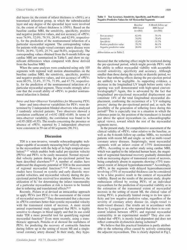
the whole, most myocardial segments displayed +PEVL,irrespective of regional wall motion score (Table 4). Arepresentative example of +PEVL in transmural MI; that is,a false positive result, is clearly illustrated in Fig1, whichshows +PEVL was manifested in the basal and mid inferiormyocardial walls, in which transmural MI was documentedby delayed-enhanced cardiac MRI.
Similar results were derived from analyses using 109 seg-ments with regional wall motion abnormalities on the base-line cardiac MRI; that is, +PEVL was consistently found inmost myocardial segments showing dysfunctional motion(18 of 25 segments (72.0%) for hypokinetic segments vs 59of 84 segments (70.2%) for akinetic segments; p=0.99).
Ability of +PEVL to Differentiate Transmural FromNontransmural MI
In an attempt to simply examine the ability of +PEVL todifferentiate transmural from nontransmural myocardialnecrosis, all myocardial segments were assigned to either anontransmural infarction group, involving only subendocar-
Table 4 Comparison of the Proportion of Segments With +PEVL According to Regional Myocardial Wall Motion Determined by Cardiac MRI
Regional myocardial wall motion
Normal Hypokinesia Akinesiap value
+PEVL 251 18 59% within PEVL category 76.5% 5.5% 18.0%% within regional wall motion 74.3% 72.0% 70.2% 0.45
–PEVL 87 7 25% within PEVL category 73.1% 5.9% 21.0%% within regional wall motion 25.7% 28.0% 29.8%
Total 338 25 84
Abbreviations see in Tables 2,3.
Fig1. A representative example of a tissue Doppler imaging (TDI) tracing of a false positive result in a patient withmyocardial infarction (MI). This patient experienced MI in the area of the right coronary artery 1 day before enrolling inthe study, which is evident based on the akinetic basal and mid inferior myocardial segments in end-diastole (A) and end-systole (B). Even if cardiac magnetic resonance imaging with delayed gadolinium enhancement well displayed the trans-mural infarction of these myocardial segments without any expectation of myocardial functional recovery (C), positivelongitudinal pre-ejectional velocity was definitely demonstrated by TDI analysis (D,E). In this patient, the left ventricularapex showed normal wall motion.
1909Clinical Validity of PEVL
Circulation Journal Vol.71, December 2007
dial layers (ie, the extent of infarct thickness is <50%), or atransmural infarction group, in which the subendocardiallayer and any degree of the epicardial layer were involved(ie, the extent of infarct thickness is ≥50%). Based on thebaseline cardiac MRI, the sensitivity, specificity, positiveand negative predictive values, and test accuracy of +PEVL
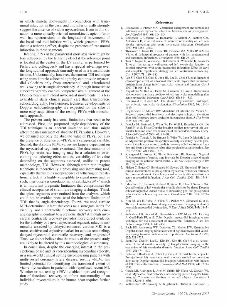
were 74.9%, 32.0%, 79.3%, 26.9%, and 62.9%, respective-ly, for the prediction of the nontransmural infarction of aparticular myocardial segment. The corresponding valuesfor patients with single-vessel coronary artery disease were70.8%, 26.9%, 72.0%, 25.7%, and 58.8%, respectively. Thecorresponding values obtained from the 6-month follow-upcardiac MRI are summarized in Table 5, and show no sig-nificant differences when compared with those derivedfrom the baseline MRI.
When the same analyses were conducted using only 105segments with regional wall motion abnormalities on thebaseline cardiac MRI, the sensitivity, specificity, positiveand negative predictive values, and test accuracy of +PEVL
were 60.5%, 32.4%, 37.7%, 71.9%, and 47.7%, respective-ly, for the prediction of the nontransmural infarction of aparticular myocardial segment. These results strongly advo-cate that the overall ability of +PEVL to predict nontrans-mural infarction is limited.
Intra- and Inter-Observer Variabilities for Measuring PEVL
Inter- and intra-observer variabilities for PEVL were de-termined by 2 independent blinded observers who analyzed5 randomly selected patients. Two observers showed acorrelation coefficient of r=0.92 (SEE=0.69). In terms ofintra-observer variability, the correlation was found to ber=0.95 (SEE=0.55). The results concerning the presence orabsence of +PEVL reported by 2 independent observerswere consistent in 59 out of 60 segments (98.3%).
DiscussionTDI is a non-invasive, versatile echocardiographic tech-
nique capable of accurately measuring brief velocity changesin the myocardium with the help of its high temporal reso-lution,15–17 which enables both radial pre-ejection velocity(PEVR) and PEVL to be easily measured. Normal myocar-dial velocity pattern during the pre-ejectional period hasbeen described elsewhere.18,19 A number of studies haveaddressed the diagnostic potential of TDI in the assessmentof regional myocardial function.15,19,20 However, moststudies have focused on systolic and early diastolic myo-cardial velocities, and myocardial velocity during the pre-ejectional period has received less attention. Moreover, theutilization of SL’ for determining the infarct transmuralityof a particular myocardium at risk is known to be limiteddue to tethering and translational effects.20,21
Recently, Pislaru et al devised an intracardiac approachto apply PEVR (not PEVL) to the assessment of the extentof myocardial necrosis.12 They demonstrated that a decreasein +PEVR correlates better than systolic myocardial velocitywith the transmural extent of necrosis. A more recentanimal experimental study suggested that PEVL is highlysensitive to changes in blood supply and that its use couldmake TDI a more powerful tool for quantifying regionalmyocardial function.13 Even more recently, using a trans-thoracic approach, Penicka et al showed the usefulness ofPEVL for predicting the recovery of contractile functionduring follow up in the setting of recent MI and a single-vessel coronary artery disease.9 In their study, they hypo-
thesized that the tethering effect might be restricted duringthe pre-ejectional period, which might provide PEVL withthe ability to reflect myocardial viability with acceptableoverall sensitivity, specificity and accuracy. Althoughsmaller than those during the systolic or diastolic period, webelieve that tethering effects during the pre-ejection periodare unlikely to be negligible. As supporting evidence, adecrease in the longitudinal LV length before aortic valveopening was well demonstrated with high-speed cineven-triculography.22 Again, this is advocated by the fact thatlongitudinal pre-ejectional myocardial displacement canconstitute 14% of the total longitudinal myocardial dis-placement, confirming the occurrence of a ‘LV reshapingprocess’ during the pre-ejectional period and, as such, thepossibility of the generation of tethering force during thisperiod.23 This is especially true in a situation in which thereference point (ie, the position of the transducer) is locatedjust above the apical myocardium (ie, echocardiographicapical views), toward which the rest of the myocardialsegments move.
In the present study, for comprehensive assessment of theclinical validity of +PEVL value relative to the baseline, aswell as the 6-month follow-up cardiac MRIs, we recruitedpatients with recent MI and single- as well as multi-vesselcoronary artery diseases. Surprisingly, most myocardialsegments with an infarct extent of >75% demonstrated+PEVL. According to an earlier study using cardiac MRI,which was applied to the infarcted human heart, the magni-tude of segmental functional recovery gradually diminishedwith an increasing degree of transmural extent of necrosis,being completely absent in segments showing >75% trans-mural extent of delayed hyperenhancement.14 Accordingly,+PEVL in the segments with delayed hyperenhancementinvolving >75% of myocardial thickness can be consideredto be a false positive result in the context of myocardialviability. Based on the results of the present study, despiteenthusiasm offered by resting PEVL of an individualmyocardium for the prediction of myocardial viability or inthe estimation of the transmural extent of myocardialnecrosis in the setting of recent MI, the clinical value ofresting PEVL is practically limited owing to its low speci-ficity and low negative predictive value, regardless of theseverity of coronary artery disease (ie, single-vessel vsmulti-vessel disease). Our results are in accordance withthose by Lyseggen et al, who reported that there is no con-sistent relation between +PEVL and regional myocardialcontractility in an experimental model.24 They also con-cluded that +PEVL is mostly load-dependent and does notreflect contractile dysfunction during ischemia.24
We believe that this practical limitation is partly attribut-able to the tethering effect caused by actively contractingthe adjacent myocardium. This is clearly depicted in Fig1,
Table 5 Test Accuracy, Sensitivity, Specificity, and Positive and Negative Predictive Values for All Myocardial Segments
Baseline 6-Month follow-upMRI-based MRI-based
(n=464) (n=368)
Test accuracy 62.9% 70.1%Sensitivity 74.9% 78.4%Specificity 32.0% 31.7%Positive predictive value 79.3% 84.1%Negative predictive value 26.9% 24.1%
Abbreviation see in Table 2.
1910 KIM H-K et al.
Circulation Journal Vol.71, December 2007
in which akinetic movements in conjunction with trans-mural infarction in the basal and mid inferior walls stronglysuggest the absence of viable myocardium. Even in this sit-uation, a more apically oriented normokinetic apicoinferiorwall has repercussions on the longitudinal movements ofthe basal and mid inferior walls, which generate +PEVL
due to a tethering effect, despite the presence of transmuralinfarction in these segments.
Resting PEVR at the parasternal short axis view might beless influenced by the tethering effect if the reference pointis located at the center of the LV cavity, as performed byPislaru and colleagues,12 and has a special advantage overthe longitudinal approach currently used in a transthoracicfashion. Unfortunately, however, the current TDI techniqueusing transthoracic echocardiography can provide myocar-dial velocities only from anteroseptal and inferolateralwalls owing to its angle-dependency. Although intracardiacechocardiography enables comprehensive alignment of theDoppler beam with radial myocardial movements, it is lessavailable in daily clinical patient care than transthoracicechocardiography. Furthermore, technical developments ofDoppler echocardiography are expected for the sake ofmore easy acquisition of the resting PEVR with transtho-racic approach.
The present study has some limitations that need to beaddressed. First, the purported angle-dependency of theTDI technique is an inherent limitation and, thus, mightaffect the measurement of absolute PEVL values. However,we obtained not only the absolute value of PEVL, but alsothe presence or absence of +PEVL as a categorical variable.Second, the absolute PEVL values are largely dependent onthe myocardial segments examined. The determination ofPEVL by strain rate imaging may be a solution to over-coming the tethering effect and the variability of its valuedepending on the segments assessed, unlike its parentmethodology, TDI. However, although strain rate imagingtechnique is theoretically a promising and interesting tool,especially thanks to its independence of tethering or transla-tional effect, it is highly susceptible to signal noise and, assuch, inter-observer correlation is not satisfactory,25–27 whichis an important pragmatic limitation that compromises theclinical acceptance of strain rate imaging technique. Third,the apical segments were omitted from the analyses, whichcould not be avoided because of the inherent limitation ofTDI; that is, angle-dependency. Fourth, we used cardiacMRI-determined infarct thickness as a surrogate index forviability, not a contractile functional recovery with cine-angiography in contrast to a previous study.6 Although myo-cardial contractile recovery provides more direct evidencefor the viability of a given myocardial segment, infarct trans-murality assessed by delayed enhanced cardiac MRI is amore sensitive and objective marker for cardiac remodeling,delayed myocardial contractile recovery, and prognosis.14
Thus, we do not believe that the results of the present studyare likely to be altered by this methodological discrepancy.
In conclusion, despite the emerging interest in the pre-ejectional phase and its corresponding myocardial velocity,in a real-world clinical setting encompassing patients withmulti-vessel coronary artery disease, resting +PEVL haslimited potential for identifying the transmural extent ofviable myocardium in patients with reperfused recent MI.Whether or not resting +PEVR enables improved recogni-tion of functional recovery or infarct transmurality of anindividual myocardium in the human heart requires furtherstudy.
References1. Braunwald E, Pfeffer MA. Ventricular enlargement and remodeling
following acute myocardial infarction: Mechanisms and management.Am J Cardiol 1991; 68: 1D–6D.
2. Bolognese L, Cerisano G, Buonamici P, Santini A, Santoro GM,Antoniucci D, et al. Influence of infarct-zone viability on left ven-tricular remodeling after acute myocardial infarction. Circulation1997; 96: 3353–3359.
3. Thanavaro S, Krone RJ, Kleiger RE, Province MA, Miller JP, deMelloVR, et al. In-hospital prognosis of patients with first nontransmuraland transmural infarctions. Circulation 1980; 61: 29–33.
4. Tani S, Nagao K, Watanabe I, Kikushima K, Watanabe K, AnazawaT, et al. Increasingly well-preserved left ventricular function inhospital survivors with acute myocardial infarction: Effect of earlyand complete reperfusion strategy on left ventricular remodeling.Circ J 2007; 71: 180–185.
5. Lee SH, Choi SH, Choi S, Jung JH, Lee N, Choi YJ, et al. Impact ofchronotropic effect of cilostazol after acute myocardial infarction:Insights from change in left ventricular volume and function. Circ J2007; 71: 106–111.
6. Nagashima M, Itoh A, Otsuka M, Kasanuki H, Haze K. Reperfusionphenomenon is a strong predictor of left ventricular remodelling afteracute myocardial infarction. Circ J 2005; 69: 884–889.
7. Braunwald E, Kloner RA. The stunned myocardium: Prolonged,postischemic ventricular dysfunction. Circulation 1982; 66: 1146–1149.
8. Heyndrickx GR, Millard RW, McRitchie RJ, Maroko PR, Vatner SF.Regional myocardial functional and electrophysiological alterationsafter brief coronary artery occlusion in conscious dogs. J Clin Invest1975; 56: 978–985.
9. Penicka M, Bartunek J, Wijns W, De Wolf I, Heyndrickx GR, DeRaedt H, et al. Tissue Doppler imaging predicts recovery of left ven-tricular function after recanalization of an occluded coronary artery.J Am Coll Cardiol 2004; 43: 85–91.
10. Penicka M, Tousek P, De Bruyne B, Wijns W, Lang O, Madaric J, etal. Myocardial positive pre-ejection velocity accurately detects pres-ence of viable myocardium, predicts recovery of left ventricular func-tion and bears a prognostic value after surgical revascularization. EurHeart J 2007; 28: 1366–1373.
11. Kjaergaard J, Hassager C, Oh JK, Kristensen JH, Berning J, SogaardP. Measurement of cardiac time intervals by Doppler tissue M-modeimaging of the anterior mitral leaflet. J Am Soc Echocardiogr 2005;18: 1058–1065.
12. Pislaru C, Bruce CJ, Belohlavek M, Seward JB, Greenleaf JF. Intra-cardiac measurement of pre-ejection myocardial velocities estimatesthe transmural extent of viable myocardium early after reperfusion inacute myocardial infarction. J Am Coll Cardiol 2001; 38: 1748–1756.
13. Edvardsen T, Urheim S, Skulstad H, Steine K, Ihlen H, Smiseth OA.Quantification of left ventricular systolic function by tissue Dopplerechocardiography: Added value of measuring pre- and postejectionvelocities in ischemic myocardium. Circulation 2002; 105: 2071–2077.
14. Kim RJ, Wu E, Rafael A, Chen EL, Parker MA, Simonetti O, et al.The use of contrast-enhanced magnetic resonance imaging to identifyreversible myocardial dysfunction. N Engl J Med 2000; 343: 1445–1453.
15. Sutherland GR, Stewart MJ, Groundstroem KW, Moran CM, FlemingA, Guell-Peris FJ, et al. Color Doppler myocardial imaging: A newtechnique for the assessment of myocardial function. J Am SocEchocardiogr 1994; 7: 441–458.
16. Bach DS, Armstrong WF, Donovan CL, Muller DW. QuantitativeDoppler tissue imaging for assessment of regional myocardial veloci-ties during transient ischemia and reperfusion. Am Heart J 1996;132: 721–725.
17. Sohn DW, Chai IH, Lee DJ, Kim HC, Kim HS, Oh BH, et al. Assess-ment of mitral annulus velocity by Doppler tissue imaging in theevaluation of left ventricular diastolic function. J Am Coll Cardiol1997; 30: 474–480.
18. Pellerin D, Berdeaux A, Cohen L, Giudicelli JF, Witchitz S, Veyrat C.Pre-ejectional left ventricular wall motions studied on consciousdogs using Doppler myocardial imaging: Relationships with indicesof left ventricular function. Ultrasound Med Biol 1998; 24: 1271–1283.
19. Garcia MJ, Rodriguez L, Ares M, Griffin BP, Klein AL, Stewart WJ,et al. Myocardial wall velocity assessment by pulsed Doppler tissueimaging: Characteristic findings in normal subjects. Am Heart J1996; 132: 648–656.
20. Wilkenshoff UM, Sovany A, Wigstrom L, Olstad B, Lindstrom L,

1911Clinical Validity of PEVL
Circulation Journal Vol.71, December 2007
Engvall J, et al. Regional mean systolic myocardial velocity estima-tion by real-time color Doppler myocardial imaging: A new techniquefor quantifying regional systolic function. J Am Soc Echocardiogr1998; 11: 683–692.
21. Hatle L, Sutherland GR. Regional myocardial function--a newapproach. Eur Heart J 2000; 21: 1337–1357.
22. Karliner JS, Bouchard RJ, Gault JH. Dimensional changes of thehuman left ventricle prior to aortic valve opening: A cineangiogra-phic study in patients with and without left heart disease. Circulation1971; 44: 312–322.
23. Lind B, Eriksson M, Roumina S, Nowak J, Brodin LA. Longitudinalisovolumic displacement of the left ventricular myocardium assessedby tissue velocity echocardiography in healthy individuals. J Am SocEchocardiogr 2006; 19: 255–265.
24. Lyseggen E, Rabben SI, Skulstad H, Urheim S, Risoe C, Smiseth OA.
Myocardial acceleration during isovolumic contraction: Relationshipto contractility. Circulation 2005; 111: 1362–1369.
25. Hanekom L, Jenkins C, Jeffries L, Case C, Mundy J, Hawley C, et al.Incremental value of strain rate analysis as an adjunct to wall-motionscoring for assessment of myocardial viability by dobutamine echo-cardiography: A follow-up study after revascularization. Circulation2005; 112: 3892–3900.
26. Marwick TH. Measurement of strain and strain rate by echocardio-graphy: Ready for prime time? J Am Coll Cardiol 2006; 47: 1313–1327.
27. Cho GY, Chan J, Leano R, Strudwick M, Marwick TH. Comparisonof two-dimensional speckle and tissue velocity based strain and vali-dation with harmonic phase magnetic resonance imaging. Am JCardiol 2006; 97: 1661–1666.