clinical utility of kras and braf mutations in a cohort of patients with colorectal neoplasms...
TRANSCRIPT
168 -
Original Study
Clinical Utility of KRAS and BRAF Mutations in aCohort of Patients With Colorectal NeoplasmsSubmitted for Microsatellite Instability Testing
Allison M. Cushman-Vokoun,1 Daniel G. Stover,2,5 Zhiguo Zhao,3
Elizabeth A. Koehler,3 Jordan D. Berlin,2,4 Cindy L. Vnencak-Jones1,6
AbstractMutations in KRAS and BRAF impact response to EGFR-based therapy. One hundred eleven paraffin-embedded tumor specimens submitted for MSI testing were screened for 7 KRAS mutations and the BRAFp.V600E mutation using fluorescent allele-specific PCR. We demonstrate patterns of KRAS and BRAF muta-tions in microsatellite-stable and -unstable tumors that are relevant and may be applied in the clinical setting.Background: Molecular analysis has become important in colorectal carcinoma (CRC) evaluation. Alterations inKRAS, BRAF, or mismatch repair (MMR) genes may determine therapeutic response or define a hereditary cancersyndrome. Correlation of DNA studies with clinical findings will further clarify the clinical utility of these markers.Patients and Methods: A retrospective study was performed on 111 paraffin-embedded tumor specimens submittedfor microsatellite instability (MSI) testing based on clinical history or histologic examination, or both. DNA sampleswere screened for 7 KRAS mutations and the BRAF p.V600E mutation using fluorescent allele-specific polymerase-chain reaction (PCR) and capillary electrophoresis. Clinical data were collected through chart review. Results: Fifty-eight male and 53 female patients were studied. The incidence of KRAS and BRAF mutations was 49.5% and 7.2%,respectively. Dideoxy sequencing verified KRAS mutation status in 46 of 49 specimens tested. There was a trendtoward significance of individual KRAS mutations on survival (P ¼ .003). Dually positive KRAS and MSI tumorsexclusively demonstrated p.G12D and p.G13D mutations (G>A transitions). BRAF-mutated tumors were predomi-nantly right-sided and associated with a borderline worse prognosis. Forty-eight percent of tumors with MSI werepresent in the left colon or rectum. Conclusion: Allele-specific PCR is an accurate and convenient method to assessKRAS and BRAF mutations and may detect mutations not identified by dideoxy sequencing. KRASmutation status, inconjunction with morphologic or clinical parameters, may be useful in determining whether a tumor should be testedfor MSI. MSI testing should not be considered exclusively in right-sided lesions. BRAF analysis may not be useful inrectal adenocarcinomas and should be evaluated in larger studies.
Clinical Colorectal Cancer, Vol. 12, No. 3, 168-78 ª 2013 Elsevier Inc. All rights reserved.Keywords: Allele specific, Missense, Mutation, Transversion, Biomarker
IntroductionColorectal carcinoma (CRC) accounted for approximately
50,000 deaths in the United States in 2013.1 There are 2 primary
Drs Cushman-Vokoun and Stover contributed equally to this work.
This work was presented in part in abstract form at the 2009 Association for MolecularPathology Meeting; November 18-22, 2009; Kissimmee, FL and at the 2011 AmericanSociety of Clinical Oncology Gastrointestinal Cancers Symposium; Jan 20-22, 2011;San Francisco, CA.
1Department of Pathology, Microbiology and Immunology, Vanderbilt-IngramComprehensive Cancer Center, Vanderbilt University Medical Center, Nashville, TN2Department of Medicine, Vanderbilt-Ingram Comprehensive Cancer Center,Vanderbilt University Medical Center, Nashville, TN3Department of Biostatistics, Division of Cancer Biostatistics, Vanderbilt-IngramComprehensive Cancer Center, Vanderbilt University Medical Center, Nashville, TN
Clinical Colorectal Cancer September 2013
molecular pathways for CRC carcinogenesis: the chromosomalinstability (CIN) pathway (80%-85% of CRC) and the microsat-ellite instability (MSI) pathway (15%-20% of CRC).2-4 The CIN
4Division of Hematology/Oncology, Vanderbilt-Ingram Comprehensive CancerCenter, Vanderbilt University Medical Center, Nashville, TN5Dana-Farber Cancer Institute/Partners CancerCare, Massachusetts General HospitalCancer Center, Boston, MA6Department of Pediatrics, Vanderbilt University Medical Center, Nashville, TN
Submitted: Aug 14, 2012; Revised: Feb 19, 2013; Accepted: Apr 15, 2013; Epub:Jun 14, 2013
Address for correspondence: Allison M. Cushman-Vokoun, MD, PhD, Department ofPathology andMicrobiology, 985454NebraskaMedicalCenter, Omaha,NE68105-5454Fax: þ1 402 559 2490; e-mail contact: [email protected]
1533-0028/$ - see frontmatter ª 2013 Elsevier Inc. All rights reserved.http://dx.doi.org/10.1016/j.clcc.2013.04.005
pathway consists of chromosomal deletions or duplications inaddition to multiple mutations in various oncogenes and tumorsuppressor genes. A well-characterized signaling pathway involved inthe development of colorectal cancer through the CIN pathway isthe epidermal growth factor receptor (EGFR) pathway. An initi-ating step in the stimulated EGFR pathway is activation of Kirstenras (KRAS), a guanosine triphosphatase (GTPase) that is activatedthrough binding of guanosine triphosphate (GTP), which subse-quently stimulates the serine/threonine kinase BRAF, triggering theMAPK/ERK pathway.5 KRAS gene mutations are thought to occurearly in the adenoma-carcinoma continuum and are estimated to bepresent in 30% to 50% of colon adenocarcinomas.2,6-9 Conservedmissense mutations in codons 12, 13, and 61 result in prolongedbinding of guanosine triphosphate (GTP) and constitutive activa-tion of the KRAS protein. In the BRAF gene, 1 specific activatingtransversion mutation, c.1799T>A (p.V600E) accounts for mostBRAF mutations associated with human cancers, including 10% to40% of colon cancers.8,10-12 Activating mutations in KRAS or BRAFhave recently been shown to render advanced CRCs insensitive toEGFR inhibitors, and KRAS and BRAF are now commonly evalu-ated for mutations before EGFR inhibitor therapy is initiated.9,13-15
Because KRAS and BRAF mutations have become increasinglyimportant in prognosis and treatment, identifying rapid and sensi-tive methods to detect these mutations is critical. Several differentapproaches demonstrate utility, including Sanger sequencing (alsoknown as dideoxy sequencing), pyrosequencing, high-resolutionmelting analysis, and allele-specific PCR.16,17 Many early studiesused Sanger sequencing to identify KRAS mutations, a techniquethat is considered the gold standard but one that also requires themutant allele to be present within the tumor at a rate of at least 15%to 20%. The sensitivity of allele-specific PCR is estimated to be 1%or better and allows for identification of target mutations in biopsyor pathologic specimens with low tumor burden. Next-generationsequencing platforms may eventually be useful for evaluating genesfor somatic mutations in large panels; however, these platforms arecurrently not easily accessible or cost-effective for single gene assaysand require sophisticated bioinformatics for analysis.
Like KRAS and BRAF, identifying MSI is important clinicallyto determine prognosis and counseling regarding inheritedcancer risks. The presence of MSI in CRC may indicate a germlineMMR defect, suggesting an autosomal dominant hereditary coloncancer syndrome—hereditary nonpolyposis colon cancer syndrome(HNPCC or Lynch syndrome).3,18 Alternatively, patients withsporadic MSI tumors caused by MLH1 gene silencing by biallelicpromoter hypermethylation have a better prognosis but may have adecreased response to 5-fluorouracil.3,19-21 Although MSI high(MSI-H) tumors have an improved prognosis, CIN has been foundto be associated with worse prognosis: In 1 meta-analysis of survival,the hazard ratio (HR) was 1.45 (95% confidence interval [CI],1.35-1.55; P < .001)4 for a CIN (defined as polyploidy or aneu-ploidy)-related neoplasm.
To assess the utility of allele-specific PCR in patients withcolorectal cancer, we used laboratory-developed allele-specific fluo-rescent PCR assays specific for KRAS and BRAF mutations toevaluate a cohort of patients diagnosed with colorectal neoplasmswhose samples were submitted for MSI analysis. We evaluated theassociation between MSI and EGFR pathway mutations. An
extensive chart review was also performed to relate mutation find-ings with patient parameters.
Patients and MethodsCohort Selection and Chart Review
A retrospective study was performed on 135 formalin-fixed,paraffin-embedded (FFPE) tumor specimens submitted to theVanderbilt University Medical Center Clinical Molecular DiagnosticLaboratory between January 1, 2007 and March 31, 2009 for MSItesting. Samples were submitted by a clinician or anatomic pathol-ogist for MSI testing loosely based on the Bethesda Criteria22: age <50 years and family history or characteristic histologic features, orboth. Twenty-four patient samples were excluded from additionalstudies because of insufficient DNA, noncolonic tumor testing, oroutside referral, disqualifying these specimens from institutional re-view board approval. Chart reviews were performed on the remaining111 patients. Parameters evaluated in the chart review included age,sex, age at diagnosis, pathologic stage, treatment (surgery, radiation,and chemotherapy), and course of disease. If germline MMR mu-tation status was known, it was also recorded. One patient had 2synchronous tumors (1 in the right colon and 1 in the left colon) thatwere tested for all 3 markers. Only the right-sided MSI-H tumor wasincluded in the study (the left tumor was microsatellite stable [MSS]).Fourteen cases were considered stage 0, which included 3 adenomas(no high-grade dysplasia), 4 adenomas with high-grade dysplasia, and3 adenomas with carcinoma in situ. Two patients with stage 0 diseasehad no residual cancer after neoadjuvant therapy, and 1 patient hadinvasive carcinoma but did not undergo resection because of othercomorbidities (no pathologic staging). In a final patient, an adenomawith high-grade dysplasia was tested, but the individual also hadinvasive carcinoma in a rectal adenoma that was not submitted fortesting because it was outside material. Overall survival (OS) wasdefined as time from surgery to death from any cause or last follow-upand was determined using chart review and the Social SecurityDeath Index. The study was approved by the Vanderbilt UniversityMedical Center Institutional Review Board. Patient demographicsand characteristics are identified in Table 1.
DNA Extraction from Peripheral Blood and FFPESpecimens
FFPE tumor blocks were selected by the surgical pathologist forclinical testing. For MSI testing, separate tumor and normal FFPEblocks were sectioned. Only cases with 30% to 50% or more tumorpresent in the block were accepted for testing. Tissue was depar-affinized using xylene, ethanol washes, and acetone dehydration,and after cell lysis and proteinase K treatment, the DNA wasextracted using the Puregene DNA Isolation or QIAquick PCRpurification kit (QIAGEN, Inc. Valencia, CA). For certain MSIcases, when normal tissue was unavailable, peripheral blood wasextracted using the EZ1 Biorobot automated extractor (QIAGEN,Inc. Valencia, CA) and used as normal DNA for comparison.
MSI AnalysisFor MSI testing, 5 to 10 ng of each tumor and normal DNA was
analyzed using the Promega MSI Analysis System (Promega Corp,Madison, WI) according to the manufacturer’s instructions.Amplicons were detected using capillary electrophoresis on an ABI
Clinical Colorectal Cancer September 2013 - 169

Table 1 Patient Characteristics and Mutation Status (KRAS and BRAF)
VariableOverall
n [ 111 (%)No Mutationn [ 48 (%)
KRAS or BRAFn [ 63 (%) P Value
Age (y) 50 (44-61)a 48 (42-56)a 51 (45-61)a .13b
< 40 16 (14.4) 10 (20.8) 6 (9.5)
40-49 39 (35.1) 18 (37.5) 21 (33.3)
50-59 27 (24.3) 9 (18.8) 18 (28.6)
> 60 29 (26.1) 11 (22.9) 18 (28.6)
Sex .85c
Female 53 (47.7) 22 (45.8) 31 (49.2)
Male 58 (52.3) 26 (54.2) 32 (50.8)
MSI .84c
MSS/MSI-L 90 (81.1) 39 (81.2) 51 (81)
MSI-H 21 (18.9) 9 (18.8) 12 (19)
Stage .15d
0 or adenoma 14 (12.6) 6 (12.5) 8 (12.7)
1 19 (17.1) 12 (25.0) 7 (11.1)
2 17 (15.3) 4 (8.3) 13 (20.6)
3 33 (29.7) 16 (33.3) 17 (27.0)
4 28 (25.2) 10 (20.8) 18 (28.6)
Location .70c
Left Colon 29 (26.1) 12 (25.0) 17 (27.0)
Right Colon 36 (32.4) 14 (29.2) 22 (34.9)
Rectum 46 (41.4) 22 (45.8) 24 (38.1)
Abbreviations: MSI-H ¼ microsatellite instability high; MSS/MSI-L ¼ microsatellite stable/microsatellite instability low.aMedian (interquartile range).bWilcoxon rank-sum test.cc2 test.dFisher exact test.
Utility of KRAS, BRAF, and MSI Mutation Patterns
170 -
3130xl Genetic Analyzer and analyzed using GeneMapper Software,version 3.7 (Applied Biosystems/LifeTechnologies, Grand Island,NY). Nine patients with MSI-H tumors underwent further evalu-ation for specific MMR testing by immunohistochemical analysis orsequencing, or both, at an outside facility. Not all patients withMSI-H were tested because of patient preference, lack of adequatetissue, loss in follow-up, or BRAF p.V600E positivity.
KRAS Mutational Analysis and SequencingMutations in KRAS codons 12 and 13 in exon 2 were identified
using allele-specific PCR with primer sequences adapted fromprimers previously reported by Takeda et al or Lecomte et al(Figure 1).23,24 The 7 most common mutations in colon cancerwere evaluated and included p.G12D, p.G12V, p.G12A, p.G12C,p.G12S, p.G12R, and p.G13D. Seven separate reactions wereperformed, each containing a fluorescently labeled upstream wild-type primer (RASWTFO), an unlabeled downstream primer(RASRO), and 1 differentially labeled fluorescent mutation-specificprimer. Each primer was used at a final concentration of 0.25 mM.The reaction conditions included 1 cycle at 95�C for 5 minutes;40 cycles at 95�C for 30 seconds, 60�C for 2 minutes, 70�C for2 minutes; and a final extension at 70�C for 10 minutes. Ampliconswere detected using capillary electrophoresis on an ABI 3130xlGenetic Analyzer (Applied Biosystems/Life Technologies, GrandIsland, NY) and analyzed using GeneMapper Software (AppliedBiosystems/Life Technologies, Grand Island, NY).
Clinical Colorectal Cancer September 2013
To validate our KRAS mutationespecific assay, a subset ofspecimens (n ¼ 49) were sequenced using the ABI BigDyeTerminator v3.1 system (Applied Biosystems/Life Technologies,Grand Island, NY) according to the manufacturer’s instructions.
BRAF p.V600E Mutational AnalysisThe c.1799T>A (p.V600E) mutation was identified using
allele-specific fluorescent PCR and capillary electrophoresis detec-tion. Five to 20 ng of genomic DNA extracted from FFPE tumorwas amplified using the following primers: BRAF-FO-50 TGTTTTCCTTTACTTACTACACCTCAGA 30 (0.05 mM final concen-tration), BRAF-RO 50 HEX-CAGTGGAAAAATAGCCTCAATTCT 30 (0.05 mM final concentration), and BRAF-Rmt 50 FAM-ACCCACTCCATCGAGATTTCT 30 (0.1 mM final concentration).The reaction conditions included 1 cycle at 94�C for 5 minutes;40 cycles at 94�C for 30 seconds, 56�C for 45 seconds, 72�C for45 seconds; and a final extension cycle at 72�C for 10 minutes.Amplicons were detected using capillary electrophoresis on an ABI3130xl Genetic Analyzer and analyzed using GeneMapper Software(Applied Biosystems/Life Technologies, Grand Island, NY).
Statistical AnalysisDemographic and pathologic characteristics were summarized
and compared across mutation group (KRAS or BRAF present orabsent). Continuous characteristic age was summarized by medianwith 25th and 75th percentiles (interquartile ratio) and compared
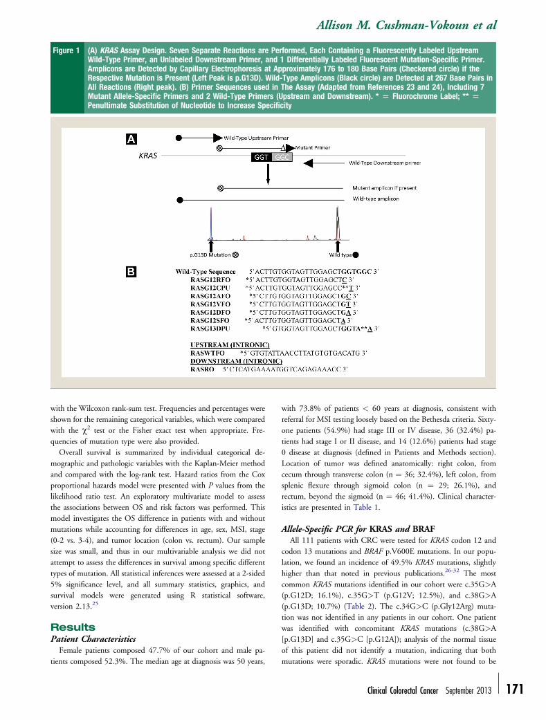
Figure 1 (A) KRAS Assay Design. Seven Separate Reactions are Performed, Each Containing a Fluorescently Labeled UpstreamWild-Type Primer, an Unlabeled Downstream Primer, and 1 Differentially Labeled Fluorescent Mutation-Specific Primer.Amplicons are Detected by Capillary Electrophoresis at Approximately 176 to 180 Base Pairs (Checkered circle) if theRespective Mutation is Present (Left Peak is p.G13D). Wild-Type Amplicons (Black circle) are Detected at 267 Base Pairs inAll Reactions (Right peak). (B) Primer Sequences used in The Assay (Adapted from References 23 and 24), Including 7Mutant Allele-Specific Primers and 2 Wild-Type Primers (Upstream and Downstream). * [ Fluorochrome Label; ** [Penultimate Substitution of Nucleotide to Increase Specificity
Allison M. Cushman-Vokoun et al
with the Wilcoxon rank-sum test. Frequencies and percentages wereshown for the remaining categorical variables, which were comparedwith the c2 test or the Fisher exact test when appropriate. Fre-quencies of mutation type were also provided.
Overall survival is summarized by individual categorical de-mographic and pathologic variables with the Kaplan-Meier methodand compared with the log-rank test. Hazard ratios from the Coxproportional hazards model were presented with P values from thelikelihood ratio test. An exploratory multivariate model to assessthe associations between OS and risk factors was performed. Thismodel investigates the OS difference in patients with and withoutmutations while accounting for differences in age, sex, MSI, stage(0-2 vs. 3-4), and tumor location (colon vs. rectum). Our samplesize was small, and thus in our multivariable analysis we did notattempt to assess the differences in survival among specific differenttypes of mutation. All statistical inferences were assessed at a 2-sided5% significance level, and all summary statistics, graphics, andsurvival models were generated using R statistical software,version 2.13.25
ResultsPatient Characteristics
Female patients composed 47.7% of our cohort and male pa-tients composed 52.3%. The median age at diagnosis was 50 years,
with 73.8% of patients < 60 years at diagnosis, consistent withreferral for MSI testing loosely based on the Bethesda criteria. Sixty-one patients (54.9%) had stage III or IV disease, 36 (32.4%) pa-tients had stage I or II disease, and 14 (12.6%) patients had stage0 disease at diagnosis (defined in Patients and Methods section).Location of tumor was defined anatomically: right colon, fromcecum through transverse colon (n ¼ 36; 32.4%), left colon, fromsplenic flexure through sigmoid colon (n ¼ 29; 26.1%), andrectum, beyond the sigmoid (n ¼ 46; 41.4%). Clinical character-istics are presented in Table 1.
Allele-Specific PCR for KRAS and BRAFAll 111 patients with CRC were tested for KRAS codon 12 and
codon 13 mutations and BRAF p.V600E mutations. In our popu-lation, we found an incidence of 49.5% KRAS mutations, slightlyhigher than that noted in previous publications.26-32 The mostcommon KRAS mutations identified in our cohort were c.35G>A(p.G12D; 16.1%), c.35G>T (p.G12V; 12.5%), and c.38G>A(p.G13D; 10.7%) (Table 2). The c.34G>C (p.Gly12Arg) muta-tion was not identified in any patients in our cohort. One patientwas identified with concomitant KRAS mutations (c.38G>A[p.G13D] and c.35G>C [p.G12A]); analysis of the normal tissueof this patient did not identify a mutation, indicating that bothmutations were sporadic. KRAS mutations were not found to be
Clinical Colorectal Cancer September 2013 - 171
Table 2 Molecular Markers: MSI Status, KRAS Mutations, andBRAF p.V600E Mutation
Molecular Markers n (%)
MSI Status (n [ 111)a,b
High 21 (18.9)
Stable 90 (81.1)
KRAS Mutations (n [ 111)
Positive 55 (49.5)
Negative 56 (50.5)
Specific KRAS Mutations (n [ 112)c
c.35G>C (p.G12A) 5 (4.5)
c.34G>T (p.G12C) 4 (3.6)
c.35G>A (p.G12D) 18 (16.1)
c.34G>A (p.G12S) 3 (2.7)
c.35G>T (p.G12V) 14 (12.5)
c.38G>A (p.G13D) 12 (10.7)
Wild type 56 (50.0)
BRAF c.1799T>A; p.V600E (n [ 111)
Positive 8 (7.2)
Negative 103 (92.8)
MSI-H Tumors (n [ 21 of 111)
KRAS positive 8 of 21 (38.1)
p.G12D 4
p.G13D 4
KRAS negative 13 of 21 (61.9)
BRAF positive 4 of 21 (19.0)
BRAF negative 17 of 21 (81.0)
MSS (n [ 90 of 111)
KRAS positive 47 of 90 (52.2)
KRAS negative 43 of 90 (47.8)
BRAF positive 4 of 90 (4.4)
BRAF negative 86 of 90 (95.6)
aMicrosatellite instability low (MSI-L) and microsatellite stable (MSS) tumors were considered asa single entity in the statistical analysis.bOne patient had synchronous tumors: 1 MSI-H and 1 MSS. Both were BRAF p.V600E mutationpositive and KRAS mutation negative. He was considered MSI-H/BRAF mutation positive forstatistical analysis.cOne patient had 2 KRAS mutations (p.G12A and p.G13D). For statistical analysis, this patientwas counted as having a p.G13D mutation because this is the more common mutation and wasthe more predominant mutation by dideoxy sequencing.
Utility of KRAS, BRAF, and MSI Mutation Patterns
172 -
significantly associated with location, either at 3 levels (left vs. rightvs. rectum; P ¼ .49) or 2 levels (colon vs. rectum; P ¼ .70)(Table 3). To validate our allele-specific PCR laboratory test, 49 ofthe specimens were also examined by dideoxy sequencing, withKRAS mutation status (mutated or wild type) verified in 46 of 49specimens by dideoxy sequencing. Three mutations identified byallele-specific PCR could not be confirmed by observing the dideoxysequencing curve amplitudes alone.
Eight patients (7.2%) had BRAF p.V600E mutations, similar tothe incidence in previously published reports (w8%).33,34 A ma-jority of BRAF mutations were identified in tumors of the rightcolon (7 of 8) compared with the left colon (1 of 8) or rectum (0 of8), which was statistically significant (P < .001) (Table 3). Therewere no concomitant BRAF and KRAS mutations, consistent with
Clinical Colorectal Cancer September 2013
previous data suggesting that these mutations are typically mutuallyexclusive.8,34,35
Microsatellite Status and KRAS/BRAF MutationsMicrosatellite status was defined in accordance with the Bethesda
guidelines: MSI-H was defined as 2 of 5 markers positive for MSIand low microsatellite instability (MSI-L) was defined as 1 of 5markers positive for MSI.22 Twenty-one patients (18.9%) wereidentified as MSI-H and 1 patient as MSI-L. Given the lack ofconclusive distinction between MSI-L and MSS tumors, we com-bined MSI-L and MSS for further analyses (Table 2).3 MSI statuswas not associated with location, at either 3 levels (left vs. right vs.rectum; P ¼ .11) or 2 levels (colon vs. rectum; P ¼ .22) (Table 3).
Within our MSI-H tumor population, 38.1% had KRAS muta-tions (8 of 21), whereas 19% (4 of 21) had a BRAF c.1799T>A(p.V600E) mutation (Table 2). Of the MSS/MSI-L tumors, 52.2%(47 of 90) had KRAS mutations and 4.4% (4 of 90) had a BRAFmutation (Table 2). Interestingly, all KRAS mutations in MSI-Htumors were a G>A transition within the second base pair of eithercodon 12 or 13 (c.35G>A [p.G12D]: GGT>GAT; c.38G>A[p.G13D]: GGC>GAC) (Table 2). Based on the data presented inthis study, there was strong evidence (P ¼ .006) to reject the nullhypothesis that MSI-H and KRAS mutations c.35G>A orc.38G>A were independent, suggesting a possible correlation be-tween the type of KRAS mutation and the presence of MSI.
Clinicopathologic CorrelationsMedian OS was 46.3 months. Only stage was found to be
significantly associated with survival in both univariate and multi-variate analyses (Table 4). Sex, age at surgery, and tumor locationwere not found to be associated with significant differences insurvival in univariate or multivariable analysis. MSI status did notshow a significant association with OS by univariate analysis (HR,0.85; 95% Cl, 0.32-2.23; P ¼ .73) or multivariate analysis (HR,1.44; 95% CI, 0.52-4.00; P ¼ .49). The presence or absence of anyKRAS mutation was not found to be significantly associated withOS on univariate analysis (P ¼ .47) (Table 4A, Figure 2A). How-ever, when the mutations were stratified by amino acid substitution,there was a trend in effect of a specific mutation on survival (P ¼.003) (Figure 2B), as has been suggested by larger studies of KRASmutation using other detection methods.36 Specifically, all 4 pa-tients with isolated p.G12A mutations appeared to exhibit aggres-sive disease with either metastasis or extensive recurrence (Table 4A,Figure 2B). The fifth patient with a p.G12A mutation also had ap.G13D mutation. On dideoxy sequencing, the p.G13D mutation,a more common KRAS mutation, was the more predominantmutant allele, and this patient was classified as having a p.G13Dmutation for these reasons (Table 2). This patient did not undergoresection because of other comorbidities (no pathologic staging).BRAF-mutated tumors trended toward worse OS than did KRAS-mutated tumors (P ¼ .051), similar to previous studies (Table 4A,Figure 2C).33,34,37
DiscussionAs we enter an era of personalized cancer medicine, identifying
mutations within patient tumors is no longer just an academicpursuit. In colorectal cancer, multiple studies have demonstrated the

Table 3 Association of Tumor Location and Presence of Molecular Markers (n [ 111)
Marker Right (32.4%, n [ 36) Left (26.1%, n [ 29) Rectum (41.4%, n [ 46)
MSI Statusa
MSS, n ¼ 90 (%) 25 (27.8) 25 (27.8) 40 (44.4)
MSI-H, n ¼ 21 (%) 11 (52.4) 4 (19.0) 6 (28.6)
KRAS (n [ 111)b
Wild type, n ¼ 56 (%) 21 (37.5) 13 (23.2) 22 (39.3)
Positive, n ¼ 55 (%) 15 (27.3) 16 (29.1) 24 (43.6)
BRAF c.1799T>A (p.V600E)c
Wild type, n ¼ 103 (%) 29 (28.2) 28 (27.2) 46 (44.7)
Positive, n ¼ 8 (%) 7 (87.5) 1 (12.5)d 0 (0.0)
Abbreviations: MSI ¼ microsatellite instability; MSI-H ¼ microsatellite instability high; MSS ¼ microsatellite stable. All comparisons were done by Fisher exact test.aP ¼ .11 at 3 locations: .22 colon vs. rectum.bP ¼ .49 (any KRAS mutation at 3 locations); .70 (any KRAS mutation colon vs. rectum).cP ¼ < .001 at 3 locations; P ¼ .02 colon vs. rectum.dOne patient had synchronous tumors, 1 in the right colon and 1 in the left colon. Only data for the right (MSI-H) was included in statistical analysis. Both tumors were BRAF p.V600E positive. Thesingle BRAF p.V600E mutation (shown here) in the left colon occurred in a different patient.
Allison M. Cushman-Vokoun et al
prognostic impact of mutations of MAP-kinase pathway compo-nents KRAS and BRAF.9,13-15,33,34 KRAS mutation analysis in pa-tients with CRC delineates those patients who respond to EGFRinhibition, providing an additional level of treatment individuali-zation. More recently, drug development efforts focusing on key“driver” mutations led to BRAF inhibitors that have shownimpressive results in melanoma; trials of similar inhibitors in BRAF-mutant CRC are ongoing. Historically, mutation detection requiredSanger sequencing. Sequencing is remarkably faster and cheaperthan it was even a decade ago, but it is still hindered by the need foran adequate sample, at least 20% to 30% of which must be tumorfor accurate detection. Given our growing awareness of tumorheterogeneity,38 Sanger sequencing may also miss small mutantclonal populations within tumors that could, if undetected, becomethe dominant population.
With the rising number of tumors that must be tested, the needfor methods beyond sequencing led to the development of severalother methods of mutation detection. Among these, allele-specificPCR mutation detection is a rapid, cost-effective, and specifictechnique that has the capacity to detect mutations from sampleswith only a small percentage tumor.16,17 In this study, we evaluateda cohort of patients identified by screening for MSI—not forpotential KRAS or BRAF mutations—and demonstrated thatallele-specific PCR identified mutations at rates close to thoseidentified in large studies. The incidence of KRAS mutations in ourstudy was slightly higher than seen in previous studies—approxi-mately 50% in the current study vs. 30% to 45% in previousstudies.7,9,14,26-32,34,39,40 This may result from the increased sensi-tivity of allele-specific PCR over that of dideoxy sequencing, or itmay also suggest variation in tumor sampling or sample bias. Thestratification of KRAS mutations in our cohort (most commonmutations: p.G12D, p.G12V, and p.G13D) mirrors that previouslyreported in the literature.7,26,27 Corroborating sequencing resultsindicate that our test is accurate and reliable. The 3 mutations thatcould not be detected by dideoxy sequencing were likely present atallelic percentages less than the dideoxy sequencing limit of detec-tion, further supporting the enhanced sensitivity of allele-specificPCR. Although all specimens were microscopically examined for
adequate tumor (30%-50%), tumor heterogeneity within the blockmay account for either decreased tumor burden in the remainingtissue that was analyzed or smaller clonal populations containingKRAS-mutated cells.
As seen in this study, allele-specific PCR is an accurate andconvenient method to assess KRAS and BRAF mutations and isknown to have enhanced sensitivity relative to dideoxy sequencing,which is potentially important in identifying mutations in thecontext of tumor heterogeneity. Allele-specific PCR is widely usedin clinical molecular diagnostic laboratories for the detection ofKRAS mutations, and recently an allele-specificebased assay wasapproved by the US Food and Drug Administration for KRASmutation detection (QIAGEN, Inc, Valencia, CA). However,although allele-specific PCR is highly sensitive for mutation detec-tion (as opposed to Sanger sequencing), there are certain limitationsto its use that should be noted. First, allele-specific PCR will detectonly the specific mutations for which the primers were designed.The assay presented in this study was designed to detect 7 of themost common and clinically relevant mutations in codons 12 and13, as is the current US Food and Drug Administrationeapprovedassay. Other variant mutations that may be present in codons 12,13, or 61 or anywhere in exons 2 or 3 of the KRAS gene would notbe detected by allele-specific PCR. Conversely, these would bedetected using the gold-standard Sanger sequencing of KRAS exons2 and 3, as long as the mutant allele was present at > 15% to 20%of the mutant allele population. Another issue that can occur withallele-specific PCR is nonspecificity of the mutant primer for anonmutated site, resulting in the presence of an artifactual mutantpeak in a wild-type specimen and a false-positive result. Primers canbe designed in a way that minimizes this issue, including a penul-timate substitution mismatch at the 30 end as was done with 2primers in this assay (Figure 1).
Additionally, this study provides an opportunity to investigatefurther the clinical significance of KRAS mutations in the setting ofMSI and MSS tumors. Eight of 21 MSI-H tumors (38%) in thiscohort had concomitant KRAS mutations. Previous reports haveindicated a variable percentage (6%-40%) of KRAS mutations inMSI-Hepositive or HNPCC-related tumors.29,30,41 Specifically, 1
Clinical Colorectal Cancer September 2013 - 173

Table 4A Univariate Survival Analysis
Variable% Alive after3 Yearsa 95% CI
Log-RankP Value HR 95% CI
LR testb
P Value
Age 0.99 0.96-1.03 .74
Sex .77 .78
Female 75.4 63.5-89.7 1
Male 67.2 51.6-87.7 1.12 0.53-2.35
Stage <.0001 <.0001
0-2 90.8 82.6-99.9 1
3-4 57.0 42.9-75.8 5.03 1.9-13.35
Location .27 .26
Colon 71.9 60.9-84.7 1
Rectum 72.4 55.3-94.8 0.63 0.27-1.43
MSI .74 .73
MSS or MSI-L 73.1 63.4-84.2 1
MSI-H 72 50.1-100 0.85 0.32-2.23
KRAS .47 .47
Negative 73.6 60.2-89.9 1
Positive 72.2 60.3-86.4 1.32 0.63-2.78
KRASa .0030
Wild type 84.5 75.2-95.0 1
p.G12A 25.0 4.6-100.0 6.51 1.78-23.73 .0046
p.G12C 100.0 100.0-100.0 NA No deaths
p.G12D 87.1 71.8-100.0 0.76 0.21-2.67 .66
p.G12S 100.0 100.0-100.0 0.70 0.09-5.63 .74
p.G12V 34.1 12.5-92.9 3.60 1.30-9.99 .01
p.G13D 79.6 57.7-100.0 0.81 0.18-3.65 .79
BRAF .05 .10
Negative 73.5 63.6-84.8 1
Positive 62.5 36.5-100 2.79 0.95-8.17
KRAS or BRAF .10 .10
Negative 74.9 59.8-93.8 1
Positive 70.7 59.5-84.0 1.93 0.87-4.28
Abbreviations: CI ¼ confidence interval; HR ¼ hazard ratio; MSI ¼ microsatellite instability: MSI-L ¼ microsatellite instability low level; MSS ¼ microsatellite stable; NA ¼ not applicable.aFor KRAS with 7 groups, the reported time is 2-year survival.bLikelihood ratio test from Cox proportional hazard model.
Table 4B Multivariable Survival Analysis
Characteristic HR 95% CI P Value
Age (Increment of 1 y) 1.01 0.97-1.05 .71
Sex (Male vs. Female) 1.35 0.62-2.90 .45
Stage (III-IV vs. 0-II) 5.69 1.98-16.34 .001
Location (Rectum vs. Colon) 0.59 0.25-1.38 .22
MSI (Positive vs. Negative) 1.44 0.52-4.00 .49
KRAS/BRAF Mutation (Positive vs. Negative) 1.51 0.68-3.37 .32
Abbreviations: CI ¼ confidence interval; HR ¼ hazard ratio; MSI ¼ microsatellite instability.
Utility of KRAS, BRAF, and MSI Mutation Patterns
174 -
study demonstrated KRAS mutations in 40% of HNPCC-associatedtumors, 22% of sporadic MSI-H tumors, and overall 31% of MMR-deficient tumors.30 Thus, KRAS mutations can be quite common inMSI tumors.
In the current study, all 8 KRAS mutations in patients withMSI-H were transition mutations (G>A) in the second position of
Clinical Colorectal Cancer September 2013
codon 12 or 13, which was significantly more likely to occur thanwould be expected by chance (P ¼ .006). Four of 21 (19.0%)MSI-H specimens were positive for p.G12D mutations and 4 of21 (19.0%) MSI-H specimens were positive for p.G13D mutations.Comparatively, 14 of 90 (15.6%) MSS and 8 of 90 (9%) MSSspecimens were positive for the p.G12D and p.G13D mutations,

Figure 2 (A) Survival Stratified by KRAS MutationePositive Tumors vs. KRAS Wild-Type Tumors. (B) Survival Stratified by IndividualKRAS Mutations. The P Value Indicates That the Difference in Overall Survival Among Different KRAS Mutation Groups isStatistically Significant. (C) Survival Stratified by BRAF MutationePositive Tumors vs. BRAF Wild-Type Tumors. NEG [Negative; POS [ Positive; WT [ Wild Type
Allison M. Cushman-Vokoun et al
respectively. If one adjusts for the number of wild-type specimensand the presence of alternative KRAS mutations, the exclusivity ofthe combined p.G12D and p.G13D KRAS mutations becomesapparent (22 of 47 [46.8%] MSS KRAS-positive tumors are p.G12Dor p.G13D mutated vs. 8 of 8 [100%] positive for a p.G12D orp.G13D mutation in MSI-H KRAS-positive tumors; Fisher exact testP¼ .006). In a large study evaluating KRASmutations in MSI CRC,81% of HNPCC-related CRC had G>A transition mutations and76% of MSI-H CRC had G>A transition mutations, more thanwould be expected based on known percentages ofKRASmutations.30
Two other smaller studies also demonstrated 5MSI-HKRAS-mutatedtumors harboring exclusively p.G12Dmutations.29,41 Early studies inEscherichia coli demonstrated that mutS-encoded protein complexeshave high affinity for G-T mismatches.42 Such mismatches, if notrepaired properly, would result in a G>A transition. The currentstudy is small and, although not definitive, supports other findingssuggesting a potential link between MSI status and specific KRASmutations. Additional studies evaluating KRAS mutations in MSI-Htumors will be important in identifying whether the presence of aG>A transition mutation (p.G12D or p.G13D) may be useful inalgorithms to identify MSI-H tumors.
Multiple studies have tried to identify the prognostic significanceof individual KRAS mutations and the general presence of KRASmutations, with contradictory results. Similar to our findings, manystudies have not been able to correlate the presence of any KRASmutation with a worse prognosis.9,34,43 Despite the lack of survivaleffect from KRAS mutations as a whole in this study, analysis ofindividual KRAS mutations suggests that specific KRAS mutationsmay be heterogeneous in their phenotype. Heterogeneity inbehavior of specific KRAS mutations has been recently reported byDe Roock et al with regard to EGFR response.44 In the currentstudy, all 4 patients with an isolated p.G12A mutation (no otherKRAS mutation) had aggressive disease (stage III or IV and extensivemetastatic or recurrent disease) and 3 of the 4 patients (75%) died
of their disease during the study. Similar to our studies, Span et aldemonstrated that all p.G12A mutations (n ¼ 15) were associatedwith metastatic disease.6 Another study found that both G>T andG>C transversions in codon 12 (p.G12V and p.G12A) wereidentified in metastatic tumors.45 Finally, a recent analysis of aprevious study comparing panitumumab and best supportive care(BSC) vs. BSC alone revealed the p.G12A mutation to be a nega-tive prognostic factor for OS in the BSC arm.46 Because KRASp.G12A mutations are less common, a meta-analysis of the pre-dictive importance of p.G12A mutations would be useful in furtherdetermining the prognostic significance of this mutation.
As in previous studies, a BRAF mutation in this cohort wasassociated with shorter OS.33 Additionally, we showed that BRAFmutations are overwhelmingly associated with right-sided coloncancers and are incredibly rare in rectal cancer, again confirmingresults presented elsewhere and supporting the validity of this dataset.34,37 In 2 large rectal cancer trials, the Dutch TME trial and aretrospective study of first primary rectal cancers, BRAF was mutatedin only 2.1% and 2.8% of patients, respectively.47,48 Several smallerstudies, like this one, failed to identify any BRAF p.V600E muta-tions among rectal cancers.49,50 These data collectively suggest thatBRAF mutation is a rare event in rectal carcinogenesis, and testingfor BRAF mutations reflexively in the setting of KRAS negativity inrectal cancers should be carefully considered and further studied.
Finally, we found that 10 of 21 of our MSI-H tumors (48%)were present in the left colon or rectum (Table 3). Seven of 9 of ourpatients with documented MMR defects and a clinical historysuggestive of HNPCC had tumors located in the left colon orrectum (Table 5). MSI is thought to be associated with right-sidedneoplasms, a criteria that is currently used in MSI scoring analysissystems51 and is often used in deciding whether to test for MSI.Additionally, certain publications have suggested that right-sided-ness should be listed as a criteria in the revised Bethesda criteriabased on their findings.52 Hoogerbrugge et al found a very low
Clinical Colorectal Cancer September 2013 - 175

Table 5 Defects in MMR-Associated Genes With KRAS Status and Tumor Location (n [ 9)a
Patient IHC Loss Sequencing Resultc KRAS Status Tumor Location
1 MLH1 and MSH2 MSH2 c.490G>A (p.G164R) Wild type Rectum
2 MSH6 MSH6 1634delAA p.G13D Left colon
3 MLH1, MSH2 MSH6, PMS2 MSH2 del exons 1-7 Wild type Rectum
4 NA MSH2 p.N127S MSH6 p.L585P p.G12D Right colon
5b MLH1/PMS2 NA Wild type Rectum
6 MSH2/MSH6 No MSH6 mutation detected p.G12D Rectum
7 NA MSH6 p.R911X p.G13D Left colon
8 NA MLH1 1489 insC p.G12D Rectum
9 NA MSH2 del exons 1-3 Wild type Right colon
Abbreviations: IHC ¼ immunohistochemistry; MMR ¼ mismatch repair; NA ¼ not available.aAs expected in patients with Lynch syndrome, all patients were negative for the BRAF p.V600E mutation.bSequencing was not performed to our knowledge. The patient had 3 metachronous tumors over 30 years, the first before the age of 50 years.cThese results were from outside reports, and therefore standard nomenclature could not always be used.
Utility of KRAS, BRAF, and MSI Mutation Patterns
176 -
incidence of MSI-positive rectal adenocarcinomas.53 Althoughsporadic MSI-H tumors are more often found in the right colon,our study is a reminder that MSI-H adenocarcinomas can and dooccur in the left colon and rectum, often in the setting of Lynchsyndrome. In a screening study for Lynch syndrome, 10 of 23patients with mutated MMR enzymes presented with tumors in theleft colon or rectum.18 Although our population was enhanced foryounger patients because of specimen collection methods, thenumber of patients with MSI-H tumors in the left colon or rectumsuggests that reflex testing for only right-sided tumors may miss asignificant number of MSI-H tumors.
This study has several limitations. Relative to the RASCAL (TheKirsten Ras In-Colorectal-Cancer Collaborative Group) and similarstudies, our cohort is small and is unique in its young average age atdiagnosis. The small cohort makes definitive conclusions regardingindividual KRAS mutations difficult; however, the observation ofG>A transitions in patients with both MSI and KRAS mutationsand the prognostic significance of the p.G12A mutation isintriguing. Because the cohort was identified for MSI testing toevaluate for inherited colon cancer syndromes, selection bias ispossible but should not impact evaluation for KRAS/BRAF muta-tions. We attempted to minimize sample heterogeneity by limitinginclusion to cases with 30% to 50% or greater tumor present inthe block.
In conclusion, we found that allele-specific PCR is an accuratemethod to identify KRAS and BRAF mutations in tumors submittedfor MSI analysis from patients with CRC. Novel techniques such asallele-specific PCR are increasingly relevant to rapidly identify thesemutations for clinical decision making. We observed that patientswith MSI and concomitant KRAS mutations exclusively had a G>Amutation in either codon 12 or 13, which in the context of otherstudies, warrants further investigation regarding mechanism andimpact on classification, algorithmic testing for MSI, and diseaseoutcomes. Additionally, BRAF mutations were absent in rectal ad-enocarcinomas in this study. We hypothesize that clinical testingfor BRAF p.V600E may not need to be done in rectal adenocarci-nomas, but this requires further validation. Conversely, MSI-H iscommonly present in left-sided tumors and therefore MSI testingshould not be limited to right-sided lesions. These findings may beuseful in improving algorithmic approaches to molecular analysis of
Clinical Colorectal Cancer September 2013
CRCs and laying the groundwork for future studies on the prog-nostic significance of specific KRAS mutations in MSS and MSI-Htumors.
ConclusionIn this study, we identified patterns of KRAS and BRAF
mutations, using allele-specific PCR, in both microsatellite-stableand -unstable CRCs that may have clinical utility in molecular di-agnostics laboratories and in determining behavior of CRCs. First,we identified a rate of KRAS mutations that is higher than that oftendescribed in the literature, indicating the need for highly sensitivemethods to detect KRAS mutations, such as allele-specific PCR.Although the presence of any KRASmutation was not associated withpoorer survival, similar to other studies, we identified that individualmutations may have an impact on survival, especially the p.G12Amutation. Although the presence of a KRAS mutation was notdependent on location, the p.V600E BRAF mutation was commonin right-sided colon cancers, rare in left-sided colorectal cancers, andabsent in rectal cancers. This raises the issue of whether testing for theBRAF mutation in rectal adenocarcinomas to identify resistance toEGFR inhibitors is beneficial or cost-effective, and requires furtherstudy. Finally, as identified in other studies, the presence of theBRAF p.V600E mutation is associated with poorer OS.
With regard to microsatellite-unstable tumors, concomitantKRAS mutations were exclusively p.G12D and p.G13D mutations,which are both G>A transition mutations in the second position ofthe codon. These findings, in conjunction with findings in a smallnumber of previous studies, suggest that the mechanism of KRASmutation is related to defective MMR enzymes. The presence ofeither of these 2 mutations in CRC may be useful in algorithmsused to identify carcinomas that should be tested for MSI. KRASmutations in microsatellite-unstable carcinomas were also identifiedin a higher percentage of MSI tumors than seen in previous studies.Finally, we found that almost half of the MSI-H tumors studiedwere in the left colon or rectum, indicating the need to test for MSIin both right- and left-sided neoplasms.
This study does have several limitations, including relatively smallsample size and bias with regard to population studied (specimenssubmitted for MSI analysis). Because of this, important findings inthis study should be confirmed by larger prospective studies or

Allison M. Cushman-Vokoun et al
meta-analysis. These findings include significance of individualKRAS mutations, especially p.G12A, and the significance of recur-rent KRAS G>A mutations in MSI-H tumors. Verification of thesefindings through additional research may help clinicians triage pa-tients for more aggressive therapies or select individuals with certainKRAS mutations for MSI analysis.
cal Practice Points
Clini� Alterations in KRAS, BRAF, or MMR genes may determinetherapeutic response or define a hereditary cancer syndrome.MSI resulting from defective MMR enzyme function has beenshown to occur more commonly in right-sided CRCs. It isknown that KRAS- and BRAF-mutated CRCs do not respond toEGFR inhibitor therapy, and mutations in these genes can bepresent in MSI-H tumors. However, the significance of KRASmutations in MSI tumors is unknown, as is prognostic infor-mation of certain KRAS mutations in CRC.� We have further identified specific G>A KRAS mutations thatare present in MSI-H tumors, indicating defective MMR as apossible mechanism of KRAS mutation in MSI-H tumors. Wehave also proposed that certain individual KRAS mutations mayhave prognostic significance and should be further studied inlarger populations. Finally, MSI is commonly present in left-sided neoplasms, whereas BRAF mutations are not, as suggestedby a few other studies. Based on these findings, algorithms usedto assess whether a CRC should be tested for MSI or MMRenzyme loss should be reviewed and evaluated to considerincluding KRAS transition mutations G>A in their assessment.MSI or MMR status should be assessed in both right- and left-sided neoplasms, and testing for the p.V600E BRAF mutation inrectal carcinomas should be carefully considered when deter-mining response to EGFR inhibition.
DisclosureThe authors have stated that they have no conflicts of interest.
References1. http://www.cancer.gov/cancertopics/types/colon-and-rectal.2. Fearon ER, Vogelstein B. A genetic model for colorectal tumorigenesis. Cell 1990;
61:759-67.3. de la Chapelle A, Hampel H. Clinical relevance of microsatellite instability in
colorectal cancer. J Clin Oncol 2010; 28:3380-7.4. Walther A, Houlston R, Tomlinson I. Association between chromosomal insta-
bility and prognosis in colorectal cancer: a meta-analysis. Gut 2008; 57:941-50.5. Karnoub AE, Weinberg RA. Ras oncogenes: split personalities. Nat Rev Mol Cell
Biol 2008; 9:517-31.6. Span M, Moerkerk PT, De Goeij AF, Arends JW. A detailed analysis of K-ras
point mutations in relation to tumor progression and survival in colorectal cancerpatients. Int J Cancer 1996; 69:241-5.
7. Bazan V, Migliavacca M, Zanna I, et al. Specific codon 13 K-ras mutations arepredictive of clinical outcome in colorectal cancer patients, whereas codon 12 K-rasmutations are associated with mucinous histotype. Ann Oncol 2002; 13:1438-46.
8. Rajagopalan H, Bardelli A, Lengauer C, Kinzler KW, Vogelstein B, Velculescu VE.Tumorigenesis: RAF/RAS oncogenes and mismatch-repair status. Nature 2002;418:934.
9. Karapetis CS, Khambata-Ford S, Jonker DJ, et al. K-ras mutations and benefitfrom cetuximab in advanced colorectal cancer. N Engl J Med 2008; 359:1757-65.
10. Davies H, Bignell GR, Cox C, et al. Mutations of the BRAF gene in humancancer. Nature 2002; 417:949-54.
11. Wang L, Cunningham JM, Winters JL, et al. BRAF mutations in colon cancer arenot likely attributable to defective DNA mismatch repair. Cancer Res 2003; 63:5209-12.
12. Deng G, Bell I, Crawley S, et al. BRAF mutation is frequently present in sporadiccolorectal cancer with methylated hMLH1, but not in hereditary nonpolyposiscolorectal cancer. Clin Cancer Res 2004; 10(1 Pt 1):191-5.
13. Khambata-Ford S, Garrett CR, Meropol NJ, et al. Expression of epiregulin andamphiregulin and K-ras mutation status predict disease control in metastaticcolorectal cancer patients treated with cetuximab. J Clin Oncol 2007; 25:3230-7.
14. Amado RG, Wolf M, Peeters M, et al. Wild-type KRAS is required for pan-itumumab efficacy in patients with metastatic colorectal cancer. J Clin Oncol 2008;26:1626-34.
15. Van Cutsem E, Kohne CH, Hitre E, et al. Cetuximab and chemotherapy as initialtreatment for metastatic colorectal cancer. N Engl J Med 2009; 360:1408-17.
16. Lang AH, Drexel H, Geller-Rhomberg S, et al. Optimized allele-specific real-timePCR assays for the detection of common mutations in KRAS and BRAF. J MolDiagn 2011; 13:23-8.
17. Sundstrom M, Edlund K, Lindell M, et al. KRAS analysis in colorectal carcinoma:analytical aspects of pyrosequencing and allele-specific PCR in clinical practice.BMC Cancer 2010; 10:660.
18. Hampel H, Frankel WL, Martin E, et al. Screening for the Lynch syndrome(hereditary nonpolyposis colorectal cancer). N Engl J Med 2005; 352:1851-60.
19. Sinicrope FA, Sargent DJ. Clinical implications of microsatellite instability insporadic colon cancers. Curr Opin Oncol 2009; 21:369-73.
20. Ribic CM, Sargent DJ, Moore MJ, et al. Tumor microsatellite-instability status asa predictor of benefit from fluorouracil-based adjuvant chemotherapy for coloncancer. N Engl J Med 2003; 349:247-57.
21. Sargent DJ, Marsoni S, Monges G, et al. Defective mismatch repair as a predictivemarker for lack of efficacy of fluorouracil-based adjuvant therapy in colon cancer.J Clin Oncol 2010; 28:3219-26.
22. Edelmann W, Yang K, Umar A, et al. Mutation in the mismatch repair gene Msh6causes cancer susceptibility. Cell 1997; 91:467-77.
23. Lecomte T, Berger A, Zinzindohoue F, et al. Detection of free-circulating tumor-associated DNA in plasma of colorectal cancer patients and its association withprognosis. Int J Cancer 2002; 100:542-8.
24. Takeda S, Ichii S, Nakamura Y. Detection of K-ras mutation in sputum bymutant-allele-specific amplification (MASA). Hum Mutat 1993; 2:112-7.
25. R Development Core Team. R: A Language and Environment for StatisticalComputing. Vienna, Austria: R Foundation for Statistical Computing; 2011.
26. Finkelstein SD, Sayegh R, Bakker A, Swalsky P. Determination of tumoraggressiveness in colorectal cancer by K-ras-2 analysis. Arch Surg 1993; 128:526-31, discussion 531-2.
27. Finkelstein SD, Sayegh R, Christensen S, Swalsky PA. Genotypic classification ofcolorectal adenocarcinoma. Biologic behavior correlates with K-ras-2 mutationtype. Cancer 1993; 71:3827-38.
28. Andreyev HJ, Norman AR, Cunningham D, Oates JR, Clarke PA. Kirsten rasmutations in patients with colorectal cancer: the multicenter “RASCAL” study.J Natl Cancer Inst 1998; 90:675-84.
29. Zhao Y, Miyashita K, Ando T, et al. Exclusive KRAS mutation in microsatellite-unstable human colorectal carcinomas with sequence alterations in the DNAmismatch repair gene, MLH1. Gene 2008; 423:188-93.
30. Oliveira C, Westra JL, Arango D, et al. Distinct patterns of KRAS mutations incolorectal carcinomas according to germline mismatch repair defects and hMLH1methylation status. Hum Mol Genet 2004; 13:2303-11.
31. Zlobec I, Kovac M, Erzberger P, et al. Combined analysis of specific KRAS mu-tation, BRAF and microsatellite instability identifies prognostic subgroups ofsporadic and hereditary colorectal cancer. Int J Cancer 2010; 127:2569-75.
32. Benhattar J, Losi L, Chaubert P, Givel JC, Costa J. Prognostic significance of K-rasmutations in colorectal carcinoma. Gastroenterology 1993; 104:1044-8.
33. Samowitz WS, Sweeney C, Herrick J, et al. Poor survival associated with theBRAF V600E mutation in microsatellite-stable colon cancers. Cancer Res 2005;65:6063-9.
34. Roth AD, Tejpar S, Delorenzi M, et al. Prognostic role of KRAS and BRAFin stage II and III resected colon cancer: results of the translational study onthe PETACC-3, EORTC 40993, SAKK 60-00 trial. J Clin Oncol 2010; 28:466-74.
35. Loupakis F, Ruzzo A, Cremolini C, et al. KRAS codon 61, 146 and BRAF mu-tations predict resistance to cetuximab plus irinotecan in KRAS codon 12 and 13wild-type metastatic colorectal cancer. Br J Cancer 2009; 101:715-21.
36. Andreyev HJ, Norman AR, Cunningham D, et al. Kirsten ras mutations inpatients with colorectal cancer: the ‘RASCAL II’ study. Br J Cancer 2001; 85:692-6.
37. Farina-Sarasqueta A, van Lijnschoten G, Moerland E, et al. The BRAF V600Emutation is an independent prognostic factor for survival in stage II and stage IIIcolon cancer patients. Ann Oncol 2010; 21:2396-402.
38. Gerlinger M, Rowan AJ, Horswell S, et al. Intratumor heterogeneity andbranched evolution revealed by multiregion sequencing. N Engl J Med 2012; 366:883-92.
39. Ogino S, Meyerhardt JA, Irahara N, et al. KRAS mutation in stage III colon cancerand clinical outcome following intergroup trial CALGB 89803. Clin Cancer Res2009; 15:7322-9.
40. Poehlmann A, Kuester D, Meyer F, Lippert H, Roessner A, Schneider-Stock R.K-ras mutation detection in colorectal cancer using the pyrosequencing technique.Pathol Res Pract 2007; 203:489-97.
41. Miyaki M, Iijima T, Yamaguchi T, Kadofuku T, Funata N, Mori T. Both BRAFand KRAS mutations are rare in colorectal carcinomas from patients with hered-itary nonpolyposis colorectal cancer. Cancer Lett 2004; 211:105-9.
42. Su SS, Modrich P. Escherichia coli mutS-encoded protein binds to mismatchedDNA base pairs. Proc Natl Acad Sci U S A 1986; 83:5057-61.
43. Bouzourene H, Gervaz P, Cerottini JP, et al. p53 and Ki-ras as prognostic factorsfor Dukes’ stage B colorectal cancer. Eur J Cancer 2000; 36:1008-15.
Clinical Colorectal Cancer September 2013 - 177
Utility of KRAS, BRAF, and MSI Mutation Patterns
178 -
44. De Roock W, Jonker DJ, Di Nicolantonio F, et al. Association of KRAS p.G13Dmutation with outcome in patients with chemotherapy-refractory metastaticcolorectal cancer treated with cetuximab. JAMA 2010; 304:1812-20.
45. Moerkerk P, Arends JW, van Driel M, de Bruine A, de Goeij A, ten Kate J. Typeand number of Ki-ras point mutations relate to stage of human colorectal cancer.Cancer Res 1994; 54:3376-8.
46. Peeters M, Douillard JY, Van Cutsem E, et al. Mutant KRAS codon 12 and13 alleles in patients with metastatic colorectal cancer: assessment as prognosticand predictive biomarkers of response to panitumumab. J Clin Oncol 2013; 31:759-65.
47. He Y, Van’t Veer LJ, Mikolajewska-Hanclich I, et al. PIK3CA mutations predictlocal recurrences in rectal cancer patients. Clin Cancer Res 2009; 15:6956-62.
48. SamowitzWS, Curtin K,Wolff RK, Tripp SR, Caan BJ, Slattery ML. Microsatelliteinstability and survival in rectal cancer. Cancer Causes Control 2009; 20:1763-8.
Clinical Colorectal Cancer September 2013
49. Kalady MF, Sanchez JA, Manilich E, Hammel J, Casey G, Church JM. Divergentoncogenic changes influence survival differences between colon and rectal adeno-carcinomas. Dis Colon Rectum 2009; 52:1039-45.
50. Gaedcke J, Grade M, Jung K, et al. KRAS and BRAF mutations in patients withrectal cancer treated with preoperative chemoradiotherapy. Radiother Oncol 2010;94:76-81.
51. Greenson JK, Huang SC, Herron C, et al. Pathologic predictors of microsatelliteinstability in colorectal cancer. Am J Surg Pathol 2009; 33:126-33.
52. Chou CL, Lin JK, Wang HS, Yang SH, Li AF, Chang SC. Microsatellite insta-bility screening should be done for right-sided colon cancer patients less than 60years of age. Int J Colorectal Dis 2010; 25:47-52.
53. Hoogerbrugge N, Willems R, Van Krieken HJ, et al. Very low incidence of mi-crosatellite instability in rectal cancers from families at risk for HNPCC. Clin Genet2003; 63:64-70.