clinical transfusion practice - who | world health ... · pdf fileiv table of contents...
TRANSCRIPT
Clinical Transfusion Practice
Guidelines for Medical Interns
ii
Foreword
Blood transfusion is an important part of day‐to‐day clinical practice. Blood and blood products provide unique and life‐saving therapeutic benefits to patients. However, due to resource constraints, it is not always possible for the blood product to reach the patient at the right time. The major concern from the point of view of both user (recipient) and prescriber (clinician) is for safe, effective and quality blood to be available when required. Standard practices should be in place to include appropriate testing, careful selection of donors, screening of donations, compatibility testing, storage of donations for clinical use, issue of blood units for either routine or emergency use, appropriate use of blood supplied or the return of units not needed after issue, and reports of transfusion reactions – all are major aspects where standard practices need to be implemented. In order to implement guidelines for standard transfusion practices, a coordinated team effort by clinicians, blood transfusion experts, other laboratory personnel and health care providers involved in the transfusion chain, is needed. Orientation of standard practices is vital in addressing these issues to improve the quality of blood transfusion services. Bedside clinicians and medical interns are in the forefront of patient management. They are responsible for completing blood request forms, administering blood, monitoring transfusions and being vigilant for the signs and symptoms of adverse reactions. These guidelines are intended to enhance the implementation of standard clinical transfusion practices for improved patient safety.

iii
Acronyms
AHF Antihaemophilic factor
APTT Activated partial thromboplastin time
DAT Direct antiglobulin test
DIC Disseminated intravascular coagulation
FFP Fresh Frozen Plasma
Hb Haemoglobin
HBV Hepatitis B virus
Hct Haematocrit
HCV Hepatitis C virus
HDN Haemolytic disease of the newborn
HIV Human immunodeficiency virus
ITP Idiopathic autoimmune thrombocytopenic purpura
MOHFW Ministry of Health and Family Welfare
MTP Massive transfusion protocol
PC Platelet concentrates
PRBC Packed red blood cells
PT Prothrombin time
PTT Partial thromboplastin time
SRO Statutory Regulation Order
TACO Transfusion associated circulatory overload
TA‐GVHD Transfusion associated graft‐versus‐host disease
TR Transfusion reaction
TRALI Transfusion related acute lung injury
TTIs Transfusion transmissible infections
TTP Thrombotic thrombocytopenic purpura

iv
Table of Contents
Foreword iii Acronyms iv
1 Introduction 7 1.1 Principles of clinical transfusion practice 7 1.2 Safe blood 7
2 Screening of blood donations 9 2.1 Steps in blood screening 9 2.2 Blood safety in the hospital setting 9 2.3 Blood donor recruitment 9 2.4 Blood collection 10
3 Blood components 11 3.1 Whole blood 11 3.2 Red cell concentrates / packed red blood cells 12 3.3 Platelet concentrates 12 3.4 Fresh frozen plasma 13 3.5 Cryoprecipitated antihaemophilic factor 14
4 Storage of blood components 16 5 Clinical transfusion procedure 17 5.1 Indications for blood transfusion 17 5.2 Transfusion trigger 17 5.3 Responsibility of attending physician 17
6 Administration of blood products 19 6.1 Blood request form 19 6.2 Blood samples 19 6.3 Red cell compatibility testing 20 6.4 Collection and receipt of blood 20 6.5 Performing the transfusion 22 6.6 Monitoring the transfusion 23 6.7 Documentation of the transfusion 23 6.8 Other aspects of transfusion 24 6.8.1 Warming blood 24 6.8.2 Use of medication at time of transfusion 25 6.8.3 Use of fresh blood 25
7 Adverse effects of transfusion 26 7.1 Guidelines for recognition and management of acute transfusion reactions 28 7.2 Investigating acute transfusion reactions 29 7.3 Haemolytic transfusion reaction 30 7.4 Bacterial contamination and septic shock 31 7.5 Transfusion associated circulatory overload 31

v
7.6 Anaphylactic reaction 31 7.7 Transfusion related acute lung injury 31 7.8 Delayed complications of transfusion 32 7.8.1 Delayed haemolytic transfusion reaction 32 7.8.2 Post‐transfusion purpura 32 7.8.3 Transfusion associated graft‐versus‐host disease 32 7.8.4 Delayed complications: transfusion transmitted infections 33
8 Massive blood transfusion 34 9 Transfusion in Paediatrics 36 9.1 Top‐up transfusions 36 9.2 Exchange transfusion 37 9.3 Haemolytic disease of the newborn 38 9.4 ABO haemolytic disease of the newborn 39 9.5 Transfusion of platelets and FFP in paediatric patients 39
10 Blood Transfusion Services in Bangladesh 43
Tables Table 1: Suggested rates of transfusion 22 Table 2: Duration times for transfusion 22 Table 3: Category 1: Mild reactions 28 Table 4: Category 2: Moderately severe reactions 28 Table 5: Category 3: Life‐threatening reactions 28 Table 6: Parameters in massive transfusion – investigation and monitoring 34
Figures Figure 1: Blood cold chain from collection to transfusion 16 Figure 2: Check points for signs of deterioration in blood and plasma 21 Figure 3: Hazards of blood transfusion 27 Figure 4: Cross match report form 40 Figure 5: Transfusion notes 41 Figure 6: Management of adverse transfusion reaction: physician’s notes 42

7
1 Introduction
It is well known that errors in blood transfusion practices can lead to serious consequences for the recipient in terms of morbidity and mortality. The majority of errors occur due to incorrect sampling of blood from a patient, fetching the wrong unit of blood for a patient and transfusing blood inappropriately. These clinical transfusion guidelines describe protocols for the collection of blood samples for blood grouping and cross matching, and for the collection, storage and administration of blood and blood products. The guidelines provide a standardized approach to transfusion so that the potential for errors is minimized and the administration of safe and efficacious blood products in the health care setting is maximized. They also contain protocols for the investigation and treatment of adverse transfusion reactions and provide guidelines for the use of specialised blood products.
1.1 Principles of clinical transfusion practice
The patient with acute blood loss should receive effective resuscitation (intravenous replace‐ment fluids, oxygen and other medication) immediately and the need for transfusion is estimated thereafter.
The patient’s haemoglobin (Hb) value, although important, should not be the sole deciding factor in the decision to transfuse blood. This decision should be supported by the need to relieve clinical signs and symptoms and to prevent significant morbidity or mortality.
Clinicians should be aware of the risk of transfusion transmissible infections in blood products prescribed for patients.
Transfusion should be prescribed only when the benefits to the patient are likely to outweigh the risks.
Clinicians should clearly record the reason for ordering a transfusion (clinical diagnosis).
Trained staff should monitor a patient undergoing transfusion and respond immediately there are signs of an adverse effect.
1.2 Safe blood
Blood for transfusion is considered safe when it is:
Donated by a carefully selected, healthy donor
Free from infections that could be harmful to the recipient
Processed by reliable methods of testing, component production, storage and transportation
Transfused only upon need and for the patient’s health and wellbeing The quality and safety of blood and blood products must be assured throughout the process from the selection of blood donors to the administration of blood into the patient. This is described in the WHO Blood Safety Initiative:
Establishment of a well‐organized blood transfusion service with quality system in all areas.
Collection of blood only from voluntary non‐remunerated donors from low‐risk populations, using rigorous procedures for donor selection.
Screening of all donated blood for transfusion transmissible infections i.e. HIV, HBV, HCV, syphilis and malaria.

8
Good laboratory practice in all aspects of blood grouping, compatibility testing, component preparation and the storage and transportation of blood and blood products.
Reduction of unnecessary transfusions through the appropriate clinical use of blood and blood products and the use of simple alternatives to transfusion when possible.
Transfusion of blood and products should be undertaken only to treat a condition that would lead to significant morbidly or mortality and that cannot be prevented or managed effectively by other means.

9
2 Screening of Blood Donations
Blood screening began in Bangladesh in 2000 at all hospital based blood transfusion centres. It is the process that starts with the recruitment of safe blood donors and is followed by the mandatory screening for five transfusion transmissible infections (TTIs) which includes HIV, Hepatitis B, Hepatitis C, syphilis and malaria. Testing for TTIs started under the purview of the Safe Blood Transfusion Act 2002, which states that prior to transfusion, all blood and its products must undergo testing. The objective of screening is to detect markers of infection, and prevent the release of infected blood and blood components for clinical use. The assay selected for screening should be highly sensitive and specific. The aim is to detect all possibly infected donations while minimizing wastage due to false positive results. Reactive donations that are confirmed positive, or in which results are indeterminate, should be discarded using methods in accordance with standard safety precautions.
2.1 Steps in blood screening Physical Screening
– Blood donor selection – Self‐exclusion, deferral
Laboratory testing - Detection of infection markers; either antibody or antigen
2.2 Blood safety in the hospital setting Low risk blood donor recruitment
Blood screening
Rational use of blood
2.3 Blood donor recruitment
It is recommended to collect blood from non‐remunerated volunteer donors. The aim of using selection guidelines for blood donors has two purposes: firstly, to protect donors from potential harm which may occur as a direct result of the donation process; secondly, to protect recipients of blood transfusion from adverse effects, such as transmission of infectious diseases or other medical conditions and unwanted effects caused by medication taken by the donor. In Bangladesh, donors are selected according the following important eligibility criteria:
General appearance: the prospective donor shall appear to be in good physical and mental health.
Age: donors shall be between 18 and 60 years of age.
Haemoglobin: Hb shall be not less than 12.5 g/dL for males and 11.5 g/dL for females.
Weight: minimum 45 kg.
Blood pressure: systolic and diastolic pressures shall be normal (systolic: 100‐140 mm Hg and diastolic: 60‐90 mm Hg is recommended), without the aid of anti‐hypertensive medication.
Temperature: oral temperature shall not exceed 37.5oC/99.5oF.
Pulse: pulse shall be between 60 and 100 beats per minute and regular.
Donation interval: the interval between blood donations shall be 3 to 4 months.
10
2.4 Blood collection
The donor should not be fasting before donation. If the last meal was taken more than four hours previously, the donor should be given something to eat and drink before donation. Blood flowing into the bag is mixed with anticoagulant in a ratio of 1:7 (anticoagulant : blood). Total collection volume is from 405‐495 mL and usually, a volume of 450 mL blood is donated, this being approximately 12% of total blood volume or 10.5 mL/kg body weight.

11
3 Blood Components
A blood component is a constituent of blood, separated from whole blood, such as:
Red cell concentrate
Plasma
Platelet concentrate
Cryoprecipitate, prepared from fresh frozen plasma; rich in Factor VIII and fibrinogen A plasma derivative is made from human plasma proteins prepared under pharmaceutical manufacturing conditions, such as:
Albumin
Coagulation factor concentrates
Immunoglobulin In Bangladesh, blood is generally collected in CPDA/CPDA1 blood bags that contain 63 mL anticoagulant solution which provides a shelf life for red cells of 35 days when stored at +2°C to +6°C. For collection of blood components in Bangladesh, double or triple bags are used. Components are prepared using a centrifuge, a piece of equipment that is available in most blood transfusion departments of medical colleges.
3.1 Whole blood Description: 450 mL whole blood in 63 mL anticoagulant‐preservative solution of which Hb will be approximately 1.2 g/dL and haematocrit (Hct) 35‐45% with no functional platelets or labile coagulation factors (V and VIII) when stored at +2°C to +6°C. Infection risk: Capable of transmitting an agent present in cells or plasma which was undetected during routine screening for TTIs, i.e. HIV, hepatitis B & C, syphilis and malaria. Storage: Between +2°C and +6°C in an approved blood bank refrigerator, fitted with a temperature monitor and alarm. Indications:
Red cell replacement in acute blood loss with hypovolaemia.
Exchange transfusion.
Contraindications: Risk of volume overload in patients with:
Chronic anaemia.
Incipient cardiac failure. Administration:
Must be ABO and RhD compatible with the recipient.
Never add medication to a unit of blood.
Complete transfusion within 4 hours of commencement.

12
3.2 Red cell concentrates [packed red blood cells (PRBC)] Description: 150‐200 mL red blood cells from which most of the plasma has been removed. Hb concentration will be approximately 20 g/100 mL (not less than 45 g per unit) and Hct 55‐75%. Infection risk: Same as for whole blood. Storage: Same as for whole blood. Indications: Replacement of red cells in anaemic patients.
3.3 Platelet concentrates (PC) Description: PCs are prepared from units of whole blood that have not been allowed to cool below +20°C. A single donor unit consists of 50‐60 mL plasma that should contain ≥55 x 109 platelets. Unit of issue: PCs may be supplied as a pooled unit, i.e. platelets prepared from 4‐6 donor units containing at least 240 x 109 platelets. Infection risk: Bacterial contamination affects about 1% of pooled units. Storage: PCs may be stored for up to 5 days at +20°C to +24°C (with agitation). PCs require continuous agitation during storage, on a platelet shaker and in an incubator that maintains the required storage temperature. Dosage: 1 unit of platelet concentrate/10 kg; for an adult of 60‐70 kg, 4‐6 single donor units containing at least 240 x 109 platelets should raise the platelet count by 20‐40 x 109/L. Increment will be less if there is splenomegaly, disseminated intravascular coagulation (DIC) or septicaemia. Indications: Treatment of bleeding due to:
Thrombocytopenia.
Platelet function defects.
Prevention of bleeding due to thrombocytopenia as in bone marrow failure. Contraindications:
Idiopathic autoimmune thrombocytopenic purpura (ITP).
Thrombotic thrombocytopenic purpura (TTP).
Untreated DIC.
Thrombocytopenia associated with septicaemia, or in cases of hypersplenism. Use in bone marrow failure:
Treatment of bleeding, thrombocytopenic patients.
Prophylactic use in thrombocytopenic patients. - Maintain platelet count >10 x 109/L in non‐bleeding, non‐infected patient. - Maintain platelet count >20 x 109/L in infected/pyrexial patient.

13
Use in DIC: For acute DIC, where bleeding is associated with thrombocytopenia, maintain platelet count above 20 x 109/L even in the absence of overt bleeding. Use in massive blood transfusion: Maintain platelet count >50 x 109/L in patients receiving massive transfusions (dilutional thrombo‐cytopenia occurs when >1.5 x blood volume of patient is transfused). Use in cardiopulmonary bypass surgery:
Platelet function defects and thrombocytopenia often occur after cardiac bypass surgery. Platelet transfusion is recommended for patients with bleeding not due to surgically correctable causes (closure time provides global indication of platelet function).
Prophylactic platelet transfusions are not required for all bypass procedures. Prophylaxis for surgery:
Ensure platelet count is >50 x 109/L for procedures such as lumbar puncture, epidural anaesthesia, insertion of indwelling lines, trans‐bronchial biopsy, liver biopsy, renal biopsy and laparotomy.
Maintain platelet count >100 x 109/L for neurological and ophthalmic surgery. Administration: Platelet concentrates after pooling should be infused as soon as possible because of the risk of bacterial proliferation. Depending on the condition of the recipient, a unit should be infused over a period of not more than 30 minutes. Do not give platelet concentrates prepared from RhD positive donors to an RhD negative female with childbearing potential. Give platelet concentrates that are ABO compatible, whenever possible. Complications: Febrile non‐haemolytic and allergic urticarial reactions are not uncommon, especially in patients receiving multiple transfusions.
3.4 Fresh Frozen Plasma (FFP) Description: FFP is plasma prepared from whole blood, either from the primary centrifugation of whole blood into red cells and plasma or from a secondary centrifugation of platelet rich plasma. The plasma is rapidly frozen to –25°C or colder within 8 hours of collection and contains normal plasma levels of stable clotting factors, albumin, immunoglobulin and Factor VIII at a level of at least 70% of normal fresh plasma. Unit of issue: 200‐300 mL. Infection risk: Capable of transmitting any agent present in cells or plasma which was undetected by routine screening TTIs, including HIV, hepatitis B and C, syphilis and malaria. Storage: FFP is stored at –25°C or colder for up to 1 year. Before use, it should be thawed in the blood transfusion centre between +30°C and +37°C.

14
Definite indications:
Replacement of a single coagulation factor deficiency, where a specific or combined factor concentrate is unavailable or contraindicated.
Immediate reversal of warfarin effect where prothrombin complex concentrate is unavailable.
Thrombotic thrombocytopenic purpura.
Inherited coagulation inhibitor deficiencies where specific concentrate is unavailable.
C1 esterase inhibitor deficiency where specific concentrate is unavailable. Conditional indications:
Massive blood transfusion.
Acute DIC if there are coagulation abnormalities and patient is bleeding.
Liver disease, with abnormal coagulation and bleeding – prophylactic use to reduce prothrombin time (PT) to 1.6‐1.8 x normal for liver biopsy.
Cardiopulmonary bypass surgery – use in the presence of bleeding but where abnormal coag‐ulation is not due to heparin. Routine perioperative use is not indicated.
Severe sepsis, particularly in neonates (independent of DIC).
Plasmapheresis. Precautions:
Acute allergic reactions are not uncommon, especially with rapid infusions.
Severe life‐threatening anaphylactic reactions occasionally occur. Dosage: 15 mL/kg. Administration:
Should be ABO compatible.
Infuse as soon as possible after thawing.
Labile coagulation factors rapidly degrade; use within 6 hours of thawing.
FFP may be beneficial if PT and/or partial thromboplastin time (PTT) >1.5 times normal.
FFP for volume expansion carries a risk of infectious disease transmission and other transfusion reactions (e.g. allergic) that can be avoided by using crystalloid or colloid solutions.
3.5 Cryoprecipitated anti‐haemophilic factor (Cryo‐AHF) Description: Cryo‐AHF is prepared from FFP by collecting the precipitate formed during controlled thawing at +4°C and re‐suspending in 10‐20 mL plasma. It is stored at –25°C or colder for up to 1 year after the date of phlebotomy. Cryo‐AHF contains about half the Factor VIII and fibrinogen as a pack of fresh whole blood: e.g. Factor VIII: 80‐100 iu/ pack; fibrinogen: 150‐300 mg/ pack. Infection risk: As for plasma, but a normal adult dose involves at least 6 donor exposures. Storage: At –25°C or colder for up to 1 year. Indications: As an alternative to Factor VIII concentrate in the treatment of inherited deficiencies of:
von Willebrand Factor (von Willebrand’s disease).
Factor VIII (haemophilia A).
As a source of fibrinogen in acquired coagulopathies; e.g. DIC.
Can be used in isolated Factor XIII deficiency.

15
Ameliorate platelet dysfunction associated with uraemia.
Used topically as a fibrin sealant. Administration:
ABO compatible product should be used.
After thawing, infuse as soon as possible.
Must be transfused within 6 hours of thawing.
16
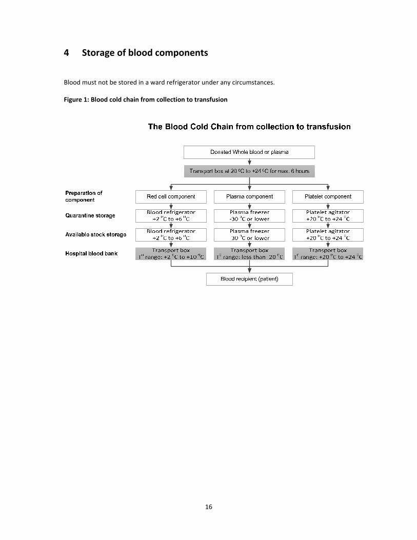
4 Storage of blood components
Blood must not be stored in a ward refrigerator under any circumstances. Figure 1: Blood cold chain from collection to transfusion

17
5 Clinical Transfusion Procedure
5.1 Indications for blood transfusion To increase the oxygen capacity of blood by giving red cells.
To restore the blood volume to maintain effective tissue perfusion.
To replace platelets, coagulation factors and other plasma proteins.
Blood may be needed in the following circumstances:
Blood loss: – Bleeding – Trauma
Inadequate production: – Diseases such as thalassemia, leukaemia
Excessive destruction of cells: – Disease – Mechanical
Transfusion of blood and products should be undertaken only to treat a condition that would lead to significant morbidly or mortality that cannot be prevented or managed effectively by other means. Blood is more often needed under the following circumstances:
Maternity: women during pregnancy and at the time of delivery – Anaemia of pregnancy; bleeding in pre‐ or post‐partum stage of delivery.
5‐29 years – Vulnerability during this age range due to infancy on the one hand (e.g. malnutrition,
malaria) and youth on the other (e.g. nature of work which may be more physical and more likely to expose individual to accidents).
Patients with chronic blood disease – e.g. thalassemia, leukaemia.
5.2 Transfusion trigger (adults) One unit of whole blood/PRBC can increase Hb by 1g/dL in an adult or Hct by 3% (Hb of unit
must be >75%).
Perioperative transfusion: – 8g/dL for patient undergoing cardiovascular surgery, orthopaedics and acute GI bleeding.
Chronic anaemia: – 7g/dL in adults.
Acute blood loss: – 30% of volume of blood.
5.3 Responsibilities of attending physician Assess patient’s clinical need for blood, and when required.
Inform patient and/or relatives about proposed transfusion and record in patient’s notes.

18
Also record indications for transfusion in patient’s notes.
Select blood product and quantity required (i.e. whole blood/PRBC/FFP/PC) and complete request form accurately and legibly.
Enter the reason for transfusion on the form, so that the blood centre can check that the product ordered is the most suitable with regard to diagnosis.
Obtain and correctly label a blood sample for compatibility testing.
Send the blood request form and blood sample to the blood bank.
When the blood product that was ordered arrives, transfuse it as soon as possible to avoid having to store it. However, if the blood product is not used immediately, store it under the correct storage conditions.
Cross check the identity of the patient and the blood product: – Patient and documentation. – Blood / blood products.

19
6 Administration of Blood Products
When blood is transfused, it is important to keep detailed records including the following in the patient’s notes:
Type and volume of each unit transfused.
Unique donation number of each unit transfused.
Blood group of each unit transfused.
Time at which the transfusion of each unit commenced.
Signature of the individual responsible for administration of the blood.
Monitor the patient before, during and on completion of the transfusion.
Record the time of completion of the transfusion.
Identify and respond immediately to any adverse effect, by stopping the transfusion.
Record the details of any transfusion reaction. The process starts with the request for blood, followed by the selection of the correct blood product for compatibility testing and finally the issuing of compatible blood for infusion into the patient.
6.1 Blood request form When blood is required for transfusion, the prescribing clinician should complete and sign a blood request form that is designed to provide all necessary information. All details requested on the blood request form must be completed accurately and legibly.
The blood request form should always be accompanied by the patient’s blood sample. The sample is placed in a sample tube that is correctly labelled and is uniquely identifiable with the patient.
The blood sample shall not be submitted in a syringe, as this could lead to errors when transferring to a test tube for grouping and compatibility testing. It may also cause haemolysis.
For a routine case, the sample and request form should be submitted to the transfusion department at least 24 hours before required, to make sure of the availability of blood.
Physicians may request those, who accompany the patient, to consider becoming blood donors if they are healthy and lead a healthy lifestyle.
6.2 Blood samples The taking of a blood sample from the patient needs supervision. If the patient is conscious at the time of taking the sample, ask him/her to identify himself/herself by given name and all other appropriate information. A 5 mL blood sample should be collected into a dry test tube and then correctly and clearly labelled with the patient’s details, and submitted to the blood centre for testing. The specimen label must include the following information:
Patient’s full name, age and sex.
Registration number.
Ward/bed number.
Date and time specimen taken.
Phlebotomist’s signature/initials.

20
– Use positive patient identification to identify the patient. NEVER pre‐label the sample tube before phlebotomy.
– Use the blood product request form, write legibly and fill in all appropriate details. – When taking a blood sample for cross match, complete the whole procedure before any
other task is undertaken – it is important that there are no interruptions during the process.
– The signature of the individual who took the sample must appear on the specimen label. Retention of blood samples:
Blood samples from recipient and donor(s) must be retained for 7 days at +2°C to +8°C after each transfusion.
Should another transfusion be necessary 72 hours after the earlier transfusion, a fresh sample shall be requested for cross match. Collection of a second 5 mL blood sample is required for re‐checking and further cross matching and must be retained in case of investigation of transfusion reaction.
6.3 Red cell compatibility testing The laboratory performs:
ABO and RhD grouping on patient and donors.
Antibody screening on patient.
Cross matching between serum of patient and red cells of donor. These procedures normally take about an hour or more to complete. Shortened procedures are possible in case of emergency, but may fail to detect some incompatibilities.
The term compatibility test and cross match are sometimes used interchangeably; they should
be clearly differentiated.
The cross match is the part of pre‐transfusion test known as compatibility testing.
The compatibility test includes: - ABO and RhD grouping of donor and recipient. - Screening for unexpected antibodies on donor and patient. - Cross match.
All pre‐transfusion test procedures should provide information on ABO and RhD grouping of both patient and units of blood to be transfused. Purpose of compatibility testing:
To select blood components that will cause no harm to the recipient and will have acceptable survival rates when transfused.
When correctly performed, compatibility tests will confirm ABO compatibility between component and recipient and will detect the most clinically significant unexpected antibodies.
Compatibility (cross match) must be performed before blood is transfused. The cross match is incompatible if there is a reaction between the patient’s serum and donor’s red cells.
6.4 Collection and receipt of blood
ALWAYS take a completed patient documentation label to the issue room of the blood trans‐fusion department when collecting the first unit of blood.
MATCH the details on the blood request form against the blood compatibility label (tag), the bag unit number and the patient documentation label.
If everything matches, sign out the unit with the date and time.

21
If there is any discrepancy, DO NOT sign out the unit; contact the staff member of the blood transfusion department immediately.
When receiving the unit of blood in the clinical area, check that it is the right unit for the right patient.
Always check patient/component compatibility/identity. Inspect pack and contents for signs of deterioration or damage. Figure 2: Check points for signs of deterioration in blood and plasma
Discoloration or signs of any leakage may be the only warning that the blood has been contaminated by bacteria and could cause a severe or fatal reaction if transfused. Blood bag should be checked for:
Any sign of haemolysis in the plasma indicating that the blood has been contaminated, allowed to freeze or to warm.
Any sign of haemolysis on the line between the red cells and plasma during storage.
Any sign of contamination, such as a change of colour in the red cells, which often look darker/ purple/ black when contaminated.
Any clot, which may mean that the blood was not mixed properly with the anticoagulant when it was collected or might also indicate bacterial contamination due to the utilization of citrate by proliferating bacteria.
Any sign that there is a leak in the bag or that it has already been opened. The blood unit must be discarded if:
It has been out of the refrigerator for longer than 30 minutes, or
The seal is broken, or
There is any sign of haemolysis, clotting or contamination.

22
6.5 Performing the transfusion Once issued by the blood centre, the transfusion of whole blood, red cells, platelet concentrate and thawed fresh frozen plasma should be commenced within 30 minutes of removal from the optimal storage conditions. If the transfusion cannot be started within this period, the unit(s) must be stored under approved optimal storage conditions. The temperature inside every blood bank refrigerator used for whole blood/ red cell storage should be monitored and recorded daily to ensure that the temperature remains between +2°C and +6°C. If the ward or operating room does not have a blood bank refrigerator that is appropriate for storing blood, the blood should only be released from the blood centre just before transfusion. Checking the patient’s identity and the blood bag before transfusion Before starting the transfusion, it is vital to make the final identity check in accordance with the hospital’s standard operating procedure. The final identity check should be undertaken at the patient’s bedside immediately before commencing the administration of the blood product. It should be undertaken by two people, at least one of whom should be a registered nurse or doctor. The final check at the patient’s bedside is the last opportunity to detect an identification error and prevent a potentially incompatible transfusion, which may be fatal. Suggested rate of transfusion Transfusion rate depends on clinical circumstances and may vary from 3‐5 mL/kg/hour to greatly increased rates for individuals in hypovolaemic shock. Table 1: Suggested rates of transfusion Adults Rate Paediatric patients Rate
Whole blood 150‐200 mL/hour Whole blood / PRBC 2‐5 mL/kg/hour
PRBC 100‐150 mL/hour Platelets / plasma 1‐2 mL/minute
Platelets / plasma 150‐300 mL/hour
Time limits for transfusion
There is a risk of bacterial proliferation or loss of function in blood products once they have been removed from the correct storage conditions.
Transfusion of a unit of blood should be completed within a maximum period of four hours after removal from the blood fridge: discard the unit if this period is exceeded.
If blood has been out of the blood bank refrigerator for more than 30 minutes and is not transfused, then the unit must be returned to the laboratory, where it will be disposed of.
Table 2: Duration times for transfusion
Blood products Start transfusion Complete transfusion
Whole blood / PRBC Within 30 minutes of removing from refrigerator
≤ 4 hoursDiscard unit if this period is exceeded
Platelet concentrate Immediately Within 30 minutes
FFP As soon as possible Within 30 minutes
Cryoprecipitate As soon as possible Within 30 minutes

23
Blood administration set:
Use a new, sterile blood administration set containing an integral 170‐200µ filter.
Change the set at least 12‐hourly during blood transfusion.
In a very warm climate, change the set more frequently and usually after every four units of blood, if given within a 12‐hour period.
Use a fresh blood administration set or special platelet transfusion set, primed with saline. All blood components can be slowly infused through small‐bore cannulas or butterfly needles, e.g. 21 to 25 G. For rapid infusion, large‐bore cannulas, e.g. 14 G, are needed.
6.6 Monitoring the transfusion
It is essential to take baseline observations and to ensure that the patient is monitored during the transfusion in order to detect any adverse event as early as possible. Before commencing the transfusion, it is essential to encourage the patient to notify a nurse or doctor immediately if he or she becomes aware of any discomfort such as shivering, flushing, pain or shortness of breath or begins to feel anxious.
Ensure that the patient is in a setting where he or she can be directly observed.
For each unit of blood transfused, monitor the patient: - Before starting the transfusion (baseline observation). - 15 minutes after starting the transfusion. - At least every hour during transfusion. - Carry out a final set of observations 15 minutes after each unit has been transfused.
6.7 Documentation of the transfusion Monitor the patient before, during and on completion of the transfusion.
At each of these stages, record the following information on the patient’s chart: - Patient’s general appearance. - Temperature. - Pulse. - Blood pressure. - Respiratory rate.
Make note of the following: - Time the transfusion started. - Time the transfusion was completed. - Volume and type of blood products transfused. - Unique donation number of all products transfused. - Any adverse effect.
Record in the patient’s notes: – Type and volume of each unit transfused. – Unique donation number of each unit transfused. – Blood group of each unit transfused. – Time at which the transfusion of each unit commenced. – Signature of the individual responsible for administration of the blood. – Record the time of completion of the transfusion. – Record the details of transfusion reaction.
Identify and respond immediately to any adverse effect, by stopping the transfusion.

24
Severe reactions most commonly present during the first 15 minutes of a transfusion. All patients and in particular, unconscious patients should be monitored during this period and for the first 15 minutes of each subsequent unit. Specific instructions concerning possible adverse events shall be provided to the patient. The transfusion of each unit of the whole blood or red blood cells should be completed within four hours of the start of the transfusion. If a unit is not fully transfused within four hours, discontinue its use and dispose of the remainder through the clinical waste system. Check that the following information has also be recorded in the patient’s notes.
Whether the patient and/or relatives were informed about the transfusion.
The reason for transfusion.
Signature of the prescribing clinician.
Pre‐transfusion checks of: - Patient’s identity. - Blood bag. - Compatibility label. - Signature of individual performing the pre‐transfusion identity check.
The transfusion: - Type and volume of each product transfused. - Unique donation number of each unit transfused. - Blood group of each unit transfused. - Time at which the transfusion of each unit commenced. - Signature of the person administering the blood component. - Monitoring of the patient before, during and on completion of transfusion. - All other details related to the transfusion process. - Informed consent. - Administration of the unit. - Set‐up time of each unit transfused. - Time of transfusion.
6.8 Other aspects of transfusion 6.8.1 Warming blood
There is no evidence that warming blood is beneficial to the patient when transfusion is slow. At transfusion rates of greater than 100 mL/minute, cold blood may be a contributing factor in cardiac arrest. However, keeping the patient warm is probably more important than warming the blood. Warmed blood is most commonly required in:
Large volume rapid transfusions: - Adults: more than 50 mL/kg/hour. - Children: more than 15 mL/kg/hour.
Exchange transfusion in infants.
Patients with clinically significant cold agglutinins. - Blood should only be warmed in a blood warmer. Blood warmers should have a visible
thermometer and an audible warning alarm and should be properly maintained. - Blood should never be warmed in a bowl of hot water as this could lead to haemolysis
of the red cells which could be life‐threatening when transfused.

25
6.8.2 Use of medication at time of transfusion
It is generally not recommended to routinely use pre‐medication like anti‐histamines, steroids or other medication before transfusion. This practice may mask or delay the signs and symptoms of an acute transfusion reaction and therefore delay recognition and action to stop the transfusion.
Addition of medicine or other fluids with blood and blood components
Medicines or other fluids should never be infused within the same line as blood and blood
components. The exception is normal saline (sodium chloride 0.9%) which may be used in special circumstances, e.g. when the flow is slow due to increased Hct, or when saline is used to prepare washed red cells.
Use a separate IV line if an intravenous fluid has to be given at the same time as blood transfusion.
6.8.3 Use of fresh blood
Stored blood less than 7 days old is termed “fresh blood”
Uses ( to avoid biochemical overload) to raise Hb: - Renal and liver dysfunction. - Patient requiring massive blood transfusion. - Patient with raised plasma potassium due to extensive burns, or intravascular
haemolysis. - Neonate requiring exchange transfusion.
There is no justification in transfusing whole blood to stop bleeding due to coagulopathies in adults, as it does not contain sufficient viable platelets, or fibrinogen, or other coagulation factors. To stop bleeding, the specific component is needed. FFP, PCs or cryoprecipitate, are the treatments of choice to stop bleeding.
Disadvantages of using blood that has not been stored between +2°C and +6°C:
Increased risk of disease transmission: - Intracellular pathogens (CMV, HTLV) survive in leukocytes present in fresh blood. - Syphilis transmission: Treponema should not survive >96 hours in stored blood. - Malaria transmission: malarial parasite should not survive > 7 hours in stored blood.

26
7 Adverse Effects of Transfusion
The very first step is to stop the transfusion immediately. If the reaction is severe, the needle should be removed to prevent any further transfusion of blood.
All suspected acute transfusion reactions should be reported immediately to the blood transfusion centre and to the doctor responsible for the patient. With the exception of urticarial allergic reactions and febrile non‐haemolytic reactions, all are potentially fatal and require urgent treatment.
Acute reactions may occur in 1% to 2% of transfused patients. Rapid recognition and manage‐ment of the reaction may save the patient’s life. Once immediate action has been taken, careful and repeated clinical assessment is essential to identify and treat the patient’s problems.
Errors and failure to adhere to correct procedures are the common causes of life‐threatening acute haemolytic transfusion reactions.
Bacterial contamination in red cells or platelet concentrates is an under‐recognized cause of acute transfusion reaction.
Patients who receive regular transfusions are particularly at risk of acute febrile reactions. These should be recognized so that transfusion is not delayed or stopped unnecessarily.
Transfusion transmitted infections are the serious delayed complications of transfusion. Since a delayed transfusion reaction may occur days, weeks or months after the transfusion, the association with the transfusion may not be recognised.
The transfusion of a large volume of blood and intravenous fluids may cause haemostatic defects or metabolic disturbances in the patient. There should be an availability of various treatments including oxygen, adrenaline, corticosteroids, bronchodilators, diuretics and an emergency team.
In an unconscious or anaesthetized patient, hypotension and uncontrolled bleeding may be the only sign of an incompatible (mismatched) transfusion. In a conscious patient undergoing an acute severe haemolytic transfusion reaction, signs and symptoms may appear within minutes of transfusion of 5‐10 mL of blood. Close observation at the start of the transfusion of each unit is therefore essential.

27
Figure 3: Hazards of blood transfusion
Transfusion reaction (TR)
Acute TR (<24 hours)
- Wrong blood, primed immunological recipient - Poor quality blood, faulty assessment
Delayed TR (>24 hours) - Diseases, other delayed immunologic reactions, metabolic effect (5‐10 days)
Investigation of a suspected TR TRs may be acute or delayed. Acute reactions range from a non‐specific febrile episode to life‐threatening intravascular haemolysis. All suspected transfusion reactions should therefore be assessed and treated appropriately.
If an acute transfusion reaction is suspected, stop the transfusion immediately.
Check the blood bag label against the patient’s identity.
If the reaction is severe or misidentification is confirmed on checking, remove the needle.
If the reaction is mild, keep the IV line open with an infusion of 0.9% sodium chloride.
At the same time, call for assistance; notify the blood bank and the senior in charge of the ward.
With the exception of urticarial allergic and febrile non‐haemolytic reactions, all are potentially fatal and require urgent treatment. The severity of the reaction and the degree of morbidity is usually related to the volume of blood transfused. The only sign in an unconscious or anesthetized patient may be hypotension and uncontrolled bleeding or oozing. In a conscious patient this may occur within minutes of transfusion of as little as 5‐10 mL blood.

28
7.1 Guidelines for recognition and management of acute transfusion reactions Table 3: Category 1: Mild reactions
Signs Symptoms Possible cause
Localized cutaneous: Urticaria Rash
Pruritus Hypersensitivity (mild)
Immediate management of Category 1: Mild reactions
Slow the transfusion.
Administer antihistamine IM.
If no clinical improvement within 30 minutes or if signs and symptoms worsen, treat as Category 2. If improved, restart transfusion slowly.
Table 4: Category 2: Moderately severe reactions
Signs Symptoms Possible cause
Flushing Urticaria Rigors Fever Restlessness Tachycardia
AnxietyPruritus Palpitations Mild dyspnoea Headache
Hypersensitivity
Immediate management of Category 2: Moderately severe reactions
Stop the transfusion and keep IV line open with normal saline in another site.
Return the blood unit with transfusion administration set, freshly collected urine and new blood samples (1 clotted and 1 anticoagulated), drawn from a vein opposite to the transfusion site, to the blood transfusion centre for laboratory investigations.
Administer antihistamine IM and oral or rectal antipyretic. Avoid aspirin in thrombocytopenic patients.
Give IV corticosteroids and bronchodilators if there are anaphylactoid features (e.g. broncho‐spasm, stridor).
If clinical improvement occurs, restart transfusion slowly with new blood unit and observe carefully.
If no clinical improvement within 15 minutes or if signs and symptoms worsen, treat as Category 3.
Collect urine for next 24 hours for evidence of haemolysis and send for laboratory investigations If available, a leucocyte reduction filter (WBC filter) may be used in repeated transfusion. Table 5: Category 3: Life‐threatening reactions
Signs Symptoms Possible cause
Rigor Fever Restlessness Hypotension (fall of 20% in systolic BP) Tachycardia (rise of 20% in heart rate) Haemoglobinuria (Hb in urine) Unexplained bleeding (DIC)
Anxiety Chest pain Pain along the transfusion line Respiratory distress/shortness of breath Loin/back pain Headache Dyspnoea
Acute intravascular haemolysis (mismatched blood transfusion)Bacterial contamination and septic shock Fluid overload Anaphylaxis Transfusion related acute lung injury (TRALI)

29
Immediate management of Category 3: Life‐threatening reactions
Stop the transfusion and keep IV line open with normal saline in another site.
Infuse normal saline to maintain systolic BP.
Maintain airway and give high flow oxygen by mask.
Give adrenaline (as 1:1000 solution) 0.01 mg/kg body weight by slow intramuscular injection.
Give IV corticosteroids and bronchodilators if there are anaphylactoid features.
Give diuretic: e.g. frusemide 1 mg/kg IV or equivalent.
Check a fresh urine specimen visually for signs of haemoglobinuria.
Notify the superior or senior doctor attending the patient, and the blood centre immediately.
Send blood unit with transfusion set, fresh urine sample and new blood samples (1 clotted and 1 anticoagulated), drawn from a vein opposite the infusion site, with the appropriate request form to the blood transfusion centre for investigation.
Start a 24‐hour urine collection and record all intake and output. Maintain fluid balance chart.
Assess for bleeding from puncture sites or wounds. If there is clinical or laboratory evidence of DIC, give platelets (adult: 4‐6 units) and either cryoprecipitate (adult: 12 units) or FFP (adult: 3 units).
Reassess. If hypotensive: – Give further saline. – Give inotrope, if available.
If urine output falls or there is laboratory evidence of acute renal failure (rising K+, urea, creatinine): – Maintain fluid balance accurately. – Give further diuretic: e.g. frusemide 1 mg/kg IV or equivalent. – Consider dopamine infusion, if available. – Seek expert help: the patient may need renal dialysis.
If bacteraemia is suspected (rigor, fever, collapse, no evidence of a haemolytic reaction), start a broad‐spectrum antibiotic IV.
7.2 Investigating acute transfusion reactions
Immediately report all acute transfusion reactions, with the exception of mild hypersensitivity (Category 1) to the doctor responsible for the patient and to the blood transfusion centre that supplied the blood. If a severe life‐threatening reaction is suspected, seek help immediately from the blood transfusion expert/anaesthetist/emergency team/whoever is available and skilled to assist.
Record the following information on the patient’s notes: – Type of transfusion reaction. – Time lapse between start of transfusion and when reaction occurred. – Volume, type and bag number of blood products transfused.
Immediately take post‐transfusion blood samples (1 clotted and 1 anti‐coagulated) from the vein opposite the transfusion site and forward to the blood centre for investigation of the following: – Repeat ABO and RhD group. – Repeat antibody screen and cross match. – Full blood count. – Coagulation screen. – Direct antiglobulin test. – Urea and creatinine. – Electrolytes.

30
Also return the following to the blood centre: – Blood bag and transfusion set containing red cell and plasma residues from the transfused
unit. – Blood culture in a special blood culture bottle. – First specimen of the patient’s urine following the reaction. – Completed transfusion reaction report form.
After the initial investigation of the reaction, send patient’s 24‐hour urine sample to the blood transfusion centre for laboratory investigation.
Record the results of the investigations for future follow‐up, if required.
7.3 Haemolytic transfusion reaction
An acute haemolytic transfusion reaction is the result of a mismatched blood transfusion, and causes acute intravascular haemolysis.
Acute intravascular haemolytic reaction is caused by the transfusion of incompatible red cells, i.e. mismatched blood. Antibodies in the patient’s plasma haemolyse the incompatible red cells transfused.
Even a small volume (5‐10 mL) of incompatible blood can cause a severe reaction and larger volume increases the risk.
The most common cause of reaction is ABO incompatible transfusion. This almost always arises from: - Errors in the blood request form. - Taking blood from the wrong patient into a pre‐labelled sample tube. - Incorrect labelling of the blood sample tube sent to the blood transfusion centre. - Inadequate checking of the blood label against the patient’s identity.
Antibodies in the patient’s plasma against other red cell antigens present on transfused blood, such as those of the Kidd, Kell or Duffy blood group systems, can also cause acute haemolysis.
In the conscious patient, signs and symptoms usually appear within minutes of commencing the transfusion, sometimes when <10 mL blood has been given.
In an unconscious or anaesthetized patient, hypotension and uncontrollable bleeding, from the transfusion site, may be the only sign of an incompatible transfusion.
It is therefore essential to monitor the patient from the commencement of the transfusion up to its completion.
Prevention:
Correctly label blood sample and request form.
Place the patient’s blood sample in the correct sample tube.
Always check the blood unit against the identity of the patient at the bedside before transfusion.

31
7.4 Bacterial contamination and septic shock
Bacterial contamination affects up to 0.4% of red cells and 1‐2% of platelet concentrates.
Blood may become contaminated by: - Bacteria from the donor’s skin entering the blood unit during collection (usually
staphylococci). - Bacteraemia present in the blood of the donor during collection (e.g. Yersinia). - Improper handling during blood processing. - Defect or damage to the blood bag. - Thawing FFP or cryoprecipitate in a water‐bath (often contaminated).
Some contaminants, particularly Pseudomonas species, grow at +2°C to +6°C and can survive or multiply in refrigerated red cell units.
Staphylococci grow in warmer conditions and are able to proliferate in PCs which are stored at +20°C to +24°C.
Signs usually appear rapidly after starting infusion, but may be delayed for a few hours.
A severe reaction may be characterized by sudden onset of high fever, rigors and hypotension.
Urgent supportive care and high‐dose intravenous antibiotics are required.
7.5 Transfusion Associated Circulatory Overload
Transfusion associated circulatory overload (TACO), i.e. fluid overload, can result in heart failure and pulmonary oedema.
May occur when: - Too much fluid is transfused. - The transfusion is given too rapidly. - Renal function is impaired.
Fluid overload is particularly likely to happen in patients with: - Chronic severe anaemia. - Underlying cardiovascular disease.
7.6 Anaphylactic reaction
This is a rare complication of transfusion of blood components or plasma derivatives.
The risk is increased by rapid infusion, typically when fresh frozen plasma is used.
IgA deficiency in the recipient is a rare cause of very severe anaphylaxis. This can be caused by any blood product since most contain traces of IgA.
Cytokines in the plasma may occasionally cause broncho‐constriction and vaso‐constriction in recipients.
Occurs within minutes of starting the transfusion and is characterized by: - Cardiovascular collapse. - Respiratory distress. - No fever.
Anaphylaxis is likely to be fatal if it is not managed rapidly and aggressively.
7.7 Transfusion Related Acute Lung Injury
Transfusion related acute lung injury (TRALI) is usually caused by donor plasma that contains antibodies against the patient’s leucocytes.

32
Rapid failure of pulmonary function usually presents within 1‐4 hours of starting transfusion, with diffuse opacity on the chest X‐ray.
There is no specific therapy. Intensive respiratory and general support in an intensive care unit is required.
7.8 Delayed complications of transfusion 7.8.1 Delayed haemolytic transfusion reaction
Signs appear 5‐10 days after transfusion:
Fever.
Anaemia.
Jaundice.
Occasionally haemoglobinuria.
Severe, life‐threatening delayed haemolytic transfusion reactions with shock, renal failure and DIC are rare.
7.8.2 Post‐transfusion purpura
This is a rare but potentially fatal complication of transfusion of red cells or platelet concentrates, caused by antibodies directed against platelet‐specific antigens in the recipient.
Most commonly seen in multigravida female patients.
Signs and symptoms: – Signs of bleeding. – Acute, severe thrombocytopenia 5‐10 days after transfusion, defined as a platelet
count of <100 x 109/L.
Management Management becomes clinically important at a platelet count of 50 x 109/L, with a danger of hidden (occult) bleeding at 20 x 109/L.
Give high dose corticosteroids.
Give high dose IV immunoglobulin, 2 g/kg or 0.4 g/kg for 5 days.
Plasma exchange.
Monitor the patient’s platelet count: normal range is 150 x 109/L to 440 x 109/L.
If available, give platelet concentrates that are negative for the platelet‐specific antigen against which the antibodies are directed.
Unmatched platelet transfusion is generally ineffective.
Recovery of platelet count after 2‐4 weeks is usual. 7.8.3 Transfusion associated graft‐versus‐host disease (TA‐GVHD)
Unlike transplant associated GVHD, TA‐GVHD it is usually a fatal condition.
Occurs in patients such as: – Immuno‐deficient recipients of bone marrow transplants. – Immuno‐competent patients transfused with blood from individuals with whom they
have a compatible HLA tissue type, usually blood relatives particularly 1st degree.
Signs and symptoms typically occur 10‐12 days after transfusion and are characterized by: – Fever. – Skin rash and desquamation. – Diarrhoea. – Hepatitis. – Pancytopenia.

33
Management Treatment is supportive; there is no specific therapy. Prevention
Do not use 1st degree relatives as donors, unless gamma irradiation of cellular blood components is carried out to prevent the proliferation of transfused lymphocytes.
7.8.4 Delayed complications: transfusion transmitted infections
The following infections may be transmitted by transfusion:
HIV, Hepatitis B and C, syphilis (Treponema pallidum), malaria.
Cytomegalovirus (CMV).
Other TTIs include human parvovirus B19, brucellosis, Epstein‐Barr virus, toxoplasmosis, Chagas disease, infectious mononucleosis and Lyme’s disease.
Since a delayed transfusion reaction may occur days, weeks or months after the transfusion, the association with the transfusion may easily be overlooked. It is essential to record all transfusions accurately in the patient’s case notes and to consider transfusion in the differential diagnosis.
34
8 Massive Blood Transfusion
Massive blood transfusion may be defined as the replacement of one blood volume (equivalent to 10 units of blood) in any 24 hour period, or half of the blood volume (5 units of blood) in any four‐hour period in an adult.
Replacement of a blood volume equivalent within 24 hours.
>10 units within 24 hours.
Transfusion >4 units in 1 hour.
Replacement of 50% of blood volume in 3‐4 hours.
A rate of loss >150 ml/hour. Massive transfusion occurs in settings such as severe trauma, ruptured aortic aneurysm, surgery and obstetric complications. The goals to the management of massive transfusion include early recognition of blood loss, maintenance of tissue perfusion, oxygenation by restoration of blood volume and Hb, and the cessation of bleeding by several means including early surgical or radiological intervention, and the judicious use of blood component therapy to correct coagulopathy.
Plasma undergoes progressive loss of coagulation factors during storage, particularly Factors V and VIII, unless stored at –30°C or colder.
Dilution of coagulation factors and platelets will occur following administration of large volumes of replacement fluids. Massive or large volume transfusions can therefore result in disorders of coagulation.
Massive Transfusion Protocol
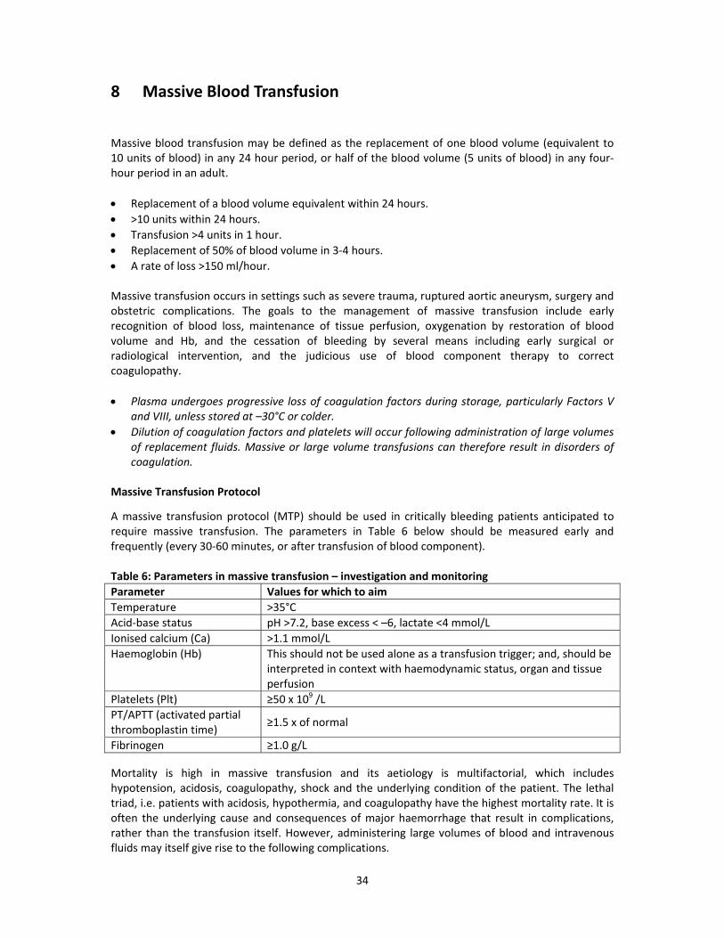
A massive transfusion protocol (MTP) should be used in critically bleeding patients anticipated to require massive transfusion. The parameters in Table 6 below should be measured early and frequently (every 30‐60 minutes, or after transfusion of blood component). Table 6: Parameters in massive transfusion – investigation and monitoring
Parameter Values for which to aim
Temperature >35°C
Acid‐base status pH >7.2, base excess < –6, lactate <4 mmol/L
Ionised calcium (Ca) >1.1 mmol/L
Haemoglobin (Hb) This should not be used alone as a transfusion trigger; and, should be interpreted in context with haemodynamic status, organ and tissue perfusion
Platelets (Plt) ≥50 x 109 /L
PT/APTT (activated partial thromboplastin time)
≥1.5 x of normal
Fibrinogen ≥1.0 g/L
Mortality is high in massive transfusion and its aetiology is multifactorial, which includes hypotension, acidosis, coagulopathy, shock and the underlying condition of the patient. The lethal triad, i.e. patients with acidosis, hypothermia, and coagulopathy have the highest mortality rate. It is often the underlying cause and consequences of major haemorrhage that result in complications, rather than the transfusion itself. However, administering large volumes of blood and intravenous fluids may itself give rise to the following complications.
35
Acidosis Acidosis in a patient receiving a large volume transfusion is more likely to be the result of inadequate treatment of hypovolaemia than due to the effects of transfusion. Under normal circumstances, the body can readily neutralize this acid load from transfusion. The routine use of bicarbonate or other alkalizing agents, based on the number of units transfused, is unnecessary. Hyperkalaemia The storage of blood results in a small increase in extra‐cellular potassium concentration, which will increase the longer it is stored. This rise is rarely of clinical significance, other than in neonatal exchange transfusions. Fresh blood (up to 7 days old) should be requested from the blood centre. Citrate toxicity and hypocalcaemia Citrate toxicity is rare, but is most likely to occur during the course of a large volume transfusion of whole blood. Hypocalcaemia, particularly in combination with hypothermia and acidosis, can cause a reduction in cardiac output, bradycardia, and other dysrhythmias. Citrate is usually rapidly metabolized to bicarbonate. It is therefore unnecessary to attempt to neutralize the acid load of transfusion. There is very little citrate in red cell concentrates. Management
If there is prolongation of PT, give ABO compatible fresh frozen plasma in a dose of 15 mL/kg.
If the APTT is also prolonged, Factor VIII/fibrinogen concentrate is recommended in addition to FFP. If none is available, give 10‐15 units of ABO compatible cryoprecipitate, which contains Factor VIII and fibrinogen.
Give PCs only when: – The patient shows clinical signs of microvascular bleeding: i.e. bleeding and oozing from
mucous membranes, wounds, raw surfaces and catheter sites. – The patient’s platelet count falls below 50 x 109/L.
Give sufficient PCs to stop microvascular bleeding and maintain an adequate platelet count.
Consider PC transfusion in cases where the platelet count falls below 20 x 109/L, even if there is no clinical evidence of bleeding, because there is a danger of occult bleeding, such as into brain tissue.
The prophylactic use of platelet concentrates in patients receiving large volume blood transfusions is not recommended.

36
9 Transfusion in Paediatrics
Paediatric anaemia is defined as a reduction of Hb concentration or red cell blood volume below the normal values for healthy children. Normal Hb/Hct values differ according to the age of the child. Indications for Transfusion
Hb ≤4 g/dL or Hct 12%.
Hb 4‐6 g/dL (or Hct 13‐18%) if any of following clinical features are present: – Clinical features of hypoxia. – Acidosis (usually causes dyspnoea). – Impaired consciousness. – Hyper‐parasitaemia (>20% malarial parasites).
Transfusion of Neonates and Infants The following recommendations apply to the transfusion of children in the first four months of life: Pre‐transfusion testing: Maternal samples: ABO and RhD group Antibody screen (5 mL clotted blood)
Infant samples: ABO and RhD group Direct antiglobulin test (DAT) Antibody screen if maternal sample unavailable (1‐2 mL clotted blood)
If the maternal antibody screen is negative and the infant’s red cells are DAT negative, cross matching is unnecessary and blood of the baby’s group can be issued. Alloantibodies are rare in the first four months of life and are related to repeated massive transfusions and to the use of fresh blood.
If the maternal antibody screen and/or the neonatal DAT are positive, serological investigation and full compatibility testing will be necessary.
After the first four months of life, cross matching procedures should conform to the require‐ments for older children/adults.
9.1 Top‐up transfusion Top‐up transfusions are carried out in order to raise Hb concentration in symptomatic chronic anaemia, often due to blood sampling in sick premature infants.
Limit donor exposure whenever possible: if repeated transfusions are likely then notify the blood transfusion department so that satellite bags (40 mL each) can be arranged.
PRBC (Hct 0.55‐0.75) should be used for top‐up transfusions. Red cells in optimal additive solutions, e.g. SAG‐M or CPD blood can be safely used for top‐up transfusion in this age group.
Complete transfusion within 4 hours. Transfusion rates of 5 mL/kg/hour are safe: increase rate if active haemorrhage and reduce if cardiac failure exists.
Due to the small volume of blood to be transfused, it is acceptable to flush the giving set with an appropriate fluid, e.g. isotonic (normal) saline to extract the full volume of red cells.

37
9.2 Exchange transfusion The main indication for neonatal exchange transfusion is to prevent neurological complications (kernicterus) caused by a rapidly‐rising unconjugated bilirubin concentration. Option for blood group is as follows:
Use group O blood that does not carry the antigen to which the maternal antibody is directed.
For HDN due to anti‐D use group O RhD negative blood.
Use blood of the ABO group of the neonate or use an alternative group which is compatible with maternal ABO antibodies, and also ABO compatible with the infant. Otherwise, use designated group O Rh compatible units.
Use blood compatible with any maternal irregular antibodies.
Storage age of blood should be within five days of collection. When carrying out an exchange transfusion use whole blood for the first exchange followed by plasma‐reduced blood (Hct 0.55‐0.60) for the second exchange. This is the only indication for the use of whole blood.
Use a blood warmer. Only approved and regularly monitored blood warming equipment should be used: fatal transfusion reactions have followed the use of inappropriate blood warming procedures.
Transfusion Procedure
If transfusion is needed, give sufficient blood to make the child clinically stable.
5 mL/kg of red cells or 10 mL/kg whole blood are usually sufficient to relieve acute shortage of oxygen‐carrying capacity. This will increase Hb concentration by approximately 2‐3 g/dL unless there is continued bleeding or haemolysis.
A red cell transfusion is preferable to whole blood for a patient at risk of circulatory overload, which may precipitate or worsen cardiac failure. 5 mL/kg of red cells gives the same oxygen‐carrying capacity as 10 mL/kg of whole blood and contains less plasma protein and fluid to overload the circulation.
Where possible, use a paediatric blood pack and a device to control the rate and volume of transfusion.
Although rapid fluid infusion increases the risk of volume overload and cardiac failure, give the first 5 mL/kg of red cells to relieve the acute signs of tissue hypoxia. Subsequent transfusion should be given slowly: e.g. 5 mL/kg of red cells over 1 hour.
Give frusemide 1 mg/kg by mouth or 0.5 mg/kg by slow IV injection to a maximum dose of 20 mg/kg if the patient is likely to develop cardiac failure and pulmonary oedema. Do not inject it into the blood pack.
Monitor during transfusion for signs of: – Cardiac failure – Fever – Respiratory distress – Tachypnoea – Hypotension – Acute transfusion reaction – Shock – Haemolysis (jaundice, hepatosplenomegaly) – Bleeding due to DIC
Re‐evaluate the patient’s Hb or Hct and clinical condition after transfusion.

38
If the patient is still anaemic with clinical signs of hypoxia or a critically low Hb level, give a second transfusion of 5–10 mL/kg of red cells or 10–15 mL/kg of whole blood.
Continue treatment of anaemia, such as with iron, to help haematological recovery. If exchange transfusion is needed:
An exchange transfusion of about two times the neonate’s blood volume (about 170 mL/kg) is most effective to reduce bilirubin and restore Hb level; this can usually be carried out with one unit of whole blood.
A unit of whole blood will normally have an Hct of 37–45%, which is more than adequate for neonatal needs.
When exchange transfusion is performed to treat haemolytic disease of the newborn (HDN), the transfused red cells must be compatible with the mother’s serum since the haemolysis is caused by maternal IgG antibodies that cross the placenta and destroy the fetal red cells.
The blood should therefore be cross matched against the mother’s serum using the antiglobulin method that detects IgG antibodies.
9.3 Haemolytic disease of the newborn HDN is caused by antibodies that are produced by the mother. These antibodies are IgG and can cross the placenta and destroy the fetal red cells. The mother may develop these antibodies:
If fetal red blood cells cross the placenta (feto‐maternal haemorrhage) during pregnancy or delivery.
As a result of a previous red cell transfusion. HDN due to ABO incompatibility between mother and infant does not affect the fetus in utero, but is an important cause of neonatal jaundice. HDN due to RhD incompatibility is an important cause of severe fetal anaemia in countries where a significant proportion of the population is RhD negative. RhD negative mothers develop antibodies to an RhD positive fetus, especially when the mother and infant are of the same or compatible ABO blood type. The fetal red cells are haemolysed, causing severe anaemia. In the most severe cases of HDN:
The fetus may die in utero.
The fetus may be born with severe anaemia that requires replacement of red cells by exchange transfusion.
There may also be severe neurological damage after birth as a result of a high bilirubin level unless this is corrected by exchange transfusion.
HDN due to other blood group antibodies can also occur, in particular anti‐c (also within the Rh blood group system) and anti‐Kell. Screening in pregnancy
The ABO and RhD group of all pregnant women should be determined when they first attend a clinic for antenatal care. The mother’s blood should also be tested for any IgG red cell antibodies that can cause HDN.

39
If no antibodies are detected at the first antenatal visit, the pregnant woman should have a further antibody check at 28‐30 weeks gestation.
If antibodies are detected at the first antenatal visit, the levels should be monitored frequently throughout the pregnancy in case they increase. Rising levels are likely to be indicative of HDN developing in the fetus.
Anti‐RhD immunoglobulin Anti‐RhD immunoglobulin prevents the sensitization and production of antibodies in an unsensitised RhD negative mother if RhD positive red cells gain entry into her circulation, either during pregnancy or during delivery.
9.4 ABO haemolytic disease of the newborn The diagnosis of ABO HDN is usually made in infants born at term who are not severely anaemic,
but who develop jaundice during the first 24 hours of life.
ABO incompatibility does not present in utero and does not cause hydrops.
The neonate should receive phototherapy and supportive treatment; treatment should be initiated promptly as jaundice can become severe enough to lead to kernicterus.
Blood units for exchange transfusion should be group O with low‐titre anti‐A and anti‐B.
A two‐volume exchange (approximately 170 mL/kg) is most effective in removing bilirubin.
If bilirubin rises again to dangerous levels, a further two‐volume exchange should be performed.
9.5 Transfusion of platelets and FFP in paediatric patients Platelet transfusion
Platelet transfusions are indicated in neonates and young infants with a count below 50 x 109/L who are experiencing bleeding.
Dose: 5‐10 ml PC/kg body weight will increase count by 50 x 109/L to 100 x 109/L. Transfusion of FFP Dose: 10‐15mL/kg in coagulopathy condition.
Paediatric blood bags are available for blood transfusion of the infant patient. It should be remembered that once a blood bag unit has been opened for transfusion it should be completed. Partial transfusion of a single bag over successive days is not permitted due to the risk of infection.
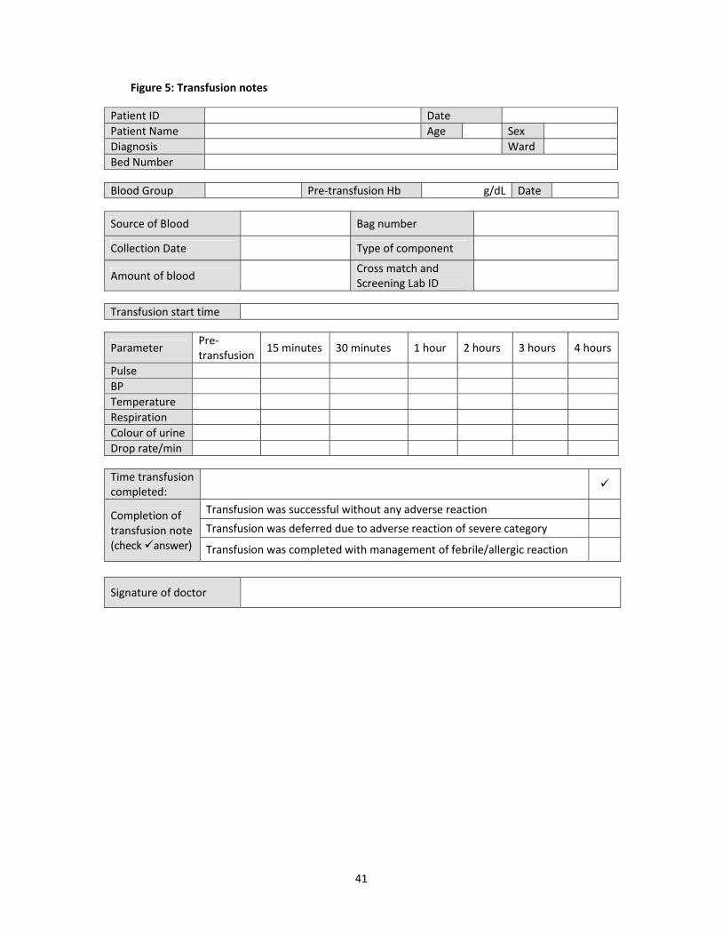
Figures 4, 5 and 6 on the following pages, provide examples of a crossmatch report form, a form for making transfusion notes and a form for making notes on the management of adverse transfusion reaction, respectively.

40
Figure 4: Cross match report form
41
Figure 5: Transfusion notes Patient ID Date
Patient Name Age Sex
Diagnosis Ward
Bed Number Blood Group Pre‐transfusion Hb g/dL Date
Source of Blood Bag number
Collection Date Type of component
Amount of blood Cross match and
Screening Lab ID
Transfusion start time
Parameter Pre‐transfusion
15 minutes 30 minutes 1 hour 2 hours 3 hours 4 hours
Pulse
BP
Temperature
Respiration
Colour of urine
Drop rate/min Time transfusion completed:
Completion of transfusion note (check answer)
Transfusion was successful without any adverse reaction
Transfusion was deferred due to adverse reaction of severe category
Transfusion was completed with management of febrile/allergic reaction
Signature of doctor

42
Figure 6: Management of adverse transfusion reaction: physician’s notes
Chief Complaint/Concern: On Examination: BP Respiratory Rate Pulse Sign of bleeding Investigation: Blood samples: blood bag, patient Urine, culture, electrolytes BT, CT, APTT, bilirubin, direct antiglobulin test Report of blood transfusion centre
Treatment:
Instruction of Experts: (transfusion specialist/ Anaesthetic/ Departmental head)
Signature of Doctor

43
10 Blood Transfusion Services in Bangladesh
To improve the safety of blood transfusion, from the year 2000 the government developed facilities, in medical college hospitals, institutes, combined military hospitals, specialized hospitals, district hospitals and at the Upazila health complex. The strengthening of blood transfusion centres started under the Safe Blood Transfusion Programme of the Ministry of Health and Family Welfare (MOHFW). A legislative framework, the ‘Safe Blood Transfusion Act, 2002’ was enacted by the Government. Blood transfusion centres supply blood after testing for markers to five TTIs i.e. HIV, Hepatitis B, Hepatitis C, syphilis and malaria, the requirement of which has been made mandatory in accordance with the ‘Safe Blood Transfusion Law’ in the country. National Safe Blood Transfusion Council This is a policy making forum for blood transfusion services under the MOHFW for developing the policy as per directives of the Safe Blood Law/Act for improvement of blood transfusion services. The honourable Health Minister, by position, is the President and Director General of Health Services and is the Member Secretary of the Council. Directors of institutes, and the head of the department of blood transfusion, are members of this council. National Safe Blood Transfusion Expert Committee The National Safe Blood Transfusion Expert Committee is an implementing body for the policy and decision making by the National Safe Blood Transfusion Council, headed by the Director General DGHS under MOHFW. It consists of transfusion specialists as members, and the Director General is the president of the committee. Local Hospital transfusion Committee Each hospital has a local blood transfusion committee which is constituted with participation of the head of the department of all disciplines in the hospital. Statutory Regulation Order The government has formulated rules for the management of blood centres which is known as SRO‐145.