clinical study the cement prosthesis-like spacer: an intermediate halt on the road to healing
TRANSCRIPT

Hindawi Publishing CorporationThe Scientific World JournalVolume 2013, Article ID 763434, 6 pageshttp://dx.doi.org/10.1155/2013/763434
Clinical StudyThe Cement Prosthesis-Like Spacer: An IntermediateHalt on the Road to Healing
Sufian S. Ahmad, Kim Huber, Dimitrios S. Evangelopoulos, Barbara Kleer,Hendrik Kohlhof, Michael Schär, Stefan Eggli, and Sandro Kohl
Department of Orthopedic Surgery and Traumatology, University of Bern, Switzerland
Correspondence should be addressed to Sufian S. Ahmad; [email protected]
Received 14 July 2013; Accepted 27 August 2013
Academic Editors: S.-Y. Kim and M. S. Lee
Copyright © 2013 Sufian S. Ahmad et al. This is an open access article distributed under the Creative Commons AttributionLicense, which permits unrestricted use, distribution, and reproduction in any medium, provided the original work is properlycited.
Background. Periprosthetic infections remain a devastating problem in the field of joint arthroplasty. In the following study, theresults of a two-stage treatment protocol for chronic periprosthetic infections using an intraoperatively molded cement prosthesis-like spacer (CPLS) are presented. Methods. Seventy-five patients with chronically infected knee prosthesis received a two-stagerevision procedure with the newly developed CPLS between June 2006 and June 2011. Based on the microorganism involved,patients were grouped into either easy to treat (ETT) or difficult to treat (DTT) and treated accordingly. Range of motion (ROM)and the knee society score (KSS) were utilized for functional assessment. Results. Mean duration of the CPLS implant in the DTTgroup was 3.6 months (range 3–5 months) and in the ETT group 1.3 months (range 0.7–2.5 months). Reinfection rates of the finalprosthesis were 9.6% in the ETT and 8.3% in the DTT group with no significant difference between both groups regarding ROMor KSS (𝑃 = 0.87, 0.64, resp.). Conclusion. The results show that ETT patients do not necessitate the same treatment protocol asDTT patients to achieve the same goal, emphasizing the need to differentiate between therapeutic regimes. We also highlight thefeasibility of CLPS in two-stage protocols.
1. Introduction
Osteoarthritis is nowadays a major cause of disability inadults with a growing trend. Alone knee osteoarthritis has aprevalence of over 30% amongst a population aged ≥60 years;this expresses the dimensions of the problem [1, 2].
Total knee arthroplasty (TKA) maintains its position as amajor treatment option for knee osteoarthritis [3–5]. A fearedcomplication of KA is infection of the prosthetic implant [6–8]. The reason for such concern is the substantial increase inmorbidity and health care expenditure [9].
At least one of the following criteria has to be fulfilledto set the diagnosis of a prosthetic infection: growth of onemicroorganism species in two or more cultures of synovialfluid or periprosthetic tissue, purulence of the synovial fluidor macroscopic changes at the site of the implant, acuteinflammation on histopathological examination of peripros-thetic tissue, or presence of a sinus tract communicating withthe prosthesis [10–12].
The gold standard for treating chronic periprostheticinfection is based on a two-stage protocol, including initialexplantation of the infected components, adequate debride-ment, and antibiotic cement spacer prostheses implanta-tion with systemic antibiotic therapy followed by secondaryTKA once the optimal condition is achieved [13, 14]. Theantimicrobial-impregnated spacer utilized in this processallows formaintenance of limb length, partialmobility duringthe recovery process, and infection control rates of 91% to100% [15, 16]. Initially, cement spacers were static, thereforenot providing sufficient range of motion (ROM); bone loss,soft tissue contracture, and increased scar tissue formation asa result have been mentioned [17–19]. The attempt to achievea degree of ROM using dynamic cement on cement spacerswith a joint geometry gained interest as a possible solutionfor the problems associated with static spacers [18, 19].
The use of either premolded spacers, intraoperative hand-crafting by the surgeon, or intraoperative molding using
2 The Scientific World Journal
standard predesigned moulds has been described in the liter-ature [20–22].
In this paper we verify the safety and efficacy of an intra-operatively produced custommade polymethyl methacrylate(PMMA) cement prostheses like spacer (CPLS) for a two-stage revision protocol of infected total KA.
2. Materials and Methods
Two molds were produced using a computerized numerical-control sinking machine (DMU 70eV-process) based on thedesign of the balanSys knee system (size B; D) (Mathys AG,Bettlach, Switzerland).Themolds for the femoral spacer con-sisted of 3 components and the tibial of 2 components madeof 100%Teflon).Thesemolds were utilized intraoperatively toproduce the spacer in its wanted shape.
The first surgical step involved explantation the pros-thetic components and extensive debridement of the infectedregion, and biopsies were taken during the process for micro-biological culture and histological examination. CPLS wasfinally performed. Initially, the components’ appropriate sizeswere assessed by means of conventional anterior posteriorand lateral knee radiographs. The parts of the femoral mouldweremounted, and themouldwas filledwith cement by hand.Due to cement expansion, the increase in pressure inside theclosed mould created a smooth surface on the final cementspacer. After polymerization, the screws which interlink themould were opened, and the femoral component was easilyremoved. The femoral component was implanted first with asmall portion of additional cement.The distance between thetibia and the femur was measured in neutral position and thetibial component has filled with PMMA cement according tothe distancemeasured. After polymerization, the tibial part ofthe spacer was removed from the mould, and the mountingcement on the posterior and lateral side of the spacer wasremoved with a Luer pincer. The tibial component was thenimplanted with a small portion of cement. The stability andrange of motion were tested, and the wound was closed.
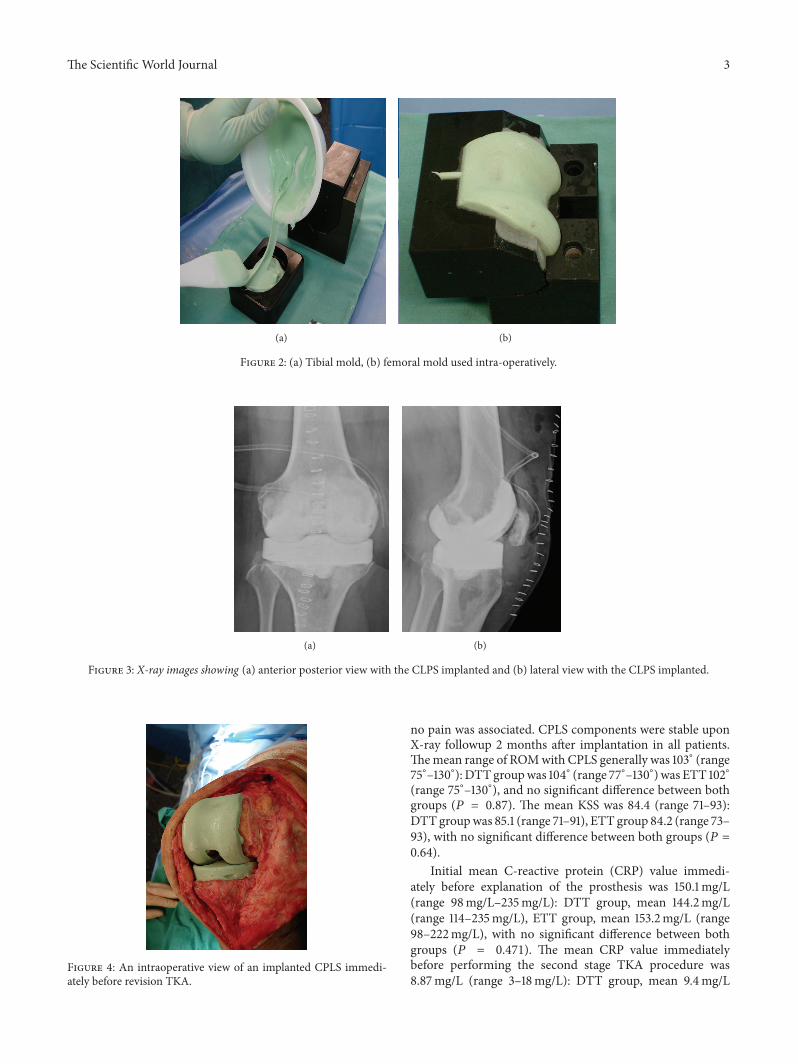
For all PMMA spacers, an antibiotic loaded cementwas applied: PALACOSR + G 40 (Heraeus Medical GmbH,Wehrheim, Germany), containing 0.5 g of Gentamycin. Thesystem permits the incorporation of different antibiotics intothe PMMA spacer according to the antibiogram obtained bythe initial puncture. Figure 2 shows the molds used intraop-eratively, Figure 3 shows X-ray view of the implanted CPLSand Figure 4 shows an intraoperative view of an implantedCPLS immediately before revision TKA.
Seventy-five patients with chronically infected TKAreceived a two-stage revision procedure with the newly devel-oped CPLS between June 2006 and June 2011 (mean age 67.5years, range 57–85 years). However, two different protocolswere considered according to the microorganism involvedand treatment response (Figure 5). Patients infected withmultidrug-resistantmicroorganisms, gramnegativemicroor-ganisms, enterococcus species, or polymicrobial infectionswere considered difficult to treat (DTT) (8), whereas patientsinfected with other microorganisms were considered easyto treat (ETT). All patients underwent joint aspiration formicrobiological examination prior to surgery.
S. a
ureu
s
Coa
g-ne
g-st
aph.
A-he
m st
rep.
B-he
m st
rep.
Gra
m-p
ositi
ve ro
ds
MRS
A
Unk
now
n
Ente
roco
ccus
spec
ies
Gra
m-n
egat
ive c
occi
Poly
mic
robi
al
01020304050
Microorganisms
(%)
Easy to treat Difficult to treat
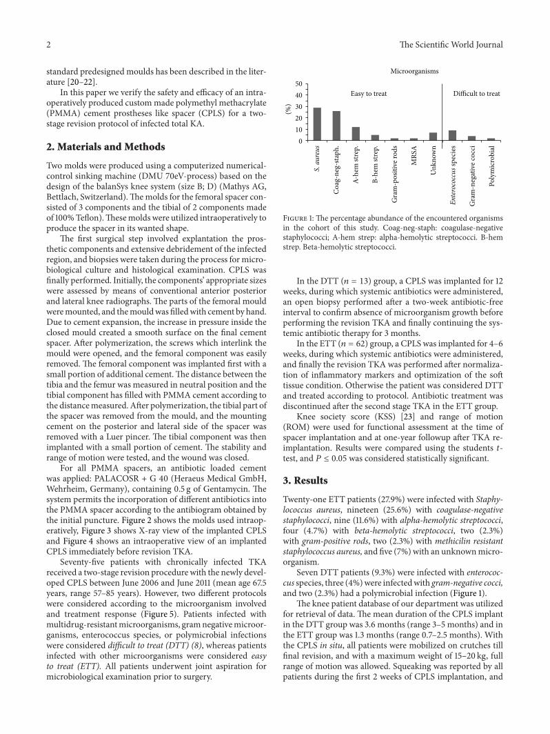
Figure 1: The percentage abundance of the encountered organismsin the cohort of this study. Coag-neg-staph: coagulase-negativestaphylococci; A-hem strep: alpha-hemolytic streptococci. B-hemstrep. Beta-hemolytic streptococci.
In the DTT (𝑛 = 13) group, a CPLS was implanted for 12weeks, during which systemic antibiotics were administered,an open biopsy performed after a two-week antibiotic-freeinterval to confirm absence of microorganism growth beforeperforming the revision TKA and finally continuing the sys-temic antibiotic therapy for 3 months.
In the ETT (𝑛 = 62) group, a CPLS was implanted for 4–6weeks, during which systemic antibiotics were administered,and finally the revision TKA was performed after normaliza-tion of inflammatory markers and optimization of the softtissue condition. Otherwise the patient was considered DTTand treated according to protocol. Antibiotic treatment wasdiscontinued after the second stage TKA in the ETT group.
Knee society score (KSS) [23] and range of motion(ROM) were used for functional assessment at the time ofspacer implantation and at one-year followup after TKA re-implantation. Results were compared using the students 𝑡-test, and 𝑃 ≤ 0.05 was considered statistically significant.
3. Results
Twenty-one ETT patients (27.9%) were infected with Staphy-lococcus aureus, nineteen (25.6%) with coagulase-negativestaphylococci, nine (11.6%) with alpha-hemolytic streptococci,four (4.7%) with beta-hemolytic streptococci, two (2.3%)with gram-positive rods, two (2.3%) with methicilin resistantstaphylococcus aureus, and five (7%)with an unknownmicro-organism.
Seven DTT patients (9.3%) were infected with enterococ-cus species, three (4%)were infectedwith gram-negative cocci,and two (2.3%) had a polymicrobial infection (Figure 1).
The knee patient database of our department was utilizedfor retrieval of data. The mean duration of the CPLS implantin the DTT group was 3.6 months (range 3–5 months) and inthe ETT group was 1.3 months (range 0.7–2.5 months). Withthe CPLS in situ, all patients were mobilized on crutches tillfinal revision, and with a maximum weight of 15–20 kg, fullrange of motion was allowed. Squeaking was reported by allpatients during the first 2 weeks of CPLS implantation, and
The Scientific World Journal 3
(a) (b)
Figure 2: (a) Tibial mold, (b) femoral mold used intra-operatively.
(a) (b)
Figure 3: X-ray images showing (a) anterior posterior view with the CLPS implanted and (b) lateral view with the CLPS implanted.
Figure 4: An intraoperative view of an implanted CPLS immedi-ately before revision TKA.
no pain was associated. CPLS components were stable uponX-ray followup 2 months after implantation in all patients.Themean range of ROMwith CPLS generally was 103∘ (range75∘–130∘): DTT groupwas 104∘ (range 77∘–130∘) was ETT 102∘(range 75∘–130∘), and no significant difference between bothgroups (𝑃 = 0.87). The mean KSS was 84.4 (range 71–93):DTT groupwas 85.1 (range 71–91), ETT group 84.2 (range 73–93), with no significant difference between both groups (𝑃 =0.64).
Initial mean C-reactive protein (CRP) value immedi-ately before explanation of the prosthesis was 150.1mg/L(range 98mg/L–235mg/L): DTT group, mean 144.2mg/L(range 114–235mg/L), ETT group, mean 153.2mg/L (range98–222mg/L), with no significant difference between bothgroups (𝑃 = 0.471). The mean CRP value immediatelybefore performing the second stage TKA procedure was8.87mg/L (range 3–18mg/L): DTT group, mean 9.4mg/L

4 The Scientific World Journal
1st stage, explant, CPLS
+ antibiotics 12 weeks
2nd stage Revision knee total
arthroplasty
2nd stage Revision total knee
arthroplasty
2-week free interval of antibiotics
+ open biopsy
3-month antibiotics
NegativePositive
Reevaluationarthrodesis
CRP (near normal) + regular soft tissue
Yes No
Prosthetic infections (n = 75)
Easy to treat (n = 62) Difficult to treat (n = 13)
1st stage,
explant + CPLS (4–6 weeks)+ antibiotics
Figure 5: Protocol upon which the treatment strategy was based.
(range 5–18mg/L), ETT group, mean 8.1mg/L (range 3–16mg/L), with no significant difference between both groups(𝑃 = 0.152).
The mean follow-up interval after the final revisionarthroplasty was 4.3 years (range 2–7 years).
The mean ROM with the final prosthesis generally was115∘ (range 90∘–125∘): DTT group, mean 112∘ (range 90∘–125∘)and ETT group, mean 117∘ (range 82∘–130∘), with no signifi-cant difference between both groups (𝑃 = 0.76). The meanKSS was generally 89.5 (range 74–95): DTT group, mean 88.4(range 76–93), ETT group, mean 90.2 (range 74–95), withno significant difference between both groups (𝑃 = 0.354).
In the ETT group, 6 reinfections (9.6%) of the final pros-thesis occurred, 3 Staphylococcus aureus, 2 Streptococci and1 coagulase-negative staphylococcus during the first 6 months.Two of these were persistent infections during the first 6weeks requiring a divert of treatment protocol from ETT toDTT, one was a reinfection within the first 6 months requir-ing a divert to a DTT treatment protocol and three required adivert during the remaining follow-up interval.
In the DTT group one reinfection (8.3%) with gram nega-tive cocci occurred 3.5 years after revision ending up in arthro-desis.
4. Discussion
Due to the devastating problem of chronic joint infections,work on the development of new strategies and material totackle the problem is necessary.
The one-stage revision arthroplasty is widely spread andmaintains its place as treatment standard in many centers[24–26]. However, a review article published recently byRomano et al. showed that two-stage procedures providebenefit over one-stage procedures regarding reinfection rates,and that far more two-stage procedures are being reportedin the literature showing the increasing popularity of two-stage procedures [27].The eradication rate of 90.7% achievedin our 75 patient of two-stage series was higher than theliterature average of 81.9 for one-stage procedures and closeto the literature average of 91.2 for two-stage procedures usingarticulating spacers [27].
In this study, we present a therapeutic plan for peri-prosthetic infections based on the microorganism involved(Figure 4). Zimmerli et al. first described the term difficultto treat DTT prosthetic infections in association with themicroorganisms mentioned above [8]. According to Zim-merli, we differentiated between ETT and DTT patients andused two different therapeutic protocols (Figure 4).The dura-tion of treatment for ETT patients was significantly less thanETT patients (1.3 months versus 3.6 moths, resp.). Same forthe antibiotic therapy that was discontinued immediatelyafter revision TKA in ETT patients and continued for threemonths in DTT patients.The results did not show any signif-icant difference in outcome regarding reinfection rates of thefinal prosthesis, ROM, or KSS scores between both groups.
The insertion of an intraoperative moulded PMMA artic-ulating spacer presents surgical advantages and apparentlyis associated with less reinfections than static spacers [27].

The Scientific World Journal 5
Wenoted that the application of such a spacer resulted in con-siderably less scar tissue formation, thus facilitating surgicalexposure during the second intervention. The fact that lessscar tissue removal had to be performed facilitated joint expo-sure avoiding complex soft tissue procedures, thus resultingin shorter operation time and easier postoperative rehabilita-tion.
In conclusion, the results show that ETT patients do notnecsseate the same treatment protocol as DTT patients toachieve the same goal, emphasizing the need to differentiatebetween therapeutic regimes.We also highlight the feasibilityof CLPS in two-stage protocols.
Conflict of Interests
The authors declare no competing interests in any means orany conflicting financial interests.
References
[1] D. T. Felson, A. Naimark, and J. Anderson, “The prevalence ofknee osteoarthritis in the elderly: the FraminghamOsteoarthri-tis Study,” Arthritis and Rheumatism, vol. 30, no. 8, pp. 914–918,1987.
[2] K. Ronn,N. Reischl, E. Gautier, andM. Jacobi, “Current surgicaltreatment of knee osteoarthritis,” Arthritis, vol. 2011, Article ID454873, 9 pages, 2011.
[3] W. Zhang, R. W. Moskowitz, G. Nuki et al., “OARSI recom-mendations for the management of hip and knee osteoarthritis,Part I: critical appraisal of existing treatment guidelines andsystematic review of current research evidence,” Osteoarthritisand Cartilage, vol. 15, no. 9, pp. 981–1000, 2007.
[4] W. Zhang, R. W. Moskowitz, G. Nuki et al., “OARSI recom-mendations for the management of hip and knee osteoarthritis,Part II: OARSI evidence-based, expert consensus guidelines,”Osteoarthritis and Cartilage, vol. 16, no. 2, pp. 137–162, 2008.
[5] W. Zhang, G. Nuki, R.W.Moskowitz et al., “OARSI recommen-dations for the management of hip and knee osteoarthritis. PartIII: changes in evidence following systematic cumulative updateof research published through January 2009,”Osteoarthritis andCartilage, vol. 18, no. 4, pp. 476–499, 2010.
[6] L. A.Whiteside, “Treatment of infected total knee arthroplasty,”Clinical Orthopaedics andRelated Research, no. 299, pp. 169–172,1994.
[7] G. Peersman, R. Laskin, J. Davis, and M. Peterson, “The Insallaward paper: Infection in total knee replacement: a retrospec-tive review of 6489 total knee replacements,” Clinical Ortho-paedics and Related Research, no. 392, pp. 15–23, 2001.
[8] W. Zimmerli, A. Trampuz, and P. E. Ochsner, “Current con-cepts: prosthetic-joint infections,” The New England Journal ofMedicine, vol. 351, no. 16, pp. 1645–1654, 2004.
[9] R. O. Darouiche, “Treatment of infections associated withsurgical implants,” The New England Journal of Medicine, vol.350, no. 14, pp. 1422–1429, 2004.
[10] E. F. Berbari, A. D. Hanssen, M. C. Duffy et al., “Risk factors forprosthetic joint infection: case-control study,”Clinical InfectiousDiseases, vol. 27, no. 5, pp. 1247–1254, 1998.
[11] A. M. Meehan, D. R. Osmon, M. C. T. Ouffy, A. D. Hanssen,and M. R. Keating, “Outcome of penicillin-susceptible strepto-coccal prosthetic joint infection treated with debridement andretention of the prosthesis,” Clinical Infectious Diseases, vol. 36,no. 7, pp. 845–849, 2003.
[12] P. Tattevin, A.-C. Cremieux, P. Pottier, D.Huten, andC. Carbon,“Prosthetic joint infection: when can prosthesis salvage beconsidered?” Clinical Infectious Diseases, vol. 29, no. 2, pp. 292–295, 1999.
[13] A. H. Wilde and J. T. Ruth, “Two-stage reimplantation ininfected total knee arthroplasty,” Clinical Orthopaedics andRelated Research, no. 236, pp. 23–35, 1988.
[14] R. P. Pitto and I. A. Spika, “Antibiotic-loaded bone cementspacers in two-stage management of infected total knee arthro-plasty,” International Orthopaedics, vol. 28, no. 3, pp. 129–133,2004.
[15] B. Fink, A. Rechtenbach, H. Buchner, S. Vogt, and M. Hahn,“Articulating spacers used in two-stage revision of infected hipand knee prostheses abrade with time,” Clinical Orthopaedicsand Related Research, vol. 469, no. 4, pp. 1095–1102, 2011.
[16] G. S. Van Thiel, K. R. Berend, G. R. Klein, A. C. Gordon, A.V. Lombardi, and C. J. Della Valle, “Intraoperative molds tocreate an articulating spacer for the infected knee arthroplasty,”Clinical Orthopaedics and Related Research, vol. 469, no. 4, pp.994–1001, 2011.
[17] T. F. Calton, T. K. Fehring, and W. L. Griffin, “Bone lossassociated with the use of spacer blocks in infected total kneearthroplasty,” Clinical Orthopaedics and Related Research, no.345, pp. 148–154, 1997.
[18] T. K. Fehring, S. Odum, T. F. Calton, and J. B.Mason, “Articulat-ing versus static spacers in revision total knee arthroplasty forsepsis,” Clinical Orthopaedics and Related Research, no. 380, pp.9–16, 2000.
[19] A. A. Hofmann, K. R. Kane, T. K. Tkach, R. L. Plaster, andM. P.Camargo, “Treatment of infected total knee arthroplasty usingan articulating spacer,” Clinical Orthopaedics and RelatedResearch, no. 321, pp. 45–54, 1995.
[20] F. S. Haddad, B. A. Masri, D. Campbell, R. W. McGraw, C. P.Beauchamp, and C. P. Duncan, “The PROSTALAC functionalspacer in two-stage revision for infected knee replacements.Prosthesis of antibiotic-loaded acrylic cement,” Journal of Boneand Joint Surgery, vol. 82, no. 6, pp. 807–812, 2000.
[21] S. Kohl, A. Krueger, C. Roeder et al., “An aluminium moldfor intraoperative production of antibiotic-loaded PMMA kneeprostheses,” Acta orthopaedica, vol. 80, no. 3, pp. 389–391, 2009.
[22] S. Kohl, D. S. Evangelopoulos, H. Kohlhof et al., “An intraop-eratively moulded PMMA prostheses like spacer for two-stagerevision of infected total knee arthroplasty,” Knee, vol. 18, no. 6,pp. 464–469, 2011.
[23] J. N. Insall, L. D. Dorr, R. D. Scott, and W. N. Scott, “Rationaleof the knee society clinical rating system,” Clinical Orthopaedicsand Related Research, no. 248, pp. 13–14, 1989.
[24] S. B. Goksan and M. A. R. Freeman, “One-stage reimplantationfor infected total knee arthroplasty,” Journal of Bone and JointSurgery, vol. 74, no. 1, pp. 78–82, 1992.
[25] L. S. Borden and P. F. Gearen, “Infected total knee arthroplasty:a protocol for management,” Journal of Arthroplasty, vol. 2, no.1, pp. 27–36, 1987.

6 The Scientific World Journal
[26] M. A. R. Freeman, R. A. Sudlow, M. W. Casewell, and S. S.Radcliff, “Themanagement of infected total knee replacements,”Journal of Bone and Joint Surgery, vol. 67, no. 5, pp. 764–768,1985.
[27] C. L. Romano, L. Gala, N. Logoluso, D. Romano, and L. Drago,“Two-stage revision of septic knee prosthesis with articulatingknee spacers yields better infection eradication rate than one-stage or two-stage revision with static spacers,” Knee Surgery,Sports Traumatology, Arthroscopy, vol. 20, pp. 2445–2453, 2012.

Submit your manuscripts athttp://www.hindawi.com
Stem CellsInternational
Hindawi Publishing Corporationhttp://www.hindawi.com Volume 2014
Hindawi Publishing Corporationhttp://www.hindawi.com Volume 2014
MEDIATORSINFLAMMATION
of
Hindawi Publishing Corporationhttp://www.hindawi.com Volume 2014
Behavioural Neurology
EndocrinologyInternational Journal of
Hindawi Publishing Corporationhttp://www.hindawi.com Volume 2014
Hindawi Publishing Corporationhttp://www.hindawi.com Volume 2014
Disease Markers
Hindawi Publishing Corporationhttp://www.hindawi.com Volume 2014
BioMed Research International
OncologyJournal of
Hindawi Publishing Corporationhttp://www.hindawi.com Volume 2014
Hindawi Publishing Corporationhttp://www.hindawi.com Volume 2014
Oxidative Medicine and Cellular Longevity
Hindawi Publishing Corporationhttp://www.hindawi.com Volume 2014
PPAR Research
The Scientific World JournalHindawi Publishing Corporation http://www.hindawi.com Volume 2014
Immunology ResearchHindawi Publishing Corporationhttp://www.hindawi.com Volume 2014
Journal of
ObesityJournal of
Hindawi Publishing Corporationhttp://www.hindawi.com Volume 2014
Hindawi Publishing Corporationhttp://www.hindawi.com Volume 2014
Computational and Mathematical Methods in Medicine
OphthalmologyJournal of
Hindawi Publishing Corporationhttp://www.hindawi.com Volume 2014
Diabetes ResearchJournal of
Hindawi Publishing Corporationhttp://www.hindawi.com Volume 2014
Hindawi Publishing Corporationhttp://www.hindawi.com Volume 2014
Research and TreatmentAIDS
Hindawi Publishing Corporationhttp://www.hindawi.com Volume 2014
Gastroenterology Research and Practice
Hindawi Publishing Corporationhttp://www.hindawi.com Volume 2014
Parkinson’s Disease
Evidence-Based Complementary and Alternative Medicine
Volume 2014Hindawi Publishing Corporationhttp://www.hindawi.com