clinical study dexmedetomidine analgesia effects...
TRANSCRIPT

Clinical StudyDexmedetomidine Analgesia Effects in PatientsUndergoing Dental Implant Surgery and Its Impact onPostoperative Inflammatory and Oxidative Stress
Sisi Li,1 Yang Yang,1 Cong Yu,1 Ying Yao,1 Yujia Wu,1 Lian Qian,2 and Chi Wai Cheung3
1Department of Anesthesia, Chongqing Key Laboratory for Oral Diseases and Biomedical Sciences, Stomatology Hospital ofChongqing Medical University, No. 426 Songshibeilu, Yubei District, Chongqing 401146, China2Duke-NUS Graduate Medical School Singapore, Singapore 1698573Department of Anesthesiology, The University of Hong Kong, Hong Kong
Correspondence should be addressed to Cong Yu; [email protected]
Received 15 September 2014; Accepted 18 November 2014
Academic Editor: Mengzhou Xue
Copyright © 2015 Sisi Li et al. This is an open access article distributed under the Creative Commons Attribution License, whichpermits unrestricted use, distribution, and reproduction in any medium, provided the original work is properly cited.
The aim of the studywas to determinewhether or not dexmedetomidine- (DEX-) based intravenous infusion in dental implantationcan provide better sedation and postoperative analgesia via suppressing postoperative inflammation and oxidative stress. Sixtypatients were randomly assigned to receive either DEX (group D) or midazolam (group M). Recorded variables were vitalsign (SBP/HR/RPP/SpO
2/RR), visual analogue scale (VAS) pain scores, and observer’s assessment of alertness/sedation scale
(OAAS) scores. The plasma levels of interleukin-6 (IL-6), tumor necrosis factor alpha (TNF-𝛼), antioxidant superoxide dismutase(SOD), and the lipid peroxidation product malondialdehyde (MDA) were detected at baseline and after 2, 4, and 24 h of drugadministration. The VAS pain scores and OAAS scores were significantly lower for patients in group D compared to group M.The plasma levels of TNF-𝛼, IL-6, and MDA were significantly lower in group D patients than those in group M at 2 h and 4 h. IngroupM, SOD levels decreased as compared to group D at 2 h and 4 h.The plasma levels of TNF-𝛼, IL-6, andMDAwere positivelycorrelated with VAS pain scores while SOD negatively correlated with VAS pain scores. Therefore, DEX appears to provide bettersedation during office-based artificial tooth implantation. DEX offers better postoperative analgesia via anti-inflammatory andantioxidation pathway.
1. Introduction
Dental implants are considered one of the most common andpopular treatment options for edentulous patients in moderndentistry. However, dental implantation remains significantlyassociated with pain and high levels of anxiety [1, 2]. Implantsurgery requires bone preparation, sometimes flap and bonegraft treatment. This usually results in tissue ischemia [3]and acute inflammation [4, 5] with concomitant increase inoxidative stress. Mild, even severe pain following implantsurgery is extremely to be expected [6]. Implant surgerycauses tissue injury, resulting in pain hypersensitivity, as aresult of peripheral sensitization (sensitization of primarysensory neurons) [7, 8] and central sensitization (sensitiza-tion of spinal cord and brain neurons) [9–11]. More andmore
dental patients choose general anaesthesia for comfortableand painless surgery. In these cases, the most widely usedform is the combination of benzodiazepine with opioid [12,13]. Our previous study indicated that the DEX/fentanylregimen appears to be better than the traditional mida-zolam/fentanyl regimen in terms of intraoperative arousal,patient-surgeon cooperation, postoperative analgesia, andsurgeon satisfaction in office-based unilateral impacted toothextraction [14].However, the effects and detailedmechanismsof postoperative analgesia effect of DEX in patients undergo-ing dental implantation surgery have yet to be revealed.
DEX, a selective agonist of 𝛼2-adrenergic receptor, selec-
tively binds to presynaptic 𝛼2adrenergic receptors nore-
pinephrine release, resulting in a reduction of postsynapticadrenergic activity [15]. DEX is a potent sedative agent and
Hindawi Publishing CorporationOxidative Medicine and Cellular LongevityVolume 2015, Article ID 186736, 11 pageshttp://dx.doi.org/10.1155/2015/186736

2 Oxidative Medicine and Cellular Longevity
also provides analgesia and anxiolytic and sympatholyticeffects and has minimal influence on respiratory physiology.Along with its beneficial effects, DEX was reported to exertpotential anti-inflammatory and antioxidant effects. Previousstudies revealed that DEX significantly decreased the levelsof inflammatory cytokines during postpartum bleeding-induced multiple organ dysfunction syndrome in rats [16], inpolymicrobial sepsis inmice [17], during cardiac surgery withcardiopulmonary bypass in human [18], and in lung injury indogs [19] and laparoscopic cholecystectomy in human [20].DEX also significantly decreased the levels of free radicals onischemia-reperfusion injury of epigastric island flaps of rats[21] and ischemic rat hippocampus [22]. Our research choseproinflammatory IL-6, TNF-𝛼, SOD, and MDA to reflectinflammatory and oxidation conditions in vivo.
It has been proposed that irrespective of the characteristicof the pain, whether it is sharp, dull, aching, burning, stab-bing, numbing, or tingling, all pains arise from inflammationand the inflammatory response [23]. Our study aims to inves-tigate the sedation and analgesia effect of DEX during andafter implant surgery comparedwithmidazolam andwhetherDEX offered better postoperative analgesia by regulating theinflammatory and oxidative stress.
2. Materials and Methods
2.1. Subjects and Study Protocol. Sixty patients enrolled in thisproject either have previously used DEX, midazolam, parac-etamol, and other nonsteroidal anti-inflammatory drugs orhad no known allergy to these drugs. All patients providedwritten informed consent. The patients were of AmericanSociety of Anesthesiology (ASA) physical status I or II,between 19 and 60 years old, andwithmandibular teeth defect(33, 34, and 35 or 43, 44, and 45). The patients were to have 3dental implants to be placed andflap andbone graftwere to beperformed during surgery. Patients were excluded if they hada clinical history or electrocardiographic evidence of heartblock, ischemic heart disease, asthma, sleep apnea syndrome,impaired liver or renal function, known psychiatric illness,diabetes, facial pain, psychological problems, smoking his-tory, or chronic use of sedative or analgesic drugs or opioids.Also excluded were those who refused to participate, werepregnant, or presented with preoperative inflammation at thesite of surgery.
The 60 patients were randomly divided into two treat-ment groups using a computer-generated random list.Patients were infused either with midazolam and fentanyl(groupM) or with DEX and fentanyl (group D). Each patienthad an intravenous cannula inserted. Investigators who werenot directly involved in the care of the patient preparedthe infusions, while the dental surgeon, anesthetist, and thepatients were blinded to the group allocation and drugs given.
Patients in groupD receivedDEX (1.0𝜇g/kg) and fentanyl(0.001mg/kg) in 20mL of normal saline for 10min and thena continuous infusion of DEX (1.0 𝜇g/kg/h) until the endof the surgery. Patients in group M received midazolam(0.05mg/kg) and fentanyl (0.001mg/kg) in 20mL of normalsaline for 10min, followed by a continuous infusion of
midazolam (0.05mg/kg/h) until the end of the surgery. Tenminutes after the start of the loading dose, local anesthesiawas provided with 4% hydrochloric articaine and 1/100000adrenaline, administered by qualified dental surgeons. Sur-geons then performed the standard surgical procedure duringwhich patients were provided with amouth prop to help keepthe mouth open when required. At the end of the opera-tion, patients were kept in the recovery area 4 h after drugadministration. Patients were prescribed one analgesic tabletcontaining 500mg of paracetamol and then oral amoxicillincapsule 500mg three times a day and ornidazole capsule500mg twice a day until 7 days after surgery.
2.2. Enzyme-Linked Immunosorbent Assay (ELISA). Venousblood samples (3.0mL each) were drawn at 0, 2, 4, and24 hours after drug administration for the measurement ofplasma cytokines. Plasma samples were immediately sepa-rated by centrifugation at 3,000 rpm for 10min at 4∘C andthen divided into aliquots and stored at −80∘C for subsequentassays by highly sensitive enzyme-linked immunosorbentassays (ELISA) kits to detect the proinflammatory cytokine(IL-6, TNF-𝛼), antioxidant enzyme superoxide dismutase(SOD), and serum levels of lipid peroxidation product(MDA).
The production lot numbers and manufacturer of ELISAkits are SODHumanELISAKit (ab119694, abcam,UK);MDAHuman ELISA Kit (E90597Hu, biorbyt, UK); TNF-𝛼HumanELISA (BMS223/4CE, eBioscience, USA); Interleukin-6Human ELISA Kit (501030-96, Cayman, USA).
2.3. Outcome Measures. All indices were recorded beforeinitiating sedation (i.e., baseline) and then at 15min intervalsuntil 4 h after the start of drug infusion. Systolic bloodpressure (SBP), heart rate (HR), rate-pressure product (RPP),breathing rate (RR), and saturation of pulse oxygen (SpO
2)
were recorded at 15min intervals until 4 h after the start ofdrug infusion. Sedation levels were assessed using observer’sassessment of alertness/sedation scale (OAAS). The patientsevaluated their level of pain subjectively using a VAS ruler,with zero representing no pain and 10 the worst pain thepatient had ever experienced.
2.4. Statistical Analyses. All variables were tested for nor-mal distribution using the Shapiro-Wilk test. The data areexpressed as themean± standard deviation (SD),median andinterquartile range (IQR), or number. The OAAS and VASscores were analyzed using the Kruskal-Wallis test. SpO
2, HR,
RR, and SBP values, age, weight, duration of surgery, numberof dental implants, total volume of local anaesthetic used,TNF-𝛼, IL-6, SOD, and MDA concentration were analyzedusing the two-sample 𝑡-test. Gender was analyzed usingthe 𝜒2 test. The correlation between VAS and SOD, VASand MDA, VAS and TNF-𝛼, TNF-𝛼 and MDA, TNF-𝛼 andSODwas analyzed using Spearman rank correlation analysis.Statistical analyses were performed using the commercialsoftware SPSS17.0 (SPSS, Institute, Chicago, IL, USA). 𝑃values of <0.05 were considered statistically significant.

Oxidative Medicine and Cellular Longevity 3
∗∗∗∗∗∗∗∗∗∗∗
∗∗∗∗
∗ ∗∗ ∗
∗ ∗ ∗ ∗ ∗ ∗ ∗ ∗ ∗ ∗∗
∗∗∗∗∗∗∗∗∗∗∗∗
∗∗∗
∗ ∗ ∗ ∗ ∗ ∗∗ ∗ ∗ ∗ ∗ ∗
∗ ∗ ∗
100
90
80
70
60
50
Hea
rt ra
te (/
min
)
140
130
120
110
100
90
80Systo
lic b
lood
pre
ssur
e(m
mH
g)
140
130
120
110
100
90
80Systo
lic b
lood
pre
ssur
e(m
mH
g)
110
105
100
95
90
85
Satu
ratio
n of
pul
se o
xyge
n (%
)
16000
14000
12000
10000
8000
6000
4000
2000
0
0 30 60 90 120 150 180 210 240
Time (min)
MD
Rate
-pre
ssur
e pro
duct
(mm
Hg/
min
)
Figure 1: Heart rate, systolic blood pressure values, respiratory rate, saturation of pulse oxygen, and rate-pressure product (mean ± SD) forthe two treatment groups (group M and group D) during the course of 240min. Time 0min = before drug administration. M: midazolam;D: dexmedetomidine. ∗𝑃 < 0.05.
3. Results
Sixty patients were recruited. The patient characteristics andoperation data of both groups are shown in Table 1.There wasno significant difference in demographic data, surgical char-acteristics, duration of operation, and total volume of localanaesthetic used between the two study groups. All patients
have no preoperative inflammation at the site of surgery.There was also no difference in the overall preoperative painscores.
3.1. SpO2, RR, and Haemodynamic Effects. Figure 1 shows the
mean SBP, HR, SpO2, RR, and RPP at different time points
in each group. The SBP, HR, and RPP of group D became

4 Oxidative Medicine and Cellular Longevity
∗
∗
∗ ∗∗
∗ ∗ ∗ ∗∗ ∗ ∗ ∗
∗
∗∗∗∗∗∗∗
∗∗
0 30 60 90 120 150 180 210 240
Time (min)
MD
10
8
6
4
2
0
10
8
6
4
2
0
OA
A/S
scor
es (1
–5)
VAS
pain
scor
es (0
–10
)
Figure 2: OAAS and VAS pain scores (median ± IQR) for the two treatment groups (group M and group D) during the course of 240min.Time 0min = before drug administration. M: midazolam; D: dexmedetomidine. ∗𝑃 < 0.05.
Table 1: Clinical characteristics of patients for the two treatmentgroups, D and M.
Variables Group D Group M𝑃 value
(𝑛 = 30) (𝑛 = 30)Age (year) 41.61 ± 9.82 43.34 ± 8.43 0.491Body weight (kg) 61.12 ± 8.63 59.20 ± 7.73 0.384Males/females 19/11 18/12 0.070Duration of surgery(min) 60.16 ± 12.21 62.83 ± 10.72 0.372
Number of dentalimplants 2.53 ± 0.51 2.50 ± 0.51 0.800
Total volume of localanaesthetic used (mL) 1.72 ± 0.38 1.85 ± 0.43 0.232
M: midazolam; D: dexmedetomidine. Data shown are the number or mean± standard deviation.
significantly lower than those of group M 30–45min afterdrug administration and remained so for the rest of the study.There was no difference in SpO
2or RR between groups D and
M.
3.2. Sedative and Analgesic Effects. Figure 2 graphically dis-plays the median ± IQR. OAAS scores and VAS pain scoresin both treatment groups were recorded at 15min intervalsuntil 4h after the start of drug infusion. The sedation level
of group D became significantly different from that of groupM 60–75min after drug administration, and the differencesremained statistically significant for the rest of the studyperiod. The OAAS scores of group M were lower than that ofgroup D 15–35min after drug administration. Main reasonsare as follows: the onset time of midazolam was 30∼60seconds and it takes 5 minutes to reach peak plasma drugconcentration. However the onset time of DEX was 10∼15min and it takes 25∼30 minutes to reach peak plasmadrug concentration. The VAS pain scores in group D andgroup M were not statistically different after 30–120min butbecame lower than that in group M after 120–240min afterdrug administration. The pain is more intense as the localanesthetic wears off. DEX has analgesic effect but midazolamhas not.Midazolamhas shorter onset time relative toDEX, sothe VAS pain score of group M was lower than that of groupD 15min after drug administration.
3.3. Anti-Inflammatory and Antioxidant Effects. As shown inFigure 3, plasma SOD levels were not statistically differenteither at baseline (0 h) or at 24 h after drug administration inboth groups. However, significant reduction of SODwas seenin group M but not in group D at 2, 4 h after drug admin-istration (𝑃 < 0.05 group D versus group M) (Figure 3(a)),indicating that DEX prevented the reduction in plasma SODlevels. Similarly, plasma MDA level was not statisticallydifferent 0, 24 h after drug administration in both groups,

Oxidative Medicine and Cellular Longevity 5
400
350
300
250
150
100
200
50
0
SOD
in p
lasm
a (U
/L)
MD
0 2 4 24
(h)
∗ ∗
(a)
MD
0 2 4 24
(h)
∗
∗
12
10
8
6
4
2
0
MD
A in
pla
sma (
nmol
/mL)
(b)
MD
0 2 4 24
(h)
∗
∗
60
50
40
30
20
10
0
TNF-𝛼
in p
lasm
a (pg
/mL)
(c)
MD
0 2 4 24
(h)
∗
∗
140
120
100
80
60
40
20
0
IL-6
in p
lasm
a (pg
/mL)
(d)
Figure 3: Plasma SOD, MDA, TNF-𝛼, and IL-6 concentrations (mean ± SD) were investigated in each plasma sample for the two treatmentgroups (group M and group D). Time 0 h = before drug administration. Time 2 h = 2 h after drug administration. Time 4 h = 4 h after drugadministration. Time 24 h = 24 h after drug administration. M: midazolam; D: dexmedetomidine. ∗𝑃 < 0.05.
while plasma MDA levels in group D were lower than groupM 2, 4 h after drug administration (𝑃 < 0.05, Figure 3(b)).Plasma levels of IL-6 and TNF-𝛼 were lower in group D thanthose in group M at 2 and 4 h after drug administration (𝑃 <0.05, 𝑃 < 0.05, Figures 3(c) and 3(d)). Plasma TNF-𝛼 andIL-6 levels were not statistically different 0, 24 h after drugadministration in both groups (Figures 3(c) and 3(d)).
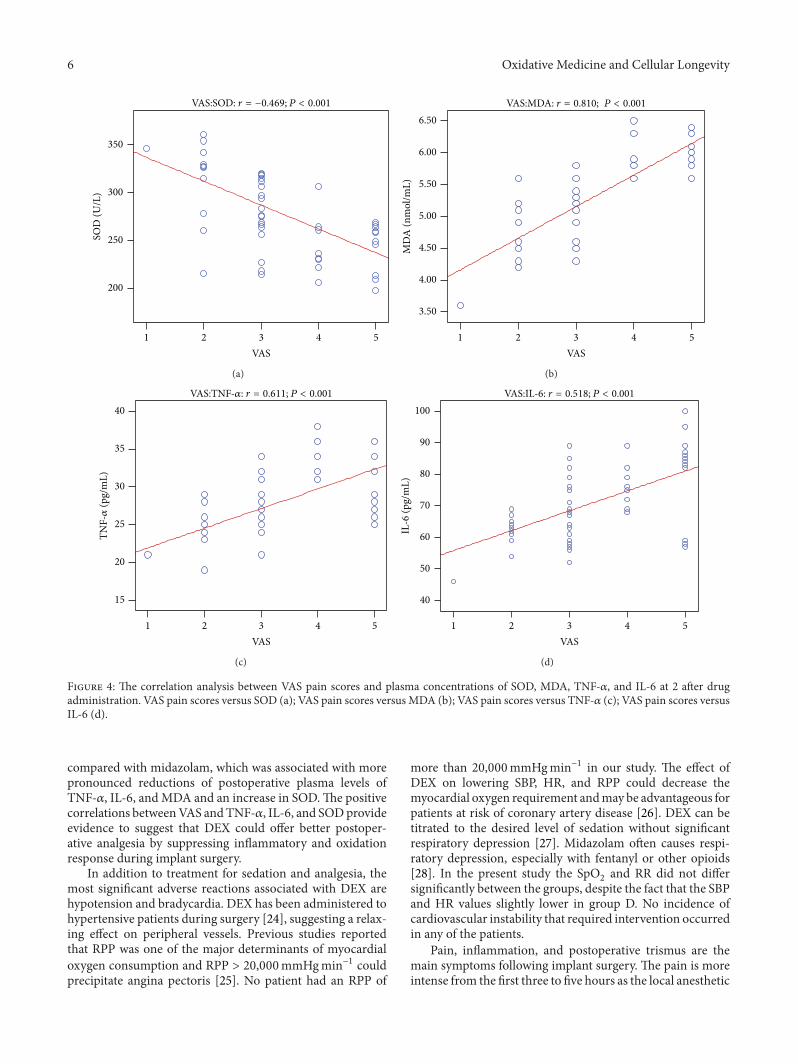
3.4. Correlation Analysis. We surmised that DEX offeredbetter postoperative analgesia by regulating the inflammatoryand oxidation factors. The correlation analyses between VASpain scores and plasma concentrations of SOD, MDA, TNF-𝛼, and IL-6 at 2, 4 after drug administration are shownin Table 2. Spearman analysis showed that VAS pain scores
and plasma SOD content of the two groups were negativelycorrelated at 2, 4 h after drug administration. VAS pain scoresand plasma MDA content were positively correlated. VASpain scores were also positively correlated with plasma TNF-𝛼 and IL-6 content (Figures 4 and 5). Inflammation maycontribute to oxidizing reaction. Our results showed thatplasma TNF-𝛼 and SOD content of the two groups werenegatively correlated, while plasma TNF-𝛼 andMDA contentof the two groups were positively correlated (Figure 6).
4. Discussion
We have shown in the current study that DEX offered bettersedation and postoperative analgesia on implant surgery
6 Oxidative Medicine and Cellular Longevity
VAS54321
SOD
(U/L
)
350
300
250
200
VAS:SOD: r = −0.469; P < 0.001
(a)
MD
A (n
mol
/mL)
6.50
6.00
5.50
5.00
4.50
4.00
3.50
VAS54321
VAS:MDA: r = 0.810; P < 0.001
(b)
VAS54321
40
35
30
25
20
15
TNF-𝛼
(pg/
mL)
VAS:TNF-𝛼: r = 0.611; P < 0.001
(c)
IL-6
(pg/
mL)
100
90
80
70
60
50
40
VAS54321
VAS:IL-6: r = 0.518; P < 0.001
(d)
Figure 4: The correlation analysis between VAS pain scores and plasma concentrations of SOD, MDA, TNF-𝛼, and IL-6 at 2 after drugadministration. VAS pain scores versus SOD (a); VAS pain scores versus MDA (b); VAS pain scores versus TNF-𝛼 (c); VAS pain scores versusIL-6 (d).
compared with midazolam, which was associated with morepronounced reductions of postoperative plasma levels ofTNF-𝛼, IL-6, andMDA and an increase in SOD.The positivecorrelations betweenVAS andTNF-𝛼, IL-6, and SODprovideevidence to suggest that DEX could offer better postoper-ative analgesia by suppressing inflammatory and oxidationresponse during implant surgery.
In addition to treatment for sedation and analgesia, themost significant adverse reactions associated with DEX arehypotension and bradycardia. DEX has been administered tohypertensive patients during surgery [24], suggesting a relax-ing effect on peripheral vessels. Previous studies reportedthat RPP was one of the major determinants of myocardialoxygen consumption and RPP > 20,000mmHgmin−1 couldprecipitate angina pectoris [25]. No patient had an RPP of
more than 20,000mmHgmin−1 in our study. The effect ofDEX on lowering SBP, HR, and RPP could decrease themyocardial oxygen requirement andmay be advantageous forpatients at risk of coronary artery disease [26]. DEX can betitrated to the desired level of sedation without significantrespiratory depression [27]. Midazolam often causes respi-ratory depression, especially with fentanyl or other opioids[28]. In the present study the SpO
2and RR did not differ
significantly between the groups, despite the fact that the SBPand HR values slightly lower in group D. No incidence ofcardiovascular instability that required intervention occurredin any of the patients.
Pain, inflammation, and postoperative trismus are themain symptoms following implant surgery. The pain is moreintense from the first three to five hours as the local anesthetic

Oxidative Medicine and Cellular Longevity 7
150
VAS543210
SOD
(U/L
)
350
300
250
200
VAS:SOD: r = −0.585; P < 0.001
(a)
12.00
10.00
8.00
6.00
VAS543210
MD
A (n
mol
/mL)
VAS:MDA: r = 0.660; P < 0.001
(b)
60
50
40
30
VAS543210
TNF-𝛼
(pg/
mL)
VAS:TNF-𝛼: r = 0.642; P < 0.001
(c)
160
140
120
100
80
60
VAS543210
IL-6
(pg/
mL)
VAS:IL-6: r = 0.624; P < 0.001
(d)
Figure 5: The correlation analysis between VAS pain scores and plasma concentrations of SOD, MDA, TNF-𝛼, and IL-6 at 4 after drugadministration. VAS pain scores versus SOD (a); VAS pain scores versus MDA (b); VAS pain scores versus TNF-𝛼 (c); VAS pain scores versusIL-6 (d).
wears off [29]. DEX can exert analgesic effects throughactivation of central 𝛼
2-adrenergic receptors in the locus
coeruleus [14]. In our study, VAS pain score was below about4 in both groups during and after implant surgery. VAS painscore in group D was lower than group M during 120min–240min. OAAS scores of group D were lower than group Mduring 60min–240min. So, comparedwithmidazolam,DEXoffered better sedation and analgesia during and after implantsurgery.
It is known that an increase in the level of proinflamma-tory cytokines, including TNF-𝛼 and IL-6, is an early featureof acute injury. Recent studies found that DEX has an anti-inflammatory effect by reducing the levels of inflammatorycytokines. A body of animal and clinical trials [30, 31] haveshown that DEX decreases cytokine (TNF-𝛼, IL-6) secretion
after endotoxin injection and that DEX reduced themortalityrate in endotoxemia-induced shock rat models in a dose-dependent manner. In addition, several studies [32–34] havedemonstrated that DEX could exert a potential protectiveeffect by suppressing inflammatory responses on ventila-tor, lipopolysaccharide, or𝛼-naphthylthiourea-induced acutelung injury. Compared to group M, our results showed thatDEX exhibited potent activity in inhibiting TNF-𝛼 and IL-6in dental surgery, particularly 4 h after drug administration.Although studies have shown the regulatory effects ofDEXoninflammatory reactions, the exact mechanisms responsiblefor these actions are not well understood.
TNF-𝛼 is a major proinflammatory cytokine producednot only in the immune system but also in the peripheraland central nervous system, especially under the pathological

8 Oxidative Medicine and Cellular Longevity
40.0035.0030.0025.0020.0015.00
MD
A (n
mol
/mL)
6.50
6.00
5.50
5.00
4.50
4.00
3.50
TNF-𝛼 (pg/mL)(2h)
TNF-𝛼:MDA: r = 0.710; P < 0.001
(a)
60.0050.0040.0030.00
MD
A (n
mol
/mL)
12.00
10.00
8.00
6.00
TNF-𝛼 (pg/mL)(4h)
TNF-𝛼:MDA: r = 0.589; P < 0.001
(b)
SOD
(U/L
)
350
300
250
200
40.0035.0030.0025.0020.0015.00TNF-𝛼 (pg/mL)(2h)
TNF-𝛼:SOD: r = −0.455; P < 0.001
(c)
150
SOD
(U/L
)350
300
250
200
60.0050.0040.0030.00TNF-𝛼 (pg/mL)(4h)
TNF-𝛼:SOD: r = −0.564; P < 0.001
(d)
Figure 6: The correlation analysis between plasma concentrations of MDA and TNF-𝛼 after 2 h of drug administration (a); the correlationanalysis between plasma concentrations of MDA and TNF-𝛼 after 4 h of drug administration (b); the correlation analysis between plasmaconcentrations of SOD and TNF-𝛼 after 2 h of drug administration (c); the correlation analysis between plasma concentrations of SOD andTNF-𝛼 after 4 h of drug administration (d).
conditions [35]. TNF-𝛼 is also known for its substantial role inperiodontitis [36]. Increasing evidence suggests a critical roleof TNF-𝛼 in the pathogenesis of pain including neuropathicpain [37, 38] and acute and persistent inflammatory pain[9, 39]. IL-6 induces muscle and joint hyperalgesia [40]and mediates the development of injury-induced hyperalge-sia [41]. Following surgery, IL-6 levels are associated withpostoperative pain [42]. In samples of patients with pain,levels of IL-6 have been shown to correlate with higher painseverity [43, 44]. Collectively, these findings support thatproinflammatory cytokines are likely to play a facilitatoryrole in the development and maintenance of persistent painsyndromes. Our results showed VAS pain scores and plasmaTNF-𝛼, IL-6 content were positively correlated at 2, 4 h after
drug administration. This suggests that postoperative painmay be caused by acute inflammation and that reducinginflammation cytokine release should have played an impor-tant role in DEXmediated reduction of postoperative pain inpatients undergoing implant surgery.
SOD has strong antioxidant and physical activity andserves as a major free radical scavenger of body [45]. MDA,the end product of lipid peroxidation [46], was assessed incombination with SOD to evaluate the effects of DEX onoxidative stress during and after dental implant surgery inthe current study. Some studies [22, 47, 48] have shown thatDEX can attenuate the increase of MDA level and enhanceSOD activities. In our study, the plasma MDA was higherin group M as compared to group D, while SOD activities

Oxidative Medicine and Cellular Longevity 9
Table 2: The correlation analysis between VAS pain scores andplasma concentrations of SOD, MDA, TNF-𝛼, and IL-6 at 2, 4 afterdrug administration.
Source Dependent Spearman Sig.variable correlation (2-tailed)
VAS painscores
SOD −0.649∗∗ (2 h) <0.001−0.585∗∗ (4 h) <0.001
MDA 0.810∗∗ (2 h) <0.0010.660∗∗ (4 h) <0.001
TNF-𝛼 0.611∗∗ (2 h) <0.0010.642∗∗ (4 h) <0.001
IL-6 0.518∗∗ (2 h) <0.0010.624∗∗ (4 h) <0.001
∗∗Correlation is significant at the 0.01 level (2-tailed).
were significantly lower in group M as compared to groupD. These results pointed to possible antioxidant effects ofDEX in the dental implant region. A few studies reportedthat various reactive oxygen species (ROS) scavengers andantioxidants reduce hyperalgesic behaviours in rat models ofpersistent pain [45]. Superoxide anion (O
2
−) is critical for
sensitization of spinal neurons and persistent pain [49, 50].Antioxidant enzyme SOD is concerned with the removal ofsuperoxide anion. One study shows that saliva and serumantioxidants and serumMDA levels were elevated in patientswith complex regional pain syndrome-type I [50]. Our resultsshowed VAS pain scores and plasma SOD content of thetwo groups were negatively correlated. VAS pain scores andplasma MDA content were positively correlated. This sug-gests that postoperative pain may be caused in part by acuteoxidative stress reaction. Thus, reduction of postoperativeoxidative stress should also play an important role in DEXmediated attenuation of pain.
An exaggerated inflammatory response to tissue injury,ischemia, and reperfusion injuries can all result in excessiveproduction of free radicals [51]. Free radicals, in turn, canincrease vascular permeability, release neuropeptides (i.e.,substance P), enhance inflammation, and cause further tissuedamage [52, 53]. TNF-𝛼 increased the levels of superoxideanion and MDA and then induced oxidative stress andcell toxicity [54, 55]. A small dose of hydrogen peroxideenhances toxicity of TNF-𝛼 in inducing human vascularendothelial cell apoptosis [56]. Our result showed that plasmaTNF-𝛼 and MDA, SOD content were correlated closely.While correlation relationship does not necessarily indicate acausal relationship, the findings of our current study providemechanistic clues for future in-depth study to elucidate themechanism of dexmedetomidine in clinical settings.
5. Conclusions
Our study demonstrates that DEX appears to provide bettersedation, postoperative analgesia than traditional medicinemidazolam during office-based artificial tooth implanta-tion. Further, our findings provide evidence to suggest that
reduction of postoperative inflammatory and oxidative stressplays important role in DEX postoperative analgesic effects,although detailed mechanism needs further study.
Conflict of Interests
The authors declare that there is no conflict of interestsregarding the publication of this paper.
Acknowledgments
The Medical Ethics Committee of the Stomatology Hospi-tal of Chongqing Medical University approved this study.This study was supported by the Research Project ofHealth Bureau of Chongqing (2013-1-031, Chongqing, China)and the Chongqing Science & Technology Commission(cstc2014yykfB10010, Chongqing, China). This study wasconducted from February 2013 to March 2014. The authorsthank surgeons from ImplantDepartment of the StomatologyHospital of Chongqing Medical University, China, and thepeers of previously published papers on this topic.
References
[1] S. C. Pani, B. AlGarni, L. M. AlZain, and N. S. AlQahtani,“Assessment of the impact of stress and anxiety on painperception in patients undergoing surgery for placement oftheir first dental implant,”Oral Health and Dental Management,vol. 13, no. 2, pp. 464–468, 2014.
[2] M. Morino, C. Masaki, Y. Seo et al., “Non-randomized con-trolled prospective study on perioperative levels of stressand dysautonomia during dental implant surgery,” Journal ofProsthodontic Research, vol. 58, no. 3, pp. 177–183, 2014.
[3] R. Gvetadze, E. K. Krechina, S. V. Abramian, A. A. Ivanov,and A. P. Nubarian, “Study of blood circulation microdynamicsin gingival mucosa after dental implantation with the use ofcustom-made healing abutments,” Stomatologiia, vol. 92, no. 3,pp. 109–111, 2013.
[4] Z. Vlahovic, A. Markovic, M. Golubovic, M. Scepanovic, M.Kalanovic, and A. Djinic, “Histopathological comparative anal-ysis of peri-implant soft tissue response after dental implantplacementwith flap and flapless surgical technique. Experimen-tal study in pigs,” Clinical Oral Implants Research, 2014.
[5] A. Figueiredo, P. Coimbra, A. Cabrita, F. Guerra, and M.Figueiredo, “Comparison of a xenogeneic and an alloplasticmaterial used in dental implants in terms of physico-chemicalcharacteristics and in vivo inflammatory response,” MaterialsScience and Engineering C, vol. 33, no. 6, pp. 3506–3513, 2013.
[6] Y. K. Kim, H. S. Kim, Y. J. Yi, and P. Y. Yun, “Evaluation ofsubjective satisfaction of dental implant patients,” Journal of theKorean Association of Oral and Maxillofacial Surgeons, vol. 40,no. 3, pp. 130–134, 2014.
[7] A. I. Basbaum, D. M. Bautista, G. Scherrer, and D. Julius,“Cellular and molecular mechanisms of pain,” Cell, vol. 139, no.2, pp. 267–284, 2009.
[8] T. Hucho and J. D. Levine, “Signaling pathways in sensitization:toward a nociceptor cell biology,”Neuron, vol. 55, no. 3, pp. 365–376, 2007.

10 Oxidative Medicine and Cellular Longevity
[9] Z. Z. Xu, L. Zhang, T. Liu et al., “Resolvins RvE1 and RvD1attenuate inflammatory pain via central and peripheral actions,”Nature Medicine, vol. 16, no. 5, pp. 592–597, 2010.
[10] P.W.Mantyh and S. P. Hunt, “Setting the tone: superficial dorsalhorn projection neurons regulate pain sensitivity,” Trends inNeurosciences, vol. 27, no. 10, pp. 582–584, 2004.
[11] R.-R. Ji, T. Kohno, K. A. Moore, and C. J. Woolf, “Centralsensitization and LTP: do pain and memory share similarmechanisms?” Trends in Neurosciences, vol. 26, no. 12, pp. 696–705, 2003.
[12] O. Goktay, T. Satilmis, H. Garip, O. Gonul, and K. Goker, “Acomparison of the effects of midazolam/fentanyl and midazo-lam/tramadol for conscious intravenous sedation during thirdmolar extraction,” Journal of Oral andMaxillofacial Surgery, vol.69, no. 6, pp. 1594–1599, 2011.
[13] P. Zeyneloglu, A. Pirat, S. Candan, S. Kuyumcu, I. Tekin, andG. Arslan, “Dexmedetomidine causes prolonged recovery whencompared with midazolam/fentanyl combination in outpatientshock wave lithotripsy,” European Journal of Anaesthesiology,vol. 25, no. 12, pp. 961–967, 2008.
[14] C. Correa-Sales, B. C. Rabin, and M. Maze, “A hypnoticresponse to dexmedetomidine, an 𝛼2 agonist, is mediated in thelocus coeruleus in rats,” Anesthesiology, vol. 76, no. 6, pp. 948–952, 1992.
[15] C. Yu, S. Li, F. Deng, Y. Yao, and L. Qian, “Comparisonof dexmedetomidine/fentanyl with midazolam/fentanyl com-bination for sedation and analgesia during tooth extraction,”International Journal of Oral and Maxillofacial Surgery, vol. 43,no. 9, pp. 1148–1153, 2014.
[16] L. Xianbao, Z. Hong, Z. Xu, Z. Chunfang, and C. Dunjin,“Dexmedetomidine reduced cytokine release during postpar-tum bleeding-inducedmultiple organ dysfunction syndrome inrats,”Mediators of inflammation, vol. 2013, Article ID 627831, 7pages, 2013.
[17] L. Xu, H. Bao, Y. Si, and X. Wang, “Effects of dexmedetomidineon early and late cytokines during polymicrobial sepsis inmice,”Inflammation Research, vol. 62, no. 5, pp. 507–514, 2013.
[18] M. Ueki, T. Kawasaki, K. Habe, K. Hamada, C. Kawasaki, andT. Sata, “The effects of dexmedetomidine on inflammatorymediators after cardiopulmonary bypass,” Anaesthesia, vol. 69,no. 7, pp. 693–700, 2014.
[19] C. Chen, Z. Zhang, K. Chen, F. Zhang, M. Peng, and Y.Wang, “Dexmedetomidine regulates inflammatory moleculescontributing to ventilator-induced lung injury in dogs,” Journalof Surgical Research, vol. 187, no. 1, pp. 211–218, 2014.
[20] S.-H. Kang, Y.-S. Kim, T.-H. Hong et al., “Effects of dexmedeto-midine on inflammatory responses in patients undergoinglaparoscopic cholecystectomy,” Acta Anaesthesiologica Scandi-navica, vol. 57, no. 4, pp. 480–487, 2013.
[21] H. Y. Uysal, S. S. Cuzdan, O. Kayıran et al., “Preventive effectof dexmedetomidine in ischemia-reperfusion injury,” Journal ofCraniofacial Surgery, vol. 23, no. 5, pp. 1287–1291, 2012.
[22] O. Eser, H. Fidan, O. Sahin et al., “The influence of dexmedeto-midine on ischemic rat hippocampus,” Brain Research, vol. 1218,pp. 250–256, 2008.
[23] S. Omoigui, “The biochemical origin of pain—proposing a newlaw of pain: the origin of all pain is inflammation and theinflammatory response. Part 1 of 3—a unifying law of pain,”Medical Hypotheses, vol. 69, no. 1, pp. 70–82, 2007.
[24] A. Koroglu, H. Teksan, O. Sagir, A. Yucel, H. I. Toprak, andO. M. Ersoy, “A comparison of the sedative, hemodynamic,
and respiratory effects of dexmedetomidine and propofol inchildren undergoing magnetic resonance imaging,” Anesthesiaand Analgesia, vol. 103, no. 1, pp. 63–67, 2006.
[25] B. F. Robinson, “Relation of heart rate and systolic bloodpressure to the onset of pain in angina pectoris,” Circulation,vol. 35, no. 6, pp. 1073–1083, 1967.
[26] J. D. Tobias, “Dexmedetomidine: applications in pediatric crit-ical care and pediatric anesthesiology,” Pediatric Critical CareMedicine, vol. 8, no. 2, pp. 115–131, 2007.
[27] Y.-W. Hsu, L. I. Cortinez, K. M. Robertson et al., “Dexmedeto-midine pharmacodynamics: part I: crossover comparison ofthe respiratory effects of dexmedetomidine and remifentanilin healthy volunteers,” Anesthesiology, vol. 101, no. 5, pp. 1066–1076, 2004.
[28] P. L. Bailey, N. L. Pace, M. A. Ashburn, J. W. B. Moll, K. A.East, and T. H. Stanley, “Frequent hypoxemia and apnea aftersedation with midazolam and fentanyl,” Anesthesiology, vol. 73,no. 5, pp. 826–830, 1990.
[29] S. E. Fisher, J. W. Frame, P. G. Rout, and D. J. McEntegart,“Factors affecting the onset and severity of pain following thesurgical removal of unilateral impactedmandibular thirdmolarteeth,” British Dental Journal, vol. 164, no. 11, pp. 351–354, 1988.
[30] M. Tasdogan, D. Memis, N. Sut, and M. Yuksel, “Results of apilot study on the effects of propofol and dexmedetomidine oninflammatory responses and intraabdominal pressure in severesepsis,” Journal of Clinical Anesthesia, vol. 21, no. 6, pp. 394–400,2009.
[31] T. Taniguchi, A. Kurita, K. Kobayashi, K. Yamamoto, and H.Inaba, “Dose- and time-related effects of dexmedetomidine onmortality and inflammatory responses to endotoxin-inducedshock in rats,” Journal of Anesthesia, vol. 22, no. 3, pp. 221–228,2008.
[32] Q.-Q. Shi, H. Wang, and H. Fang, “Dose-response and mech-anism of protective functions of selective alpha-2 agonistdexmedetomidine on acute lung injury in rats,” Saudi MedicalJournal, vol. 33, no. 4, pp. 375–381, 2012.
[33] V. Hanci, G. Yurdakan, S. Yurtlu, I. O. Turan, and E. Y. Sipahi,“Protective effect of dexmedetomidine in a rat model of 𝛼-naphthylthiourea-induced acute lung injury,” Journal of SurgicalResearch, vol. 178, no. 1, pp. 424–430, 2012.
[34] C.-L. Yang, C.-H. Chen, P.-S. Tsai, T.-Y.Wang, and C.-J. Huang,“Protective effects of dexmedetomidine-ketamine combinationagainst ventilator-induced lung injury in endotoxemia rats,”Journal of Surgical Research, vol. 167, no. 2, pp. e273–e281, 2011.
[35] M. Schafers, C. Geis, C. I. Svensson, Z. D. Luo, and C.Sommer, “Selective increase of tumour necrosis factor-alpha ininjured and spared myelinated primary afferents after chronicconstrictive injury of rat sciatic nerve,” European Journal ofNeuroscience, vol. 17, no. 4, pp. 791–804, 2003.
[36] B. F. Boyce, P. Li, Z. Yao et al., “TNF𝛼 and pathologic boneresorption,” Keio Journal of Medicine, vol. 54, no. 3, pp. 127–131,2005.
[37] C. Sommer and M. Kress, “Recent findings on how proinflam-matory cytokines cause pain: peripheral mechanisms in inflam-matory andneuropathic hyperalgesia,”Neuroscience Letters, vol.361, no. 1–3, pp. 184–187, 2004.
[38] M. Schafers, C. I. Svensson, C. Sommer, and L. S. Sorkin,“Tumor necrosis factor-𝛼 induces mechanical allodynia afterspinal nerve ligation by activation of p38 MAPK in primarysensory neurons,” Journal of Neuroscience, vol. 23, no. 7, pp.2517–2521, 2003.

Oxidative Medicine and Cellular Longevity 11
[39] L. Zhang, T. Berta, Z.-Z. Xu, T. Liu, J. Y. Park, and R.-R. Ji,“TNF-alpha contributes to spinal cord synaptic plasticity andinflammatory pain: distinct role of TNF receptor subtypes 1 and2,” Pain, vol. 152, no. 2, pp. 419–427, 2011.
[40] O. A. Dina, P. G. Green, and J. D. Levine, “Role of interleukin-6in chronic muscle hyperalgesic priming,”Neuroscience, vol. 152,no. 2, pp. 521–525, 2008.
[41] G. J. Summer, E. A. Romero-Sandoval, O. Bogen, O. A. Dina,S. G. Khasar, and J. D. Levine, “Proinflammatory cytokinesmediating burn-injury pain,” Pain, vol. 135, no. 1-2, pp. 98–107,2008.
[42] B. Lisowska, P. Małdyk, E. Kontny, C. Michalak, L. Jung, andR. Cwiek, “Postoperative evaluation of plasma interleukin-6concentration in patients after total hip arthroplasty,”OrtopediaTraumatologia Rehabilitacja, vol. 8, no. 5, pp. 547–554, 2006.
[43] A. Koch, K. Zacharowski, O. Boehm et al., “Nitric oxide andpro-inflammatory cytokines correlate with pain intensity inchronic pain patients,” Inflammation Research, vol. 56, no. 1, pp.32–37, 2007.
[44] A. L. Davies, K. C. Hayes, and G. A. Dekaban, “Clinicalcorrelates of elevated serum concentrations of cytokines andautoantibodies in patients with spinal cord injury,” Archives ofPhysical Medicine and Rehabilitation, vol. 88, no. 11, pp. 1384–1393, 2007.
[45] H. K. Kim, J. H. Kim, X. Gao et al., “Analgesic effect of vitaminE is mediated by reducing central sensitization in neuropathicpain,” Pain, vol. 122, no. 1-2, pp. 53–62, 2006.
[46] M. Uchiyama and M. Mihara, “Determination of malonalde-hyde precursor in tissues by thiobarbituric acid test,” AnalyticalBiochemistry, vol. 86, no. 1, pp. 271–278, 1978.
[47] X. Dong, Q. Xing, Y. Li, X. Han, and L. Sun, “Dexmedetomidineprotects against ischemia-reperfusion injury in rat skeletalmuscle,” Journal of Surgical Research, vol. 186, no. 1, pp. 240–245,2014.
[48] M. Cosar, O. Eser, H. Fidan et al., “The neuroprotectiveeffect of dexmedetomidine in the hippocampus of rabbits aftersubarachnoid hemorrhage,” Surgical Neurology, vol. 71, no. 1, pp.54–59, 2009.
[49] E. S. Park, X. Gao, J. M. Chung, and K. Chung, “Levels of mito-chondrial reactive oxygen species increase in rat neuropathicspinal dorsal horn neurons,” Neuroscience Letters, vol. 391, no.3, pp. 108–111, 2006.
[50] E. Eisenberg, S. Shtahl, R. Geller et al., “Serum and salivaryoxidative analysis in Complex Regional Pain Syndrome,” Pain,vol. 138, no. 1, pp. 226–232, 2008.
[51] T. J. Coderre, D. N. Xanthos, L. Francis, and G. J. Bennett,“Chronic post-ischemia pain (CPIP): a novel animal model ofcomplex regional pain syndrome-Type I (CRPS-I; reflex sym-pathetic dystrophy) produced by prolonged hindpaw ischemiaand reperfusion in the rat,” Pain, vol. 112, no. 1-2, pp. 94–105,2004.
[52] N. Yonehara and M. Yoshimura, “Effect of nitric oxide onsubstance P release from the peripheral endings of primaryafferent neurons,” Neuroscience Letters, vol. 271, no. 3, pp. 199–201, 1999.
[53] L. van der Laan, P. J. C. Kapitein, W. J. G. Oyen, A. A. J.Verhofstad, T. Hendriks, and R. J. A. Goris, “A novel animalmodel to evaluate oxygen derived free radical damage in softtissue,” Free Radical Research, vol. 26, no. 4, pp. 363–372, 1997.
[54] S. Lei, W. Su, H. Liu et al., “Nitroglycerine-induced nitratetolerance compromises propofol protection of the endothelial
cells against TNF-𝛼: the role of PKC- 𝛽2 and nadph oxidase,”Oxidative Medicine and Cellular Longevity, vol. 2013, Article ID678484, 9 pages, 2013.
[55] J. Han, D. Wang, B. Yu et al., “Cardioprotection againstischemia/reperfusion by licochalcone B in isolated rat hearts,”Oxidative Medicine and Cellular Longevity, vol. 2014, Article ID134862, 11 pages, 2014.
[56] T. Luo and Z. Xia, “A small dose of hydrogen peroxide enhancestumor necrosis factor-alpha toxicity in inducing human vascu-lar endothelial cell apoptosis: reversal with propofol,”Anesthesia& Analgesia, vol. 103, no. 1, pp. 110–116, 2006.

Submit your manuscripts athttp://www.hindawi.com
Stem CellsInternational
Hindawi Publishing Corporationhttp://www.hindawi.com Volume 2014
Hindawi Publishing Corporationhttp://www.hindawi.com Volume 2014
MEDIATORSINFLAMMATION
of
Hindawi Publishing Corporationhttp://www.hindawi.com Volume 2014
Behavioural Neurology
EndocrinologyInternational Journal of
Hindawi Publishing Corporationhttp://www.hindawi.com Volume 2014
Hindawi Publishing Corporationhttp://www.hindawi.com Volume 2014
Disease Markers
Hindawi Publishing Corporationhttp://www.hindawi.com Volume 2014
BioMed Research International
OncologyJournal of
Hindawi Publishing Corporationhttp://www.hindawi.com Volume 2014
Hindawi Publishing Corporationhttp://www.hindawi.com Volume 2014
Oxidative Medicine and Cellular Longevity
Hindawi Publishing Corporationhttp://www.hindawi.com Volume 2014
PPAR Research
The Scientific World JournalHindawi Publishing Corporation http://www.hindawi.com Volume 2014
Immunology ResearchHindawi Publishing Corporationhttp://www.hindawi.com Volume 2014
Journal of
ObesityJournal of
Hindawi Publishing Corporationhttp://www.hindawi.com Volume 2014
Hindawi Publishing Corporationhttp://www.hindawi.com Volume 2014
Computational and Mathematical Methods in Medicine
OphthalmologyJournal of
Hindawi Publishing Corporationhttp://www.hindawi.com Volume 2014
Diabetes ResearchJournal of
Hindawi Publishing Corporationhttp://www.hindawi.com Volume 2014
Hindawi Publishing Corporationhttp://www.hindawi.com Volume 2014
Research and TreatmentAIDS
Hindawi Publishing Corporationhttp://www.hindawi.com Volume 2014
Gastroenterology Research and Practice
Hindawi Publishing Corporationhttp://www.hindawi.com Volume 2014
Parkinson’s Disease
Evidence-Based Complementary and Alternative Medicine
Volume 2014Hindawi Publishing Corporationhttp://www.hindawi.com