clinical prospective study on the use of subcutaneous
TRANSCRIPT
DDiabetic footfoot ulcer (DFU) is a serious complication of diabetes mellitus, and possibly the major mor-
bidity of the diabetic foot. It is the most common foot injury in diabetic patients and can lead to lower-
extremity amputation. Management of DFU requires a systematic knowledge of the major risk factors
for amputation, frequent routine evaluation, scrupulous preventive maintenance, and correction of periph-
eral arterial insufficiency.
Carboxytherapy refers to the subcutaneous injection of CO2 to improve the microcirculation and promote
wound-healing by stimulating the microcirculation. Since optimal ulcer-healing requires adequate tissue
perfusion, it is considered that carboxytherapy could be useful in the treatment of DFU.
The present prospective clinical study included 40 patients with different sizes and types of chronic DFU. In
addition to cleaning of the wound, antibiotics and debridement as necessary, the treatment protocol
included blood sugar control, medication, healthy habits, no weight-bearing, and carboxytherapy.
The results showed that this treatment that included carboxytherapy promoted wound-healing and
prevented amputation.
These positive effects should be confirmed through a complete study that includes different clinical and
instrumental parameters.
Clinical Prospective Study on the Use ofSubcutaneous Carboxytherapy in the
Treatment of Diabetic Foot Ulcer LYNDA KHIAT, MD
GENERAL PRACTITIONERFACULTÉ DE MÉDECINE D'ORAN
ORAN, ALGERIA
GUSTAVO H. LEIBASCHOFF, MDSPECIALIST IN OBSTETRICS AND GYNECOLOGY
FACULTY OF MEDICINE, UNIVERSITY OF BUENOS AIRESBUENOS AIRES, ARGENTINA
- 81 -
#915-Leibaschoff FINAL
ABSTRACT
Advanced Wound HealingSURGICAL TECHNOLOGY INTERNATIONAL Volume 32

- 82 -
Diabetic foot ulcer (DFU) is a majorcomplication of diabetes mellitus. Thewound-healing process involves foursteps: hemostasis, inflammation, prolif-eration and remodeling. The prolifera-tion step includes angiogenesis, collagendeposition, granulation, tissue formationand epithelialization. While wound-heal-ing works effectively most of the time,diabetes mellitus delays the normalphases of wound-healing.1,2 The alter-ation of the healing cascade in diabeticpatients is associated with changes in theextracellular matrix through changes infibroblasts, which lengthen and decreasein quantity. These changes in fibroblasts,in turn, lead to the reduced productionof collagen, elastin and glycosaminogly-can (GAG), and the consequent delay inthe healing and contraction of wounds.3Diabetic patients also exhibit a reducedability to generate nitric oxide from L-arginine, which compromises angiogen-esis and vasodilatation of the bloodvessels.4Uncontrolled diabetic ulcers in the
limbs are responsible for more than 25%of hospital admissions among diabeticpatients in the U.S. and U.K.5 Diabetesis the principal cause of nontraumaticlower-limb amputation in the U.S.6Everett and Mathioudakis recentlyreported that “Mortality rates associatedwith the development of a DFU are estimatedto be 5% in the first 12 months, and 5-yearmortality rates have been estimated at42%.”7In many countries with low levels of
diagnostic technology, the most impor-tant measures for avoiding foot compli-
cations in diabetic patients are carefulinspection and proper care of the diabet-ic foot. The education of diabeticpatients regarding foot hygiene, nail careand proper footwear can help reduce thepossibility of an injury that could lead tothe development of an ulcer in theextremities.8,9Adequate glycemic control, periodic
foot inspection, early recognition andproper medical treatment of these ulcerscould prevent up to 85% of amputa-tions,10,11 and the sooner a diabetic footulcer is treated, the better the result.12The risk factors for ulcer formation
include diabetic neuropathy, structuralfoot deformity and peripheral arterialocclusive disease (PAOD). PAOD is fourtimes more predominant in diabeticsthan in non-diabetics,13 and arterialocclusion typically involves the tibial andperoneal arteries. Aggravating condi-tions, like hypertension, hyperlipidemiaand smoking, also contribute to theincrease in vascular problems in diabeticpatients.14Treatment for diabetic wounds
includes both general guidelines (noweight-bearing, blood sugar control,and the promotion of healthy habits)and local treatments (e.g., removingpressure from the foot ulcer and usingtopical and general medications forwound-healing). Surgical procedures,such as skin grafts, debridement toremove dead tissue, and bypasssurgery, can be useful to restore circu-lation.Ultimately, optimal healing of diabet-
ic ulcers requires satisfactory tissue per-fusion and a healthy, oxygenated woundbed.15-17 A deficiency of oxygen (hypox-ia) in the wound bed slows or stops the
normal healing process. Hyperbaric oxy-gen therapy (HBOT) can be used as anadjunct to comprehensive wound care,however, there is some controversyregarding its effectiveness. Löndahl andco-workers 18 concluded that HBOTpromotes wound repair and enhanceshealing by improving blood circulation,encouraging angiogenesis, and providingmore oxygen to tissue in the woundbed; it also helps kill anaerobic bacteria,which cause some of the worst infec-tions in chronic wounds. On the otherhand, Fedorko and co-workers 19 wrotethat “HBOT does not offer an additionaladvantage to comprehensive wound care inreducing the indication for amputation orfacilitating wound healing in patients withchronic DFUs (diabetic foot ulcers).”Carboxytherapy is an alternative to
HBOT that consists of the therapeuticuse of carbon dioxide (CO2) in itsgaseous state via either transcutaneousapplication or subcutaneous injection.The clinical use of CO2 is not new. In
both Argentina and France, the injectionof CO2 gas has been used to treatperipheral arteriopathies, especiallyarteriopathy obliterans, in the lowerlimbs.20 When CO2 is injected subcuta-neously, it immediately diffuses at thecutaneous and muscular microcirculato-ry levels, increases microcirculatoryvasodilatation and improves flowthrough a direct action on arteriolesmooth muscle cells.21 Via the Bohreffect, it also increases the tissue PO2 atthe injection site.22-24In addition to these effects, the tis-
sue-stretching during CO2 injectioninduces slight inflammation, which trig-gers tissue repair and regeneration, andincreases macrophages, fibroblasts, and
#915-Leibaschoff FINAL
Clinical prospective study on the use of subcutaneous carboxytherapy in the treatment of diabetic foot ulcerKHIAT/LEIBASCHOFF
INTRODUCTION
Figure 1. Carboxytherapy device. (a) side view. (b) control panel.
a b

- 83 -
endothelial cells, resulting in neo-vascu-larization and remodeling of the extra-cellular matrix.25-30Based on these effects of car-
boxytherapy on the microcirculation ofthe skin, we decided to start using thistechnique in diabetic patients withulcers in their limbs. In this report, wedescribe our experience with car-boxytherapy in 40 patients with diabeticfoot ulcers.
Methods and Materials
SubjectsThis prospective clinical study includ-
ed 40 patients (13 female/27 male) withdifferent sizes and types of chronic dia-betic foot ulcers who were treatedbetween January 2015 and January 2017.The treatment protocol included
blood sugar control, medication, promo-
tion of healthy habits, no weight-bearing,and carboxytherapy. All of the patientswere volunteers from the communityfrom different regions of Algeria. They allprovided their written informed consentfor the treatment.Of the total 40 patients, 6 patients had
previously received hyperbaric oxygentherapy and 3 had received LED therapy.The remaining 31 patients had been treat-ed only with local dressings, which wasthe only treatment available in that part ofAlgeria. The average duration of car-boxytherapy was 7 months (Case 6 wastreated with CO2 for only three weeks).Healing was evaluated according to
the following criteria: a change in theodor of the dressing, the presence ofangiogenesis, new networks of blood ves-sels, changes in the exudate, a change inlocal wound color, re-epithelializationfrom the edges of the wound, andimprovement in the general condition ofthe patient (reduction of fever and betterglycemic balance).
CarboxytherapyThe CO2 device (Carboxy-pen®,
Anti-Aging Medical Systems, Montrodat,France) consists of a handpiece and a con-trol panel (Fig. 1). The electronic controlallows the user to select different para-meters, including the flow velocity, thegas volume and the dose. The CO2 itselfmust be sterile and suitable for medicaluse. CO2 is injected both systemically (at
the blue sites in Fig. 2) and locally at thewound site.Figure 3 shows an example of where
CO2 was injected in and around a wound.
Results
Clinical casesOur experiences with 6 of the 40
cases are summarized below. All of thecarboxytherapy patients were treatedwith metronidazole (500 mg, 2tablets/day) to prevent anaerobicinfection. In all cases, treatment was asfollows:75 cc or less (depending on tolerabili-
ty by the patient) of CO2 was injected ateach injection site (Fig. 2) on the internaland external sides of the right and leftlegs along the route of the large vessels ata depth of 4mm (intradermic).The same dose of CO2 was then
injected in and around the wound.
#915-Leibaschoff FINAL
Advanced Wound HealingSURGICAL TECHNOLOGY INTERNATIONAL Volume 32
MATERIALS AND METHODS
RESULTS
Figure 2. Sites for general CO2 injection (blue).
- 84 -
Case 1
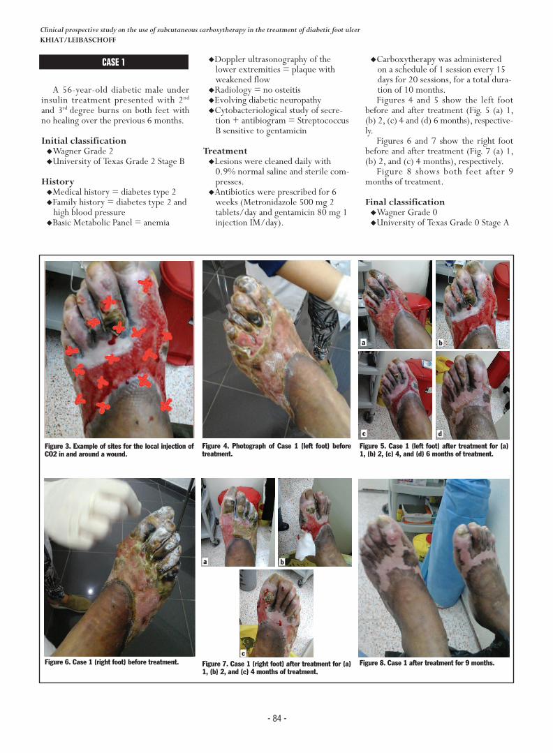
A 56-year-old diabetic male underinsulin treatment presented with 2ndand 3rd degree burns on both feet withno healing over the previous 6 months.
Initial classification�Wagner Grade 2�University of Texas Grade 2 Stage B
History�Medical history = diabetes type 2 �Family history = diabetes type 2 andhigh blood pressure
�Basic Metabolic Panel = anemia
�Doppler ultrasonography of thelower extremities = plaque withweakened flow
�Radiology = no osteitis�Evolving diabetic neuropathy�Cytobacteriological study of secre-tion + antibiogram = StreptococcusB sensitive to gentamicin
Treatment�Lesions were cleaned daily with0.9% normal saline and sterile com-presses.
�Antibiotics were prescribed for 6weeks (Metronidazole 500 mg 2tablets/day and gentamicin 80 mg 1injection IM/day).
�Carboxytherapy was administeredon a schedule of 1 session every 15days for 20 sessions, for a total dura-tion of 10 months.Figures 4 and 5 show the left foot
before and after treatment (Fig. 5 (a) 1,(b) 2, (c) 4 and (d) 6 months), respective-ly. Figures 6 and 7 show the right foot
before and after treatment (Fig. 7 (a) 1,(b) 2, and (c) 4 months), respectively. Figure 8 shows both feet after 9
months of treatment.
Final classification�Wagner Grade 0�University of Texas Grade 0 Stage A
#915-Leibaschoff FINAL
Clinical prospective study on the use of subcutaneous carboxytherapy in the treatment of diabetic foot ulcerKHIAT/LEIBASCHOFF
Figure 3. Example of sites for the local injection ofCO2 in and around a wound.
Figure 6. Case 1 (right foot) before treatment.
Figure 5. Case 1 (left foot) after treatment for (a)1, (b) 2, (c) 4, and (d) 6 months of treatment.
Figure 4. Photograph of Case 1 (left foot) beforetreatment.
Figure 7. Case 1 (right foot) after treatment for (a)1, (b) 2, and (c) 4 months of treatment.
Figure 8. Case 1 after treatment for 9 months.
CASE 1
a
dc
b
a
c
b

- 85 -
Case 2
A 62-year-old diabetic male underinsulin treatment presented with ampu-tation of the left great toe with poorwound-healing over the previous 6months.
Initial classification�Wagner Grade 3�University of Texas Grade 3 Stage B
History�Medical history = diabetes type 2 �Surgical history = amputation of theleft great toe
�Family history = diabetes type 2 andhigh blood pressure
�Basic Metabolic Panel = anemia�Doppler ultrasonography of thelower extremities = plaque withweakened flow
�Radiology = osteitis �Evolving diabetic neuropathy�Cytobacteriological study of secre-tion+ antibiogram = Proteus sensi-tive to ciprofloxacin
Treatment�The lesion was cleaned daily with0.9% normal saline and sterile com-presses with medical honey.
�Antibiotics were administered for 8weeks (Metronidazole 500 mg 2tablets/day and ciprofloxacin 500mg 2 tablets/day)
�Carboxytherapy was administered on
a schedule of 1 session every 7 daysuntil the lesion started to improve(at 2 months), and then 1 sessionevery 15 days, for a total of 23 ses-sions over a duration of 10 months.
Figure 9 shows a photograph at theinitiation of treatment. In the photo-graph at 2 weeks (Fig. 10), osteitis isvisible. Figures 11-15 show the results after
treatment for 3, 3, 5, 6 and 8 months,respectively.
Final classification�Wagner Grade 1�University of Texas Grade 0 Stage A
#915-Leibaschoff FINAL
Advanced Wound HealingSURGICAL TECHNOLOGY INTERNATIONAL Volume 32
Figure 9. Photograph of Case 2 before treatment
Figure 10. Case 2 after treatment for 2 weeks.Osteitis is visible.
Figure 11. Case 2 after treatment for 3 months. Figure 12. Case 2 after treatment for 3 months.
Figure 13. Case 2 after treatment for 5 months. Figure 14. Case 2 after treatment for 6 months. Figure 15. Case 2 after treatment for 8 months.Final result with hyperkeratosis.
CASE 2
07/01/2015
20/01/2015
02/04/2015
28/05/2015 25/06/2015 03/09/2015

- 86 -
CASE 3
A 54-year-old diabetic male underinsulin treatment presented withaltered wound healing at 9 months afteramputation of the left fourth toe withmany fibr in filaments and plantarinfection.
Initial classification�Wagner Grade 2�University of Texas Grade 2 Stage B
History�Medical = diabetes type 2 and highblood pressure.
�Surgical = amputation
�Family = diabetes type 2 and highblood pressure
Exam results�Basic Metabolic Panel = normal�Doppler ultrasonography of thelower extremities = plaque withweakened flow
�Radiology = No osteitis �Evolving diabetic neuropathy�ECB pus + antibiogram =Haemophilus influenza sensitive toCiprolon
Treatment�Lesions were cleaned daily with0.9% normal saline and sterile com-presses.
�Antibiotics were prescribed for 8weeks (Metronidazole 500 mg 2tablets/day and Ciprolon 500 mg 2tablets/day)
�Carboxytherapy was administeredon a schedule of 1 session every 15days for 10 sessions, for a total dura-tion of 5 months.
Figures 16-22 show photographs atthe initiation of treatment, and aftertreatment for 1, 2, 3, 3.5, 4 and 48months, respectively.
Final classification�Wagner Grade 0�University of Texas Grade 0 Stage A
#915-Leibaschoff FINAL
Clinical prospective study on the use of subcutaneous carboxytherapy in the treatment of diabetic foot ulcerKHIAT/LEIBASCHOFF
Figure 16. Case 3 before treatment. Figure 17. Case 3 after treatment for 1month.
Figure 18. Case 3 after treatment for 2months.
Figure 19. Case 3 after treatment for 3months.
Figure 20. Case 3 after treatment for3.5 months.
Figure 21. Case 3 after treatment for 4months.
Figure 22. Case 3 final control photoone year after the last session of car-boxytherapy.
CASE 3
18/01/2016 16/02/2016 15/03/2016 14/04/2016
28/04/2016 November 201718/05/2016
- 87 -
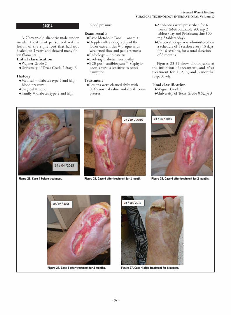
CASE 4
A 70-year-old diabetic male underinsulin treatment presented with alesion of the right foot that had nothealed for 3 years and showed many fib-rin filaments.Initial classification �Wagner Grade 2�University of Texas Grade 2 Stage B
History�Medical = diabetes type 2 and highblood pressure.
�Surgical = none�Family = diabetes type 2 and high
blood pressure
Exam results�Basic Metabolic Panel = anemia�Doppler ultrasonography of thelower extremities = plaque withweakened flow and pedis stenosis
�Radiology = no osteitis �Evolving diabetic neuropathy�ECB pus+ antibiogram = Staphylo-coccus aureus sensitive to pristi-namycine
Treatment�Lesions were cleaned daily with0.9% normal saline and sterile com-presses.
�Antibiotics were prescribed for 6weeks (Metronidazole 500 mg 2tablets/day and Pristinamycine 500mg 2 tablets/day)
�Carboxytherapy was administered ona schedule of 1 session every 15 daysfor 16 sessions, for a total durationof 8 months.
Figures 23-27 show photographs atthe initiation of treatment, and aftertreatment for 1, 2, 3, and 6 months,respectively.
Final classification�Wagner Grade 0�University of Texas Grade 0 Stage A
#915-Leibaschoff FINAL
Advanced Wound HealingSURGICAL TECHNOLOGY INTERNATIONAL Volume 32
Figure 23. Case 4 before treatment. Figure 24. Case 4 after treatment for 1 month. Figure 25. Case 4 after treatment for 2 months.
Figure 26. Case 4 after treatment for 3 months. Figure 27. Case 4 after treatment for 6 months.
CASE 4
- 88 -
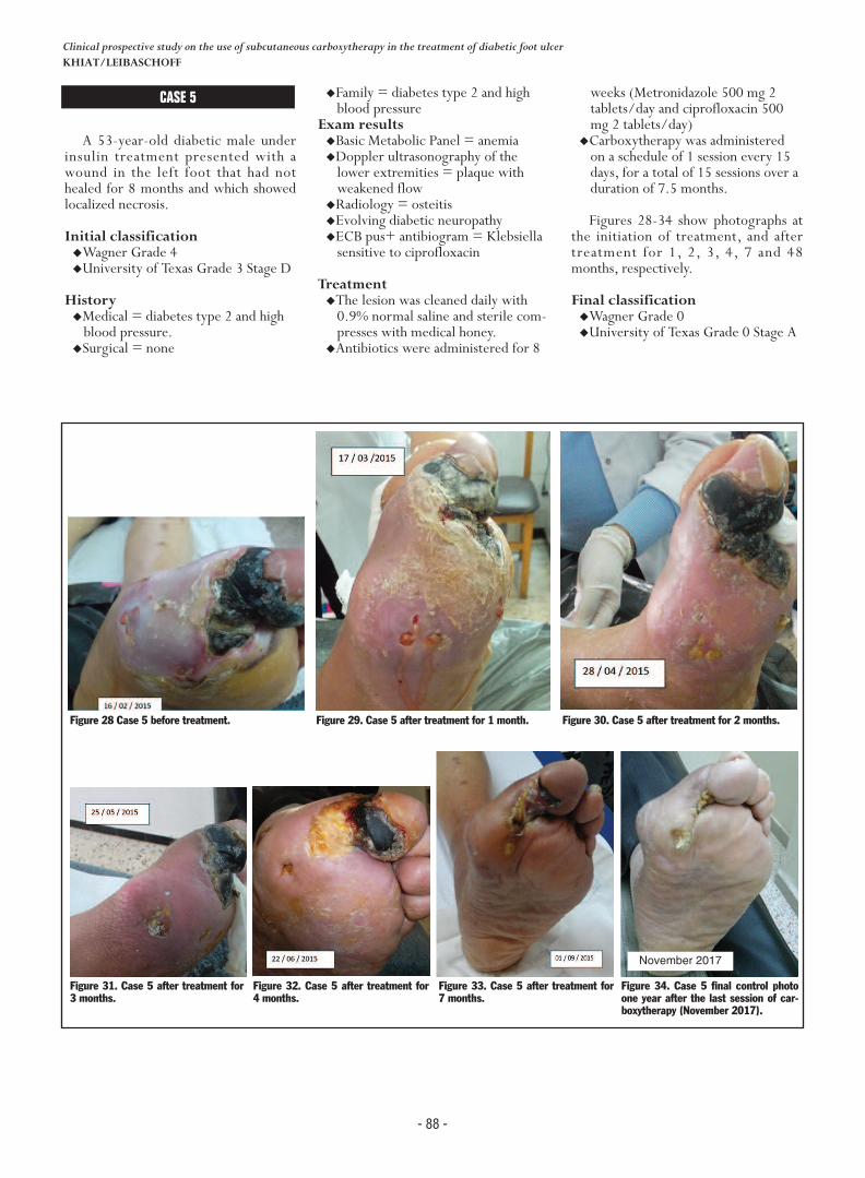
CASE 5
A 53-year-old diabetic male underinsulin treatment presented with awound in the left foot that had nothealed for 8 months and which showedlocalized necrosis.
Initial classification�Wagner Grade 4�University of Texas Grade 3 Stage D
History�Medical = diabetes type 2 and highblood pressure.
�Surgical = none
�Family = diabetes type 2 and highblood pressure
Exam results�Basic Metabolic Panel = anemia�Doppler ultrasonography of thelower extremities = plaque withweakened flow
�Radiology = osteitis �Evolving diabetic neuropathy�ECB pus+ antibiogram = Klebsiellasensitive to ciprofloxacin
Treatment�The lesion was cleaned daily with0.9% normal saline and sterile com-presses with medical honey.
�Antibiotics were administered for 8
weeks (Metronidazole 500 mg 2tablets/day and ciprofloxacin 500mg 2 tablets/day)
�Carboxytherapy was administeredon a schedule of 1 session every 15days, for a total of 15 sessions over aduration of 7.5 months.
Figures 28-34 show photographs atthe initiation of treatment, and aftertreatment for 1, 2, 3, 4, 7 and 48months, respectively.
Final classification�Wagner Grade 0�University of Texas Grade 0 Stage A
#915-Leibaschoff FINAL
Clinical prospective study on the use of subcutaneous carboxytherapy in the treatment of diabetic foot ulcerKHIAT/LEIBASCHOFF
Figure 28 Case 5 before treatment. Figure 29. Case 5 after treatment for 1 month. Figure 30. Case 5 after treatment for 2 months.
Figure 31. Case 5 after treatment for3 months.
Figure 32. Case 5 after treatment for4 months.
Figure 33. Case 5 after treatment for7 months.
Figure 34. Case 5 final control photoone year after the last session of car-boxytherapy (November 2017).
CASE 5
November 2017
- 89 -
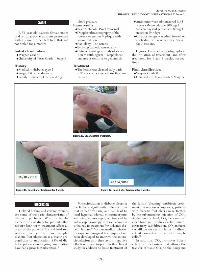
CASE 6
A 54-year-old diabetic female underoral antidiabetic treatment presentedwith a lesion on her left foot that hadnot healed for 6 months.
Initial classification�Wagner Grade 1�University of Texas Grade 1 Stage B
History�Medical = diabetes type 2�Surgical = appendectomy�Family = diabetes type 2 and high
blood pressure Exam results�Basic Metabolic Panel =normal �Doppler ultrasonography of thelower extremities = plaque withweakened flow
�Radiology = no osteitis�Evolving diabetic neuropathy�Cytobacteriological study of secre-tion + antibiogram = Staphylococ-cus aureus sensitive to gentamicin
Treatment�The lesion was cleaned daily with0.9% normal saline and sterile com-presses.
�Antibiotics were administered for 3weeks (Metronidazole 500 mg 2tablets/day and gentamicin 80mg 1injection IM/day)
�Carboxytherapy was administered ona schedule of 1 session every 7 daysfor 2 sessions.
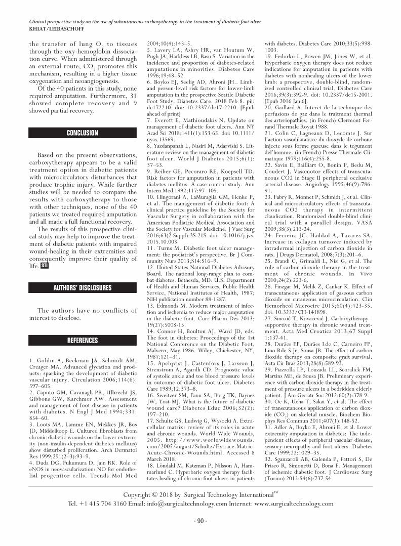
Figures 35-37 show photographs atthe initiation of treatment, and aftertreatment for 1 and 3 weeks, respec-tively.
Final classification�Wagner Grade 0�University of Texas Grade 0 Stage A
#915-Leibaschoff FINAL
Advanced Wound HealingSURGICAL TECHNOLOGY INTERNATIONAL Volume 32
Figure 35. Case 6 before treatment.
Figure 36. Case 6 after treatment for 1 week. Figure 37. Case 6 after treatment for 3 weeks.
CASE 6
Discussion
Delayed healing and chronic woundsare some of the basic characteristics ofdiabetic patients. Wounds in theextremities of diabetic patients thatrequire long-term treatment affect allareas of the patient’s life and lead to areduced quality of life. For example,diabetic foot ulceration is a major pre-condition to amputation; 85% of dia-betic patients undergoing amputationhave had a prior foot ulceration.31
Microcirculation in diabetic ulcers inthe limbs is significantly different fromthat in healthy skin, and can lead tolocal hypoxia, edema, microaneurysmsand microhemorrhages, as observed byvideo capillaroscopy. Revascularizationis the key to treatment for ischemic dia-betic lesions.32 Various medical, physio-therapy and surgical techniques havebeen developed to improve the micro-circulation and thus avoid negativeeffects on tissue tropism. In this clinicalstudy, in addition to basic treatment of
the lesion (cleaning, antibiotic treat-ment, correction of support), patientswith diabetic foot ulcers were treatedby the subcutaneous injection of CO2.At the vascular level, CO2 increases vas-cular tone and produces active micro-circulatory vasodilatation. CO2-inducedvasodilatation results from its directactivity on arteriole smooth musclecells.21In addition, CO2 promotes Bohr’s
effect, a mechanism that allows thetransfer of tissue CO2 to the lungs and
DISCUSSION
the transfer of lung O2 to tissuesthrough the oxy-hemoglobin dissocia-tion curve. When administered throughan external route, CO2 promotes thismechanism, resulting in a higher tissueoxygenation and neoangiogenesis.Of the 40 patients in this study, none
required amputation. Furthermore, 31showed complete recovery and 9showed partial recovery.
Conclusion
Based on the present observations,carboxytherapy appears to be a validtreatment option in diabetic patientswith microcirculatory disturbances thatproduce trophic injury. While furtherstudies will be needed to compare theresults with carboxytherapy to thosewith other techniques, none of the 40patients we treated required amputationand all made a full functional recovery.The results of this prospective clini-
cal study may help to improve the treat-ment of diabetic patients with impairedwound-healing in their extremities andconsequently improve their quality oflife.
Authors Disclose
The authors have no conflicts ofinterest to disclose.
References
1. Goldin A, Beckman JA, Schmidt AM,Creager MA. Advanced glycation end prod-ucts: sparking the development of diabeticvascular injury. Circulation 2006;114(6):597–605.2. Caputo GM, Cavanagh PR, Ulbrecht JS,Gibbons GW, Karchmer AW. Assessmentand management of foot disease in patientswith diabetes. N Engl J Med 1994;331:854–60. 3. Loots MA, Lamme EN, Mekkes JR, BosJD, Middelkoop E. Cultured fibroblasts fromchronic diabetic wounds on the lower extrem-ity (non-insulin-dependent diabetes mellitus)show disturbed proliferation. Arch DermatolRes 1999;291(2–3):93–9.4. Duda DG, Fukumura D, Jain RK. Role ofeNOS in neovascularization: NO for endothe-lial progenitor cells. Trends Mol Med
2004;10(4):143–5.5. Lavery LA, Ashry HR, van Houtum W,Pugh JA, Harkless LB, Basu S. Variation in theincidence and proportion of diabetes-relatedamputations in minorities. Diabetes Care1996;19:48–52.6. Boyko EJ, Seelig AD, Ahroni JH.. Limb-and person-level risk factors for lower-limbamputation in the prospective Seattle DiabeticFoot Study. Diabetes Care. 2018 Feb 8. pii:dc172210. doi: 10.2337/dc17-2210. [Epubahead of print]7. Everett E, Mathioudakis N. Update onmanagement of diabetic foot ulcers. Ann NYAcad Sci 2018;1411(1):153-65. doi: 10.1111/nyas.13569.8. Yazdanpanah L, Nasiri M, Adarvishi S. Lit-erature review on the management of diabeticfoot ulcer. World J Diabetes 2015;6(1):37–53.9. Reiber GE, Pecoraro RE, Koepsell TD.Risk factors for amputation in patients withdiabetes mellitus. A case-control study. AnnIntern Med 1992;117:97–105.10. Hingorani A, LaMuraglia GM, Henke P,et al. The management of diabetic foot: Aclinical practice guideline by the Society forVascular Surgery in collaboration with theAmerican Podiatric Medical Association andthe Society for Vascular Medicine. J Vasc Surg2016;63(2 Suppl):3S-21S. doi: 10.1016/j.jvs.2015.10.003.11. Turns M. Diabetic foot ulcer manage-ment: the podiatrist’s perspective. Br J Com-munity Nurs 2013;S14:S16–9.12. United States National Diabetes AdvisoryBoard. The national long-range plan to com-bat diabetes. Bethesda, MD: U.S. Departmentof Health and Human Services, Public HealthService, National Institutes of Health, 1987;NIH publication number 88-1587. 13. Edmonds M. Modern treatment of infec-tion and ischemia to reduce major amputationin the diabetic foot. Curr Pharm Des 2013;19(27):5008-15.14. Connor H, Boulton AJ, Ward JD, eds.The foot in diabetes: Proceedings of the 1stNational Conference on the Diabetic Foot,Malvern, May 1986. Wiley, Chichester, NY,1987:121–31. 15. Apelqvist J, Castenfors J, Larsson J,Strenstrom A, Agardh CD. Prognostic valueof systolic ankle and toe blood pressure levelsin outcome of diabetic foot ulcer. DiabetesCare 1989;12:373–8.16. Sweitzer SM, Fann SA, Borg TK, BaynesJW, Yost MJ. What is the future of diabeticwound care? Diabetes Educ 2006;32(2):197–210.17. Schultz GS, Ludwig G, Wysocki A. Extra-cellular matrix: review of its roles in acuteand chronic wounds. World Wide Wounds,2005. http://www.worldwidewounds.com/2005/august/Schultz/Extrace-Matric-Acute-Chronic-Wounds.html. Accessed 8March 2018.18. Löndahl M, Katzman P, Nilsson A, Ham-marlund C. Hyperbaric oxygen therapy facili-tates healing of chronic foot ulcers in patients
with diabetes. Diabetes Care 2010;33(5):998-1003.19. Fedorko L, Bowen JM, Jones W, et al.Hyperbaric oxygen therapy does not reduceindications for amputation in patients withdiabetes with nonhealing ulcers of the lowerlimb: a prospective, double-blind, random-ized controlled clinical trial. Diabetes Care2016;39(3):392-9. doi: 10.2337/dc15-2001.[Epub 2016 Jan 6].20. Gaillard A. Interet de la technique desperfusions de gaz dans le traitment thermaldes arteriopathies. (in French) Clermont Fer-rand Thermale Royat 1988.21. Colin C, Lagneaux D, Lecomte J. Surl’action vasodilatatrice du dioxyde de carboneinjecte sous forme gazeuse dans le tegumentdel’homme. (in French) Presse Thermale Cli-matique 1979;116(4):255-8.22. Savin E, Bailliart O, Bonin P, Bedu M,Coudert J. Vasomotor effects of transcuta-neous CO2 in Stage II peripheral occlusivearterial disease. Angiology 1995;46(9):786-91.23. Fabry R, Monnet P, Schmidt J, et al. Clin-ical and microcirculatory effects of transcuta-neous CO2 therapy in intermittentclaudication. Randomized double-blind clini-cal trial with a parallel design. VASA2009;38(3):213-24. 24. Ferreira JC, Haddad A, Tavares SA.Increase in collagen turnover induced byintradermal injection of carbon dioxide inrats. J Drugs Dermatol, 2008;7(3):201–6.25. Brandi C, Grimaldi L, Nisi G, et al. Therole of carbon dioxide therapy in the treat-ment of chronic wounds. In Vivo2010;24(2):223-6.26. Finzgar M, Melik Z, Cankar K. Effect oftranscutaneous application of gaseous carbondioxide on cutaneous microcirculation. ClinHemorheol Microcirc 2015;60(4):423-35.doi: 10.3233/CH-141898.27. Sinozić T, Kovacević J. Carboxytherapy -supportive therapy in chronic wound treat-ment. Acta Med Croatica 2013;67 Suppl1:137-41.28. Durães EF, Durães Lde C, Carneiro FP,Lino Rde S Jr, Sousa JB. The effect of carbondioxide therapy on composite graft survival.Acta Cir Bras 2013;28(8):589-93.29. Piazzolla LP, Louzada LL, Scoralick FM,Martins ME, de Sousa JB. Preliminary experi-ence with carbon dioxide therapy in the treat-ment of pressure ulcers in a bedridden elderlypatient. J Am Geriatr Soc 2012;60(2):378-9.30. Oe K, Ueha T, Sakai Y, et al. The effectof transcutaneous application of carbon diox-ide (CO2) on skeletal muscle. Biochem Bio-phys Res Commun 2011;407(1):148-52.31. Adler A, Boyko E, Ahroni E, et al. Lowerextremity amputation in diabetes: The inde-pendent effects of peripheral vascular disease,sensory neuropathy and foot ulcers. DiabetesCare 1999;22:1029–35.32. Sganzaroli AB, Galenda P, Fattori S, DePrisco R, Simonetti D, Bona F. Managementof ischemic diabetic foot. J Cardiovasc Surg(Torino) 2013;54(6):737-54.
- 90 -
#915-Leibaschoff FINAL
Clinical prospective study on the use of subcutaneous carboxytherapy in the treatment of diabetic foot ulcerKHIAT/LEIBASCHOFF
REFERENCES
STI
CONCLUSION
AUTHORS’ DISCLOSURES
Copyright © 2018 by Surgical Technology International™
Tel. +1 415 704 3160 Email: [email protected] Internet: www.surgicaltechnology.com