clinical presentation and management of endometriosis
TRANSCRIPT

Review began 06/04/2021 Review ended 06/12/2021 Published 06/22/2021
© Copyright 2021Chan-Tiopianco et al. This is an openaccess article distributed under the termsof the Creative Commons AttributionLicense CC-BY 4.0., which permitsunrestricted use, distribution, andreproduction in any medium, provided theoriginal author and source are credited.
Clinical Presentation and Management ofEndometriosis-Related Hemorrhagic Ascites: ACase Report and Systematic Review of theLiteratureMareesol Chan-Tiopianco , Wei-Ting Chao , Patrick R. Ching , Ling-Yu Jiang , Peng-Hui Wang ,Yi-Jen Chen
1. Division of Obstetrics and Gynecology, San Lazaro Hospital, Manila, PHL 2. Department of Obstetrics andGynecology, ManilaMed - Medical Center Manila, Manila, PHL 3. Department of Obstetrics and Gynecology, TaipeiVeterans General Hospital, Taipei, TWN 4. Department of Medicine, University of Maryland Medical Center MidtownCampus, Baltimore, USA 5. Institute of Clinical Medicine, National Yang-Ming University, Taipei, TWN
Corresponding author: Yi-Jen Chen, [email protected]
AbstractThis study aims to analyze the patient profile and presentation of endometriosis-related hemorrhagicascites and review its management to raise awareness among gynecologists and improve treatmentstrategies. We present a case report and engage in a systematic review involving human cases ofhistologically proven endometriosis with hemorrhagic ascites. Keywords were searched inPubMed/MEDLINE, Cochrane Library, EMBASE, and Ovid Discovery databases from inception untilDecember 2018. Studies that did not include a description of ascites or histopathologic results confirmingendometriosis or those that involved patients with other conditions that may contribute to ascites wereexcluded.
The review yielded 73 articles describing 84 premenopausal women with histologically provenendometriosis-related hemorrhagic ascites. Of note, 83% (65/78) of the patients were nulliparous and69.35% (43/62) were of African descent. The most common chief complaint was abdominal enlargement(58.33%, 49/84) but a host of other symptoms were also reported. Pleural effusion was reported in 32.14%(27/84), and elevated CA-125 was seen in 74.42% (32/43). The majority (64.29%, 54/84) of the patientsunderwent laparotomy, and an increasing trend of minimally invasive surgical approaches (p<0.001) andfertility-sparing techniques (p<0.001) was observed. The mean ascites volume was 4228.27 mL (SD:2625.66). Moderate to severe endometriosis was seen in 97.44% (76/78) of cases. The majority of the patientswho received medical treatment were given gonadotropin-releasing hormone (GnRH) agonists (63.79%,37/58). The rate of recurrence after termination or suppression of ovarian function was 8.33% (7/84), andthere was a mortality rate of 1.19% (1/84). Diagnosis of endometriosis-related hemorrhagic ascites may bechallenging because it mimics several disease entities that cause ascites, thereby warranting a heightenedclinical suspicion. Minimally invasive techniques are usually employed to establish a histologic diagnosis.The prevention of recurrence involves the recognition of endometriosis-related hemorrhagic ascites as amanifestation of severe endometriosis, which should prompt therapies directed at suppressing ovarianfunction. Since affected women are of childbearing age, ovary-preserving surgeries are generally preferred.The rate of recurrence is low after appropriate surgical and medical interventions.
Categories: Obstetrics/GynecologyKeywords: ascites, bloody ascites, endometriosis, hemorrhage, hemorrhagic ascites
IntroductionHemorrhagic ascites is a rare complication of endometriosis. The first description of endometriosis-relatedascites has been attributed to Brews in 1954 [1]. However, it was not until 1957 that Charles first chronicled acase of blood-stained ascites in association with endometriosis [2]. Since then, fewer than 100 reports ofhemorrhagic ascites related to endometriosis have been published in the literature.
Endometriosis-related hemorrhagic ascites may manifest with varying symptoms. Recognizing it may bedifficult as it may present with similar disease processes such as malignancy, infection, cirrhosis, or trauma[3-6]. In light of this, we conducted this study to examine and elucidate the patient profiles and presentationof the disease to raise clinical awareness among gynecologists regarding the diagnosis of hemorrhagicascites associated with endometriosis.
Case PresentationA 34-year-old Taiwanese nulligravida woman presented to the outpatient department with a one-yearhistory of irregular dysmenorrhea that was 5/10 in severity. She had no other associated complaints such as
1, 2 3 4 3, 5 3, 5
3, 5
Open Access CaseReport DOI: 10.7759/cureus.15828
How to cite this articleChan-Tiopianco M, Chao W, Ching P R, et al. (June 22, 2021) Clinical Presentation and Management of Endometriosis-Related HemorrhagicAscites: A Case Report and Systematic Review of the Literature. Cureus 13(6): e15828. DOI 10.7759/cureus.15828
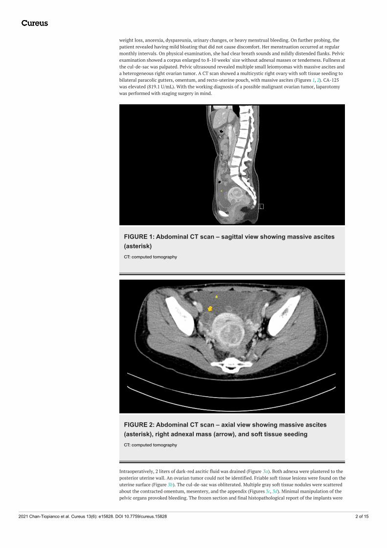
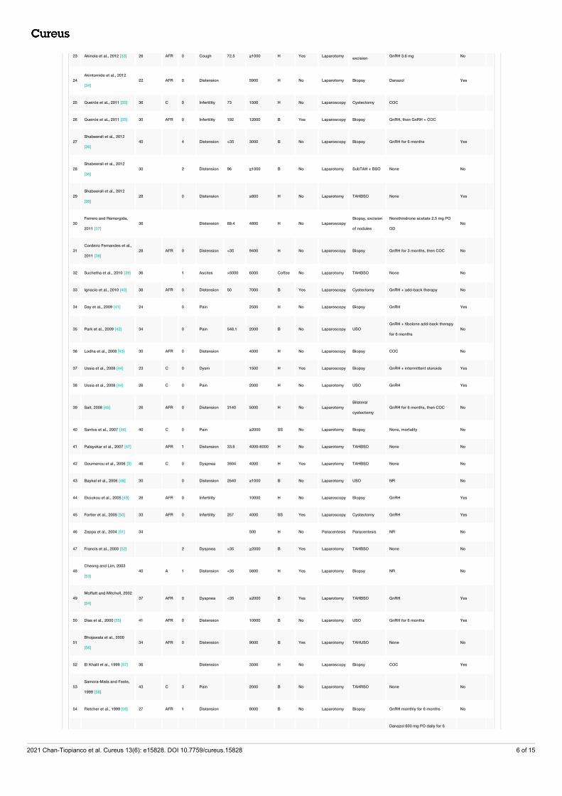
weight loss, anorexia, dyspareunia, urinary changes, or heavy menstrual bleeding. On further probing, thepatient revealed having mild bloating that did not cause discomfort. Her menstruation occurred at regularmonthly intervals. On physical examination, she had clear breath sounds and mildly distended flanks. Pelvicexamination showed a corpus enlarged to 8-10 weeks' size without adnexal masses or tenderness. Fullness atthe cul-de-sac was palpated. Pelvic ultrasound revealed multiple small leiomyomas with massive ascites anda heterogeneous right ovarian tumor. A CT scan showed a multicystic right ovary with soft tissue seeding tobilateral paracolic gutters, omentum, and recto-uterine pouch, with massive ascites (Figures 1, 2). CA-125was elevated (819.1 U/mL). With the working diagnosis of a possible malignant ovarian tumor, laparotomywas performed with staging surgery in mind.
FIGURE 1: Abdominal CT scan – sagittal view showing massive ascites(asterisk)CT: computed tomography
FIGURE 2: Abdominal CT scan – axial view showing massive ascites(asterisk), right adnexal mass (arrow), and soft tissue seedingCT: computed tomography
Intraoperatively, 2 liters of dark-red ascitic fluid was drained (Figure 3a). Both adnexa were plastered to theposterior uterine wall. An ovarian tumor could not be identified. Friable soft tissue lesions were found on theuterine surface (Figure 3b). The cul-de-sac was obliterated. Multiple gray soft tissue nodules were scatteredabout the contracted omentum, mesentery, and the appendix (Figures 3c, 3d). Minimal manipulation of thepelvic organs provoked bleeding. The frozen section and final histopathological report of the implants were
2021 Chan-Tiopianco et al. Cureus 13(6): e15828. DOI 10.7759/cureus.15828 2 of 15

consistent with endometriosis. A diagnosis of stage IV endometriosis was made.
The patient had an uncomplicated postoperative course and was started on leuprorelin injections once amonth for six months. After two months, a repeat ultrasound showed mild ascites (~100 mL). The patientremained otherwise asymptomatic on her monthly follow-up visits.
FIGURE 3: Operative findingsa. Hemorrhagic fluid. b. Friable soft tissue lesions on the uterine surface. c. Granular lesions on intestines,soft tissue nodules at the base of the appendix. d. Contracted omentum with numerous gray soft tissuenodules
DiscussionMethodsLiterature Search Strategy
An extensive literature search of all case reports, case series, and letters to the editor was performed.PubMed/MEDLINE, Cochrane Library, EMBASE, and Ovid Discovery were searched with the keywords,“endometriosis” OR “endometriotic “OR “endometrioma” AND “ascites” OR “bloody ascites” OR“hemorrhagic ascites” OR “serosanguinous “OR “chocolate” OR “brown fluid” OR “chocolate ascites” OR“brown ascites” OR “serosanguinous ascites”. Human studies involving women with biopsy-provenendometriosis published in any language were included, from inception until December 2018.
2021 Chan-Tiopianco et al. Cureus 13(6): e15828. DOI 10.7759/cureus.15828 3 of 15

Eligibility Criteria
Studies with no available full-texts, non-histologically proven cases of endometriosis, non-hemorrhagicascites, or those without a description of ascites were excluded. Patients with conditions that may causeascites or hemorrhage (current tuberculosis, malignancy, other infections, ovulation induction, end-stagerenal disease, HIV), history of trauma, pregnancy, were likewise excluded.
Screening and Data Extraction
Two independent reviewers (MCT and WTC) reviewed all titles and abstracts of articles obtained through theonline database search. The full-text articles of abstracts that were deemed relevant were retrieved online orby manual searching. Reviewed articles were entered into a standardized data collection matrix. Informationon authors, country/continent of origin, year of publication, patient characteristics such as age, parity, andethnicity were entered into the data matrix. Chief complaint, character and volume of the ascites,interventions, intraoperative findings, severity of endometriosis, and outcomes were likewise recorded. Incases where the exact volume of ascites was not stated in a study, ascites was quantified based on thedefinitions from the existing literature and consensus reports [7-9]. The severity of endometriosis wasrecorded in each case or assessed based on intraoperative descriptions vis-a-vis the revised AmericanSociety for Reproductive Medicine (ASRM) classification of endometriosis [10].
Quality Assessment of Case Reports
MCT and WTC independently assessed the quality of individual studies based on the checklist for casereports and case series from the Joanna Briggs Institute Critical Appraisal tools for systematic reviews [11].
PRISMA Flow Diagram
The literature search strategy was summarized in a flow diagram based on the protocol laid out by thePreferred Reporting Items for Systematic Reviews and Meta-analyses (PRISMA) Statement [12] (Figure 4).
FIGURE 4: PRISMA flow diagramPRISMA: Preferred Reporting Items for Systematic Reviews and Meta-analyses
Statistical Analysis
2021 Chan-Tiopianco et al. Cureus 13(6): e15828. DOI 10.7759/cureus.15828 4 of 15

Descriptive statistics were used to report study and patient characteristics, including symptoms andperitoneal involvement. Spearman rank correlation was used. Analyses were done using the Stata softwareversion 16.0 (StataCorp, College Station, TX).
ResultsThe literature search initially yielded 1,341 citations for review. After a screening based on the inclusion andexclusion criteria, 73 case reports involving 84 women of endometriosis-related hemorrhagic ascites wereincluded in the final analysis. These were published from 1957 to 2018. The patient demographics, clinicalpresentation, and management as described in these reports are summarized in Table 1.
Study
Patient
age
(years)
Race ParityChief
complaint
CA-125
(U/mL)
Ascites
volume
(mL)
Ascites
color
Pleural
effusionSurgery Main procedure Medical management Recurrence
1 Soyman et al., 2018 [13] 31 0 Pain <35 3000 H No Laparotomy Biopsy GnRH No
2 Mendes et al., 2018 [14] 31 AFR 0 Distension 192 8500 H No LaparoscopyBS, excision of
peritoneumGnRH, then COC Yes
3 Mendes et al., 2018 [14] 26 C 0 Distension 86 ≥2000 H Yes Laparoscopy Biopsy GnRH, then desogestrel No
4 Mendes et al., 2018 [14] 37 AFR 0 Distension 5700 H No LaparoscopyBiopsy, excision
of nodules
GnRH for 3 months, then
desogestrelNo
5 Walker et al., 2018 [15] 33 A 0 Distension 239 6000 SS Yes Laparotomy Biopsy GnRH, then dienogest Yes
6 O'yandjo et al., 2018 [16] 31 AFR 0 Distension 5000 H Yes Laparotomy Cyst excision GnRH No
7Magalhães et al., 2018
[17]28 AFR 0
Weight
loss889.6 8000 H No Laparoscopy Biopsy GnRH for 6 months Yes
8Petrosellini et al., 2018
[18]44 AFR 0 Mass 89.8 2000 B No Laparotomy
Partial
cystectomyNone No
9 Pereira et al., 2018 [19] 21 0 Distension 4000 H No Laparoscopy Biopsy Monophasic COC Yes
10N'Guessan et al., 2017
[20]26 AFR 0 Distension 63 6000 H No Laparoscopy Biopsy GnRH, then COC No
11Varun and Tanwar, 2016
[21]26 A 0 Distension 36.3 3000 H No Laparotomy Cystectomy GnRH No
12 Dun et al., 2016 [22] 26 AFR 0 Distension 7800 H No Laparoscopy
Biopsy,
peritoneal
stripping
None Yes
13 Hinduja et al., 2016 [23] 34 1 Distension <35 4500 SS No Laparotomy TAHBSO GnRH 250mcg/day for 6 weeks Yes
14 Setubal et al., 2015 [24] 26 C 0 Dysm 100 3500 H No Laparoscopy Biopsy COC Yes
15 Bignall et al., 2014 [25] 36 AFR 0 Pain 1123 3500 H No Laparoscopy Biopsy GnRH + tibolone Yes
16 Cosma et al., 2014 [26] 36 0 Dysm 184 4200 B No LaparoscopyBiopsy, excision
of all lesionsNone Yes
17 Hasdemir et al., 2015 [27] 32 0 Distension 41.7 2500 H Yes Laparoscopy Biopsy GnRH for 6 moths, then dienogest Yes
18 Park and Kim, 2014 [28] 44 0 Pain >10000 ≥2000 B No LaparotomyUSO,
cystectomyNR No
19 Asano et al., 2014 [29] 35 A 0 Dysm 22 5500 H No Laparoscopy Biopsy GnRH, then dienogest 2 mg PO OD Yes
20 Appleby et al., 2014 [30] 34 AFR 0 Distension 4000 H No Laparoscopy Biopsy GnRH for 6 months No
21Mumtahana et al., 2014
[31]36 A 0 Distension 5009 3000 H No Laparoscopy
Bilateral
cystectomyGnRH No
22Packard and Adamson,
2013 [32]22 AFR 0 Dyspnea 61 2700 B Yes Paracentesis Biopsy GnRH, then DMPA No
Ovarian mass
2021 Chan-Tiopianco et al. Cureus 13(6): e15828. DOI 10.7759/cureus.15828 5 of 15
23 Akinola et al., 2012 [33] 26 AFR 0 Cough 72.5 ≥1000 H Yes Laparotomy excision GnRH 3.6 mg No
24Akintomide et al., 2012
[34]22 AFR 0 Distension 5900 H No Laparotomy Biopsy Danazol Yes
25 Queirós et al., 2011 [35] 36 C 0 Infertility 73 1500 H No Laparoscopy Cystectomy COC
26 Queirós et al., 2011 [35] 30 AFR 0 Infertility 192 12000 B Yes Laparoscopy Biopsy GnRH, then GnRH + COC
27Shabeerali et al., 2012
[36]40 4 Distension <35 3000 B No Laparoscopy Biopsy GnRH for 6 months Yes
28Shabeerali et al., 2012
[36]30 2 Distension 96 ≥1000 B No Laparotomy SubTAH + BSO None No
29Shabeerali et al., 2012
[36]28 0 Distension ≥800 H No Laparotomy TAHBSO None Yes
30Ferrero and Remorgida,
2011 [37]36 Distension 89.4 4800 H No Laparoscopy
Biopsy, excision
of nodules
Norethindrone acetate 2.5 mg PO
ODNo
31Cordeiro Fernandes et al.,
2011 [38]28 AFR 0 Distension <35 9400 H No Laparoscopy Biopsy GnRH for 3 months, then COC No
32 Suchetha et al., 2010 [39] 36 1 Ascites >5000 6000 Coffee No Laparotomy TAHBSO None No
33 Ignacio et al., 2010 [40] 38 AFR 0 Distension 50 7000 B Yes Laparoscopy Cystectomy GnRH + add-back therapy No
34 Day et al., 2009 [41] 24 0 Pain 2500 H No Laparoscopy Biopsy GnRH Yes
35 Park et al., 2009 [42] 34 0 Pain 548.1 2000 B No Laparoscopy USOGnRH + tibolone add-back therapy
for 6 monthsNo
36 Lodha et al., 2008 [43] 30 AFR 0 Distension 4000 H No Laparoscopy Biopsy COC No
37 Ussia et al., 2008 [44] 23 C 0 Dysm 1500 H Yes Laparoscopy Biopsy GnRH + intermittent steroids Yes
38 Ussia et al., 2008 [44] 26 C 0 Pain 2000 H No Laparotomy USO GnRH Yes
39 Sait, 2008 [45] 26 AFR 0 Distension 3140 5000 H No LaparotomyBilateral
cystectomyGnRH for 6 months, then COC No
40 Santos et al., 2007 [46] 40 C 0 Pain ≥2000 SS No Laparotomy Biopsy None, mortality No
41 Palayekar et al., 2007 [47] AFR 1 Distension 33.6 4000-6000 H No Laparotomy TAHBSO None No
42 Goumenou et al., 2006 [3] 46 C 0 Dyspnea 3504 4000 H Yes Laparotomy TAHBSO None No
43 Baykal et al., 2006 [48] 30 0 Distension 2540 ≥1000 B No Laparotomy USO NR No
44 Ekoukou et al., 2005 [49] 28 AFR 0 Infertility 10000 H No Laparoscopy Biopsy GnRH Yes
45 Fortier et al., 2005 [50] 33 AFR 0 Infertility 257 4000 SS Yes Laparoscopy Cystectomy GnRH Yes
46 Zeppa et al., 2004 [51] 34 500 H No Paracentesis Paracentesis NR No
47 Francis et al., 2003 [52] 2 Dyspnea <35 ≥2000 B Yes Laparotomy TAHBSO None No
48Cheong and Lim, 2003
[53]40 A 1 Distension <35 5600 H Yes Laparotomy Biopsy NR No
49Moffatt and Mitchell, 2002
[54]37 AFR 0 Dyspnea <35 ≥2000 B Yes Laparotomy TAHBSO GnRH Yes
50 Dias et al., 2000 [55] 41 AFR 0 Distension 10000 B No Laparotomy USO GnRH for 6 months Yes
51Bhojawala et al., 2000
[56]34 AFR 0 Distension 9000 B Yes Laparotomy TAHUSO None No
52 El Khalil et al., 1999 [57] 36 Distension 3500 H No Laparoscopy Biopsy COC Yes
53Samora-Mata and Feste,
1999 [58]43 C 3 Pain 2000 B No Laparotomy TAHRSO None No
54 Fletcher et al., 1999 [59] 27 AFR 1 Distension 8000 B No Laparotomy Biopsy GnRH monthly for 6 months No
Danazol 600 mg PO daily for 6
2021 Chan-Tiopianco et al. Cureus 13(6): e15828. DOI 10.7759/cureus.15828 6 of 15

55Muneyyirci-Delale et al.,
1998 [60]26 AFR Pain 455 2000 H Yes Laparotomy
Bilateral
cystectomymonths, then norethindrone
acetate
Yes
56Muneyyirci-Delale et al.,
1998 [60]31 AFR 0
Shortness
of breath 10000 B Yes Laparotomy TAHBSO None Yes
57Muneyyirci-Delale et al.,
1998 [60]32 AFR 0 Distension 4900 H No Laparotomy
Ovarian wedge
resectionGnRH No
58Muneyyirci-Delale et al.,
1998 [60]35 AFR 1 Dysm 266 3000 H No Laparotomy
Adnexal mass
resection
GnRH for 6 months, then
norethindrone acetateNo
59 Mejia et al., 1997 [61] 44 AFR 0 Distension <35 10000 H No Laparotomy TAHBSO None No
60Flanagan and Barnes,
1996 [62]30 AFR Distension 49 2000 B Yes Laparotomy
USO, ovarian
wedge
resection
GnRH Yes
61 el-Newihi et al., 1995 [63] 32 AFR 0 Distension 118 4000 B Yes Laparotomy TAHBSO GnRH IM monthly for 6 months No
62Schlueter and
McClennan, 1994 [64]20 AFR 0 Distension 5000 H No Laparoscopy Biopsy GnRH monthly No
63 Jose et al., 1994 [65] 30 0 Distension 5000 B Yes Laparotomy USO Danazol 200 mg TID No
64London and Parmley,
1993 [66]29 AFR 0 Distension 3000 B No Laparotomy TAHBSO None No
65 Chen et al., 1992 [67] 20 A 0 Distension 46 5600 B Yes Laparotomy USO
Danazol 400 mg PO daily +
Duphaston 10 mg PO OD for 6
months
No
66 Tsvelev et al., 1990 [68] 31 Pain 8000 B No Laparotomy USO NR No
67 Yu and Grimes, 1991 [69] 26 A 0 Pain 3000 H Yes Laparotomy USO GnRH for 6 months No
68 Hattori et al., 1990 [70] 50 A 2 Distension 36 3800 B No Laparotomy TAHBSO MPA Yes
69 Taub et al., 1989 [6] 32 AFR 1 Distension 3400 H Yes Laparotomy BSO DMPA No
70 Olubuyide et al., 1988 [71] 19 AFR 0 Distension 4600 H No Laparotomy BiopsyNorethisterone acetate 5 mg PO
TID for 1 week, then 10 mg BIDNo
71
Chichareon and
Wattanakitkrailert, 1988
[72]
31 0 Distension 1800 H No Laparotomy TAHUSO DMPA Yes
72 Iwasaka et al., 1985 [73] 35 A 0 Distension 17 2500 B No Laparotomy TAHBSO None No
73 Iwasaka et al., 1985 [73] 25 A 0 Pain 150 H No Laparotomy
USO, Ovarian
wedge
resection
Danazol 400 mg PO daily for 3
monthsNo
74Naraynsingh et al., 1985
[74]24 AFR 0 Distension 6000 H No Laparotomy Biopsy DMPA IM q2 weeks for 6 months No
75 Halme et al., 1985 [75] 23 AFR 0 Distension 7500 SS No Laparotomy Biopsy Danazol 400 mg PO BID No
76 Jenks et al., 1984 [76] 33 AFR 0 Distension 5000 H No Laparotomy TAHBSO None No
77 Gaulier et al., 1983 [77] 22 AFR 0 Pain ≥2000 B Yes LaparotomyOvarian
resectionDanazol No
78Chervenak et al., 1981
[78]20 0 Distension 1500 B No Laparotomy BSO None No
79Chervenak et al., 1981
[78]26 AFR 0 Distension 4000 B No Laparotomy BSO
Danazol 400 mg daily for 10
monthsNo
80 Irani et al., 1976 [79] 32 AFR 0 Distension 2000 H Yes Laparotomy TAHBSO None No
81 Collier et al., 1962 [80] 34 AFR 0 Distension 4000 B No Laparotomy TAHBSO None Yes
82 Bernstein et al., 1961 [81] 29 AFR 1 Distension 3900 B No Laparotomy TAHBSO None No
2021 Chan-Tiopianco et al. Cureus 13(6): e15828. DOI 10.7759/cureus.15828 7 of 15

83 Ripstein et al., 1959 [82] 24 AFR 0Chest
discomfort 100-150 B Yes Laparotomy Biopsy COC No
84 Charles, 1957 [2] 33 0 Pain 3000 H Yes Laparotomy USO Deep X-ray therapy Yes
TABLE 1: Case reports of endometriosis-related hemorrhagic ascitesA: Asian; AFR: of African descent; B: brown/dark brown/brownish/chocolate-colored; BS: bilateral salpingectomy; BSO: bilateral salpingo-oophorectomy; C: Caucasian; COC: combined oral contraceptive pills; coffee: coffee-colored; distension: abdominal distension; DMPA:depot medroxyprogesterone acetate; Dysm: dysmenorrhea; GnRH: gonadotropin-releasing hormone agonists; H: hemorrhagic/bloody; mass:abdominal mass; MPA: medroxyprogesterone acetate; pain: abdominal pain; SS: serosanguinous/blood-stained/haemoserous; TAHBSO: totalabdominal hysterectomy with bilateral salpingo-oophorectomy; USO: unilateral salpingo-oophorectomy; RSO: right salpingo-oophorectomy
Patient characteristics are shown in Table 2. The mean age of the patients at diagnosis was 31.16 years (SD:6.57; range: 19-50). There was no relationship between the year of publication/presentation and age(p=0.193) or age distribution (p=0.600).
Characteristics Values
Age, years, mean (SD) 31.16 (6.57)
Age range, years 19-50
Age distribution, number (%), N=82
<20 years 1 (1.22)
20-29 years 31 (37.80)
30-39 years 40 (48.78)
40-49 years 9 (10.98)
≥50 years 1 (1.22)
Parity, number (%), N=78
Nulliparous 65 (83.33)
Parous 13 (16.67)
Race distribution, number (%), n=62
African 43 (69.35)
Asian 10 (16.13)
Caucasian 9 (14.52)
Ascitic fluid volume, mL, mean (SD) 4228.27 (2625.66)
TABLE 2: Endometriosis-related hemorrhagic ascites – patient characteristicsSD: standard deviation
The most common presenting symptom was abdominal distension (Table 1). Other initial complaintsreported by patients are presented in Table 3. The majority (91.67%, 77/84) of the symptoms were gradual inonset. Pleural effusion was reported in 32.14% (27/84) of cases. The ascitic fluid was predominantly massivewith a mean volume of 4228.27 mL (SD: 2625.66; range: 100-10000). CA-125 was elevated in 32 out of 43patients, with a median value of 86 U/mL (range: 17->10000 U/mL).
2021 Chan-Tiopianco et al. Cureus 13(6): e15828. DOI 10.7759/cureus.15828 8 of 15

Symptom Number (%)
Abdominal distension 66 (78.57)
Dysmenorrhea 47 (55.95)
Abdominal pain 28 (33.33)
Weight loss 18 (21.43)
Primary infertility 17 (20.24)
Nausea and/or vomiting 13 (15.48)
Anorexia 11 (13.10)
Dyspnea 9 (10.71)
Deep dyspareunia 6 (7.14)
Fatigue/malaise 6 (7.14)
Chronic pelvic pain 5 (5.95)
Constipation 5 (5.95)
Shortness of breath 4 (4.76)
Early satiety 4 (4.76)
Cough 3 (4.57)
Dyschezia 3 (3.57)
Menorrhagia 3 (3.57)
Right-sided chest discomfort 3 (3.57)
Weight gain 2 (2.38)
Loose stools 2 (2.38)
Dysuria 2 (2.38)
Orthopnea 1 (1.19)
Abdominal mass 1 (1.19)
Thoracic pain 1 (1.19)
TABLE 3: Symptoms of hemorrhagic ascites associated with endometriosis (N=84)
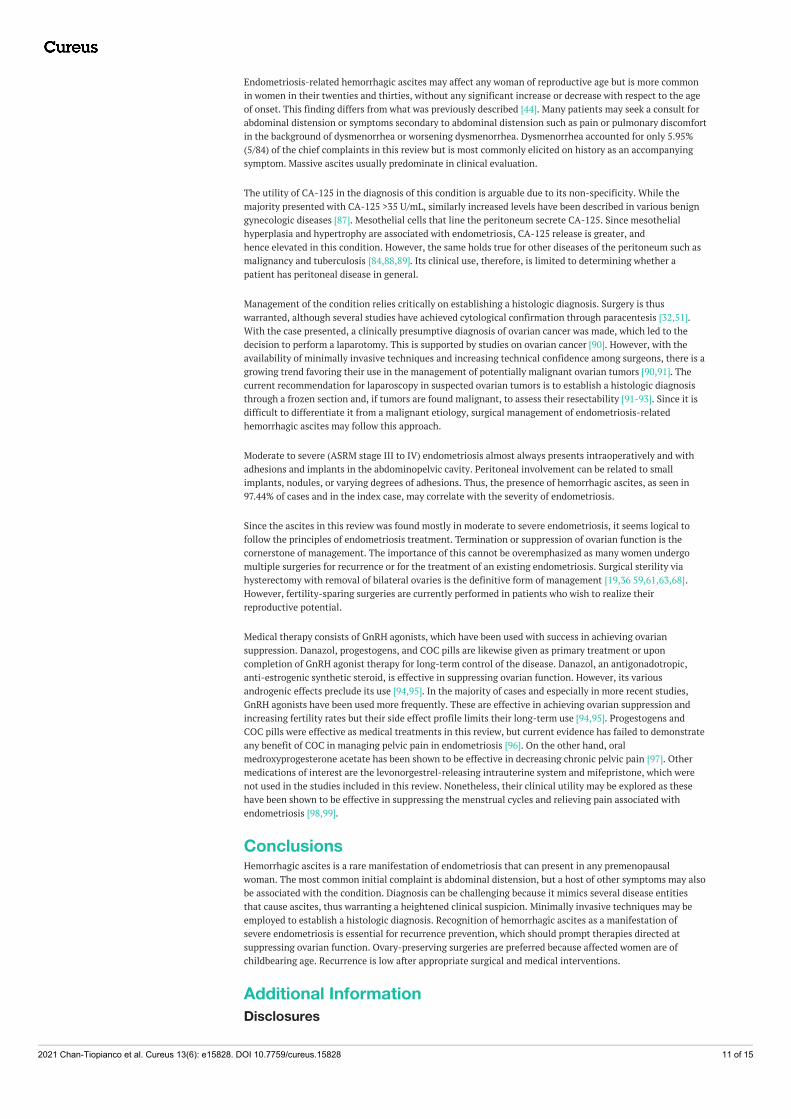
Moderate to severe endometriosis (ASRM stage III to IV) was seen in 97.44% (76/78) of the cases, andadhesions were described in 78.05% (64/82). In 43.90% (36/82) of the cases, an ovarian cyst was identified;11.11% (4/36) of the cases were ruptured. Peritoneal implants scattered about the abdominopelvic cavity in42.68% (35/82), while peritoneal nodules were seen in 20/82 (24.39%). Other abdominopelvic areas involvedare shown in Table 4.
2021 Chan-Tiopianco et al. Cureus 13(6): e15828. DOI 10.7759/cureus.15828 9 of 15
Organ involved Number (%)
Intestines 52 (63.41)
Recto-sigmoid 27 (32.93)
Omentum (caking/nodule/retraction/implants) 25 (30.49)
Cul-de-sac 23 (28.05)
Liver 10 (12.20)
Diaphragm 7 (8.54)
Appendix 6 (7.32)
Rectovaginal area 5 (6.10)
Umbilicus (nodule/mass/cyst) 4 (4.88)
TABLE 4: Peritoneal involvement in endometriosis-related hemorrhagic ascites (N=82)
At the time of presentation, 64.29% (54/84) underwent laparotomy, and laparoscopy was performed in33.33% (28/84). Two cases (2/84) had paracentesis. Almost half (44.05%, 37/84) of the cases had repeatabdominal surgeries, while 76.19% (64/84) required multiple procedures that included repeat abdominalsurgeries (laparoscopy and/or laparotomy), paracentesis, thoracostomy, or thoracotomy. On the other hand,less invasive surgical approaches (p<0.001) and fertility-sparing procedures (p<0.001) are observed to beincreasingly favored in recent years.
A cure was reported in 95.45% (21/22) who went through definitive surgery via hysterectomy with bilateralsalpingo-oophorectomy. Medical treatment was not given to 68.18% (15/22) after surgery. Four patientstolerated stripping or excision of the peritoneum of all endometriotic implants with no recurrence. Two ofthese received no additional medical therapy.
Patients who were offered medical therapy post-surgery received gonadotropin-releasing hormone (GnRH)agonists (63.79%, 37/58), either alone, with add-back therapy, or as a preliminary treatment that waseventually transitioned to either a progestogen or a combined oral contraceptive (COC) pill. In 86.49%(32/37) who received GnRH agonists, no recurrences were observed. Other therapies included danazol(13.79%, 8/58), progestogens alone (10.34%, 6/58), or COC alone (10.34%, 6/58). The cure rate with danazolwas 100% (eight out of eight), while COC and progestogens were equally effective, each with an 83.33% (fiveout of six) cure rate.
The recurrence rate observed at the time of presentation or after initial management was 36.90% (31/84),while that after definitive surgery and/or ovarian function suppression was 8.33% (7/84). Five of these casesreported significant ascites upon the cessation of GnRH therapy [35,49,50,62] or upon shifting from GnRH toprogestogen therapy [15]. The other two had reaccumulating minimal ascites while on oral COC [35] or oralprogestogen [70]. Of note, 71.42% (five out of seven) of recurrences had undergone ovary-preservingprocedures (oophorocystectomy or biopsy) prior to medical therapy. Mortality was reported in one case. TheMedian follow-up period was eight months.
AnalysisVery little is known about the pathogenesis of endometriosis-related hemorrhagic ascites. One putativemechanism is peritoneal irritation from the rupture of ovarian cysts. The endometrial cells from this spillagepropagate the spread of implants in the pelvic cavity and cause inflammation, which in turn leads toadhesions and ascites [81]. This theory assumes the presence of ovarian cysts. However, in this review, lessthan half of the study population were found to have ovarian endometriotic cysts, and only four out of 36 ofthese cysts were ruptured. Alternative hypotheses such as alterations in vascular permeability, lymphaticchannel obstruction, as well as individual variations in susceptibility to the disease may be explored[44,49,83,84].
The rubor of ascites may be due to increased angiogenesis seen in endometriosis. Erosions from affectedfriable soft tissue, serosal, peritoneal surfaces, and implants cause micro-bleeding or frank bleeding, leadingto the hemorrhagic character of ascites [49,84]. Pleural effusions associated with the hemorrhagic ascitesmay be due to several mechanisms. However, based on the presentation of massive ascites in the majority ofcases, the most plausible cause is anatomic defects in the diaphragm that allow for the passage ofhemorrhagic fluid into the pleural space [85,86].
2021 Chan-Tiopianco et al. Cureus 13(6): e15828. DOI 10.7759/cureus.15828 10 of 15
Endometriosis-related hemorrhagic ascites may affect any woman of reproductive age but is more commonin women in their twenties and thirties, without any significant increase or decrease with respect to the ageof onset. This finding differs from what was previously described [44]. Many patients may seek a consult forabdominal distension or symptoms secondary to abdominal distension such as pain or pulmonary discomfortin the background of dysmenorrhea or worsening dysmenorrhea. Dysmenorrhea accounted for only 5.95%(5/84) of the chief complaints in this review but is most commonly elicited on history as an accompanyingsymptom. Massive ascites usually predominate in clinical evaluation.
The utility of CA-125 in the diagnosis of this condition is arguable due to its non-specificity. While themajority presented with CA-125 >35 U/mL, similarly increased levels have been described in various benigngynecologic diseases [87]. Mesothelial cells that line the peritoneum secrete CA-125. Since mesothelialhyperplasia and hypertrophy are associated with endometriosis, CA-125 release is greater, andhence elevated in this condition. However, the same holds true for other diseases of the peritoneum such asmalignancy and tuberculosis [84,88,89]. Its clinical use, therefore, is limited to determining whether apatient has peritoneal disease in general.
Management of the condition relies critically on establishing a histologic diagnosis. Surgery is thuswarranted, although several studies have achieved cytological confirmation through paracentesis [32,51].With the case presented, a clinically presumptive diagnosis of ovarian cancer was made, which led to thedecision to perform a laparotomy. This is supported by studies on ovarian cancer [90]. However, with theavailability of minimally invasive techniques and increasing technical confidence among surgeons, there is agrowing trend favoring their use in the management of potentially malignant ovarian tumors [90,91]. Thecurrent recommendation for laparoscopy in suspected ovarian tumors is to establish a histologic diagnosisthrough a frozen section and, if tumors are found malignant, to assess their resectability [91-93]. Since it isdifficult to differentiate it from a malignant etiology, surgical management of endometriosis-relatedhemorrhagic ascites may follow this approach.
Moderate to severe (ASRM stage III to IV) endometriosis almost always presents intraoperatively and withadhesions and implants in the abdominopelvic cavity. Peritoneal involvement can be related to smallimplants, nodules, or varying degrees of adhesions. Thus, the presence of hemorrhagic ascites, as seen in97.44% of cases and in the index case, may correlate with the severity of endometriosis.
Since the ascites in this review was found mostly in moderate to severe endometriosis, it seems logical tofollow the principles of endometriosis treatment. Termination or suppression of ovarian function is thecornerstone of management. The importance of this cannot be overemphasized as many women undergomultiple surgeries for recurrence or for the treatment of an existing endometriosis. Surgical sterility viahysterectomy with removal of bilateral ovaries is the definitive form of management [19,36 59,61,63,68].However, fertility-sparing surgeries are currently performed in patients who wish to realize theirreproductive potential.
Medical therapy consists of GnRH agonists, which have been used with success in achieving ovariansuppression. Danazol, progestogens, and COC pills are likewise given as primary treatment or uponcompletion of GnRH agonist therapy for long-term control of the disease. Danazol, an antigonadotropic,anti-estrogenic synthetic steroid, is effective in suppressing ovarian function. However, its variousandrogenic effects preclude its use [94,95]. In the majority of cases and especially in more recent studies,GnRH agonists have been used more frequently. These are effective in achieving ovarian suppression andincreasing fertility rates but their side effect profile limits their long-term use [94,95]. Progestogens andCOC pills were effective as medical treatments in this review, but current evidence has failed to demonstrateany benefit of COC in managing pelvic pain in endometriosis [96]. On the other hand, oralmedroxyprogesterone acetate has been shown to be effective in decreasing chronic pelvic pain [97]. Othermedications of interest are the levonorgestrel-releasing intrauterine system and mifepristone, which werenot used in the studies included in this review. Nonetheless, their clinical utility may be explored as thesehave been shown to be effective in suppressing the menstrual cycles and relieving pain associated withendometriosis [98,99].
ConclusionsHemorrhagic ascites is a rare manifestation of endometriosis that can present in any premenopausalwoman. The most common initial complaint is abdominal distension, but a host of other symptoms may alsobe associated with the condition. Diagnosis can be challenging because it mimics several disease entitiesthat cause ascites, thus warranting a heightened clinical suspicion. Minimally invasive techniques may beemployed to establish a histologic diagnosis. Recognition of hemorrhagic ascites as a manifestation ofsevere endometriosis is essential for recurrence prevention, which should prompt therapies directed atsuppressing ovarian function. Ovary-preserving surgeries are preferred because affected women are ofchildbearing age. Recurrence is low after appropriate surgical and medical interventions.
Additional InformationDisclosures
2021 Chan-Tiopianco et al. Cureus 13(6): e15828. DOI 10.7759/cureus.15828 11 of 15

Human subjects: Consent was obtained or waived by all participants in this study. Taipei Veterans GeneralHospital Institutional Review Board issued approval VGH IRB: 2017 10 012AC. This study has been approvedby the Taipei Veterans General Hospital Institutional Review Board. Conflicts of interest: In compliancewith the ICMJE uniform disclosure form, all authors declare the following: Payment/services info: Allauthors have declared that no financial support was received from any organization for the submitted work.Financial relationships: All authors have declared that they have no financial relationships at present orwithin the previous three years with any organizations that might have an interest in the submitted work.Other relationships: All authors have declared that there are no other relationships or activities that couldappear to have influenced the submitted work.
AcknowledgementsAccess to databases and procurement of journal article copies were made possible through the TaipeiVeterans General Hospital Medical Library, the University of Maryland Medical Center Midtown CampusMedical Staff Library, and Dr. Howard H. Lee. Assistance for the translation of foreign articles was providedby Mr. Andre Anton A. Altea and Dr. Maria Patricia Medina-Kiziler. Many thanks to the various authors ofthe included studies and to those whose correspondences proved valuable in the creation of this paper.
References1. Brews A: Endometriosis including endometriosis of the diaphragm and Meigs' syndrome . Proc R Soc Med.
1954, 47:461.2. Charles D: Endometriosis and hemorrhagic pleural effusion. Obstet Gynecol. 1957, 10:309-12.3. Goumenou A, Matalliotakis I, Mahutte N, Koumantakis E: Endometriosis mimicking advanced ovarian
cancer. Fertil Steril. 2006, 86:219. 10.1016/j.fertnstert.2005.12.0444. Myers TJ, Arena B, Granai CO: Pelvic endometriosis mimicking advanced ovarian cancer: presentation with
pleural effusion, ascites, and elevated serum CA 125 level. Am J Obstet Gynecol. 1995, 173:966-7.10.1016/0002-9378(95)90381-x
5. Urrunaga NH, Singal AG, Cuthbert JA, Rockey DC: Hemorrhagic ascites. Clinical presentation and outcomesin patients with cirrhosis. J Hepatol. 2013, 58:1113-8. 10.1016/j.jhep.2013.01.015
6. Taub WH, Rosado S, Kalaycioglu M, Booher D, Barnes DS: Hemorrhagic ascites secondary to endometriosis .J Clin Gastroenterol. 1989, 11:458-60. 10.1097/00004836-198908000-00023
7. Goldberg BB, Goodman GA, Clearfield HR: Evaluation of ascites by ultrasound . Radiology. 1970, 96:15-22.10.1148/96.1.15
8. Moore KP, Wong F, Gines P, et al.: The management of ascites in cirrhosis: report on the consensusconference of the International Ascites Club. Hepatology. 2003, 38:258-66. 10.1053/jhep.2003.50315
9. Moore CM, Van Thiel DH: Cirrhotic ascites review: pathophysiology, diagnosis and management . World JHepatol. 2013, 5:251-63. 10.4254/wjh.v5.i5.251
10. No authors listed: Revised American Society for Reproductive Medicine classification of endometriosis:1996. Fertil Steril. 1997, 67:817-21. 10.1016/s0015-0282(97)81391-x
11. Moola S, Munn Z, Tufanaru C, et al.: Systematic reviews of etiology and risk . JBI Manual for EvidenceSynthesis. Aromataris E, Munn Z (ed): The Joanna Briggs Institute, Adelaide, Australia; 2017. 1:20-30.10.46658/JBIMES-20-08
12. Page MJ, McKenzie JE, Bossuyt PM, et al.: The PRISMA 2020 statement: an updated guideline for reportingsystematic reviews. BMJ. 2021, 372:n71. 10.1136/bmj.n71
13. Soyman Z, Bacanakgil BH, Kaya S, et al.: An uncommon presentation of endometriosis a case report . JReprod Med. 2018, 63:317-8.
14. Mendes S, Carvalho C, Rodrigues G, Barata S, Calhaz-Jorge C, Osório F: Successful treatment ofendometriosis-related hemorrhagic ascites: a report of three cases. Surg Technol Int. 2018, 32:150-5.
15. Walker PJ, Johnson NP: Benign endometriosis masquerading as intra-abdominal malignancy: one of themost extreme cases reported and a review of the literature. J Endometr Pelvic Pain Disord. 2018, 10:174-7.10.1177/2284026518780820
16. O’yandjo AM, Bosenge NJD, Kadima NJ: Umbilical nodule and hemorrhagic ascites of endometriosis origin:a clinical case report. Gynecol Obstet Case Rep. 2018, 4:62. 10.21767/2471-8165.1000062
17. Magalhães TF, Augusto KL, Mota LP, Costa ARD, Puster RA, Bezerra LRPS: Ascites and encapsulatingperitonitis in endometriosis: a systematic review with a case report. Rev Bras Ginecol Obstet. 2018, 40:147-55. 10.1055/s-0038-1626700
18. Petrosellini C, Abdalla S, Oke T: The many guises of endometriosis: giant abdominal wall endometriosismasquerading as an incisional hernia. Int J Fertil Steril. 2018, 11:321-5. 10.22074/ijfs.2018.5126
19. Pereira N, Gunnala V, Palermo GD, Elias RT: Laparoscopic management of severe endometriosis-relatedhemorrhagic ascites. J Minim Invasive Gynecol. 2018, 25:8-9. 10.1016/j.jmig.2017.03.010
20. N’Guessan E, Kouamé N, Dia JM, Gbeli F, Guié P, Anongba S: Endometriosis revealed by recurrenthemorrhagic ascites. Open J Obstet Gynecol. 2017, 7:1160-5. 10.4236/ojog.2017.712117
21. Varun N, Tanwar RT: A rare presentation of endometriosis with massive haemorrhagic ascites: a case report .Gynecol Obstet Case Rep. 2016, 3:1. 10.21767/2471-8165.1000047
22. Dun EC, Wong S, Lakhi NA, Nehzat CH: Recurrent massive ascites due to mossy endometriosis . Fertil Steril.2016, 106:e14. 10.1016/j.fertnstert.2016.07.1119
23. Hinduja I, Kapadia K, Udwadia F, Bhilawadikar R, Adhe A, Zaveri K: Unusual presentation of endometriosiswith haemorrhagic ascites - a case report. J Obstet Gynaecol. 2016, 36:133-4.10.3109/01443615.2015.1030605
24. Setubal A, Sidiropoulou Z, Soares S, Barbosa C: Endometriosis and ascites: a strategy to achieve pregnancy . JMinim Invasive Gynecol. 2015, 22:1104-8. 10.1016/j.jmig.2015.05.013
25. Bignall J, Arambage K, Vimplis S: Endometriosis: a rare and interesting cause of recurrent haemorrhagic
2021 Chan-Tiopianco et al. Cureus 13(6): e15828. DOI 10.7759/cureus.15828 12 of 15

ascites. BMJ Case Rep. 2014, 2014:bcr2013010052. 10.1136/bcr-2013-01005226. Cosma S, Ceccaroni M, Benedetto C: A pseudoneoplastic finding of deep endometriosis: laparoscopic triple
segmental bowel resection. Wideochir Inne Tech Maloinwazyjne. 2014, 9:463-7. 10.5114/wiitm.2014.4161727. Hasdemir PS, Ikiz N, Ozcakir HT, Kara E, Guvenal T: Endometriosis associated with relapsing ascites and
pleural effusions. J Obstet Gynaecol. 2015, 35:419. 10.3109/01443615.2014.94882328. Park CM, Kim SY: Rupture of an endometrioma with extremely high serum CA-125 level (> 10,000 IU/ml)
and ascites resembling ovarian cancer. Eur J Gynaecol Oncol. 2014, 35:469-72.29. Asano R, Nakazawa T, Hirahara F, Sakakibara H: Dienogest was effective in treating hemorrhagic ascites
caused by endometriosis: a case report. J Minim Invasive Gynecol. 2014, 21:1110-2.10.1016/j.jmig.2014.04.014
30. Appleby R, Saroya H, Postgate A, Meer Z: A young woman with abdominal distension . BMJ Case Rep. 2014,2014:bcr2014203726. 10.1136/bcr-2014-203726
31. Mumtahana F, Jiao J, Cui B: A rare presentation of endometriosis with recurrent massive hemorrhagicascites which can mislead. Int J Women’s Health Reproduction Sci. 2014, 2:30-4. 10.15296/ijwhr.2014.05
32. Packard LK, Adamson GD: Endometriosis presenting with massive ascites and pleural effusion: a casereport. J Endometr Pelvic Pain Disord. 2013, 5:123-5. 10.5301/je.5000162
33. Akinola RA, Akinola OI, Alakija A, Wright KO: Widespread endometriosis mimicking ovarian malignancy: acase report. Niger Postgrad Med J. 2012, 19:46-9.
34. Akintomide AO, Bassey DB, Ekanem EI, Omotosho AJ: Omental endometriosis: a rare site and an unusualassociation with ovarian fibroma and haemorrhagic ascites. A case report and review of the imagingtechniques. IOSR JDMS. 2012, 2:22-6. 10.9790/0853-0212226
35. Queirós A, Correia L, Pinto G, Rosa D, Silva G, Simões T: Endometriosis with hemorrhagic ascites: two casesreport (Article in Portuguese). Rev Iberoam Fert Rep Hum. 2011, 28:141-5.
36. Shabeerali TU, Rajan R, Kuruvilla AP, et al.: Hemorrhagic ascites: are we missing endometriosis? . Indian JGastroenterol. 2012, 31:195-7. 10.1007/s12664-012-0221-1
37. Ferrero S, Remorgida V: Endometriosis presenting with hemorrhagic ascites . Arch Gynecol Obstet. 2011,283:1429-30. 10.1007/s00404-010-1796-3
38. Cordeiro Fernandes LF, Podgaec S, Castro Cotti GC, Abrao MS: Severe endometriosis may be considered inthe differential diagnosis in young women presenting massive hemorrhagic ascites. Gynecol Surg. 2011,8:459. 10.1007/s10397-011-0690-8
39. Suchetha S, Rema P, Mathew AP, Sebastian P: Endometriosis with massive hemorrhagic ascites . Indian JCancer. 2010, 47:224-5. 10.4103/0019-509X.63004
40. Ignacio MM, Joseph N, Hélder F, Mamourou K, Arnaud W: Massive ascites, pleural effusion, anddiaphragmatic implants in a patient with endometriosis. Eur J Obstet Gynecol Reprod Biol. 2010, 149:117-8.10.1016/j.ejogrb.2009.10.017
41. Day T, Hui K, Perkins S, Pelletier P: Ascites and ileus due to endometriosis . J Pelvic Med Surg. 2009, 15:471-5. 10.1097/SPV.0b013e3181c6e90b
42. Park BJ, Kim TE, Kim YW: Massive peritoneal fluid and markedly elevated serum CA125 and CA19-9 levelsassociated with an ovarian endometrioma. J Obstet Gynaecol Res. 2009, 35:935-9. 10.1111/j.1447-0756.2009.01122.x
43. Lodha A, Klein T, Elish D, Tarkovsky R: Endometriosis: a rare presentation as hemorrhagic ascites . PractGastroenterol. 2008, 32:48-9.
44. Ussia A, Betsas G, Corona R, De Cicco C, Koninckx PR: Pathophysiology of cyclic hemorrhagic ascites andendometriosis. J Minim Invasive Gynecol. 2008, 15:677-81. 10.1016/j.jmig.2008.08.012
45. Sait KH: Massive ascites as a presentation in a young woman with endometriosis: a case report . Fertil Steril.2008, 90:2015. 10.1016/j.fertnstert.2008.07.021
46. Santos VM, Barbosa ER Jr, Lima SH, Porto AS: Abdominal cocoon associated with endometriosis . SingaporeMed J. 2007, 48:e240-2.
47. Palayekar M, Jenci J, Carlson JA Jr: Recurrent hemorrhagic ascites: a rare presentation of endometriosis .Obstet Gynecol. 2007, 110:521-2. 10.1097/01.AOG.0000268283.99315.58
48. Baykal C, Arioglu P, Kalayci M, Özkan F, Çetinkaya N, Fiçicioglu C: Giant endometrioma mimicking ovariancarcinoma: case report. Turk J Obstet Gynecol. 2006, 3:356-8.
49. Ekoukou D, Guilherme R, Desligneres S, Rotten D: Endometriosis with massive hemorrhagic ascites: a casereport and review of the literature (Article in French). J Gynecol Obstet Biol Reprod (Paris). 2005, 34:351-9.10.1016/s0368-2315(05)82841-8
50. Fortier D, Dedecker F, Gabriele M, Graesslin O, Barau G: Endometriosis with ascites and pleural effusion: acase report (Article in French). Gynecol Obstet Fertil. 2005, 33:508-10. 10.1016/j.gyobfe.2005.05.014
51. Zeppa P, Vetrani A, Cozzolino I, Palombini L: Endometrial glands in ascites secondary to endometriosis .Diagn Cytopathol. 2004, 30:131-2. 10.1002/dc.10390
52. Francis M, Badero OO, Borowsky M, Lee YC, Abulafia O: Pericardial effusion, right-sided pleural effusionand ascites associated with stage IV endometriosis. A case report. J Reprod Med. 2003, 48:463-5.
53. Cheong EC, Lim DT: Massive ascites--an uncommon presentation of endometriosis . Singapore Med J. 2003,44:98-100.
54. Moffatt SD, Mitchell JD: Massive pleural endometriosis. Eur J Cardiothorac Surg. 2002, 22:321-3.10.1016/s1010-7940(02)00277-4
55. Dias CC, Andrade JM, Ferriani RA, Villanova MG, Meirelles RS: Hemorrhagic ascites associated withendometriosis. A case report. J Reprod Med. 2000, 45:688-90.
56. Bhojawala J, Heller DS, Cracchiolo B, Sama J: Endometriosis presenting as bloody pleural effusion andascites-report of a case and review of the literature. Arch Gynecol Obstet. 2000, 264:39-41.10.1007/pl00007484
57. El Khalil T, Mourad FH, Barada K, Uthman S: Massive hemorrhagic ascites secondary to endometriosis . J ClinGastroenterol. 1999, 29:344-5. 10.1097/00004836-199912000-00010
58. Samora-Mata J, Feste JR: Endometriosis ascites: a case report . JSLS. 1999, 3:229-31.59. Fletcher H, McFarlane M, Shirley SE, Clarke WF, Lyon K: Massive ascites secondary to severe endometriosis .
2021 Chan-Tiopianco et al. Cureus 13(6): e15828. DOI 10.7759/cureus.15828 13 of 15
West Indian Med J. 1999, 48:158-9.60. Muneyyirci-Delale O, Neil G, Serur E, Gordon D, Maiman M, Sedlis A: Endometriosis with massive ascites .
Gynecol Oncol. 1998, 69:42-6. 10.1006/gyno.1998.495361. Mejia EM, Alvarez OA, Lee M: Endometriosis with massive bloody ascites . J Am Board Fam Pract. 1997,
10:59-61.62. Flanagan KL, Barnes NC: Pleural fluid accumulation due to intra-abdominal endometriosis: a case report
and review of the literature. Thorax. 1996, 51:1062-3. 10.1136/thx.51.10.106263. el-Newihi HM, Antaki JP, Rajan S, Reynolds TB: Large bloody ascites in association with pelvic
endometriosis: case report and literature review. Am J Gastroenterol. 1995, 90:632-4.64. Schlueter FJ, McClennan BL: Massive ascites and pleural effusions associated with endometriosis . Abdom
Imaging. 1994, 19:475-6. 10.1007/BF0020694565. Jose R, George SS, Seshadri L: Massive ascites associated with endometriosis . Int J Gynaecol Obstet. 1994,
44:287-8. 10.1016/0020-7292(94)90185-666. London S, Parmley T: Endometriosis and ascites. South Med J. 1993, 86:1173-5. 10.1097/00007611-
199310000-0002267. Chen FF, Chow NH, Chou CY, Lin MF: Hemorrhagic ascites associated with endometriosis: a rare clinical
presentation. J Gynecol Surg. 1992, 8:43-7. 10.1089/gyn.1992.8.4368. Tsvelev IuV, Lishchuk VD, Kolosov AE: Ascites as a manifestation of generalized endometriosis (Article in
Russian). Vestn Khir Im I I Grek. 1990, 145:48-50.69. Yu J, Grimes DA: Ascites and pleural effusions associated with endometriosis . Obstet Gynecol. 1991, 78:533-
4.70. Hattori S, Tamakoshi K, Oguchi H, Kodama H: A case of endometriosis with massive ascites (Article in
Japanese). Nihon Sanka Fujinka Gakkai Zasshi. 1990, 42:291-4.71. Olubuyide IO, Adebajo AO, Adeleye JA, Solanke TF: Massive ascites associated with endometriosis in a
Nigerian African. Int J Gynaecol Obstet. 1988, 27:439-41. 10.1016/0020-7292(88)90127-072. Chichareon SB, Wattanakitkrailert S: Endometriosis with ascites . Acta Obstet Gynecol Scand. 1988, 67:187-
8. 10.3109/0001634880900419873. Iwasaka T, Okuma Y, Yoshimura T, Kidera Y, Sugimori H: Endometriosis associated with ascites . Obstet
Gynecol. 1985, 66:72S-5.74. Naraynsingh V, Raju GC, Ratan P, Wong J: Massive ascites due to omental endometriosis . Postgrad Med J.
1985, 61:539-40. 10.1136/pgmj.61.716.53975. Halme J, Chafe W, Currie JL: Endometriosis with massive ascites . Obstet Gynecol. 1985, 65:591-2.76. Jenks JE, Artman LE, Hoskins WJ, Miremadi AK: Endometriosis with ascites . Obstet Gynecol. 1984, 63:75S-7.77. Gaulier A, Jouret-Mourin A, Marsan C: Peritoneal endometriosis. Report of a case with cytologic,
cytochemical and histopathologic study. Acta Cytol. 1983, 27:446-9.78. Chervenak FA, Greenlee RM, Lewenstein L, Tovell HM: Massive ascites associated with endometriosis .
Obstet Gynecol. 1981, 57:379-81.79. Irani S, Atkinson L, Cabaniss C, Danovitch SH: Pleuroperitoneal endometriosis. Obstet Gynecol. 1976,
47:72S-4.80. Collier HA, Gonzales LL, Bossert LJ: Cyclic ascites as a manifestation of endometriosis. Report of a case .
Obstet Gynecol. 1962, 19:681-3.81. Bernstein JS, Perlow V, Brenner JJ: Massive ascites due to endometriosis . Am J Digest Dis. 1961, 6:1-6.
10.1007/BF0223924082. Ripstein CB, Robman M, Wallach JB: Endometriosis involving the pleura . J Thorac Surg. 1959, 37:464-71.83. Koninckx PR, Renaer M, Brosens IA: Origin of peritoneal fluid in women: an ovarian exudation product . Br J
Obstet Gynaecol. 1980, 87:177-83. 10.1111/j.1471-0528.1980.tb04514.x84. Koninckx PR, Kennedy SH, Barlow DH: Pathogenesis of endometriosis: the role of peritoneal fluid . Gynecol
Obstet Invest. 1999, 47:23-33. 10.1159/00005285685. Hwang SM, Lee CW, Lee BS, Park JH: Clinical features of thoracic endometriosis: a single center analysis .
Obstet Gynecol Sci. 2015, 58:223-31. 10.5468/ogs.2015.58.3.22386. Nwiloh J: Diaphragmatic patch: a useful adjunct in surgical treatment of recurrent catamenial hemothorax .
Rev Port Pneumol. 2011, 17:278-80. 10.1016/j.rppneu.2011.06.00687. Daoud E, Bodor G: CA-125 concentrations in malignant and nonmalignant disease . Clin Chem. 1991,
37:1968-74.88. Chen DX, Schwartz PE, Li XG, Yang Z: Evaluation of CA 125 levels in differentiating malignant from benign
tumors in patients with pelvic masses. Obstet Gynecol. 1988, 72:23-7.89. Oparka R, McCluggage WG, Herrington CS: Peritoneal mesothelial hyperplasia associated with
gynaecological disease: a potential diagnostic pitfall that is commonly associated with endometriosis. J ClinPathol. 2011, 64:313-8. 10.1136/jcp.2010.086074
90. Rimbach S, Neis K, Solomayer E, Ulrich U, Wallwiener D: Current and future status of laparoscopy ingynecologic oncology. Geburtshilfe Frauenheilkd. 2014, 74:852-9. 10.1055/s-0034-1383075
91. Angeles MA, Martínez-Gómez C, Migliorelli F, et al.: Novel surgical strategies in the treatment ofgynecological malignancies. Curr Treat Options Oncol. 2018, 19:73. 10.1007/s11864-018-0582-5
92. Ratnavelu ND, Brown AP, Mallett S, et al.: Intraoperative frozen section analysis for the diagnosis of earlystage ovarian cancer in suspicious pelvic masses. Cochrane Database Syst Rev. 2016, 3:CD010360.10.1002/14651858.CD010360.pub2
93. Morton R, Anderson L, Carter J, Pather S, Saidi SA: Intraoperative frozen section of ovarian tumors: a 6-yearreview of performance and potential pitfalls in an Australian tertiary referral center. Int J Gynecol Cancer.2017, 27:17-21. 10.1097/IGC.0000000000000851
94. Brown J, Farquhar C: Endometriosis: an overview of Cochrane Reviews . Cochrane Database Syst Rev. 2014,2014:CD009590. 10.1002/14651858.CD009590.pub2
95. Berlanda N, Somigliana E, Viganò P, Vercellini P: Safety of medical treatments for endometriosis . ExpertOpin Drug Saf. 2016, 15:21-30. 10.1517/14740338.2016.1121991
96. Brown J, Crawford TJ, Datta S, Prentice A: Oral contraceptives for pain associated with endometriosis .
2021 Chan-Tiopianco et al. Cureus 13(6): e15828. DOI 10.7759/cureus.15828 14 of 15

Cochrane Database Syst Rev. 2018, 5:CD001019. 10.1002/14651858.CD001019.pub397. Brown J, Kives S, Akhtar M: Progestagens and anti-progestagens for pain associated with endometriosis .
Cochrane Database Syst Rev. 2012, 2012:CD002122. 10.1002/14651858.CD002122.pub298. Fu J, Song H, Zhou M, Zhu H, Wang Y, Chen H, Huang W: Progesterone receptor modulators for
endometriosis. Cochrane Database Syst Rev. 2017, 7:CD009881. 10.1002/14651858.CD009881.pub299. Abou-Setta AM, Houston B, Al-Inany HG, Farquhar C: Levonorgestrel-releasing intrauterine device (LNG-
IUD) for symptomatic endometriosis following surgery. Cochrane Database Syst Rev. 2013, 1:CD005072.10.1002/14651858.CD005072.pub3
2021 Chan-Tiopianco et al. Cureus 13(6): e15828. DOI 10.7759/cureus.15828 15 of 15