clinical practice guidelines for the diagnosis and ... · pdf fileclinical practice guidelines...
TRANSCRIPT

Clinical Practice Guidelines for the Diagnosis and Management of Acute Otitis
Media (AOM) in Children
Subcommittee on Clinical Practice Guidelines for the Diagnosis and Management of
Acute Otitis Media in Children
(Japan Otological Society, Japan Society for Pediatric Otorhinolaryngology, Japan
Society for Infectious Diseases in Otolaryngology)
1. Summary
Objective: To 1) indicate methods of diagnosis and testing for acute otitis media
(AOM) in children (<15 years); and 2) recommend methods of treatment in accordance with the evidence-based consensus reached by the Subcommittee on
Clinical Practice Guidelines for the Diagnosis and Management of AOM in Children
(Subcommittee on Clinical Practice Guidelines), in light of the causative bacteria of
AOM in Japan and their susceptibility to antimicrobial agents. Methods: We
investigated the most recently detected bacteria causing childhood AOM in Japan as
well as their antimicrobial susceptibility, developed Clinical Questions(CQ)
concerning the diagnosis, testing methods, and treatment of AOM, searched the
literature published during 2000–2004, and issued the 2006 Guidelines.1-4) In the 2009
Guidelines we performed the same investigation with the addition of literature that
was published during 2005–2008 and that was not included in the 2006 Guidelines.
Results: We categorized AOM as mild, moderate, or severe on the basis of otoscopic
findings and clinical symptoms, and presented a recommended treatment for each
degree of severity. Conclusion: Accurate assessment of otoscopic findings is important
for judging the degree of severity and selecting a method of treatment.
2. Authors
The membership of the Subcommittee on Clinical Practice Guidelines for the
Diagnosis and Management of Acute Otitis Media (AOM) in Children is shown in
Table 1. This committee is composed of three organizations: the Japan Otological
Society (JOS), the Japan Society for Infectious Diseases in Otolaryngology (JSIDO),
and the Japan Society for Pediatric Otorhinolaryngology (JSPO). The first committee
meeting was held on January 8, 2003, and the 2006 Guidelines were published in
1

March of that year on the website of the JSIDO, in the journals of the JOS and the
JSPO, on the website of the Japan Council for Quality Healthcare, and in printed
form.1-4) The 2006 Guidelines underwent evaluation, and work on the production of a
revised edition began at the 13th committee meeting on January 7, 2007.
3. Financial Backers and Sponsors
Production of these Guidelines was funded by JOS operating expenses. The JOS
does not receive support from any specific organizations or companies. A list of
organizations and companies that posed non-personal financial conflicts of interest to
members of the Subcommittee on Clinical Practice Guidelines during the production
of these Guidelines is provided (attachment).
4. Introduction
AOM is a typical upper respiratory inflammation commonly affecting children and
is mainly treated by otolaryngologists. Its exact frequency of occurrence in Japan is
unknown, however. According to reports from Europe and the US, 62% of children
aged less than one year and 83% of those up to the age of three have suffered from at
least one bout of AOM.5) Faden et al.6) have reported that it affects 75% of children up
to the age of one.
Some authors in Europe and the US do not recommend the use of antimicrobial
agents for AOM. In the Netherlands, it has been proposed that antimicrobial agents
are unnecessary in at least 90% of cases, and that patients should be observed for 3-4
days without antimicrobial agent administration.7,8) Rosenfeld et al. have also
reported observation as a management option,9-11) and more recent studies have also
found no significant difference in clinical outcome if antimicrobial agents are not
given immediately but rather are prescribed if there is no improvement in symptoms
after 48 or 72 hours.12,13) A Cochrane Review that examined randomized controlled
trials of antimicrobial agent administration versus placebo also found that
antimicrobial agents had little effect on childhood AOM.14) In addition, a double-blind
randomized controlled trial of amoxicillin (AMPC) and a placebo found no significant
difference in therapeutic efficacy between the two.15,16) Dagan et al.17,18) and Toltzis et
al.,19) in a review and case-control study, advised that antimicrobial agent use would be
reduced because the use of a wide variety of antimicrobial agent increases the survival
2

of resistant Streptococcus pneumonia (S. pneumonia) in the nasopharynx, which can
cause additional infections in middle-ear (ME) fluid.
In Japan, regular nationwide surveys are performed of the causative bacteria for
AOM, acute sinusitis, acute tonsillitis, and peritonsillar abscess. These surveys have
reported that multidrug-resistant bacteria are now being detected more frequently,20,21)
which means that the recommendation to avoid administration of antimicrobial agents
proposed in Europe and the US does not apply. In addition, the criteria and assessment
levels used in conventional clinical assessment are not necessarily uniform even
within Europe and the US.22) Investigation and unified evaluation of the diagnosis and
treatment of childhood AOM are therefore required, based on the actual situation in
Japan. Based on this perspective, the JOS, the JSIDO, and the JSPO produced 2006
Clinical Practice Guidelines consistent with evidence-based medicine (EBM) with the
aim of supporting the diagnosis and treatment of childhood AOM.23)
A survey of otolaryngologists and pediatricians in Ishikawa Prefecture showed that
85% of otolaryngologists and 52% of pediatricians were aware of the 2006 edition of
the Guidelines, with 56% of those otolaryngologists and 49% of those pediatricians
reporting that they used them in practice.24) Therapeutic outcomes of clinical practice
that adhered to the guidelines were also generally good.25,26) In light of these results,
the JOS, JSIDO, and JSPO decided to revise the 2006 Guidelines and issue a new
edition in 2009.
These Clinical Practice Guidelines are issued only to assist clinical practice, and
have no binding authority on treatment (Note 1). How they should actually be used for
patients in the clinical setting is a matter to be decided in light of the patient’s wishes
and values, and based on the medical practitioner’s specialist knowledge and
experience. The fact that there is insufficient evidence of a treatment method’s efficacy
does not necessarily mean that treatment is ineffective or should not be carried out.
When using such methods of treatment, however, an extra degree of consideration is
required with respect to the evaluation of clinical efficacy and communication with the
patient. It must be re-emphasized that recommendations in clinical practice guidelines
are not legal grounds for dictating the particular types of medical treatment that should
be practiced in individual clinical situations.27) These Guidelines will be periodically
revised to reflect the opinions of users and patients and as a result of external
evaluation, in the same way as the 2006 Guidelines were revised after their
3

publication.
The differences between the 2006 Guidelines and 2009 Guidelines comprise
changes in the symptoms and findings used in the severity categories and changes in
the criteria for determining the degree of severity. No major changes were made to
other items. To enable the 2009 Guidelines to be used as clinical practice guidelines
without reference to the 2006 edition, however, they also include material published in
the 2006 Guidelines.
Note 1: The Guidelines are ranked as follows:
Regulations > directives > recommendations ≥ guidelines (From A Dictionary of Epidemiology, trans. Japan Epidemiological Association ed. J. Last, with additions)
5. Objective and Aim of Production
These Guidelines were produced to describe diagnostic and testing methods for
childhood AOM (below the age of 15* [see note]), and represent the evidence-based
consensus of the members of the Subcommittee on Clinical Practice Guidelines on
recommended treatment methods in light of the causative bacteria and their
susceptibility to antimicrobial agents in cases of AOM in Japan. The aim is for these
Guidelines to be used to assist clinical decision-making in the care of childhood AOM,
and for them to prove beneficial in the diagnosis and treatment of patients with AOM.
*Note: In the Ministry of Health, Labour and Welfare’s Pharmaceutical Affairs Bureau
Notification No. 1334, Guidance Concerning Clinical Trials of Drugs in Pediatric
Populations, released on December 15, 2000,28) the following have been proposed as
age categories for the design of clinical trials of drugs on pediatric patients: preterm
neonates, full-term neonates (0–27 days), infants (28 days to 23 months), children
(2–11 years), and adolescents (12–16 or 12–18 years). In these Guidelines, we have
defined children using a general criterion of <15 years.
6. Users
The main users of these Guidelines will be otolaryngologists who perform
otological procedures including the accurate evaluation of otoscopic findings and
myringotomy.
4

7. Subjects
The subjects of these Guidelines are AOM patients aged <15 years who were free from AOM or otitis media with effusion (OME) within one month prior to onset, who
do not have a tympanostomy tube inserted, who have no craniofacial abnormality, and
who do not suffer from immunodeficiency. Patients with the following conditions are
excluded as subjects: AOM with complications including facial palsy and inner ear
disorder, elevated pinna with acute mastoiditis, and AOM with Gradenigo’s syndrome
or similar findings. It can be difficult to distinguish between AOM and bullous
myringitis, but the latter is not covered by these Guidelines.
The consensus reached by the Subcommittee on Clinical Practice Guidelines with
respect to recurrent otitis media (ROM) (using the definition proposed below) has
been included as an additional statement.
Treatment algorithms are included at the end of the Guidelines, in which cases that
have not improved after tertiary treatment according to each treatment algorithm are
classed as intractable. The care of intractable cases is not covered in these Guidelines.
*Additional Statement: Proposals for the Treatment of ROM
(a) Definition of ROM
The definition of ROM has yet to be standardized either in Japan or internationally,
but in these Guidelines it has been defined as three or more occurrences of AOM
within the previous six months, or four or more within the previous 12 months, as
generally used in comparatively recent studies.29-31)
(b) Pathophysiology of and risk factors for ROM
The pathophysiology of ROM can be categorized into two types: recurrent simple
AOM, and recurrent AOM occurring as an acute exacerbation in patients suffering
from OME.
Proposed risk factors for ROM include young age, multidrug-resistant causative
bacteria, immunity of the affected individual, and lifestyle and environmental factors.
Genetic make-up has also been reported as a risk factor in young children aged <2 years.32) In terms of causative bacteria, multidrug-resistant pneumococci are
reportedly responsible in many cases,33) with incomplete elimination from the
nasopharynx owing to reduced antimicrobial agent efficacy regarded as one cause of
recurrence. The involvement of decreased immune response by the host to the
5

causative bacteria is also important.34) It has also been conjectured that there is a link
between immunity received from the mother via breast milk and the onset of ROM,
with the absence of breastfeeding constituting a strong risk factor for ROM.35)
Lifestyle and environmental risk factors include having siblings, attending daycare,
and pacifier use.35)
(c) Treatment of ROM
With the factors described above assumed to constitute risk factors for ROM,
bacterial sensitivity tests must always be carried out prior to antimicrobial agent
administration to counteract resistant causative bacteria, and an appropriate dose of
antimicrobial agents must be selected. Recommended antimicrobial agents are listed
in these Guidelines.
Pneumococcal conjugate vaccine is used in Europe and the US to prevent ROM. In
a double-blind randomized controlled trial of a 7-valent pneumococcal conjugate
vaccine and pneumococcal polysaccharide vaccine in Holland, there was no
significant reduction in the frequency of occurrence of ROM.36) Although a Cochrane
Review accepts the utility of pneumococcal polysaccharide vaccine, it does not
recommend the conjugate vaccine.37) In a double-blind randomized controlled trial in
the Czech Republic, however, 11-valent pneumococcal capsular polysaccharide
vaccine conjugated to H. influenzae-derived protein D had a significant protective
effect against AOM caused by pneumococci or non-typable H. influenzae.38) In Japan,
7-valent pneumococcal conjugate vaccine was approved for use in 2010. This vaccine
covers 60.6% of pneumococcal serotypes isolated from the middle ears of childhood
AOM patients in Japan and 87% of multidrug-resistant bacteria, and is anticipated to
provide up to about 17% protection against all forms of AOM.
One form of treatment unique to Japan that has been proposed is the use of Chinese
herbal medicines for their protective effect in boosting immunity, and juzentaihoto has
been reported as effective.39)
Adenoidectomy has not been shown to reduce the frequency of ROM as a surgical
treatment in double-blind randomized controlled trials, nor is it regarded as having any
preventive effect.40-42) Myringotomy has not been shown to have any significant effect
in reducing the frequency of occurrence of ROM in research on patients in Japan,43) but
insertion of a tympanostomy tube for one year and short-term insertion for one month
significantly reduce the frequency of occurrence.44,45) As measures to deal with
6

lifestyle and environmental factors, discontinuation of attendance of group daycare
and breastfeeding are desirable.
8. Definition of AOM
In these Clinical Practice Guidelines, AOM is defined as “an acute occurrence of
middle ear infection that may be associated with otalgia, fever, or otorrhea.” The
following notes are also added.
Notes:
(i) Acute occurrence is defined as a case in which the individual complains of an
acute symptom or in which an acute symptom is observed by his/her
parent/guardian, and the individual is seen in a clinic within 48 hours.46) The
duration of acute inflammation is commonly defined as not longer than three
weeks, though there is no clear evidence serving as the basis for this definition.
These Guidelines also adopt these common definitions. It should be noted that
acute aggravation of chronic otitis media is excluded from these definitions
because its pathological condition differs.
(ii) The Clinical Practice Guidelines for the Diagnosis and Management of AOM
reported by the American Academy of Pediatrics (Subcommittee on
Management of Acute Otitis Media 2004)47 provides that a diagnosis of AOM
requires the following signs and symptoms:
(1) Recent, usually abrupt, onset of signs and symptoms of middle-ear
inflammation and middle ear effusion (MEE)
(2) The presence of MEE, indicated by any bulging of the tympanic
membrane, limited or absent mobility of the tympanic membrane,
air-fluid level behind the tympanic membrane, and otorrhea
(3) Signs or symptoms of middle-ear inflammation as indicated by either
distinct erythema of the tympanic membrane or distinct otalgia.
9. Bacteria isolated from children with acute otitis media in Japan and
antimicrobial activity against them
(1) Bacteria isolated from children with AOM
The report of the Fourth Nationwide Surveillance of Clinical Isolates from Patients
with Otorhinolaryngological Infections in 2007 (conducted from January through June
7

2007)48) showed changes in the frequencies of bacteria isolated from patients of all
ages with AOM based on the four previous surveillances conduced in 1994, 1998,
2003, and 2007 (Figure 1 and Table 2). S. pneumoniae tended to increase and was
detected in 34.1% of isolates in the surveillance of 2007. Haemophilus influenzae (H.
influenzae) tended to increase during the surveillance of 2003, but slightly decreased
to 24.2% in 2007. Staphylococcus aureus (S. aureus) decreased to 4.4%. Moraxella
catarrhalis (M. catarrhalis) was detected in 7.1% of isolates in 2003 and in 4.4% in
2007. Of 45 specimens of pus taken from the middle ear of children aged <15 years with AOM not associated with tympanic membrane perforation, H. influenzae was
detected in 22.2%, S. pneumoniae in 46.7%, and M. catarrhalis in 4.4%. Of 23
specimens of pus leaked from the middle ear of which the tympanic membrane was
perforated due to spontaneous rupture, S. aureus was found to be increased to 8.7%. H.
influenzae and S. pneumoniae were detected in 47.8% and 8.7% of isolates,
respectively, in 2007 (Table 3). H. influenzae, S. pneumoniae, M. catarrhalis, and
Streptococcus pyogenes are thought to be significant bacteria causing AOM. However,
S. aureus appears contaminant from the external ear canal and is unlikely to be a
causative bacterium. Reports from the US and Europe also show that H. influenzae, S.
pneumoniae, and M. catarrhalis are the three predominant causative bacteria. Turner
et al.49) reported that H. influenzae had been detected in 34%, S. pneumoniae in 46%,
and M. catarrhalis in 2% of isolates from 109 infants who experienced 122 episodes of
AOM within two months after birth. Commisso et al.50) also reported from Argentina
that S. pneumoniae and H. influenzae had been detected in the majority of isolates
(39.4% and 32.7%, respectively).
The cases reported in the 2007 Nationwide Surveillance of Clinical Isolates from
Patients with Otorhinolaryngological Infections, including adult patients, consisted of
AOM (94 patients), acute sinusitis (95 patients), acute tonsillitis (91 patients),
peritonsillar abscess (69 patients), chronic otitis media (95 patients), and chronic
sinusitis (90 patients). Of 63 H. influenzae strains isolated from these patients, 26
strains (41.3%) were β-lactamase-non-producing ampicillin-susceptible H. influenzae
(BLNAS), 33 strains (52.4%) were β-lactamase-non-producing ampicillin-resistant H.
influenzae (BLNAR), and four strains (6.3%) were β-lactamase-producing
ampicillin-resistant H. influenzae (BLPAR). BLNAR strains, which are significant as
multidrug-resistant bacteria, were recovered from 52.4% of the patients, and showed
8

an increasing trend each year. Of the S. pneumoniae strains, 42 strains (53.8%) were
penicillin-susceptible S. pneumoniae (PSSP), 26 strains (33.3%) were
penicillin-intermediately resistant S. pneumoniae (PISP), and 10 strains (12.8%) were
penicillin-resistant S. pneumoniae (PRSP). Multidrug-resistant bacteria—i.e., PISP
and PRSP combined—accounted for approximately 50% of the strains, showing a
decreasing trend from 60% in 2004.
A multicenter clinical study was conducted in 701 patients in Japan from February
2005 to February 2008. Among isolates in nasopharyngeal swab specimens of 684
patients, S. pneumoniae was detected in 486 patients, H. influenzae in 427 patients,
and M. catarrhalis in 333 patients. Among isolates in MEE of 592 patients, S.
pneumoniae was detected in 183 patients, H. influenzae in 208 patients, and M.
catarrhalis in 38 patients. Combining these results for the total 701 patients, S.
pneumoniae was detected in 490 patients (69.9%), H. influenzae in 438 patients
(62.4%), and M. catarrhalis in 340 patients (48.5%). Of the 183 S. pneumoniae strains
detected in MEE, 65 strains (35.5%) were PSSP, 68 strains (37.2%) were PISP, and 50
strains (27.3%) were PRSP. Multidrug-resistant bacteria—i.e., PISP and PRSP
combined—accounted for a large proportion (approximately 65%) of the isolates
(Figure 2). This analysis also showed that, of the 208 H. influenzae strains derived
from MEE, 62 strains (29.8%) were BLNAS, 144 strains (69.3%) were BLNAR, and
two strains (0.9%) were BLPAR. BLNAR strains, which are significant as
multidrug-resistant bacteria, accounted for a large proportion (approximately 70%) of
the isolates (Figure 3).
Uno collected isolates from the nasopharynx of patients with AOM or acute sinusitis
below the age of 15 years who visited his clinic from 2003 to 2007 and analyzed
antimicrobial activity against 5,720 S. pneumoniae strains and 5,297 H. influenzae
strains. PRSP was detected in 51.2%, PISP in 40.1%, and PSSP in 8.7% of the isolates
in 2003. PRSP was detected in 37.1%, PISP in 36.8%, and PSSP in 26.1% of those in
2007. The proportion of S. pneumoniae strains that were resistant to antimicrobial
agents tended to decrease (Figure 4). BLNAS strains were detected in 55.1%, low
BLNAR strains in 18.1%, BLNAR strains in 21.1% and BLPAR strains in 5.7% of the
isolates in 2003. BLNAS strains were detected in 76.7%, low BLNAR strains in 9.6%,
BLNAR strains in 2% and BLPAR strains in 11.7% in 2007. The proportion of
9

BLPAR strains of H. influenzae tended to increase but that of BLNAR strains tended
to decrease (Figure 5).
The 2006 Guidelines reported that the proportion of resistant bacteria—i.e., PISP and
PRSP combined—ranged from 60% to 92% and that of BLNAR strains ranged from
25% to 47% both at the nationwide level and among patients who visited a clinic.
However, the subsequent analysis showed that the proportion of resistant S.
pneumoniae strains decreased and the isolation frequency of resistant H. influenzae
strains tended to increase. In the Nationwide Surveillance of Clinical Isolates from
Patients with Otorhinolaryngological Infections, yearly changes in the proportions of
resistant S. pneumoniae and H. influenzae strains were compared between children
below the age of six years and those six years of age and older. The results are shown
in Figures 6 and 7. The isolation frequencies of PRSP and PISP were higher in children
below the age of six years than in those six years of age and older. The isolation
frequency of resistant S. pneumoniae strains was highest in the Third Nationwide
Surveillance in 2003 and was slightly lower thereafter in both age groups of children
(Figure 6). Meanwhile, the isolation frequency of BLNAR strains was higher in
children below the age of six years than in those six years of age and older, as with
resistant S. pneumoniae strains, and tended to increase in both age groups (Figure 7).
Kobayashi et al.51) also tested bacteria in the upper pharynx of children with AOM in
2001 and 2004 and reported that the isolation frequency of resistant H. influenzae
strains had increased.
(2) Antimicrobial activity of various antimicrobial agents against prevalently detected
bacteria
a. Antimicrobial activity against S. pneumoniae
The results of the activity of oral β-lactam antimicrobial agents against S. pneumoniae
reported in the 2007 Nationwide Surveillance showed that amoxicillin (AMPC) and
clavulanate/amoxicillin (CVA/AMPC [1:14] formulation) are superior to
ampicillin/sulbactam (ABPC/SBT) by one tube. Cefditoren pivoxil (CDTR-PI),
cefcapene pivoxil (CFPN-PI), and faropenem (FRPM) are also effective (Table 4).
Macrolide antimicrobial agents are ineffective. New quinolone antimicrobial agents,
particularly sitafloxacin (STFX), tosfloxacin (TFLX), and moxifloxacin (MFLX), are
relatively effective. Telithromycin (TEL) is also effective. However, none of these
antimicrobial agents are indicated in children at this point. Among injections, cephems,
10
such as cefpirom (CPR) and ceftriaxone (CTRX), and carbapenems, such as
panipenem (PAPM), meropenem (MEPM), and doripenem (DRPM), are very useful.
Cefmenoxime (CMX), approved as an eardrop and the only approved nebulizer agent,
also has relatively high antimicrobial activity.
The results of the multicenter clinical study show that S. pneumoniae has relatively
high susceptibility to AMPC and CVA/AMPC (1:14 formulation) (Table 5).
CDTR-PL and CFPN-PI also have high antimicrobial activity. Levofloxacin (LVFX),
a new quinolone antimicrobial agent, has high antimicrobial activity but is not
indicated in children. Injections, such as CTRX and PAPM, a carbapenem
antimicrobial agent, also exhibit high antimicrobial activity.
b. Antimicrobial activity against H. influenzae
According to the 2007 Nationwide Surveillance, ABPC is superior to AMPC against H.
influenzae by one tube among oral penicillin antimicrobial agents, but a high dose is
required based on the MIC (Table 6). Among cephem antimicrobial agents, CDTR-PI
and cefteram pivoxil (CFTM-PI) have favorable MIC values, but susceptibility of H.
influenzae to other antimicrobial agents is low. Although minocycline hydrochloride
(MINO) is effective, caution is required when using it in children due to deposition of
pigments on the teeth. All new quinolones have very high antimicrobial activity, but
cannot be used in children at this point. Among injections, CTRX and CMX, cephem
antimicrobial agents, and MEPM, a carbapenem antimicrobial agent, are very useful.
According to the analysis in the multicenter clinical study, AMPC does not necessarily
have high antibacterial activity, with an MIC ≥8 μg/mL against more than half of all H.
influenzae strains (Table 7). Approximately 40% of H. influenzae strains are
susceptible to CVA/AMPC (1:14 formulation). The antimicrobial activity of
CDTR-PI is favorable. Approximately 96% of H. influenzae strains are susceptible to
azithromycin (AZM). LVFX, a new quinolone antimicrobial agent, has high
antimicrobial activity but is not indicated in children. Injections, such as CTRX and
MEPM, a carbapenem antimicrobial agent, also have high antimicrobial activity.
c. Antimicrobial activity against M. catarrhalis
While M. catarrhalis has a low pathogenicity, 94% of M. catarrhalis strains produce
β-lactamase, as shown in the Third Nationwide Surveillance. When they are present
with pathogens, they inactivate β-lactam antimicrobial agents. They are therefore
significant as so-called indirect causative bacteria. As shown in Table 8, the results of
11

the 2007 Nationwide Surveillance showed that there are a total of only 20 strains, and
all antimicrobial agents except ABPC, AMPC, PIPC, CPR, and fosfomycin (FOM)
can be used against M. catarrhalis. All antimicrobial agents are effective if they are
stable in the presence of β-lactamase (Table 8).
In the multicenter clinical study, CVA/AMPC (1:14 formulation), CDTR-PI,
CFPN-PI, CTRX, and LVFX showed effective antimicrobial activity (Table 9).
d. Antimicrobial activity against S. pyogenes
S. pyogenes is not detected at high frequency, but is a significant causative bacterium
with strong pathogenicity (Figure 1). As all antimicrobial agents except macrolides
and FOM are expected to be effective, safe agents can be selected for use (Table 10).
The abbreviations and descriptions used for the detected bacteria are provided in a
separate attachment (Appendix 1).
10. Gathering evidence
During the preparation of these Guidelines, existing evidence (literature) was
gathered with respect to the following clinical issues by means of the procedures
described below:
(a) Diagnosis
(b) Testing methods
(c) Treatment
(i) Databases used
For the 2006 Guidelines, PubMed and Japan Centra Revuo Medicina Web version
3 were used, and for the 2009 Guidelines, PubMed, the Cochrane library, and Japan
Centra Revuo Medicina Web version 4 were used.
(ii) Search period
For the 2006 Guidelines, searches were performed in the databases of the literature
published during 2000–2004. And for the 2009 Guidelines, articles published in 2004
but not included in the 2006 Guidelines were added, along with articles published after
2005 and searchable on April 10, 2008.
(iii) Criteria for use
12

Priority was given to articles comprising systematic reviews of randomized
controlled trials or describing individual randomized controlled trials, and if these
were not available then articles describing observational studies such as cohort studies
and case controlled studies were used. If these were insufficient, the scope was
widened to include articles describing case series. Articles concerning animal
experiments and basic science were excluded.
(iv) Method of use
For the 2006 Guidelines, the keyword 中耳炎 (chuujien, “otitis media”) was used
to search the Japan Centra Revuo Medicina Web version 3 database with the
“meta-analysis,” “randomized controlled trial,” “controlled clinical trial,” and
“comparative research” research design tags checked, but no articles suitable for use in
these Clinical Practice Guidelines were found. In PubMed, searches were performed
with the following keywords: (1) otitis media, treatment; (2) otitis media, antibiotics;
(3) acute otitis media, treatment; and (4) acute otitis media, antibiotics. For
meta-analyses and systematic reviews using the Cochrane Collaboration, the search
format “English [la] AND otitis media[ti] AND (Cochrane Database Syst Rev[jour]
OR meta-analysis[pt]) AND 2000:2004[dp]”was used. Articles cited in the American
Academy of Pediatrics Guidelines (2004) were also analyzed. In addition to the
literature searches described above, articles published before 2000, those published
during 2003–2005 while the Guidelines were in preparation, and those published in
Japanese and international journals that were considered to be required for the
preparation of the Guidelines were also identified, resulting in a total of 82 articles for
investigation.
For the 2009 Guidelines, the search format (中耳炎/TH or 中耳炎/AL) and (PT=
会議録除く and RD=メタアナリシス,ランダム化比較試験,準ランダム化比較
試験,比較研究,診療ガイドライン) (otitis media/TH or otitis media/AL) and (PT =
NOT conference report and RD = metaanalysis, randomized controlled trial,
semi-randomized controlled trial, controlled study, clinical practice guidelines) was
used to search Japan Centra Revuo Medicina Web version 4, yielding hits for 104
articles (2003–2008). The abstracts or main texts of these articles were studied, and
seven articles were selected for inclusion. In PubMed, searches were performed using the following keywords: Search
13

(English[la] OR Japanese[la]) AND (otitis media) AND (treatment OR antibiotics)
AND (randomized controlled trial[pt]) AND 2004:2007[dp]; and Search (English[la]
OR Japanese[la]) AND (otitis media) AND (treatment OR antibiotics) AND
(meta-analysis[pt] OR Cochrane Database Syst Rev[ta]) AND 2004:2007[dp],
yielding 118 articles. A further 268 articles published between 2004 and April 2007
and containing “otitis media” in their title, abstract, or keywords were also identified
from the Cochrane Reviews, Clinical Trials, Other Reviews, Technology Assessments,
and Economic Evaluations included in the Cochrane Library. A total of 386 articles
found by the above searches were studied and 60 of 386 articles were added to the
2009 Guidelines, excluding those already used in the 2006 Guidelines. In addition,
with the cooperation of the Japan Council for Quality Healthcare Medical Information
Network Distribution System EBM Medical Information Department, a search of
PubMed for articles published after April 1, 2007 was performed on April 10, 2008
using the search format ((“otitis media”[MeSH] AND “therapy”[Subheading]) OR
(“otitis media”[MeSH] AND antibiotics) OR (“acute otitis media” AND
“therapy”[Subheading]) OR (“acute otitis media” AND antibiotics)) AND
((“meta-analysis”[pt] OR “randomized controlled trial”[pt]) NOT ”Cochrane database
of systematic reviews (Online)”[Jour] AND “humans”[MeSH] AND (english[la] OR
japanese[la]) AND 2007/4/1[edat]: 2008/3/31[edat]. This identified 11 articles, of
which five were selected for study.
In addition to the literature searches described above, three other articles were
added that were considered required for preparation of the Guidelines, resulting in 75
articles being added to those used in the 2006 Guidelines and giving a final total of 157
articles in the new Guidelines (Abstract Table not attached).
11. Criteria for deciding recommendation grades
The method proposed by the Japan Stroke Society to indicate the level of evidence
was used in the preparation of these Guidelines, as shown below.
Level of evidence
Ia Meta-analysis (with homogeneity) of randomized controlled trials
Ib At least one randomized controlled trial
IIa At least one well-designed, controlled study but without randomization
14
IIb At least one well-designed, quasi-experimental study
III At least one well-designed, non-experimental descriptive study
(e.g., comparative studies, correlation studies, case studies)
IV Expert committee reports, opinions and/or experience of respected authorities
Recommendation grades were determined based on the evidence obtained by the
search policies described above and the anticipated degree of benefit or harm. During
this process, reference was made to items according to the proposed grades outlined
below. Five levels of recommendation grades were established, based on the US
Preventive Services Task Force report
(http://www.uspreventiveservicestaskforce.org/uspstf08/methods/proctab4.htm).
A: (strongly recommended: strong evidence is available, benefits substantially
outweigh harms)
B: (recommended: fair evidence is available, benefits outweigh harms)
C: (no recommendation made: fair evidence is available, but the balance of benefits
and harms is close)
D: (recommended against: harms outweigh benefits)
I: (insufficient evidence to determine the balance of benefits and harms)
The specification of recommendation grades is one of the most important roles
expected of clinical practice guidelines, but there is great debate concerning the sort of
factors that should be taken into account when determining recommendation grades.
The Subcommittee on Clinical Practice Guidelines made overall judgments taking into
consideration the factors below, with reference to the proposals of Fukui and Tango
(Shinryou gaidorain sakusei no tebiki dai 4-pan, “Guide to the Preparation of Clinical
Practice Guidelines, 4th edition”)52) and of the Grading of Recommendations
Assessment, Development and Evaluation (GRADE) Working Group.53)
Level of evidence
Quality of evidence
Consistency of evidence (supported by multiple studies)
Directness (magnitude of clinical efficacy, external validity, indirect evidence,
evaluation by surrogate outcomes)
15

Clinical applicability
Evidence concerning harm or costs
No Level I study reports on AOM in Japan were found. Accordingly, Grade A
recommendations were determined based on the existence of at least one piece of
Level I evidence from Europe or the US that was judged by the committee to be
applicable to Japanese circumstances. The condition for determination of Grade B
recommendations was the existence of at least one piece of Level II evidence
demonstrating efficacy that was judged by the committee to be applicable to Japanese
circumstances.
Opinions on these recommendations were solicited from the directors and
executive committee members of the JOS, the JSIDO, and the JSPO before the final
decision was made by the Subcommittee on Clinical Practice Guidelines. The
committee endeavored to maintain objectivity and transparency when deciding on
recommendation grades, but it was not possible to guarantee this in every case.
A system will be put in place in the future for accepting comments and suggestions
from users concerning the content of recommendations and recommendation grades,
with a view to the future revision of these Guidelines.
12. Procedures for consolidating evidence
To consolidate the evidence, the main findings from each article were identified
and an evidence table was prepared. The features of each finding were compared and
evaluated. When meta-analyses were found during the literature search, their results
were used as a reference. No new meta-analyses or decision analyses were conducted
in the preparation of these Guidelines.
13. Pre-release review
Before these Guidelines were released for general use, they were reviewed with
reference to the Conference on Guideline Standardization (COGS) proposals
concerning publication format54) and the Appraisal of Guidelines for Research &
Evaluation (AGREE) appraisal instrument for assessing content.55)
Before publication of the 2006 edition of the Guidelines, opinions were solicited
from the JOS, JSIDO, and JSPO, and pediatricians, and corrections were made where
16

necessary. Otolaryngologists, regarded as the general users of the Guidelines, were
also surveyed regarding the utility of the Guidelines in the clinical setting, and the
results were reflected where appropriate.
14. Planned updates
These Guidelines are scheduled to be updated in around 3–5 years. After their
publication, work will begin toward the organization of a new Subcommittee on
Clinical Practice Guidelines. Newly published evidence will be systematically
assessed and reviewed, with a Working Group established to contribute resources for
the updated Guidelines. Should partial updates to the Guidelines be required, these
will be published on the societies’ websites as appropriate.
15. Recommendations and explanation of reasons
These Guidelines were formulated for otolaryngologists as users, but they are also
expected to be used as a reference in all situations in which clinical judgments are
made concerning the diagnosis and treatment of childhood AOM, by all medical
professionals involved in the treatment of this condition, in a wide variety of clinical
settings. The specific relationships between the recommendations and the literature on
which they are based are described in each section of the Guidelines. It must again be
emphasized that the recommendation grades indicated by these Guidelines do not
constitute an alternative to the judgment of an experienced medical practitioner, but
are only provided to assist his or her decision-making.
16. Patients’ wishes
In the process of deciding the recommendations for the 2006 edition of the
Guidelines, the wishes of patients or their parents or guardians were listened to but not
actively incorporated. When dealing with individual patients and clinical situations,
however, to apply the recommendations in the Guidelines without exception in every
case is to mistake the spirit in which they were written, namely, as an aid to
decision-making in actual clinical situations. It must again be emphasized that
decision-making in actual clinical situations must always be carried out with reference
to the evidence and recommendations contained in the Guidelines and elsewhere, the
experience and specialist knowledge of the medical practitioner, and the wishes and
17
values of the patient and his or her parents or guardians. Future revisions of the
Guidelines will consider efforts to reflect the wishes of patients and their parents and
guardians to a greater extent.
17. Algorithms
The generally recommended algorithms according to the degree of severity of
AOM are included at the end of the Guidelines(Figure 8, 9, 10).
18. Practical consideration
In principle, in these Guidelines medications are referred to by their generic names
rather than brand names. The reasons for this include concerns that it would be unfair
to refer only to selected products by name in the Guidelines as well as the strong
influence of expert opinion. In addition, all generic products are fully included, and
updating this information to include brand names would pose too great a burden on the
Subcommittee on Clinical Practice Guidelines. For this reason, we advise the
preparation of clinical paths or manuals that take account of the status of medications
used and other specific attributes of individual facilities, to enable the smooth
acceptance of the recommendations in these Guidelines in actual clinical settings.
19. Diagnosis and examinations
CQ 19-1: Under what conditions is AOM diagnosed?
Recommendation
AOM is diagnosed when the following tympanic membrane findings are
recognized, and thus, detailed inspection of the tympanic membrane is indispensable
for its diagnosis (typical otoscopic findings are shown in Figure 11, by Kamide56))
(level of recommendation grade: B; Hyperemia, protrusion, diminishment of the light
reflex, thickening, bullar formation, cloudiness (turbidity), and perforation of the
tympanic membrane, MEE, otorrhea, edema of middle-ear mucosa; references used to
assess this recommendation level: Rosenfeld et al., 2001.57) (Level IIb)).
[Addendum] Otomicroscopic or otoendoscopic observation of the tympanic
membrane is most desirable, but a recent modeling with a pneumatic otoscope is also
18

acceptable.
Background
As AOM is acute inflammation of the middle-ear mucosa, confirmation by
inspection of the tympanic membrane findings manifesting middle-ear inflammatory
effusion and/or inflammatory change is indispensable for its diagnosis.
Comments
As for the findings of the tympanic membrane suitable for the diagnosis of
AOM, a range of findings have been observed, including hyperemia, cloudiness
(turbidity), protrusion, thickening, bullar formation, perforation, and change of the
light reflex of the tympanic membrane, but no uniform standard has been established
and applied in the studies reported to date. Among these findings, protrusion of the
tympanic membrane is frequently observed and is considered suspicious for the
existence of MEE. Tympanic membrane protrusion is, therefore, in combination with
color and mobility of the tympanic membrane, the finding considered most suggestive
of AOM.58-60) Turbidity of the tympanic membrane frequently represents an edema,
except when it is due to scar tissue. Although hyperemia of the tympanic membrane
by acute inflammation is also frequently observed with AOM, those due to crying or
systemic fever should be discriminated, and the differential diagnosis should also
include viral otitis media.61) It is sometimes the case that hyperemia of the tympanic
membrane is not distinct in spite of apparent protrusion of the AOM in infants under
the age of one.
Diagnosis of AOM is almost precise when findings of the tympanic
membrane related to AOM such as MEE and/or various inflammatory findings are
observed by otoscopic examination.57) It is a strong sign of MEE when the mobility of
the tympanic membrane is observed to be diminished or lost by pneumatic otoscopy.
For the appropriate and precise observation of the tympanic membrane, the cerumen
should be removed to allow for adequate illumination. As the external ear canal of 0-
to 2-year-old children who frequently suffer from AOM is sometimes extremely
narrow, a magnifying otoscope with a sufficient amount of light is useful for precise
inspection of the tympanic membrane. Although it has been reported that the use of a
surgical microscope did not result in a more precise diagnosis of AOM compared to
19
that achieved using a magnifying otoscope,62) observation of the tympanic membrane
by a surgical otomicroscope or an otoendoscope (especially one equipped with a CCD
video camera) is desirable for detailed inspection of the tympanic membrane and the
chronological recordings and preservation of the data. A prospective clinical trial
reported that video endoscopy was a better modality for identifying MEE than
pneumatic otoscopy, video endoscopy, tympanometry, or acoustic reflectometry.63) In
our country, where optical instruments are highly developed and distributed, tympanic
membrane inspection by using a surgical microscope and/or a rigid endoscope
equipped with a CCD video camera is recommended.
CQ 19-2: How is the severity of AOM assessed?
Recommendation
Severity of AOM is classified as mild, moderate and severe according to
otoscopic findings and clinical manifestations.(Level of recommendation grade A)
References used to assess the recommendation level: Hotomi et al., 200464), 200565)
(Level IIa), Friedman et al., 200666) (Level Ib), Biner et al., 200767) (Level Ib)
Manifestations and findings and their scores used for classification of the severity
of AOM (proposal from the Subcommittee on Clinical Practice Guidelines)
3 points are automatically given below the age of 24 months
Otalgia is scored as 0, 1, or 2.
0: absent; 1: present; 2: present - continuous severe pain.
Fever (axilla) is scored as 0, 1, or 2.
0: under 37.5 degrees centigrade (oC); 1: higher than 37.5oC but under
38.5oC; 2: higher than 38.5oC.
Crying and/or bad temper is scored as 0 or 1.
0: absent; 1:present.
Hyperemia of the tympanic membrane is scored as 0, 2, or 4.
0: absent; 2: present at the manubrium of malleus, or in a part of the
eardrum; 4: present in the whole tympanic membrane .
Protrusion of the tympanic membrane is scored as 0, 4, or 8.
0: absent; 4: present in a part of the tympanic membrane ; 8: present in the
20
whole tympanic membrane (Figure 1256)).
Otorrhea is scored as 0, 4, or 8.
0: absent; 4: present but the tympanic membrane is visible; 8: present and
obstructing visibility of the tympanic membrane .
Condition of the light reflex of the tympanic membrane is scored as 0 or
4.
0: normal; 4: diminished or absent due to turbidity.
Classification of severity of AOM according to the total score
Mild - ≦ 9
Moderate – 10 - 15
Severe - ≧ 16
A sample of a score chart used for assessing the severity of AOM in the clinic
is shown in Table 11.
Background
For AOM, the treatment must be matched appropriately to the disease
severity. In patients of younger age, there is often a discrepancy between the general
condition and the tympanic membrane findings during the convalescent stage of
AOM; that is, the general condition is often much improved even though the tympanic
membrane findings are not.64,65) Thus, a precise assessment of the tympanic membrane
findings and thereby the severity of AOM will lead to a more appropriate choice of
treatment.66)
Comments
In these Guidelines, the severity of otoscopic findings and clinical
manifestations was scored, and the severity of AOM was assessed by summing those
scores. Friedman et al.66) used similar method; they assessed the severity of AOM by
summing the scores of both the total impressions of the child by the guardian and the
tympanic membrane findings, and concluded that such an assessment is important for
an appropriate choice of treatment. In our previous Guidelines issued in 2006, 3
tympanic membrane findings were chosen to assess the severity of AOM—hyperemia
21

(yellowish change), protrusion, and otorrhea—while turbidity of the tympanic
membrane and diminishment of the light reflex were reported to be important for
choosing an appropriate treatment for AOM.64,65) For these reasons, in the present
Guidelines, we reconsidered the factors of tympanic membrane findings used for
assessing the severity of AOM by way of the normal group technique (NGT; see the
note for details), and decided to pick up “diminishment of the light reflex” in addition
to hyperemia, protrusion and otorrhea. Then, in stratifying these factors, we assigned a
score of 0 (normal) or 4 (diminished or absent) to the newly-added factor of
“diminishment of the light reflex.” Analysis of the influence of this factor (i.e.,
addition of the light reflex) on the score of severity of AOM using 721 children
revealed that it did not significantly affect the distribution of the severity.
In our previous issue of the Guidelines in 2006, we recommended that the
protrusion of the tympanic membrane and otorrhea should not be scored
simultaneously, since they cannot exist at the same time—in other words, protrusion
must disappear when otorrhea occurs. But in reality, there were not a few children
showing both of them, and many clinicians were found to score both of them. This is
the reason why we recommended that both factors be scored in the present Guidelines.
Analysis of the influence of this change on the distribution of the severity of AOM
using the clinical data from two groups respectively consisting of 1196 and 721
children with AOM revealed that this change did not substantially affect the
distribution of the severity of AOM.
In the present Guidelines, as important clinical manifestations for assessing
the severity of AOM, we chose three factors—otalgia, fever, and crying/bad temper—
which were also chosen for the previous 2006 Guidelines by NGT, and we used the
same scoring system as used in those earlier Guidelines.
As for the body temperature, Kaleida et al.68) classified mild and severe
AOM at 39.0oC (oral cavity) and 39.5oC (anus). Considering that the body temperature
is measured at the axilla in our country, the grades of fever were classified into three
groups, <37.5oC, ≦37.5 – <38.5oC, and 38.5oC≦, based on discussions within the
committee using NGT. It was decided that the fever score would be determined based
on the body temperature measured at the first visit to a clinic. Although it is possible
that fever is not necessarily related with the severity of AOM, fever was adopted as
one of the factors determining the severity of AOM, because it is one of the basic signs
22
and symptoms for diagnosing acute pediatric febrile diseases, including AOM. This
topic will be discussed again at the next revision of these Guidelines.
Since younger age appears to be one of the risk factors aggravating and/or
prolonging AOM,64,69-71) “under 3 years of age” was adopted as one of the factors
determining the severity of AOM in our previous Guidelines in 2006. In this edition,
considering the results of several other reports,69-72) we changed this factor to “under
24 months of age.” Analysis of the clinical data of 681 children with AOM revealed
that this change caused only a slight shift of the distribution of the severity of AOM.
Based on the results of all the reviews included, we defined the scores for
each factor as follows: otalgia: 0, 1, 2; fever: 0, 1, 2; crying and/or bad temper: 0, 1;
hyperemia of the tympanic membrane: 0, 2, 4; protrusion of the tympanic membrane: 0.
4, 8; otorrhea: 0, 4, 8; changes of light reflex: 0, 4; and 3 points were added for age of
under 24 months. Total scores of less than 9, of 10 to 15, and of greater than 16 were
defined as mild, moderate, and severe AOM, respectively.
[Note]: normal group technique (NGT)
The consensus method is used to decide issues on which general agreement
(consensus) is not obtained due to a lack of positive scientific evidence or due to the
existence of adverse evidence. In the consensus method, final consensus is attained by
intensively discussing and assessing individual opinions on an issue. In the fields of
medicine or health care, Delphi’s method and the Normal group technique (NGT) are
often used. Both are characterized by collection and quantification of opinions and
providing feedback to the participants. In NGT, specialists in the field on a particular
issue get together, directly give their opinions, receive feedback from the other
specialists, and then provide new opinions in response to the feedback. A consensus is
obtained by repeating these processes. Although this method has the disadvantage that
relationships between the participants may affect the final conclusion, it has a greater
advantage that all the participants can directly exchange opinions. In these Guidelines,
by using this method, agreement was attained on the selection of necessary items of
tympanic membrane findings and clinical manifestations for assessing the severity of
AOM, stratification of each item, scores allotted to the three levels of severity (mild,
moderate, severe), and the selection of treatment for AOM for of the three levels of
severity. Although the present consensus is not an objective result based on definitive
23

scientific evidence, it is expected to be a useful tool for reaching appropriate
judgments in clinical practice, since it effectively distills and refines the opinions of
experienced specialists.
CQ19-3: Is tympanometry useful to diagnose acute otitis media?
Recommendation
Tympanometry is recommended to identify the presence of MEE after the diagnosis
of AOM is confirmed by a precise otoscopic finding (level of recommendation grade:
B; references used to assess the recommendation level: Saeed et al. 200473) (Level
IIa)).
Background
Tympanometry is a reliable test to identify the presence of MEE in the tympanic
cavity. Acoustic reflectometry, which has been recommended to identify the effusion
in European countries and the US, is not recommended in Japan because it has not
been available since 1994.
Comments
Tympanometry is a tool to measure the compliance change of the middle ear
conduction system consisting of the tympanic membrane, ossicles and tympanic cavity
by forcing positive and negative pressures in the sealed external ear canal.
Tympanograms can be roughly distributed into three types: types A, C, and B.
Tympanometry is a very reliable method for detecting the presence of MEE and the
negative pressure in the middle ear.73-75) Although MEE can be detected using
tympanometry, the stage of AOM, i.e., the acute stage or resolution stage, cannot be
identified. Therefore, it is necessary to observe the tympanic membrane precisely
using an otomicroscope or an otoendoscope. As children with AOM are usually
younger than those with OME, it is important to be vigilant for the following
conditions when performing tympanometry: presence of pain, impact cerumen, crying,
insufficiency of an ear probe insertion and lack of a patient’s compliance. In addition,
one report has indicated that antimicrobial agents are not always used even when MEE
24

is detected by tympanometry.76) For these reasons, the reliability of tympanometry for
the diagnosis of AOM might be limited.
CQ 19-4 Is it necessary to acquire the patient’s history when diagnosing AOM?
Recommendation
It is very important to ask patients for their background, past history and family
history in order to predict the carriage of multidrug-resistant bacteria and intractability
of AOM (level of recommendation grade: B; references used to assess the
recommendation level: Hotomi et al. 200464) (level IIa); Damiseaux et al. 20068) (level
IIa)).
A sample questionnaire is shown in Table 12.
Background
AOM is mostly seen in infants. Younger children and children attending day-care are
frequently infected with multidrug-resistant bacteria, and these bacterial strains tend to
be invasive.64,77) It is also important to determine whether such children have siblings,
since bacterial infection is also transferred at home.78)
Comments
Multidrug-resistant bacteria are frequently detected as pathogens infecting children
who attend day-care.77) A home-based child also has a chance to be exposed to
multidrug-resistant bacterial infection if his or her siblings are enrolled in a nursery
school. There have been reports from Japan and European countries that AOM in
young children tends to be severe or intractable.66,72) The history of recurrent AOM
suggests the carriage of multidrug-resistant bacteria and the presence of
immunological weakness in each child. These data can be provided by a detailed
questionnaire, which is very valuable to predict the severity of AOM and the presence
of an immunological condition in a child.
Additional remarks
It is desirable to determine the presence of sensorineural hearing loss by pure tone
audiometry in patients with AOM. Sensorineural hearing loss is a well-known
25
complication of AOM. The association of sensorineural hearing loss suggests the
extension and severity of acute inflammation in the temporal bone. The recommended
method for obtaining a specimen for a microbial test is shown in Table 13. AOM is
caused by the invasion of nasopharyngeal pathogens into the middle ear via the
eustachian tube. Therefore, it is valuable to take a specimen of the nasopharynx not
orally but nasally. In one report, the pathogens detected from otorrhea and the
nasopharynx were the same in 90% of S.pneumonia and in 80% of H.influenzae
samples. 79)
20. Treatment
The outcome of the treatment recommended by the present Guidelines is defined by
improvement of otoscopic findings such as hyperemia, protrusion, diminishment of
the light reflex, thickening, bullar formation, cloudiness (turbidity), and perforation of
the tympanic membrane, MEE, otorrhea, and edema of middle-ear mucosa at the time
point of 3 weeks after onset. A score of 0 for the tympanic membrane and clinical
manifestations except for age factor (under 24 months) is judged as cure of AOM.
A patient who has already received antimicrobial agents is also classified as having
mild, moderate or severe AOM based on the prescribed antimicrobial agents, tympanic
membrane findings, and clinical manifestations at the examination. In addition, the
proposed algorithm in these Guidelines should be adopted in consideration of the
severity of AOM.
CQ 20-1 Is it reasonable not to administer antimicrobial agents for mild AOM?
Recommendation
Watchful waiting for 3 days without use of antimicrobial agents is recommended for
mild AOM (level of recommendation grade: A; references used to assess the
recommendation level: Glasziou et al. 200080) (level Ia), Little et al. 200613) (level IIa)).
Background
It has been reported that most cases of AOM improve without use of antimicrobial
agents.7,8,10,80,81) However, as the incidence of AOM caused by multidrug-resistant
bacteria is high in Japan, it is important for us to diagnose mild AOM precisely by the
26

findings of the tympanic membrane, and to follow a child strictly when we do not use
antimicrobial agents.
Comments
The use of antimicrobial agents is closely related with the increase of
multidrug-resistant bacteria in the treatment of infectious diseases. It has been reported
that most of cases of AOM improve without use of antimicrobial agents.7,8,10,80-82)
Takata et al.83) performed a meta-analysis of 74 randomized controlled trials (RCT)
and concluded that the cases with simple AOM treated without antimicrobial agents
rarely showed complications and the effect of AMPC was minimal in these cases. In a
double-blind RCT comparing AMPC and placebo for children with AOM, in which
80% of the cases were classified as moderate AOM, the placebo group did not show
poorer recovery compared with the AMPC group.15) McCormick et al.16) reported a
comparative study between groups with and without administration of AMPC for
cases with non-severe AOM, and found that the eradication rate was high but the
carriage rate of multidrug-resistant S. pneumoniae increased in cases treated by AMPC.
They concluded that watchful waiting should be recommended when the following
items were resolved: evaluation of the severity of AOM, education for parents,
treatment for relief of the symptoms, easy access to the hospital for follow-up, and
antimicrobial agent use if necessary.
Little et al.84) reported on multicenter RCT between two groups, one with immediate
use of antimicrobial agents and the other with use of antimicrobial agents only when
symptom relief was not obtained for 72 hours, and the results were as follows: there
was no significant difference between the two groups if patients showing high fever,
restlessness and vomiting were excluded from the analysis. However, immediate use
of antimicrobial agents significantly decreased the incidence of restlessness and sleep
disturbance in the patients with high fever(≧37.5℃), restlessness and vomiting. In
addition, Little et al.13) reported the long-term outcome of otalgia and functional scale,
i.e., the results at three months and one year after the randomized trial on
antimicrobial-agent use in the two groups described above. The results showed that
there was no statistically significant difference in the long-term outcome between the
two groups. In another report, a policy of wait-and-see for 48 hours without
prescription of antimicrobial agents was found to be useful in patients whose
27
symptoms had not worsened.85)
Rover et al.72) reported a meta-analysis of RCT, in which the prognosis of the two
groups, i.e., a wait-and-see group and an immediate antimicrobial agent use group,
was compared with respect to pain and /or fever at 3 and 7 days. They concluded that
the symptoms in children with bilateral AOM under 2 years persisted twice longer than
those in children with unilateral AOM at the age of 2 years and older. In the RCT
between the immediate administration of antimicrobial agents and administration after
72 hours based on the judgment of guardians on otalgia, fever, and other persistent
symptoms, the group with immediate use of antimicrobial agents showed significantly
lower incidence of otalgia than the other group at 3 months after the trial in cases of
recurrent AOM. However, one year after the trial, there was no significant difference
in the incidence of otalgia between the two groups (Little et al. 200613)). From the
results of these reports, it is possible to do watchful waiting for children with good
general condition, but it is necessary to evaluate the clinical signs and symptoms in
children with risk factors if they are not administered antimicrobial agents.
In Japan, AOM caused by multidrug-resistant bacteria has been increasing. Hotomi et
al.65) classified children with AOM into two categories, mild and severe cases, and
children with mild AOM were not given antimicrobial agents. The results showed that
it was possible not to use antimicrobial agents for mild AOM as long as 5 days after the
onset. Although they showed that the clinical symptoms improved in 94% of the
children with both mild and severe AOM irrespective of whether or not antimicrobial
agents were administered, otoscopic findings were improved in 55% of the patients
with mild AOM and in only 10% of those with severe AOM at day five. Therefore,
when a child is followed without antimicrobial agent use, it is necessary to follow the
child watchfully and to be prepared to administer antimicrobial agents at any time if
he/she fails to show improvement. In another trial, parents were permitted to
administer pre-prescribed antimicrobial agents to their children on an as-needed basis.
As a result, only 31 %86) and 34%16) of children were administered antimicrobial agents.
Rover et al.72) also reported that some children improved without the use of
antimicrobial agents, but that it was important to strictly follow these cases for 2~3
days after the onset.
28

CQ 20-2 Are antimicrobial agents useful for the analgesic treatment of AOM?
Recommendation
The efficacy of antimicrobial agents specifically for otalgia is unknown (level of
recommendation grade: I).
Background
Otalgia is the main clinical symptom of AOM to be treated, but contradictory results
concerning the analgesic effect of antimicrobial agents have been reported.
Comment
Glasziou et al.80) reported that antimicrobial agents did not significantly improve
otalgia compared to the natural course. In contrast, Bascelli et al.87) reported that the
duration of subjective otalgia was significantly shorter and the consumption of
analgesics was significantly reduced in an antimicrobial agents-treated group in an
RCT in which antimicrobial agents were administered immediately after onset and 3
days after onset in cases showing no remission tendency. Therefore, the effect of
antimicrobial agents on otalgia is unclear. The effect of analgesics on otalgia has also
not been fully investigated. In a multicenter double-blind RCT performed by Bertin et
al.,88) the effect of ibuprofen was significant compared to a placebo, but no significant
analgesic effect of acetaminophen was observed.
Note: At the present time in Japan, acetaminophen is selected for analgesic treatment
for infants aged 3 years or younger.
Additional statement
It was reported in 2008 that the local administration of 2% lignocaine into the external
acoustic meatus significantly reduced pain to 50% of the pretreatment level at 10 and
29

30 minutes after ear drop treatment in a double-blind RCT,89) for which additional
study results are anticipated.
CQ 20-3 Which antimicrobial agents should be used for AOM?
Recommendation
Recommended antimicrobial agents depending on bacterial resistance and the severity
of AOM are as follows: P.O.: Amoxicillin (AMPC), clavulanate / amoxicillin (CVA
/AMPC [1:14] formulation), cefditoren pivoxil (CDTR-PI); and DIV: Ampicillin
(ABPC), ceftriaxone (CTRX) (level of recommendation grade: A) (references used to
assess the recommendation level: Ghaffar et al. 2002,90) 200091) (Level Ib), Piglansky
et al. 200392) (Level Ib), Haiman et al. 200293) (Level Ib)).
Background
Currently in Japan: about 50-60% of S.pneumoniae and about 50-70% of H. influenzae
strains are multidrug-resistant, and it is recommended that the above antimicrobial
agents should be chosen corresponding to the severity of AOM based on the
susceptibility against pathogens. This does not mean that other antimicrobial agents
are not recommendable, but rather that the above antimicrobial agents are
recommended in consideration of the current antimicrobial susceptibility in Japan.
Comment
Based on bacteria detected in infants with AOM and the activities of antimicrobial
agents against them, oral AMPC, CVA / AMPC, and CDTR-PI and CTRX injection
are selected corresponding to the severity. Reports on AMPC treatment in Japan have
demonstrated their usefulness.46,65)
Regarding studies on AMPC and AMPC/CVA in Western countries, a
30

prospective observational study reported significant therapeutic results of AMPC.94)
Lund et al.95) investigated changes in bacterial flora in the oropharynx and
nasopharynx in 12 patients treated with AMPC/CVA and 17 patients treated with
cefuroxime axetil (CMX-AX) in an RCT, and observed similar effects on
S.pneumoniae, H.influenzae, and M. catarrhalis in the nasopharynx in both groups.
The administration of AMPC/CVA at an increased dose (90/6.4 mg/kg/day, 10 days)
was effective with regard to the eradication rates of S.pneumoniae, PRSP, and
H.influenzae in a multicenter RCT.96) Piglansky et al.92) also reported that high-dose
AMPC (80 mg/kg/day, 10 days) was effective as an early treatment in a prospective
observational study. In contrast, high-dose AMPC was neither beneficial nor
non-beneficial for multidrug-resistant low-risk AOM in a double-blind RCT.97)
Casellas et al.98) observed no difference in efficacy between AMPC/SBT and
AMPC/CVA in a multicenter single-blind RCT. Regarding non-AMPC responders,
Block et al.99) reported that many patients in whom the disease was not remitted by
AMPC were infected by S.pneumoniae in a retrospective observational study.
Regarding the dosing frequency, Damrikarnlert et al.100) observed no significant
difference between twice- and 3 times-per-day administrations of AMPC/CVA in a
multicenter RCT.
Haiman et al.93) investigated changes in S.pneumoniae in the nasopharynx
(before, in the middle of, and after treatment) in an RCT in which ceftriaxone (CTRX)
was injected 3 times, and observed a significant reduction of S.pneumoniae. Heikkinen
et al.101) also reported that the intramuscular injection of CTRX significantly decreased
H.influenzae in the nasopharynx. Toltzis et al.102) reported that the potentiation of fecal
gram-negative bacterial growth by a single intramuscular injection of CTRX was
similar to that by cefprozil (CFPZ), AMPC, and azithromycin (AZM) in an RCT.
Wang et al.103) also reported that a single administration of CTRX was as effective as
the 10-day administration of AMPC/CVA (40 mg/kg). The intramuscular injection of
31

CTRX is not indicated in Japan, but once-a-day administration to infants was approved
on November 13, 2007, and intravenous antimicrobial agent injection at outpatient
clinics became available for infants, as in adults.
The results of a single-blind study of cefaclor (CCL) and AZM104) and
efficacy of 5-day administration of CCL (50 mg/kg)105) have been reported, but these
are not included in the recommendations due to the current state of pathogens in Japan.
Ioannidis et al.106) performed a meta-analysis of multicenter RCTs, and
identified no significant differences in the efficacy or safety of AZM for upper airway
infection compared to other antimicrobial agents. Hoberman et al.107) compared
high-dose CVA/AMPC (6.4/90 mg/day) and AZM, and observed that CVA/AMPC
was more useful than AZM, but Guven et al.108) noted no significant difference
between AZM and AMPC/CVA (45/6.4 mg/day) in a single-blind RCT. Arguedas et
al.109) reported that AZM exhibited an effect equivalent to that of high-dose AMPC (90
mg/kg) and was superior in compliance in a multicenter double-blind RCT. Regarding
the current state of pathogens in Japan, AZM may be selected for H.influenzae.
Cohen110) performed a meta-analysis of 5 RCTs, and observed that
cefpodoxime (CPDX) was effective. Fulton et al.111) reported that the clinical effect of
a 10-day administration of CPDX was significantly more favorable than 5-day
administration, but not significantly different at 1-1.5 months after completion of
treatment completion in a literature review. These data on CPDX do not meet the
challenge of the actual state of pathogens of AOM in Japan, and, accordingly, CPDX is
not a treatment option.
The relevant findings on cefdinir (CFDN) are as follows. In a multicenter
RCT comparing CFDN administered once at 14 mg/kg, CFDN administered twice at 7
mg/kg, and AMPC/CVA administered 3 times at 40/10 mg/kg, the cure rate was
similar in all groups.112) In a case-control study, twice-a-day administration of 7 mg/kg
CFDN for 5 days was effective for the eradication of pathogens of less intractable
32

AOM, including those caused by moderately-resistant S.pneumoniae and β-
lactamase-producing bacteria.113) In a multicenter RCT involving the 5-day
administration of CFDN at 14 mg/kg/day and 10-day administration of CFPZ
(cefprozil) at 33 mg/kg/day, no significant differences were noted in the clinical effect,
incidence of diarrhea, or recurrence rate based on the otoscopic findings and clinical
symptoms.71) Adler et al.114) compared the clinical effect among CFDN administered
twice at 7 mg/kg, once at 14 mg/kg, and AMPC/CVA administered twice at 13.3 mg/kg
in a multicenter RCT, but observed no difference in the cure rate. Klein et al.115)
reported that CFDN was advantageous with respect to several factors in the
meta-analysis, such as the in-vitro activity, taste, smell, low dosing-frequency
requirement of only 1-2 times/day, and low rate of adverse events, indicating its
usefulness for AOM and hemolytic streptococcus-induced pharyngitis in infants.
Furthermore, Block et al.116) reported that the effects of a 10-day administration of
CFDN (14 mg/kg) and a high-dose administration of AMPC/CVA (90/6.4 mg/kg) were
equivalent. However, CFDN is not recommended based on the current state of
pathogens of AOM and antimicrobial agent activities in Japan.
Hedrick et al.117) observed no significant difference between CFPZ and
high-dose AMPC/CVA (45/6.4 kg/day) in an RCT, but CFPZ has not been approved in
Japan.
Saes-Llorens et al.118) and Sher et al.29) reported that the therapeutic effects of
GFLX and AMPC/CVA (90/6.4 mg/kg/day) were equivalent in an RCT, but GFLX has
not been approved in Japan.
CQ20-4. How long should the antimicrobial agents be administered?
Recommendation
In moderate and severe cases, the patient should be treated with 5-day
33

administration of an antimicrobial agent and the disease status should be evaluated at
the third or fourth day of the treatment (level of recommendation grade: A) (references
used to assess the recommendation level: Ovetchkine et al. 200370) (level Ia),
Kozyrskyj et al. 2000119) (level Ia).
Background
Although the period of antimicrobial therapy is generally 5, 7, or 10 days, the period
is recommended according to the pathogenicity of bacteria and the efficacy of
antimicrobial therapy.
Discussion and Conclusion
In a prospective study, Pichichero et al.120) evaluated 5-day, 7-day and 10-day
periods of antimicrobial therapy for AOM. Although 10-day treatment was found to
have a better cure and improvement rate in cases with more than one episode of AOM
in the preceding one month, no significant difference in the outcome was observed
among the period and the type of the antimicrobial agent, and ages.
On the other hand, a meta-analysis based on 7 RCTs revealed that a longer period of
antimicrobial therapy was associated with a better outcome than a shorter course in
children under 2 years of age, attending daycare centers and those with perforated
tympanic membrane, while a shorter course was recommended in children older than 2
years even with previous antimicrobial treatment or recurrent AOM history
(Ovetchkine et al.70)). In another meta-analysis of 32 RCTs, Kozyrskyj et al.119)
compared the clinical efficacy of antimicrobial therapies of less than seven days and
those with a longer treatment course (8 – 19 days). The five-day antimicrobial therapy
was found to be effective for uncomplicated AOM. Leibovitz et al.121) compared the
efficacy of 1-day treatment with 50 mg/kg and 3-day treatment with 50 mg/kg/day
intramuscular CTRX in non-responsive AOM and found the 3-day regime to be
34

significantly more effective, especially for PISP and PRSP. However, intramuscular
administration of CTRX is not approved in Japan. Pichicero et al.122) found no
significant difference in clinical efficacy between 5-day and 10-day treatment of
cefuroxime axetil (CXM-AX) but concluded that the short-course treatment was more
effective because of the better drug compliance. However, CXM-AX is not a treatment
alternative in Japan when considering the etiologic bacterial agents. In a multicenter
single-blind (doctor-blinded) randomized study, Roos et al.123) compared 5-day and
10-day treatment with ceftibuten (CETB) 9 mg/kg/day for recurrent AOM and
reported that the 10-day treatment achieved a lower recurrence rate in short-term
follow-up. Another prospective study on the duration of antimicrobial therapy for
patients who had not previously received antimicrobial agents suggested that 5-day
treatment for AOM was effective in children over 2 years of age (Manarey et al.124)).
CQ20-5. What are appropriate indications for myringotomy?
Recommendation
The indications should be considered depending on the severity of AOM (level of
recommendation grade: I).
Background
In AOM, there is fluid accumulation due to inflammatory pathology in the middle ear,
and therefore drainage of the inflammatory fluid by myringotomy would be efficient
for early cure of the disease. However, currently there are only a limited number of
studies about the clinical efficacy of myringotomy for the early cure of the disease.
Discussion and Conclusion
All of the reports showing the clinical efficacy of myringotomy have been
35

retrospective studies. Myringotomy was performed for cases with infection signs after
48 hours of antimicrobial therapy and all cases showed improvement 48 hours after the
procedure.125) Hotomi et al.126) concluded that myringotomy was necessary depending
on the severity of the disease. In a case-control study, Nomura et al.43) reported that
myringotomy significantly decreased the transition rate of AOM into OME, while it
was not effective for prevention of early relapse or recurrence of AOM. An RCT
comparing three groups—a group receiving myringotomy only, antimicrobial therapy
only, or myringotomy with antimicrobial therapy in severe cases—revealed that there
was no clinically significant difference among them.7) In another randomized trial of
severe cases, Kalaida et al.68) compared three regimens, i.e., amoxicillin only,
amoxicillin and myringotomy, and placebo and myringotomy (2 years and older), and
found that treatment failure in severe patients aged two years and older was higher in
the placebo and myringotomy group than in the other group. Based on this trial, it can
be inferred that myringotomy only may not be an effective treatment; nevertheless,
myringotomy would be effective in combination with antimicrobial therapy.
CQ20-6. What are appropriate indications for topical otic treatment?
Recommendation
In patients who have undergone tympanostomy tube insertion, antimicrobial
eardrops are recommended only for the condition securing full access of antimicrobial
solution with high antimicrobial activity into middle ear through the tube (level of
recommendation grade: A) (references used to assess the recommendation level:
Dohar J et al. 2006127) (level 1B)).
Background
Topical otic treatment can achieve a high concentration of antimicrobial agent in the
36

middle ear and may be indicated in selected cases.
Discussion and Conclusion
In a prospective study, 4 drops of topical ciprofloxacin (CPFX 0.3%) / dexamethasone
(0.1%) twice daily for 7 days and oral suspension of AMPC/CVA (600mg/42.9mg)
every 12 hours for 10 days was compared in AOM cases with otorrhea through
tympanostomy tubes. Otic drops obtained a significantly earlier cure and the otic
treatment was found to be effective in earlier resolution of otorrhea through
tympanostomy tubes.127) Concerning the type of otic treatment, an RCT comparing
CPFX/dexamethasone and ofloxacin (OFLX) revealed CPFX/ dexamethasone to be
significantly superior.128)
Supplemental note:
In a double-blind RCT in 2008 evaluating eardrops for pain relief, topical 2%
lignocaine was found to reduce significantly pain scores at 10 and 30 minutes by 50%
from the baseline.89)
CQ20-7. Risk factors deteriorating AOM and medications other than
antimicrobial agents
Recommendation
Since younger age and day-care attendance have an important role on deterioration
of the disease, attention should be paid during the treatment (level of recommendation
grade: A).
In cases of AOM associated with nasal disease, nasal treatments should be
considered as complementary to the treatment of AOM (level of recommendation
grade: I) (references used to assess the recommendation level: Ovetchkine et al.
37
200370) (Level Ia)).
Background
It is requisite to treat AOM as an upper respiratory infection in considering the
background of AOM being to be serious.
Discussion and Conclusion
Flynn et al.129) compared anti-inflammatory drug, antihistamine agents and combined
treatment of both agents with placebo in a meta-analysis including 13 RCTs, and they
did not find any benefit in either the group receiving anti-inflammatory agents or that
receiving antihistamine agents. Furthermore, in a recent meta-analysis of Flynn et
al.130) that included 15 RCTs, a combination of decongestants and antihistamine agents
was evaluated and no benefit was found in the improvement of AOM. Therefore,
decongestants are not recommended and standard administration of antihistamine
agents is also not recommended. An age of under 2 years is a risk factor for recurrent
AOM and persistent MEE after AOM.131,132) Hotomi et al.64,65) found that severe AOM
cases were more common in children with younger age and male gender, while there
was no relation between day-care attendance and severity of AOM. However,
Ovetchkine et al.70) showed that in addition to tympanic membrane perforation and age
below 2 years, day-care attendance played a significant role in the severity of AOM.
In a meta-analysis of Glasziou et al. (2000)80) consisting of 7 RCTs, antimicrobial
agent use was reported to be unnecessary in mild AOM cases, while antimicrobial
agent administration was effective in the risk group for mastoiditis.
Pacifier use was reported to be a risk factor for both AOM and respiratory infections
in an RCT133) and it was also found to be a risk factor for recurrent AOM.78)
The role of nasal intervention in the treatment of AOM is not clearly defined. Ito et
al.134) evaluated the effects of nasal intervention on bacterial flora of the nasopharynx
38

39
in AOM cases by a prospective study. They found that the bacterial population of the
nasopharynx showed lower rates of PRSP (57%) and BLNAR (60%) when the nasal
intervention was performed. In a prospective study, Irimada et al.135) evaluated the
change of bacterial population in nasal discharge in a patient group performing nasal
wash without antimicrobial agent treatment for two weeks. They reported that the
amounts of nasal discharge and postnasal drip decreased and were normalized in 55%
and 71% of cases, respectively. Moreover, the bacteria quantity decreased to 80% for S.
pneumoniae and 60% for H. influenzae. Although the level of evidence is not high
enough, normalization of bacterial flora in the nasopharnx by nasal intervention as
well as better eustachian tube function is very likely to be of benefit in the treatment of
AOM.
1. Clinical Practice Guideline for Diagnosis and Management of Acute Otitis Media in
Children(2006). Otol Jpn. 2006;16;(3), Suppl 1 (in Japanese).
2. Clinical Practice Guideline for Diagnosis and Management of Acute Otitis Media in
Children. Pediatric Otorhinolaryngology Japan 2006;27;(1):71-107 (in Japanese).
3. Japan Council for Quality Healthcare Minds(http://minds.jcqhc.or.jp/).
4. Clinical Practice Guideline for Diagnosis and Management of Acute Otitis Media in
Children. Japan Otological Society, Japan Society for Pediatric Otorhinolaryngology,
Japan Society for Infectious Diseases in Otolaryngology (eds) Kanehara Shuppan,
Tokyo, 2006 (in Japanese).
5. Teele DW, Klein JO, Rosner B, The Greater Boston Otitis Media Study Group.
Epidemiology of otitis media during the first seven years of life in children in Greater
Boston: a prospective cohort study. J Infect Dis. 1989;160:83-94.
6. Faden H, Duffy L, Boeve M. Otitis media: back to basics. Pediatr Infect Dis J.
1998;17:1105-13.
7. van Buchem FL, Peeters NIF, van't Hof MA. Acute otitis media: a new treatment
strategy. BMJ. 1985;290:1033-7.
8. Damoiseaux R, van Balen FAM, Hoes A, Verheij T, deMelker R. Primary care based
randomised, double blind trial of amoxicillin versus placebo for acute otitis media in
children aged under 2 years. BMJ. 2000;320:350-4.
9. Rosenfeld RM, Bluestone CD eds. Evidence-Based Otitis Media 2nd ed.
2003(a):199-226.
10. Rosenfeld RM, Kay D. Natural history of untreated otitis media. In:Rosenfeld RM,
Bluestone CD eds. Evidence-Based Otitis Media 2nd ed. 2003(b):180-98.
11. Rosenfeld RM. Kay D. Natural history of untreated otitis media. Laryngoscope.
2003(c);113 (10):1645-57.
12. Spiro DM, Tay KY, Arnold DH, Dziura JD, Baker MD, Shapiro ED. Wait-and-see
prescription for the treatment of acute otitis media: a randomized controlled trial.
JAMA. 2006;296:1235-41.
13. Little P, Moore M, Warner G, Dunleavy J, Williamson I. Longer term outcomes from
a randomised trial of prescribing strategies in otitis media. Br J Gen Pract.
2006;56:176-82.
14. Glasziou PP, Del Mar CB, Sanders SL, Hayem M. Antibiotics for acute otitis media
in children. Cochrane Database of Systematic Reviews:2004, Issue 1.
40

15. Le Saux N, Gaboury I, Baird M, Klassen TP, MacCormick J, Blanchard C, Pitters C,
Sampson M, Moher D. A randomized, double-blind, placebo-controlled
noninferiority trial of amoxicillin for clinically diagnosed acute otitis media in
children 6 months to 5 years of age. CMAJ. 2005;172 (3):335-41.
16. McCormick DP, Chonmaitree T, Pittman C, Saeed K, Friedman NR, Uchida T,
Baldwin CD. Nonsevere acute otitis media: a clinical trial comparing outcomes of
watchful waiting versus immediate antibiotic treatment.
Pediatrics.2005;115:1455-65.
17. Dagan R. Treatment of acute otitis media - challenges in the era of antibiotic
resistance. Vaccine. 2000;19 Suppl 1:S9-S16.
18. Dagan R, Leibovitz E, Cheletz G, Leiberman A, Porat N. Antibiotic treatment in
acute otitis media promotes superinfection with resistant Streptococcus pneumoniae
carried before initiation of treatment. J Infect Dis. 2001;183 (6):880-6.
19. Toltzis P, Dul M, O'Riordan MA, Toltzis H, Blumer JL. Impact of amoxicillin on
pneumococcal colonization compared with other therapies for acute otitis media.
Pediatr Infect Dis J. 2005;24:24-8.
20. Suzuki K. The status quo of drug-resistant bacteria in pediatric otolaryngological
infectious diseases. Pediatric Otorhinolaryngology Japan 2000;21(2):26-31 (in
Japanese).
21. Nishimura T, Suzuki K, Oda M, Kobayashi T, Yajin K, Yamanaka N, Ubukata K,
Fujisawa T, Baba S. The third nationwide survey of clinical isolates from patients
with otolaryngological field infections. J Jpn Soc Infect Dis Otolaryngol. 2004;22
(2):12−23(in Japanese).
22. Chan LS, Takata GS, Shekelle P, Morton SC, Mason W, Marcy SM. Evidence
assessment of management of acute otitis media: II. Research gaps and priorities for
future research. Pediatrics. 2001;108 (2):248-54.
23. Nakayama T. Clinical Practice Guideline Consistent with Evidence-based Medicine
Manual for Development and Application. Kanehara Shuppan, Tokyo、2004 (in
Japanese).
24. Ito M, Furukawa M. Usefulness of the Guideline of Diagnosis and Management of
Acute Otis Media. Pediatric Otorhinolaryngology Japan. 2008;29(1):20-4 (in
Japanese).
25. Hayashi T, Abe Y, Ueda S, Otaka R, Bandoh N, Katada A, Harabuchi Y. Treatment
41

outcome of acute otitis media in children treated with the newly released guideline in
Japan. Otol Jpn. 2007;17:118-23 (in Japanese).
26. Sugahara K, Yamashita H. Treatment based on the guidelines for pediatric acute
otitis media. Pediatric Otorhinolaryngolgoy Japan 2008;28(3):206-10 (in
Japanese).
27. Hurwitz B. Legal and political considerations of clinical practice guidelines. BMJ.
1999;318:661-4.
28. Guidance Concerning Clinical Trials of Drugs in Pediatric Populations, The Ministry
of Health, Labour and Welfare’s Pharmaceutical Affairs Bureau Notification No.
1334, released on December 15, 2000 (in Japanese)
29. Sher L, Arguedas A, Husseman M, Pichichero M, Hamed KA, Biswas D, Pierce P,
Echols R. Randomized, investigator-blinded, multicenter, comparative study of
gatifloxacin versus amoxicillin/clavulanate in recurrent otitis media and acute otitis
media treatment failure in children. Pediatr Infect Dis J. 2005;24(4):301-8.
30. Ables AZ, Warren PK. High-dose azithromycin or amoxicillin-clavulanate for
recurrent otitis media? J Fam Pract.2004;53:186-8.
31. Arrieta A, Singh J. Management of recurrent and persistent acute otitis media: new
options with familiar antibiotics. Pediatr Infect Dis J.2004;23:S115-24.
32. Wiertsema SP, Herpers BL, Veenhoven RH, Salimans MM, Ruven HJ, Sanders EA,
Rijkers GT. Functional polymorphisms in the mannan-binding lectin 2 gene: effect
on MBL levels and otitis media. J Allergy Clin Immunol.2006;117:1344-50.
33. van Kempen MJ, Vaneechoutte M, Claeys G, Verschraegen GL, Vermeiren J,
Dhooge IJ. Antibiotic susceptibility of acute otitis media pathogens in otitis-prone
Belgian children. Eur J Pediatr.2004;163:524-9.
34. Yamanaka N, Hotomi M, Billal DS. Clinical bacteriology and immunology in acute
otitis media in children. J Infect Chemother. 2008;14(3):180-7.
35. Lubianca Neto JF, Hemb L,Silva DB. Systematic literature review of modifiable risk
factors for recurrent acute otitis media in childhood. J Pediatr (Rio J). 2006;82:87-96.
36. Brouwer CN, Maille,AR, Rovers MM, Veenhoven RH, Grobbee DE, Sanders EA,
Schilder AG. Effect of pneumococcal vaccination on quality of life in children with
recurrent acute otitis media: a randomized, controlled trial.
Pediatrics.2005;115:273-9.
37. Straetemans M, Sanders EAM, Veenhoven RH, Schilder AGM, Damoiseaux RAMJ,
42

Zielhuis GA. Pneumococcal vaccines for preventing otitis media. Cochrane
Database of Systematic Reviews.2004 issue 1.
38. Prymula R, Peeters P, Chrobok V, Kriz P, Novakova E, Kaliskova E, Kohl I, Lommel
P, Poolman J, Prieels JP, Schuerman L. Pneumococcal capsular polysaccharides
conjugated to protein D for prevention of acute otitis media caused by both
Streptococcus pneumoniae and non-typable Haemophilus influenzae: a randomised
double-blind efficacy study. Lancet. 2006;367:740-8.
39. Maruyama Y, Hoshida S, Furukawa M, Ito M. Effects of Japanese herbal medicine,
Juzen-taiho-to, in otitis-prone children -a preliminary study. Acta Otolaryngol
2009;129:14-8.
40. Oomen KP, Rovers MM, van den Akker EH, van Staaij BK, Hoes AW, Schilder AG.
Effect of adenotonsillectomy on middle ear status in children.
Laryngoscope.2005;115:731-4.
41. Hammaren-Malmi S, Saxen H, Tarkkanen J, Mattila PS. Adenoidectomy does not
significantly reduce the incidence of otitis media in conjunction with the insertion of
tympanostomy tubes in children who are younger than 4 years: a randomized
trial.Pediatrics.2005;116(1):185-9.
42. Koivunen P, Uhari M, Luotonen J, Kristo A, Raski R, Pokka T, Alho OP.
Adenoidectomy versus chemoprophylaxis and placebo for recurrent acute otitis
media in children aged under 2 years: randomised controlled trial. BMJ (Clinical
research ed.).2004;328:487.
43. Nomura Y, Ishibashi T, Yano J, Ichikawa T, Shinogami M, Monobe H, Hirai R, Kaga
K.Effect of myringotomy on prognosis in pediatric acute otitis media. Int J Pediatr
Otorhinolaryngol.2005;69:61-4.
44. Uno Y. Effects of long-term tympanostomy tubes in children with intractable otitis
media. Otol Jpn. 2007(a);17:16-25(in Japanese).
45. Uno Y. Effects of short-term tympanostomy tube on intractable recurrent otitis media
in children. Otol Jpn .2007(b);17:194-202(in Japanese).
46. Harabuchi Y, Kodama H, Faden H. Outcome of acute otitis media and its relation to
clinical features and nasopharyngeal colonization at the time of diagnosis. Acta
Otolaryngol. 2001;121 (8):908-14.
47. American Academy of Pediatrics Subcommittee on Management of Acute Otitis
Media 2004. Diagnosis and management of acute otitis media. Subcommittee on
43
Management of Acute Otitis Media: Pediatrics. 2004;113 (5): 1451-65.
48. Suzuki K, Kurono Y, Kobayashi T, Nishimura T, Baba S, Harabuchi Y, Fujisawa T,
Yamanaka N, Ubukata K, Kobayashi I. The fourth nationwide survey of clinical
isolates from patients with otolaryngological field infections. J Jpn Soc Infect Dis
Otolaryngol. 2008;26(1):15−26(in Japanese).
49. Turner D, Leibovitz E, Aran A, Piglansky L, Raiz S, Leiberman A, Dagan R. Acute
otitis media in infants younger than two months of age: microbiology, clinical
presentation and therapeutic approach. Pediatr Infect Dis J. 2002 ;21 (7):669-74.
50. Commisso R, Romero-Orellano F, Montanaro PB, Romero-Moroni F, Romero-Diaz
R. Acute otitis media: bacteriology and bacterial resistance in 205 pediatric patients.
Int J Pediatr Otorhinolaryngol. 2000;56 (1):23-31.
51. Kobayashi H, Suzaki H, Kudo Y, Suzuki A, Fujitani S, Okabe E. Pediatric acute
otitis media study-trend in the past 4 years-. J Jpn Soc Infect Dis Otolaryngol.
2006;24:73-8 (in Japanese).
52. Fukui T, Tango T. Guide to the Preparation of Clinical Practice Guidelines, 4th
edition EBM Journal 2003;4(3):284-92 (in Japanese).
53. Atkins D, Best D, Briss PA, Eccles M, Falck-Ytter Y, Flottorp S, Guyatt GH,
Harbour RT, Haugh MC, Henry D, Hill S, Jaeschke R, Leng G, Liberati A, Magrini N,
Mason J, Middleton P, Mrukowicz J, O'Connell D, Oxman AD, Phillips B,
Schunemann HJ, Edejer TT, Varonen H, Vist GE, Williams JW Jr, Zaza S; GRADE
Working Group. Grading quality of evidence and strength of recommendations. BMJ.
2004;328 (7454):1490.
54. Shiffman RN, Shekelle P, Overhage JM, Slutsky J, Grimshaw J, Deshpande AM.
Standardized reporting of clinical practice guidelines: a proposal from the
Conference on Guideline Standardization. Ann Intern Med. 2003;139(6):493-8.
55. Appraisal of Guidelines, Research, and Evaluation in Europe (AGREE) Collaborative Group.
Guideline development in Europe. An international comparison. Int J Technol Assess Health
Care. 2000;16 (4):1039-49.
56. Kamide Y. Classification of tympanic membrane finding son acute otitis media in
children and their verification. Oto-Rhino-Laryngology Tokyo. 2003;46:17-30(in
Japanese).
57. Rosenfeld RM, Casselbrant ML, Hannley MT. Implications of the AHRQ evidence
report on acute otitis media. Otolaryngol Head Neck Surg. 2001;125(5):440-8.
44

58. Karma PH, Penttila MA, Sipila MM, Kataja MJ. Otoscopic diagnosis of middle ear
effusion in acute and non-acute otitis media. I. The value of different otoscopic
findings. Int J Pediatr Otolaryngol. 1989;17:37–49.
59. Pelton SI. Otoscopy for the diagnosis of otitis media. Pediatr Infect Dis J.
1998;17:540–3.
60. Pichichero ME. Acute otitis media: Part I. Improving diagnostic accuracy. Am Fam
Physician. 2000; 61(7):2051-6.
61. Weiss JC, Yates GR, Quinn LD. Acute otitis media: making an accurate diagnosis.
Am Fam Physician. 1996;53(4):1200-6.
62. Hemlin C, Hassler E, Hultcrantz M, Papatziamos G, Krakau I. Aspects of diagnosis
of acute otitis media. Fam Practice. 1998;15(2):133-7.
63. Guo YC, Shiao AS. Diagnostic methods for otitis media with effusion in children.
Zhonghua Yi Xue Za Zhi (Taipei). 2002;65(8):372-7.
64. Hotomi M, Yamanaka N, Shimada J, Ikeda Y, Faden H. Factors associated with
clinical outcome in acute otitis media. Ann Otol Rhinol Laryngol. 2004;113: 846-52.
65. Hotomi M, Yamanaka N, Samukawa T, Suzumot M, Sakai A, Shimada J, Ikeda Y,
Faden H. Treatment and outcome of severe and non-severe acute otitis media. Eur J
Pediatr. 2005; 164: 3-8.
66. Friedman NR, McCormick DP, Pittman C, Chonmaitree T, Teichgraeber DC, Uchida
T, Baldwin CD, Saeed KA. Development of a practical tool for assessing the severity
of acute otitis media. Pediatr Infect Dis J. 2006;25:101-7.
67. Biner B, Celtik C, Oner N, Küçükuğurluoğlu Y, Güzel A, Yildirim C, Adali MK. The
comparison of single-dose ceftriaxone, five-day azithromycin, and ten-day
amoxicillin/clavulanate for the treatment of children with acute otitis media.Turk J
Pediatr. 2007;49; (4) 390-6.
68. Kaleida PH, Casselbrant ML, Rockette HE, Paradise JL, Bluestone CD, Blatter MM,
Reisinger KS, Wald ER, Supance JS. Amoxicillin or myringotomy or both for acute
otitis media: Results of a randomized clinical trial. Pediatrics. 1991;87: 466-74.
69. Rovers MM, Schilder AGM, Zielhuis GA, Rosenfeld RM. Otitis Media. Lancet.
2004;363:465-73.
70. Ovetchkine P, Cohen R. Shortened course of antibacterial therapy for acute otitis
media. Paediatr Drugs. 2003;5(2):133-40.
71. Block SL, Kratzer J, Nemeth MA, Tack KJ. Five-day cefdinir course vs. ten-day
45

cefprozil course for treatment of acute otitis media. Pediatr Infect Dis J. 2000
(c);19(12 Suppl):S147-52.
72. Rovers MM, Glasziou P, Appelman CL, Burke P, McCormick DP, Damoiseaux RA,
Little P, Le Saux N, Hoes AW. Predictors of pain and/or fever at 3 to 7 days for
children with acute otitis media not treated initially with antibiotics: a meta-analysis
of individual patient data. Pediatrics. 2007;119:579-85.
73. Saeed K, Coglianese CL, McCormick DP, Chonmaitree T. Otoscopic and
tympanometric findings in acute otitis media yielding dry tap at tympanocentesis.
Pediatr Infect Dis J. 2004;23(11):1030-4.
74. American Academy of Pediatrics Subcommittee on Management of Acute Otitis
Media 2004. Diagnosis and management of acute otitis media. Subcommittee on
Management of Acute Otitis Media: Pediatrics. 2004;113(5): 1451-65.
75. Sakaguchi M, Taguchi K, Ishiyama T, Netsu K, Katsuno S. Tympanometric changes
following acute otitis media in Japanese children. Eur Arch Otorhinolaryngol.
1994;251(2):113-6.
76. Spiro DM, King WD, Arnold DH, Johnston C, Baldwin S. A randomized clinical
trial to assess the effects of tympanometry on the diagnosis and treatment of acute
otitis media. Pediatrics. 2004 ;114(1):177-81.
77. Ito M, Shirai A, Tatsumi A, Yoshizaki T, Furukawa M. Incidence of penicillin
resistant streptococcus pneumonia in a nursery school. Practica Otologica. 1999;
92:1071-19(in Japanese).
78. Lubianca Neto JF, Hemb L,Silva DB. Systematic literature review of modifiable risk
factors for recurrent acute otitis media in childhood. J Pediatr (Rio J). 2006;82:87-96.
79. Konno M, Ubukata K. Background of demonstration of streptococcus pneumonia.
Revised Penicillin Resistant Streptococcus Pneumonia. Kyowa Kikaku Tushin 1999:
pp53-63(in Japanese).
80. Glasziou PP, Del Mar CB, Hayem M, Sanders SL. Update of: Cochrane Database
Syst Rev. 2000;(2):CD000219. Antibiotics for acute otitis media in children.
Cochrane Database Syst. Rev 2000;(4):CD000219.6.
81. Jacobs J, Springer DA, Crothers D. Homeopathic treatment of acute otitis media in
children: a preliminary randomized placebo-controlled trial. Pediatr Infect Dis J.
2001;20(2):177-83.
82. Wald ER, Mason EO Jr, Bradley JS, Barson WJ, Kaplan SL, US Pediatric
46

Multicenter Pneumococcal Surveillance Group. Acute otitis media caused by
Streptococcus pneumoniae in children’s hospitals between 1994 and 1997. Pediatr
Infect Dis J. 2001;20:34-9.
83. Takata GS, Chan LS, Shekelle P, Morton SC, Mason W, Marcy SM. Evidence
assessment of management of acute otitis media: I. The role of antibiotics in
treatment of uncomplicated acute otitis media. Pediatrics. 2001;108(2):239-47.
84. Little P, Gould C, Williamson I, Moore M, Warner G, Dunleavey J. Pragmatic
randomised controlled trial of two prescribing strategies for childhood acute otitis
media. BMJ. 2001;322(7282):336-42.
85. Spiro DM, Tay KY, Arnold DH, Dziura JD, Baker MD, Shapiro ED. Wait-and-see
prescription for the treatment of acute otitis media: a randomized controlled trial.
JAMA. 2006;296:1235-41.
86. Siegel RM, Kiely M, Bien JP, Joseph EC, Davis JB, Mendel SG, Pestian JP, DeWitt
TG. Treatment of otitis media with observation and a safety-net antibiotic
prescription. Pediatrics. 2003;112:527-31.
87. Bascelli LM, Losh DP. How does a "wait and see" approach to prescribing antibiotics
for acute otitis media (AOM) compare with immediate antibiotic treatment? J Fam
Pract. 2001;50(5):469.
88. Bertin L, Pons G, d’Athis P, Duhamel JF, Maudelonde C, Lasfargues G, Guillot M,
Marsac A, Debregeas B, Olive G. A randomized, double-blind, multicentre
controlled trial of ibuprofen versus acetaminophen and placebo for symptoms of
acute otitis media in children. Fundam Clin Pharmacol. 1996;10:387-92.
89. Bolt P, Barnett P, Babl FE, Sharwood LN.Topical lignocaine for pain relief in acute
otitis media: results of a double-blind placebo-controlled randomised trial. Arch Dis
Child. 2008:93; (1) 40-4.
90. Ghaffar F, Muniz LS, Katz K, Smith JL, Shouse T, Davis P, McCracken GH Jr.
Effects of large dosages of amoxicillin/clavulanate or azithromycin on
nasopharyngeal carriage of Streptococcus pneumoniae, Haemophilus influenzae,
nonpneumococcal alpha-hemolytic streptococci, and Staphylococcus aureus in
children with acute otitis media. Clin Infect Dis. 2002;34(10):1301-9.
91. Ghaffar F, Muniz LS, Katz K, Reynolds J, Smith JL, Davis P, Friedland IR,
McCracken GH Jr. Effects of amoxicillin/clavulanate or azithromycin on
nasopharyngeal carriage of Streptococcus pneumoniae and Haemophilus influenzae
47

in children with acute otitis media. Clin Infect Dis. 2000;31(4):875-80.
92. Piglansky L, Leibovitz E, Raiz S, Greenberg D, Press J, Leiberman A, Dagan R.
Bacteriologic and clinical efficacy of high dose amoxicillin for therapy of acute
otitis media in children. Pediatr Infect Dis J. 2003;22:405-13.
93. Haiman T, Leibovitz E, Piglansky L, Press J, Yagupsky P, Leiberman A, Dagan R.
Dynamics of pneumococcal nasopharyngeal carriage in children with nonresponsive
acute otitis media treated with two regimens of intramuscular ceftriaxone. Pediatr
Infect Dis J. 2002;21(7):642-7.
94. Brook I, Gober AE. Effect of amoxicillin and co-amoxiclav on the aerobic and
anaerobic nasopharyngeal flora. J Antimicrob Chemother. 2002;49(4):689-92.
95. Lund B, Edlund C, Rynnel-Dagoo B, Lundgren Y, Sterner J, Nord CE. Ecological
effects on the oro- and nasopharyngeal microflora in children after treatment of acute
otitis media with cefuroxime axetil or amoxycillin-clavulanate as suspensions. Clin
Microbiol Infect. 2001;7(5):230-7.
96. Dagan R, Hoberman A, Johnson C, Leibovitz EL, Arguedas A, Rose FV, Wynne BR,
Jacobs MR. Bacteriologic and clinical efficacy of high dose amoxicillin/clavulanate
in children with acute otitis media. Pediatr Infect Dis J. 2001;20(9):829-37.
97. Garrison GD, Sorum PC, Hioe W, Miller MM. High-dose versus standard-dose
amoxicillin for acute otitis media. Ann Pharmacother.2004;38:15-9.
98. Casellas JM, Israele V, Marin M, Ishida MT, Heguilen R, Soutric J, Arenoso H,
Sibbald A, Stamboulian D. Amoxicillin-sulbactam versus amoxicillin-clavulanic
acid for the treatment of non-recurrent-acute otitis media in Argentinean children. Int
J Pediatr Otorhinolaryngol. 2005;69(9):1225-33.
99. Block SL, Hedrick JA, Tyler RD, Smith RA, Harrison CJ. Microbiology of acute
otitis media recently treated with aminopenicillins. Pediatr Infect Dis J.
2001 ;20(11):1017-21.
100. Damrikarnlert L, Jauregui AC, Kzadri M. Efficacy and safety of
amoxycillin/clavulanate (Augmentin) twice daily versus three times daily in the
treatment of acute otitis media in children. The Augmentin 454 Study Group. J
Chemother. 2000;12(1):79-87.
101. Heikkinen T, Saeed KA, McCormick DP, Baldwin C, Reisner BS, Chonmaitree T. A
single intramuscular dose of ceftriaxone changes nasopharyngeal bacterial flora in
children with acute otitis media. Acta Paediatr. 2000;89(11):1316-21.
48

102. Toltzis P, Dul M, O'Riordan MA, Toltzis H, Blumer JL. Comparative effects of
single-dose ceftriaxone versus three oral antibiotic regimens on stool colonization by
resistant bacilli in children. Pediatr Infect Dis J. 2007;26:25-30.
103. Wang CY, Lu CY, Hsieh YC, Lee CY, Huang LM. Intramuscular ceftriaxone in
comparison with oral amoxicillin-clavulanate for the treatment of acute otitis media
in infants and children. J Microbiol Immunol Infect. 2004;37:57-62.
104. Oguz F, Unuvar E, Suoglu Y, Erdamar B, Dundar G, Katircioglu S, Sidal M. Etiology
of acute otitis media in childhood and evaluation of two different protocols of
antibiotic therapy: 10 days cefaclor vs. 3 days azithromycin. Int J Pediatr
Otorhinolaryngol. 2003;67(1):43-51.
105. Catania S, Gallo A. Clinical efficacy and tolerability of short course therapy with
cefaclor compared with long-term therapy for treatment of acute otitis media in
children. Infez Med.2004;12:259-65.
106. Ioannidis JP, Contopoulos-Ioannidis DG, Chew P, Lau J. Meta-analysis of
randomized controlled trials on the comparative efficacy and safety of azithromycin
against other antibiotics for upper respiratory tract infections. J Antimicrob
Chemother. 2001;48(5):677-89.
107. Hoberman A, Dagan R, Leibovitz E, Rosenblut A, Johnson CE, Huff A, Bandekar R,
Wynne B. Large dosage amoxicillin/clavulanate, compared with azithromycin, for
the treatment of bacterial acute otitis media in children. Pediatric Infect Dis
J.2005;24(6):525-32.
108. Guven M, Bulut Y, Sezer T, Aladag I, Eyibilen A, Etikan I. Bacterial etiology of
acute otitis media and clinical efficacy of amoxicillin-clavulanate versus
azithromycin. Int J Pediatr Otolaryngol. 2006;70:915-23.
109. Arguedas A, Emparanza P, Schwartz RH, Soley C, Guevara S, de Caprariis. A
randomized, multicenter, double blind, double dummy trial of single dose
azithromycin versus high dose amoxicillin for treatment of uncomplicated acute
otitis media. Pediatric Infect Dis J. 2005;24:153-61.
110. Cohen R. Clinical efficacy of cefpodoxime in respiratory tract infection. J
Antimicrob Chemother. 2002;50 Suppl:23-7.
111. Fulton B, Perry CM. Cefpodoxime proxetil: a review of its use in the management of
bacterial infections in paediatric patients. Paediatr Drugs. 2001;3(2):137-58.
112. Block SL, McCarty JM, Hedrick JA, Nemeth MA, Keyserling CH, Tack KJ; Cefdinir
49

Otitis Media Study Group. Comparative safety and efficacy of cefdinir vs
amoxicillin/clavulanate for treatment of suppurative acute otitis media in children.
Pediatr Infect Dis J. 2000 (a);19(12 Suppl):S159-65.
113. Block SL, Hedrick JA, Kratzer J, Nemeth MA, Tack KJ. Five-day twice daily
cefdinir therapy for acute otitis media: microbiologic and clinical efficacy. Pediatr
Infect Dis J. 2000(b);19(12 Suppl):S153-8.
114. Adler M, McDonald PJ, Trostmann U, Keyserling C, Tack K. Cefdinir vs.
amoxicillin/clavulanic acid in the treatment of suppurative acute otitis media in
children. Pediatr Infect Dis J. 2000;19(12 Suppl):S166-70.
115. Klein JO, McCracken GH Jr. Summary: role of a new oral cephalosporin, cefdinir,
for therapy of infections of infants and children. Pediatr Infect Dis J. 2000;19(12
Suppl):S181-3.
116. Block SL, Schmier JK, Notario GF, Akinlade BK, Busman TA, Mackinnon GE 3rd,
Halpern MT, Nilius AM. Efficacy, tolerability, and parent reported outcomes for
cefdinir vs. high-dose amoxicillin/clavulanate oral suspension for acute otitis media
in young children. Curr Med Res Opin. 2006;22:1839-47.
117. Hedrick JA, Sher LD, Schwartz RH, Pierce P. Cefprozil versus high-dose
amoxicillin/clavulanate in children with acute otitis media. Clin Ther.
2001;23(2):193-204
118. Saez-Llorens X, Rodriguez A, Arguedas A, Hamed KA, Yang J, Pierce P, Echols R.
Randomized, investigator-blinded, multicenter study of gatifloxacin versus
amoxicillin/clavulanate treatment of recurrent and nonresponsive otitis media in
children. Pediatr Infect Dis J. 2005;24:293-300.
119. Kozyrskyj AL, Hildes-Ripstein GE, Longstaffe SE, Wincott JL, Sitar DS, Klassen
TP, Moffatt ME. Short course antibiotics for acute otitis media. Cochrane Database
Syst Rev. 2000;(2):CD001095.
120. Pichichero ME, Marsocci SM, Murphy ML, Hoeger W, Francis AB, Green JL. A
prospective observational study of 5-, 7-, and 10-day antibiotic treatment for acute
otitis media. Otolaryngol Head Neck Surg. 2001;124(4):381-7.
121. Leibovitz E, Piglansky L, Raiz S, Press J, Leiberman A, Dagan R. Bacteriologic and
clinical efficacy of one day vs. three day intramuscular ceftriaxone for treatment of
nonresponsive acute otitis media in children. Pediatr Infect Dis J.
2000;19(11):1040-5.
50

122. Pichicero M. Short courses of antibiotic in acute otitis media and sinusitis infections.
J Int Med Res. 2000;28 Suppl 1:25A-36A.
123. Roos K, Larsson P. Efficacy of ceftibuten in 5 versus 10 days treatment of recurrent
acute otitis media in children. Int J Pediatr Otorhinolaryngol. 2000;55(2):109-15.
124. Manarey CR, Westerberg BD, Marion SA. Clinical decision analysis in the treatment
of acute otitis media in a child over 2 years of age. J Otolaryngol. 2002;31(1):23-30.
125. Babin E, Lemarchand V, Moreau S, Goullet de Rugy M, Valdazo A, Bequignon A.
Failure of antibiotic therapy in acute otitis media. J Laryngol Otol.
2003;117(3):173-6.
126. Hotomi M, Yamanaka N. Antimicrobial therapy for intractable otitis media caused
by drug-resistant bacteria. Monthly Book ENTONI 2002;15:15-22(in Japanese).
127. Dohar J, Giles W, Roland P, Bikhazi N, Carroll S, Moe R, Reese B, Dupre S, Wall M,
Stroman D, McLean C, Crenshaw K. Topical ciprofloxacin/dexamethasone superior
to oral amoxicillin/clavulanic acid in acute otitis media with otorrhea through
tympanostomy tubes. Pediatrics. 2006;118:e561-9.
128. Roland PS, Dohar JE, Lanier BJ, Hekkenburg R, Lane EM, Conroy PJ, Wall. Topical
ciprofloxacin/dexamethasone otic suspension is superior to ofloxacin otic solution in
the treatment of granulation tissue in children with acute otitis media with otorrhea
through tympanostomy tubes. Otolaryngol Head Neck Surg. 2004;130:736-41.
129. Flynn CA, Griffin G, Tudiver F. Decongestants and antihistamines for acute otitis
media in children. Cochrane Database of Systematic Reviews. 2002;(1):CD001727.
130. Flynn CA, Griffin GH, Schultz JK. Decongestants and antihistamines for acute otitis
media in children. Cochrane Database of Systematic Reviews.2007,Issue 2.
131. Kibayashi N, Fukushima K, Kariya S, Kataoka Y, Nishizaki K. The trend of otitis
media in the municipal hospital. J Jpn Soc Infec Dis Otolaryngol. 2006;24:83-6(in
Japanese).
132. Damoiseaux RA, Rovers MM, Van Balen FA, Hoes AW, de Melker RA. Long-term
prognosis of acute otitis media in infancy: determinants of recurrent acute otitis
media and persistent middle ear effusion. Fam Pract.2006;23:40-5.
133. Niemela M, Pihakari O, Pokka T, Uhari M. Pacifier as a risk factor for acute otitis
media: A randomized, controlled trial of parental counseling. Pediatrics. 2000;106
(3): 483-8.
134. Ito M, Shirai A, Yoshizaki T, Nishimura T, Miwa T, Furukawa M. The efficacy of
51
52
nasopharyngeal topical treatment. Practica Otologica. 2002 ; 95 : 145-51(in
Japanese).
135. Irimada M, Suetake M, Takayanagi R, Endo H. The effectiveness of nasal irrigation
in the treatment of infant nasal problems. Jpn J Rhinol. 1999;38:230-4(in Japanese).

Figure Legends
Figure 1: Nationwide surveillance of clinical isolates from patients with
otorhinolaryngological infections
The frequencies of bacteria isolated from patients with AOM in 1994, 1998, 2003, and
2007 are shown.
Figure 2: Antimicrobial susceptibility of Streptococcus pneumoniae(183 cases) detected
in MEE(Multicenter clinical study)
Multidrug-resistant bacteria—i.e., PISP and PRSP combined—accounted for a large
proportion (approximately 65%) of the isolates.
Figure 3: Antimicrobial susceptibility of Haemophilus influenzae (208 cases) detected in
MEE(Multicenter clinical study)
BLNAR strains, which are significant as multidrug-resistant bacteria, accounted for a
large proportion (approximately 70%) of the isolates.
Figure 4:Antimicrobial susceptibility of Streptococcus pneumoniae(5,720 strains)
detected from the nasopharynx of patients with AOM or acute sinusitis(Uno)
The proportion of S. pneumoniae strains that were resistant to antimicrobial agents tended
to decrease.
Figure 5: Antimicrobial susceptibility of Haemophilus influenzae(5,297 strains) detected
from the nasopharynx of patients with AOM or acute sinusitis(Uno)
The proportion of BLPAR strains of H. influenzae tended to increase but that of BLNAR
strains tended to decrease.
Figure 6: Antimicrobial susceptibility of S. pneumoniae between children below the age
of six years and those six years of age and older(Nationwide Surveillance of Clinical
Isolates from Patients with Otorhinolaryngological Infections)
The isolation frequencies of PRSP and PISP were higher in children below the age of six
years than in those six years of age and older.

Figure 7: Antimicrobial susceptibility H. influenzae between children below the age of
six years and those six years of age and older(Nationwide Surveillance of Clinical
Isolates from Patients with Otorhinolaryngological Infections)
The isolation frequency of BLNAR strains was higher in children below the age of six
years than in those six years of age and older and tended to increase in both age groups.
Figure 8:Recommended treatment algorithm(Mild AOM)
Figure 9:Recommended treatment algorithm(Moderate AOM)
Figure 10:Recommended treatment algorithm(Severe AOM)
Figure 11:Typical otoscopic findings of AOM
A: bullar formation of the tympanic membrane, B: thickening of the tympanic membrane,
C: presence of the middle ear effusion, D: perforation of the tympanic membrane
Figure12: Protrusion of the tympanic membrane
Upper low: protrusion present in a part of the tympanic membrane is scored as 4.
Lower low: protrusion present in whole tympanic membrane is scored as 8.
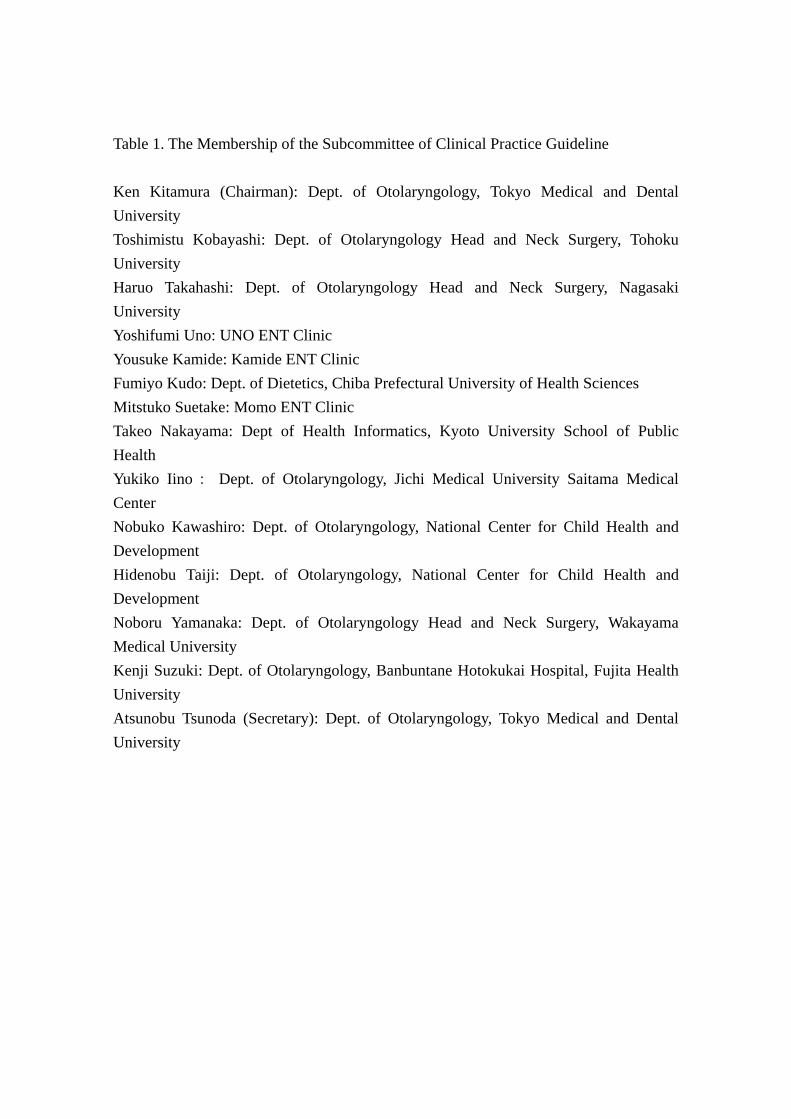
Table 1. The Membership of the Subcommittee of Clinical Practice Guideline Ken Kitamura (Chairman): Dept. of Otolaryngology, Tokyo Medical and Dental University Toshimistu Kobayashi: Dept. of Otolaryngology Head and Neck Surgery, Tohoku University Haruo Takahashi: Dept. of Otolaryngology Head and Neck Surgery, Nagasaki University Yoshifumi Uno: UNO ENT Clinic Yousuke Kamide: Kamide ENT Clinic Fumiyo Kudo: Dept. of Dietetics, Chiba Prefectural University of Health Sciences Mitstuko Suetake: Momo ENT Clinic Takeo Nakayama: Dept of Health Informatics, Kyoto University School of Public Health Yukiko Iino: Dept. of Otolaryngology, Jichi Medical University Saitama Medical Center Nobuko Kawashiro: Dept. of Otolaryngology, National Center for Child Health and Development Hidenobu Taiji: Dept. of Otolaryngology, National Center for Child Health and Development Noboru Yamanaka: Dept. of Otolaryngology Head and Neck Surgery, Wakayama Medical University Kenji Suzuki: Dept. of Otolaryngology, Banbuntane Hotokukai Hospital, Fujita Health University Atsunobu Tsunoda (Secretary): Dept. of Otolaryngology, Tokyo Medical and Dental University

Attachment
List of Organizations and Companies that posed non-personal financial conflicts of
interest to members of the Subcommittee
Astellas Pharma Inc.
AstraZeneca K.K.
Eisai Co. Ltd.
Otsuka Pharmaceutical Co. Ltd.
Ono Pharmaceutical Co. Ltd.
Lumenis Co. Ltd.
Kissei Pharmaceutical Co. Ltd.
Kyorin Pharmaceutical Co. Ltd.
Kyowa Hakko Kirin Co. Ltd.
GlaxoSmithKline K.K.
Kowa Shinyaku Co. Ltd.
Sanofi-aventis K.K.
Shionogi & Co. Ltd.
Senju Pharmaceutical Co. Ltd.
Daiichi Sankyo Co. Ltd
Taisho Toyama Pharmaceutical Co. Ltd.
Dainippon Sumitomo Pharma Co. Ltd.
Taiho Pharmaceutical Co. Ltd.
Takeda Pharmaceutical Co. Ltd.
Mitsubishi Tanabe Pharma
Chugai Pharmaceutical Co. Ltd.
Nikken Chemical Laboratory Co. Ltd.
Nippon Shinyaku Co. Ltd.
Nippon Boehringer Ingelheim Co. Ltd.
Bayer Pharmaceutical Co. Ltd.
Pfizer Co. Ltd.
Meiji Seika Pharma Co.Ltd.

27.7%
17.0%
4.4%
15.5%18.3%
24.1%34.1%
15.3%
17.5%27.4%
24.2%
25.1%
0%
20%
40%
60%
80%
100%
others
Candida spp.
other G(-) rod
other NFGNR
P.aeruginosa
Enterobacteriaceae
other Haemophilus spp.
H.influenzae
M.(B.) catarrhalis
Enterococcus spp.
other Streptococcus spp.
S.agalactiae
S.pyogenes
S.pneumoniae
CNS
other CNS
S.epidermidis
S.aureus
1994 200720031998
Fig. 1
Nationwide surveillance of clinical isolates from patients with otorhinolaryngological infections


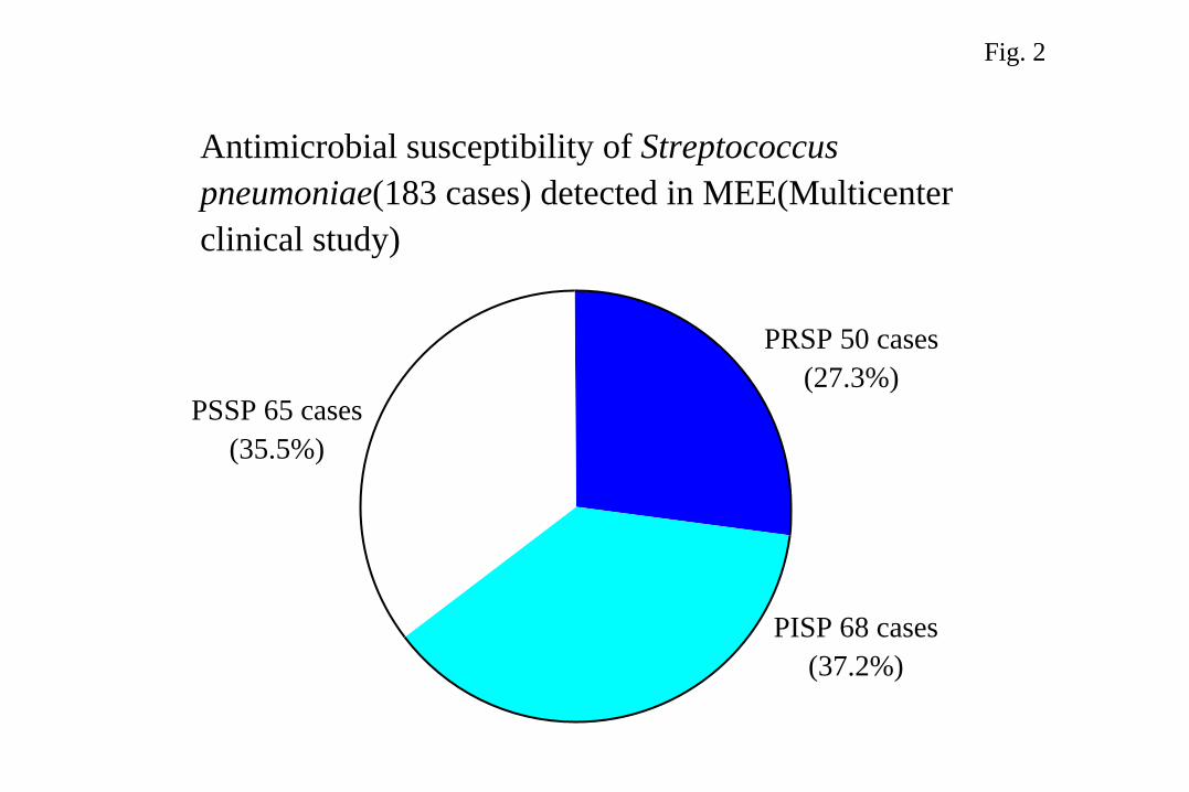
PRSP 50 cases(27.3%)
PISP 68 cases(37.2%)
PSSP 65 cases(35.5%)
Fig. 2
Antimicrobial susceptibility of Streptococcus pneumoniae(183 cases) detected in MEE(Multicenter clinical study)

BLNAR 144 cases(69.3%)
BLNAS 62 cases(29.8%)
BLPAR 2 cases (0.9%)
Fig. 3
Antimicrobial susceptibility of Haemophilus influenzae (208 cases) detected in MEE(Multicenter clinical study)
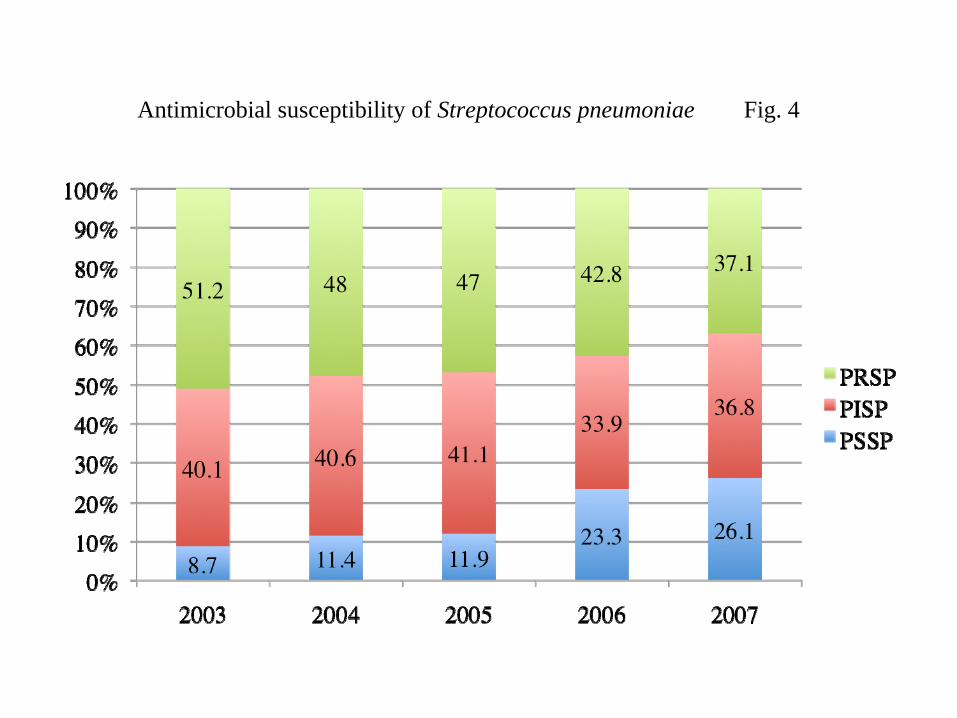
Fig. 4Antimicrobial susceptibility of Streptococcus pneumoniae

Fig. 5Antimicrobial susceptibility of Haemophilus influenzae
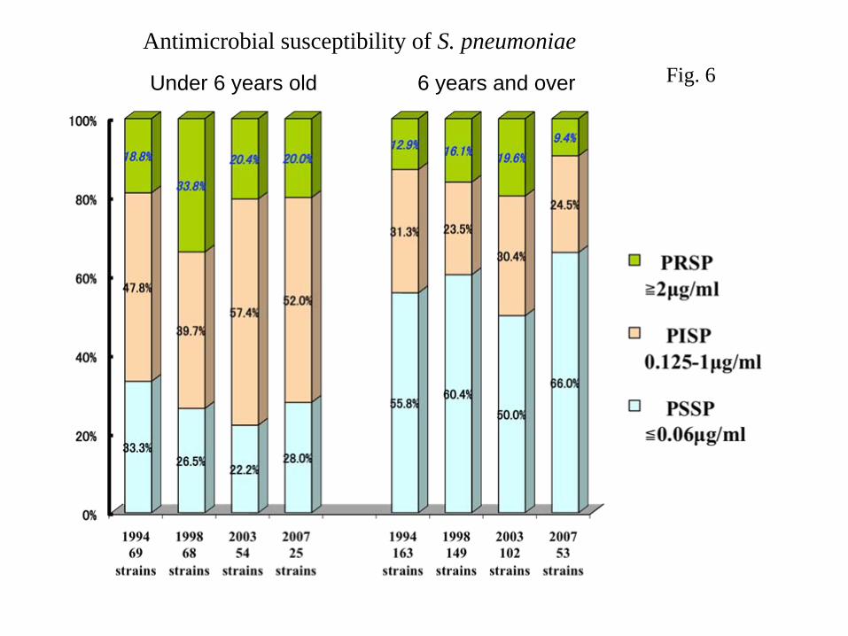
Under 6 years old 6 years and over Fig. 6Antimicrobial susceptibility of S. pneumoniae

Under 6 years old 6 years old and over
1998 101
strains
2003 110
strains
2007 17
strains
2003 81
strains
2007 46
strains
1998 178
strains
Fig. 7Antimicrobial susceptibility of H. influenzae


Appendix 1
The abbreviations and descriptions used for the detected bacteria are as follows.
Streptococcus pneumoniae is classified according to the minimum inhibitory
concentration (MIC) of Penicillin G.
Penicillin susceptible Streptococcus pneumoniae (PSSP) ≦0.06 µg/mL
Penicillin intermediate-resistant Streptococcus pneumoniae (PISP) 0.125~1.0 µg/mL
Penicillin resistant Streptococcus pneumoniae (PRSP) ≧2 µg/mL
Haemophilus influenzae is classified according to the minimum inhibitory concentration
(MIC) of Ampicillin (ABPC)
susceptible bacterium ≦1 µg/mL
resistant bacterium ≧2 µg/mL
and is further classified according to the production of β- lactamase
β-lactamase negative ampicillin resistant Haemophilus influenzae (BLNAR)
β-lactamase negative ampicillin susceptible Haemophilus influenzae (BLNAS)
β- lactamase productive ampicillin resistant Haemophilus influenzae (BLPAR)

No antimicrobial, watchful waiting for 3 days
Mild (Score
0~9)
Observation
Observation
High dose AMPC or CVA/AMPC or CDTR‐PI for 5 days
No improvement Improvement
No improvementImprovement
Treatment Algorithm
AMPC 5 days
*Otalgia、fever(
≥
38.5℃)→Acetaminophen 10mg/kg
*Sign of nasal disorder→treatment*Bacterial culture from nasopharynx or otorrhea
# Lactobacillus bifidus when
administrating antimicrobials# Do not exceed adult dose# Follow‐up period is 3 weeks from
the first visit
Fig. 8

Observation Bacterial culture
No improvement Improvement Myringotomy
Observation
Bacterial cultureImprovementNo improvement
Moderate (Score 10~15)
Any of ① to ④ based on the susceptibility①High dose AMPC ②CVA/AMPC
③High dose CDTR‐PI ④myringotomy+AMPC
for 5 days
Myringotomy+High dose AMPC 5 days or Myringotomy+CVA/AMPC 5 days or ABPC
150mg/kg/day, CTRX60mg/kg/day (premature, neonate<50mg/kg/day) 3 days
AMPC 5 days Severe otoscopic findings
*Otalgia、fever(
≥38.5℃)→Acetaminophen 10mg/kg
*Sign of nasal disorder→treatment*Bacterial culture from nasopharynx or otorrhea
# Lactobacillus bifidus when
administrating antimicrobials# Do not exceed adult dose# Follow‐up period is 3 weeksfrom the first visit
Fig. 9

Observation
ABPC150mg/kg/day 3 days or CTRX60mg/kg/day
(premature, neonate <50mg/kg/day)
3 days
No improvementImprovement
Severe(Score
16≦)
Observation
Any of AMPC、CVA/AMPC、CDTR‐PI based on susceptibility、
for 5 days with high dose + myringotomy
①High dose of AMPC
②CVA/AMPC
③High dose of CDTR‐PI
Any of ①②③ for 5 days + myringotomy
No improvement Improvement
# Lactobacillus bifidus when
administrating antibiotics# Do not exceed adult dose# Follow‐up period is 3 weeks from
the first visit
*Otalgia、fever(
≥38.5℃)→Acetaminophen 10mg/kg
*Sign of nasal disorder→treatment*Bacterial culture for nasopharynx or otorrhea
Fig. 10

A: bullar formation B: thickening
C: presence of middle ear effusion D: perforation
Fig. 11Typical otoscopic findings of AOM

Fig. 12Protrusion of the tympanic membraneScore 4: protrusion present in a part of the tympanic membrane
Score 8: protrusion present in the whole tympanic membrane

Table 11. AOM Clinical Score Sheet (2009) Patient ID: Name: Age: year months Date: Body weight: Body temperature: oC Sex: male female Other features: [Scores chart] Age (<24 mo.) 0 3 Otalgia 0 1 (present) 2 (continuous & severe) Fever 0 (Temp<37.5 oC), 1 (37.5≦ Temp<38.5 oC) 2 (38.5 oC≦Temp) Crying/Bad temper 0 1 Hyperemia of the tympanic membrane
0 2 (at the manubrium or partial) 4(whole tympanic membrane) Protrusion of the tympanic membrane
0 4 (partial) 8 (whole tympanic membrane) Otorrhea
0 4 (visible tympanic membrane) 8(invisible tympanic membrane) Light reflex
0 4 (diminished or absent due to turbidity)
Total score [Assessment] Mild (0-9) Moderate (10-15) Severe (≧16)

Table 12. Questionnaire for a child with acute otitis media Check appropriate items, and ffill out descriptive items, if applicable. Family history: Chronic otitis media (yes –paternal grandparents, maternal grandparents, father, mother siblings, others, none, unknown) Chronic sinusitis (yes –paternal grandparents, maternal grandparents, father, mother siblings, others, none, unknown) Allergic rhinitis (yes –paternal grandparents, maternal grandparents, father, mother siblings, others, none, unknown) Bronchial asthma (yes –paternal grandparents, maternal grandparents, father, mother siblings, others, none, unknown) Atopic dermatitis (yes –paternal grandparents, maternal grandparents, father, mother siblings, others, none, unknown) Other diseases (yes –paternal grandparents, maternal grandparents, father, mother siblings, others, none, unknown) Disease name , none, unknown Past history: Acute otitis media (yes – first episode year months old, times until now, no, unknown) Pneumonia (yes – first episode year months old, times until now, no, unknown) Otitis media with effusion (yes – first episode year months old, times until now, no, unknown) Rhinosinusitis (yes – first episode year months old, times until now, no, unknown) Allergic diseases Bronchial asthma (yes – first episode year months old, no, unknown) Atopic dermatitis (yes – first episode year months old, no, unknown) Allergic rhinitis (yes – first episode year months old, no, unknown) Food allergy (yes – first episode year months old, no, unknown) Drug allergy (yes – name of the drug(s) , no, unknown) Congenital diseases (yes – name of the disease(s) , no, unknown) Other diseases (yes – name of the disease(s) at year months old, no, unknown) Hospitalization (yes – name of the disease(s) at year months old, no, unknown) Often get fever? (yes, no, unknown) Growth and life history: Birth weight grams, gestational weeks at birth , days earlier or later than the full term Nutrition at neonatal and infantile period – mainly by milk, mainly by breast feed, both mixed Day care (yes – from year months old to year months old, no, unknown) Siblings (brother(s) year old, sister(s) year old) Family living together (father, mother, brother(s), sister(s), paternal grandparent(s), maternal grandparent(s)) Smoking of family member (yes, no) Signs and symptoms: ① Ear
Otalgia (earache) (yes, no, unknown) Only for infants and small children often touch his or her ear (yes, no, unknown) Only for older children Ear fullness (yes, no, unknown) Hearing loss (yes, no, unknown) Pulsatile tinnitus (yes, no, unknown) Disequilibrium (dizziness) (yes, no, unknown)
Otorrhea (ear dscharge) (yes, no, unknown) ② General Flu symptoms (yes, no, unknown) Fever (yes, no, unknown) Cough (yes, no, unknown) Nasal discharge or stuffing (yes, no, unknown) Nausea, vomiting (yes, no, unknown) Diarrhea (yes, no, unknown) Bad temper or low activity (yes, no, unknown)

Table 13. The method of sampling for microbial test To detect pathogens of AOM, it is desirable to take specimens not only from middle ear effusion or otorreha but also from nasopharynx. The main pathogens of AOM such as Streptococcus pneumoniae and Heamophilus influenzae are very weak under dry condition. A swab should be immediately inoculated into a porter for bacteriological examination. 1) A case without perforation of the tympanic membrane
After sterilization of the external ear canal with alcohol or popvidone-iodine, the myringotomy is performed. The middle ear effusion is taken for a microbial test using Seed-Swab No. 2.
2) A case with perforation of the tympanic membrane After removal of otorrhea in the external ear canal followed by the sterilization of the external ear canal with alcohol or popvidone-iodine, the middle ear effusion is taken for a microbial test using Seed-Swab No. 2.
3) Nasopharynx After removal of nasal discharge followed by the sterilization of a nostril, nasopharyngeal smear is obtained endonasally.