clinical management of adult patients with complications...
TRANSCRIPT
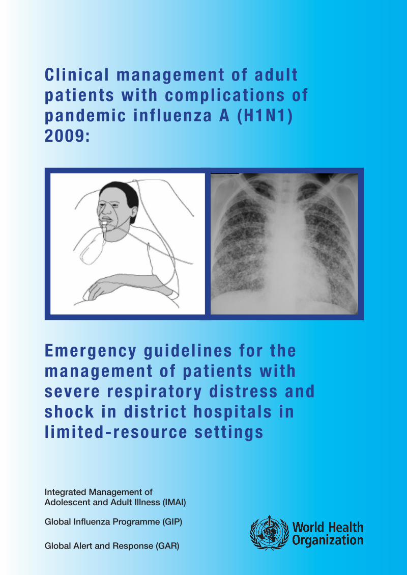
Cl in ical management of adul t pat ients with compl icat ions of pandemic inf luenza A (H1N1) 2009:
Emergency guidel ines for the management of pat ients with severe respiratory d istress and shock in d istr ict hospi ta ls in l imited-resource set t ings
Integrated Management of Adolescent and Adult Illness (IMAI)
Global Influenza Programme (GIP)
Global Alert and Response (GAR)
For further information please contact:IMAI Team Department of HIV/AIDS World Health Organization Avenue Appia, 20CH–1211 Geneva 27Switzerland
[email protected]/hiv/capacity/en

Table of Contents1. Introduction 3
2. Preparing district hospitals and clinicians to manage severe complications
of pandemic influenza A (H1N1) in limited-resource settings 3
3. Quick check and emergency treatments 5
4. Summary of management of septic shock and severe respiratory distress 10
First 2 hours 11
2-6 hours 12
6-24 hours 13
Post resuscitation 14
5. Treatment instructions 15
Manage airway 16
Give oxygen 17
Give antimicrobials: antivirals, antibiotics (and antimalarials if appropriate)1 21
Treat wheezing 23
Give glucose 24
Give pressors 25
6. General principles in caring for the severely ill patient 26
7. Monitoring forms for severely ill patients:
First 6 hours 30
7- 24 hours 32
Annex: Background/scientific basis 34
Process of development 36
1 These are generic guidelines for country adaptation.
WHO Library Cataloguing-in-Publication Data:
Clinical management of adult patients with complications of pandemic influenza A (H1N1) 2009 influenza: emergency guidelines for the management of patients with severe respiratory distress and shock in district hospitals in limited- resource settings.
1.Influenza, Human - complications. 2. Influenza A Virus, H1N1 subtype - complications. 3.Respiratory insufficiency - therapy. 4.Emergency medical services. 5.Adult. 6.Guidelines. I.World Health Organization.
ISBN 978 92 4 159961 0 (NLM classification: WC 515)
© World Health Organization 2010All rights reserved. Publications of the World Health Organization can be obtained from WHO Press, World Health Organization, 20 Avenue Appia, 1211 Geneva 27, Switzerland (tel.: +41 22 791 3264; fax: +41 22 791 4857; e-mail: [email protected]). Requests for permission to reproduce or translate WHO publications – whether for sale or for noncommercial distribution – should be addressed to WHO Press, at the above address (fax: +41 22 791 4806; e-mail: [email protected]). The designations employed and the presentation of the material in this publication do not imply the expression of any opinion whatsoever on the part of the World Health Organization concerning the legal status of any country, territory, city or area or of its authorities, or concerning the delimitation of its frontiers or boundaries. Dotted lines on maps represent approximate border lines for which there may not yet be full agreement.
The mention of specific companies or of certain manufacturers’ products does not imply that they are endorsed or recommended by the World Health Organization in preference to others of a similar nature that are not mentioned. Errors and omissions excepted, the names of proprietary products are distinguished by initial capital letters.
All reasonable precautions have been taken by the World Health Organization to verify the information contained in this publication. However, the published material is being distributed without warranty of any kind, either expressed or implied. The responsibility for the interpretation and use of the material lies with the reader. In no event shall the World Health Organization be liable for damages arising from its use.
Printed in Switzerland
3
1 WHO CAH: Emergency triage assessment and treatment guidelines are included in the Pocket book of hospital care for children: Guidelines for the management of common illnesses with limited resources and are accompanied by a training course implemented in multiple countries. The Pocket Book guides management of severe pneumonia and shock. 2 Integrated Management of Adolescent and Adult Illness (IMAI) is a collaborative project among multiple WHO departments and partners including WHO GIP and GAR, based in the WHO Department of HIV/AIDS. 3 http://www.who.int/csr/resources/publications/swineflu/clinical_management/en/index.html 4 http://www.who.int/csr/resources/publications/swineflu/h1n1_guidelines_pharmaceutical_mngt.pdf
1. Introduction The preparation of these emergency guidelines for the management of severe acute illness, specifically respiratory distress and shock, in district hospitals in limited-resource settings has been prompted by an outbreak of pandemic influenza A (H1N1). Although most cases of pandemic influenza A (H1N1) (or other influenzas) are mild, a small proportion will develop severe, potentially fatal, complications. This is especially of concern in patients with co-morbidities such as chronic lung disease or diabetes mellitus, and extreme obesity, and in pregnant women. District hospitals and clinicians should be prepared to assess and treat patients with these severe complications when they occur. The WHO Integrated Management of Childhood Illness (IMCI),1 with which there is substantial country experience, and the more recently introduced Integrated Management of Adolescent and Adult Illness (IMAI)2 have relevant tools for the management of patients with severe acute illness, both for first-level facilities and district hospitals. These tools include protocols for emergency triage assessment and treatment and guidelines for patient management. These approaches simplify and put into operational terms standard guidelines. The guidelines presented in this document are taken from the draft IMAI District Clinician Manual: Hospital Care for Adolescents and Adults, which is currently undergoing field testing. In some instances specific parts of the full guidelines have not been included in this shortened version and other guidelines need to be consulted to consider, for example, the full differential diagnosis or detailed management of underlying conditions. As with any set of generic guidelines, country or local adaptation will be necessary. For example, if malaria is not endemic in the country or area, the antimalarial recommendations should be omitted. Additionally the differential diagnosis will be influenced by the local prevalence of HIV and of tuberculosis. Consequently, local epidemiological circumstances, facilities, equipment, and staffing must be taken into account in adapting the guidelines. A training course on these guidelines is nearing completion. Some management recommendations are based on Clinical management of human infection with pandemic (H1N1) 2009: revised guidance issued in November 2009.3 In addition oseltamivir recommendations are based on WHO Guidelines for Pharmacological Management of Pandemic Influenza A (H1N1) 2009 and other Influenza Viruses Revised February 2010.4 These guidelines will be revised in December 2010. 2. Preparing district hospitals and clinicians to manage severe
complications of pandemic influenza A (H1N1) in limited-resource settings
District hospital staff should be made aware of the current patterns of influenza epidemiology to (when appropriate) heighten the diagnostic suspicion for pandemic (H1N1) 2009 influenza (or other influenza viruses). District hospitals should have written guidelines for the diagnosis of pandemic influenza A (H1N1) and employ infection control measures

4
when the diagnostic criteria are met in a given patient. Uncomplicated influenza should be suspected among patients whose symptoms include: fever, cough, sore throat, rhinorrhoea, headache, muscle pain, and malaise, but no shortness of breath. Patients may present with some or all of these symptoms. Complicated or severe influenza is indicated by clinical (e.g. shortness of breath/dyspnoea, fast breathing, hypoxia) and/or radiological signs of lower respiratory tract involvement (e.g. pneumonia), central nervous system involvement (e.g. encephalopathy, encephalitis), severe dehydration, or secondary complications, such as renal failure, multi-organ failure, and septic shock. Other complications can include rhabdomyolysis and myocarditis, exacerbations of underlying chronic disease, including asthma, COPD, chronic hepatic or renal failure, diabetes mellitus, or cardiovascular conditions. Patients who present initially with uncomplicated influenza may progress to more severe disease. Progression can be rapid (i.e. within 24 hours). The following are some of the indicators of progression, which would necessitate an urgent review of patient management: • Symptoms and signs suggesting oxygen impairment or cardiopulmonary insufficiency:
- shortness of breath (with activity or at rest), difficulty in breathing, turning blue, bloody or coloured sputum, chest pain, and low blood pressure;
- fast or laboured breathing; - hypoxia, as indicated by pulse oximetry.
• Symptoms and signs suggesting CNS complications: - altered mental status, unconsciousness, drowsiness, or difficult to awaken
recurring or persistent convulsions (seizures), confusion, severe weakness, paralysis.
• Evidence of sustained virus replication or invasive secondary bacterial infection based on laboratory testing or clinical signs (e.g. persistent high fever and symptoms beyond three days).
• Severe dehydration, manifested as decreased activity, dizziness, decreased urine output, or lethargy.
Clinical teams need to have the training equipment and facilities to enable triage, clinical assessment, and appropriate treatment for patients with severe influenza. Key lifesaving treatments include:
oxygen fluids Antimicrobials – antivirals and antibiotics (and antimalarials, if appropriate) salbutamol if wheezing is present
These guidelines should not be viewed as being applicable only to pandemic influenza A (H1N1), or even limited to viral illnesses. The facilities, training and approaches to clinical management are generally applicable to any patient with shock or respiratory distress and will also benefit patients with severe illnesses caused by bacterial pneumonia, tuberculosis (TB), and opportunistic infections associated with HIV infection as well as malaria in endemic areas.

5
Section 3: QUICK CHECK and Emergency Treatments for Adolescents and Adults
This section describes the triage assessment and emergency treatments relevant to the management of severe respiratory distress and septic shock. (It does not include all priority and emergency signs or trauma).* Existing guidelines should be used for the development of a full differential diagnosis at the time of every patient encounter to ensure that processes presenting with signs and symptoms similar to those of influenza or concurrent processes are recognized and treated appropriately. The assessment in the Quick Check should be performed for any patient on arrival at the facility. The ABC emergency signs (Airway, Breathing, Circulation, Consciousness, Convulsions) are a special set of emergency signs that are checked rapidly and repeatedly. Triage is the process of rapidly screening patients soon after arrival in hospital to identify:
• patients with emergency signs, who require immediate emergency treatment • patients with priority signs, who should be given priority while waiting in the queue so that
they can be assessed and treated without delay • non-urgent patients, who have neither emergency or priority signs.
This section should guide the entire hospital team. The Quick Check should be used both for the immediate, first assessment on arrival in hospital and to re-assess sick patients in hospital, or waiting in the emergency department. This section summarizes how to check the ABC signs and how to give emergency treatments; detailed treatment instructions are provided in sections 4 and 5. These sections of the Quick Check and Emergency Treatments relevant for assessing and providing emergency treatments for patients with complications of influenza have been extracted from the full guidelines. • Patients with severe respiratory distress without shock or septic shock should then be managed
according to the summary guidelines in Section 4 of this document. • Section 5 provides the detailed treatment instructions. Use infection control precautions during triage, Quick Check and emergency treatments. Standard precautions should be followed for all patients. Droplet and contact precautions should be added when pandemic influenza A (H1N1) is suspected. For patients with acute respiratory diseases as appropriate, add droplet, contact, airborne and special precautions for aerosol generating procedures as appropriate – see current WHO infection control guidelines. * This is a partial Quick Check and Emergency Treatments extracted from the draft IMAI District Clinician Manual that does not include trauma or other priority signs.
Abbreviations: SBP 90 = systolic blood pressure 90 mm Hg SpO2 90 = oxygen saturation 90% NS = normal saline LR = lactated ringers L = litres

6
EMERGENCY SIGNS All staff should be able to assess. If any sign positive patient is severely ill. Call for help. Clinical staff should immediately give emergency treatment(s).
FIRST LINE EMERGENCY TREATMENT If any emergency sign is positive, nurse and others on clinical team should give the treatments, call for help, establish IV access.
After the Quick Check, send blood for glucose, malaria smear, haemoglobin. Make sure all patients with positive emergency signs have full set of vital signs and pulse oximetry and that these are acted on.
FIRST ASSESS: AIRWAY AND BREATHING
If obstructed breathing • If foreign body aspiration, treat choking patient • If suspect anaphylaxis (wheezing and facial
swelling), give 0.5 ml 1:1000 epinephrine (adrenaline) IM (p.24)
Manage airway (see p.7) Give oxygen 5 L/minute (see p.19) Help patient assume position of comfort If wheezing, give salbutamol (see p.24) If inadequate breathing, assist ventilation with bag-valve mask
• Appears obstructed
or • Central cyanosis or • Severe
respiratory distress
3. Quick Check for Adolescents and Adults
Check for obstruction (noisy breathing), wheezing, choking, not able to speak)
Do not move neck if cervical spine injury possible
THEN ASSESS: CIRCULATION
• Weak or fast pulse or
• Capillary refill longer than three seconds or
• Heavy bleeding from any site or
• Severe trauma
Check BP, HR Is she pregnant?
If systolic BP < 90 mmHg or pulse >100 per minute or heavy bleeding: Give oxygen 5L/minute if respiratory distress or
SpO2<90 Insert IV, give 1 bag (500 ml to 1 litre) bolus
crystalloid (RL or NS) then reassess Keep warm (cover) If pregnant, place on side
If anaphylaxis (rash, wheezing and facial swelling, low BP, give 0.5 ml 1:1000 epinephrine (adrenaline) IM (p.24) If suspect heart failure, cardiogenic shock or severe anaemia, be cautious with fluids.
IF ALTERED LEVEL CONSCIOUSNESS or CONVULSING, go to page 8 for emergency treatments

7
Use this chart for rapid triage assessment then emergency treatments. Assess pregnancy status for women of childbearing age to appropriately manage.
Use standard precautions for all patients. Use droplet precautions if acute respiratory infection of concern. Add aerosol precautions if airway management or intubation.
CONTINUE WITH URGENT MANAGEMENT OF PATIENTS WITH EMERGENCY SIGNS
Finish rest of Quick Check then: • Count HR, RR; measure oxygen saturation • Titrate oxygen to SpO2 90 • Give antibiotics and (if appropriate) antimalarials (p.22) if fever and RR >30/minute and/or
suspect pneumonia • Give antiviral (p.23) • Insert IV, give 500 ml RL or NS unless suspect heart failure. Consider differential diagnosis. If severe respiratory distress from pneumonia or acute lung injury and no heart failure, follow management instructions in Section 4 of this document.
Decide on type of shock and treat accordingly. Consider differential diagnosis (septic, haemorrhagic, hypovoemic, cardiogenic). If fever or low temperature, consider septic shock. Give urgent empiric antibiotics (p.22) and antiviral treatment if suspect influenza (p.23). If falciparum malaria is a possibility, do malaria smear and give antimalarial treatment. (p.21). Also give glucose (if blood glucose is low or unknown) (p.25). Send blood culture if feasible before starting antibiotics. See Section 4 in this guideline summary for management of septic shock.

8
For all: Protect from fall or injury Manage airway (p.17) and assist into recovery
position Give oxygen 5 L/minute Call for help but do not leave patient alone Give glucose (if blood glucose is low or unknown) Check (then monitor and record) level of
consciousness on AVPU scale. If convulsing: Give diazepam IV or rectally
If convulsing in second half pregnant or post-partum
up to 1 week, also give magnesium sulphate. Then check BP, HR, RR, temperature If fever, give antibiotics and (if appropriate) antimalarial (p.21). If convulsions continue after 10 minutes: Continue to monitor airway, breathing, circulation Recheck glucose Give second dose diazepam Consult to add second antiepileptic drug Consider differential diagnosis.
• Altered level
consciousness or • Convulsing
ALTERED LEVEL CONSCIOUSNESS/CONVULSING
FIRST LINE EMERGENCY TREATMENT EMERGENCY SIGNS
Do not move neck if cervical spine injury possible
Is she pregnant?

9
PRIORITY SIGNS AND SYMPTOMS
Check BP, pulse, respiratory rate, temperature, oxygen saturation
PRIORITY SIGNS FOR URGENT CARE OF RESPIRATORY ILLNESS – these patients should not wait in queue: • If any respiratory distress/complaint of
difficulty breathing. • Very weak/ill.
If any respiratory distress/complaint of difficulty breathing: • Give oxygen 5L/minute if SpO2 <90. • Assess for other signs of pneumonia
(see IMAI Acute Care*). If wheezing, give salbutamol. The patient needs clinical evaluation and should not wait in queue. Repeat Quick Check every 20 minutes.
If H1N1 cases are occurring and antivirals are available, treat patients in risk groups, and those who are not improving by day 3 of the illness, or who have progressive disease or any signs of pneumonia; give oseltamivir 75 mg twice daily (p. 23).**
• Patient can wait in queue. • Provide routine care using
appropriate guidelines (IMAI or other sources).
If no emergency signs and no difficult breathing (or other priority signs of illness) NON-URGENT
After screening for emergency signs, screen all patients for priority signs. PRIORITY SIGNS FOR INFECTION CONTROL: if cough or acute febrile respiratory illness of concern, use source control (tissues or mask, hand hygiene, place in room or separate area and evaluate as soon as possible – see infection control guidelines)
* IMAI Acute Care http://www.who.int/hiv/pub/imai/en/acutecarerev2_e.pdf ** See WHO Clinical management of human infection with pandemic influenza H1N1 2009: Revised guidance November 2009 http://www.who.int/csr/resources/publications/swineflu/clinical_management/en/index.html

10
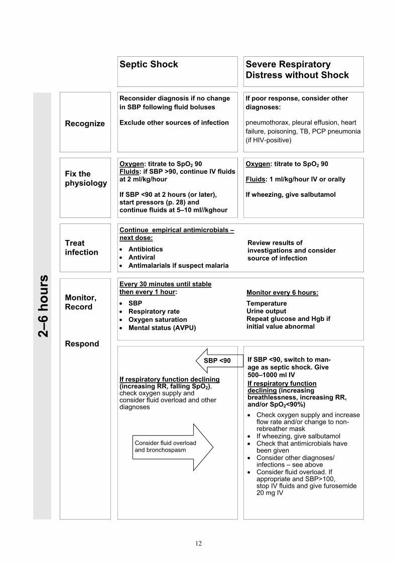
4. Summary of management of septic shock and
severe respiratory distress without shock
This summarizes management for septic shock and severe respiratory distress without shock during the first 2 hours, 2 to 6 hours, 6 to 24 hours, and post-resuscitation. The treatment instructions are in section 5.

11
Septic Shock Severe Respiratory Distress without Shock
Clinical diagnosis of severe sepsis or septic shock:
• Hypotension (systolic blood pressure <90 mmHg) and 1 or more of the following: - Heart Rate >100 bpm - Respiratory Rate >24 - Abnormal temperature (<36oC
or >38oC) • Suspected infection
Clinical diagnosis of severe respiratory distress without shock:
• Respiratory rate >30 or SpO2 <90 and
• SBP >90 mm Hg and • Heart failure excluded and • Suspect pneumonia or acute lung
injury
Recognize
Fix the physiology
Treat infection
Monitor, Record, Respond
What has already happened: Quick Check showed emergency signs —> Manage airway, O2 started at 5 litres, IV 500–1000 ml LR or NS. Emergency labs sent: glucose, haemoglobin, malaria smear/rapid test. Consider differential diagnosis of shock and/or respiratory distress. Exclude heart failure.
Oxygen: titrate to SpO2 90 Fluids: after initial bolus of 1000 ml, continue rapid IV fluids LR or NS at 20 ml/kg/hour – up to 60 ml/kg within first 2 hours
Urgent empirical antimicrobials: Give both: • Antibiotics (p.18) • Antiviral (p.19) If falciparum malaria possible, do malaria smear and give antimalarial (p.18)
Firs
t 2 h
ours
Identify source of infection: Use signs/symptoms and these investigations to consider source Malaria screen Chest X-ray Send blood cultures Gram stain sputum, other body fluids as appropriate
Every 30 minutes until stable then every 1 hour: • SBP • Respiratory rate • Oxygen saturation • Mental status (AVPU)
Check results of emergency laboratory: If haemoglobin <5 mg/dL (Hct <15) consider transfusion If glucose <3 mM/L (54 mg/dl) then give 50% dextrose 25–50 ml
If respiratory function worsening (increasing RR, falling SpO2), check oxygen supply and consider fluid overload and other diagnoses
If SBP less than 90, switch to manage as septic shock SBP <90
Consider fluid overload and bronchospasm
Oxygen: titrate to SpO2 90 Fluids: after initial 500 ml, give IV fluids at 1 ml/kg/hour IV or orally If wheezing, give salbutamol
Possible pandemic influenza A (H1N1) with severe complications:
12
Septic Shock Severe Respiratory Distress without Shock
2–6
hour
s
Recognize
Fix the physiology
Treat infection
Monitor, Record Respond
Oxygen: titrate to SpO2 90 Fluids: if SBP >90, continue IV fluids at 2 ml/kg/hour If SBP <90 at 2 hours (or later), start pressors (p. 28) and continue fluids at 5–10 ml//kghour
Oxygen: titrate to SpO2 90 Fluids: 1 ml/kg/hour IV or orally If wheezing, give salbutamol
Continue empirical antimicrobials – next dose: • Antibiotics • Antiviral • Antimalarials if suspect malaria
Every 30 minutes until stable then every 1 hour: • SBP • Respiratory rate • Oxygen saturation • Mental status (AVPU)
Review results of investigations and consider source of infection
Monitor every 6 hours: Temperature Urine output Repeat glucose and Hgb if initial value abnormal
If respiratory function declining (increasing RR, falling SpO2), check oxygen supply and consider fluid overload and other diagnoses
Reconsider diagnosis if no change in SBP following fluid boluses
Exclude other sources of infection
If poor response, consider other diagnoses:
pneumothorax, pleural effusion, heart failure, poisoning, TB, PCP pneumonia (if HIV-positive)
SBP <90 If SBP <90, switch to man-age as septic shock. Give 500–1000 ml IV If respiratory function declining (increasing breathlessness, increasing RR, and/or SpO2<90%) • Check oxygen supply and increase
flow rate and/or change to non-rebreather mask
• If wheezing, give salbutamol • Check that antimicrobials have
been given • Consider other diagnoses/
infections – see above • Consider fluid overload. If
appropriate and SBP>100, stop IV fluids and give furosemide 20 mg IV
Consider fluid overload and bronchospasm

13
Septic Shock Severe Respiratory Distress without Shock
Reconsider diagnosis if no change in SBP following fluid boluses
Establish source of infection
Consider surgical cause –
is drainage required?
Recognize
Fix the physiology
Treat infection
Monitor, Record Respond
Oxygen: titrate to SpO2 90 Fluids: when SBP >90, continue fluids at 2 ml/kg/hour. If on pressors, reduce rate and continue fluids. If SBP <90, continue or increase pressors (p. 28) and LR/NS at 2 ml/kg/hour IV.
Continue antimicrobials – next doses: • Antibiotics • Antiviral • Antimalarials
6–24
hou
rs
Every hour if SBP <90 or on pressors, otherwise every 2 hours:
• SBP • Respiratory rate • Oxygen saturation • Mental status (AVPU)
Monitor every 6 hours: Temperature Urine output Repeat glucose and Hgb if initial value abnormal or if clinical state worsens
Respond to changes as indicated for 2–6 hours on prior page Consider broader antimicrobial coverage if not improving.
Oxygen: titrate to SpO2 90 Fluids: continue at 1 ml/kg/hour IV or orally If wheezing, give salbutamol
If poor response, consider other diagnoses:
pneumothorax, pleural effusion, heart failure, poisoning, TB, PCP pneumonia associated with HIV infection, etc.
Revisit differential diagnosis.
14
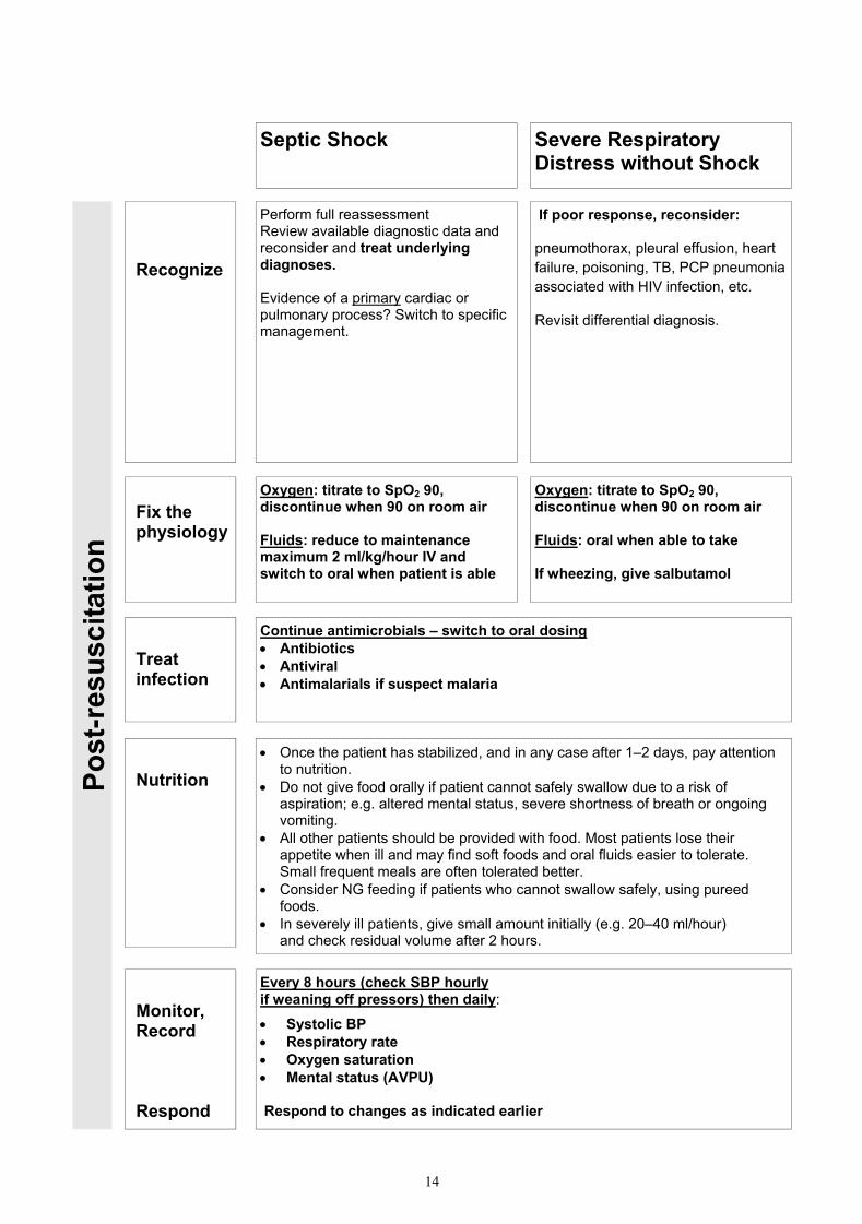
Septic Shock Severe Respiratory Distress without Shock
Perform full reassessment Review available diagnostic data and reconsider and treat underlying diagnoses. Evidence of a primary cardiac or pulmonary process? Switch to specific management.
If poor response, reconsider:
pneumothorax, pleural effusion, heart failure, poisoning, TB, PCP pneumonia associated with HIV infection, etc.
Revisit differential diagnosis.
Recognize
Fix the physiology
Treat infection
Monitor, Record Respond
Oxygen: titrate to SpO2 90, discontinue when 90 on room air Fluids: reduce to maintenance maximum 2 ml/kg/hour IV and switch to oral when patient is able
Continue antimicrobials – switch to oral dosing • Antibiotics • Antiviral • Antimalarials if suspect malaria
Post
-res
usci
tatio
n
Every 8 hours (check SBP hourly if weaning off pressors) then daily:
• Systolic BP • Respiratory rate • Oxygen saturation • Mental status (AVPU) Respond to changes as indicated earlier
Oxygen: titrate to SpO2 90, discontinue when 90 on room air Fluids: oral when able to take If wheezing, give salbutamol
Nutrition
• Once the patient has stabilized, and in any case after 1–2 days, pay attention to nutrition.
• Do not give food orally if patient cannot safely swallow due to a risk of aspiration; e.g. altered mental status, severe shortness of breath or ongoing vomiting.
• All other patients should be provided with food. Most patients lose their appetite when ill and may find soft foods and oral fluids easier to tolerate. Small frequent meals are often tolerated better.
• Consider NG feeding if patients who cannot swallow safely, using pureed foods.
• In severely ill patients, give small amount initially (e.g. 20–40 ml/hour) and check residual volume after 2 hours.
15
5. Treatment instructions Section 5 presents a simplified summary of the management of septic shock and respiratory distress without septic shock, in patients without signs suggesting heart failure. It specifies the frequency of monitoring, response to abnormal signs, fluid administration, and antimicrobial treatment.

16
Manage the airway
Emergency Treatments➤ Manage the airway
After only a few minutes a patient without oxygen can sustain brain damage and die. It ismandatory for ALL health care providers to be competent in basic airway and breathingmanagement (giving oxygen, opening the airway, bag valve mask ventilation). Most patientscan be managed with oxygen and simple manoeuvres, and it is rare for a patient to requireadvanced airway management and intubation.
ASSESS AIRWAY• Talk to the patient. If the patient is
speaking the airway is clear.• Look/listen for signs of airway obstruction:
• Snoring or gurgling• Stridor or noisy breathing• Foreign body or vomit in mouth
IF SEVERE HEAD ORNECK TRAUMAPatients with severe head orneck trauma often havesignificant associated injuriesto airway and cervical spine.When caring for thesepatients:• Protect cervical spine• Give oxygen 5 L• Open airway using jaw thrust• Remove foreign body if visible• Suction secretions• Place oral airway• A definitive airway including
intubation or surgical cricothyroidotomy may be required
If airway obstructed, clear obstruction as follows:If no obstruction, go to STEP 4
NO TRAUMA• Position patient on
firm surface Tilt the head• Lift the chin to open
the airway Remove foreign
body if visible• Clear secretions• Lie patient on side
in recovery position
TRAUMA• Stabilize cervical
spine– do not lift head
• Open airway using jaw thrust
• Remove foreign body if visible Clear secretions
STEP 1
STEP 2
IF AIRWAY IS STILL OBSTRUCTED, INSERT AIRWAY THEN GO TO STEP 4IF AIRWAY IS NO LONGER OBSTRUCTED GO TO STEP 4
Oropharyngeal airway• Use if patient unconscious• Use appropriate size (measure from
front of ear to corner of mouth)• Slide airway over tongue• Give oxygen after placing airway
device• If patient resists, gags, or vomits
remove immediately
STEP 3
INSERT AIRWAY DEVICE
Nasopharyngeal airway• Better tolerated if patient conscious• Pass well lubricated airway into
one nostril directed posterior towards the throat
• Give oxygen after placing airway device

17
Manage the airway
ASSESS OXYGENATION
• Is patient in severe respiratory distress?• Is patient convulsing?• Is patient altered or confused?• Is patient cyanotic?• Is the measured oxygen saturation is <90%
THEN GIVE OXYGEN
IF PATIENT IS OXYGENATING ADEQUATELY, THEN PROCEED TO THE REST OF QUICK CHECK
STEP 5
STEP 6
STEP 4
If ventilation is inadequate (if ventilation appears inadequate, or patient is, cyanotic or unconscious with respiratory distress), then assist breathing via bag valve mask ventilation (STEP 5)
If ventilation is adequate go to STEP 6
ASSESS VENTILATION
• Attach bag valve mask to highest available flow oxygen• Place mask over patient’s mouth and nose* If 2 people: one person holds bag
and other holds mask on patient’s face• Create seal so that air does not leak out• Squeeze bag to give one breath every 6 seconds• If unable to effectively ventilate, insert oral or nasal airway (see STEP 3)
How to bag patient:
• Hold the bag in one hand and depress a 2 L bag to about 1/3 of its volume• Make sure that after each breath, the patient completely exhales before giving
another breath• Watch to make sure that the chest is rising and falling evenly with each breath• If the patient is breathing on his or her own, deliver breaths during inspiration.
Do not attempt to deliver a breath as the patient exhales• Avoid overaggressive bagging, as it will result in damage to lungs
ASSIST VENTILATION WITH BAG VALVE MASK

18
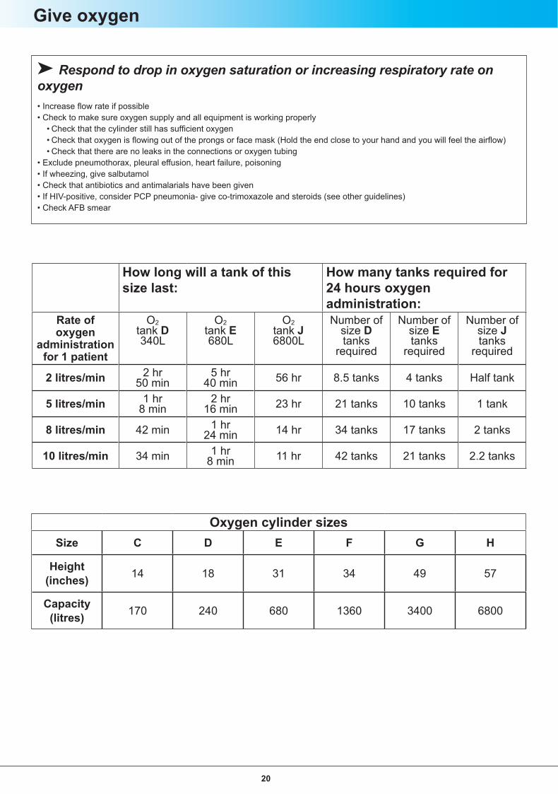
➤ Give oxygen Start flow rate at 5 L/minute. Measure oxygen saturation and titrate flow (see next box). Make sure you have adequate oxygen supply (see p. 21)
Set up the oxygen equipment: either a concentrator with cylinder back-up or cylinder may be used• Connect the non-crush oxygen delivery tube to the tubing adaptor at the oxygen outlet of the concentrator or cylinder firmly
(if concentrator: make sure to plug into power source)• Fully open the cylinder by turning the keywheel anti-clockwise• Turn the knob on the flow controller to adjust the flow based on the flowmeter reading (check
manufacture directions for reading)• Check that oxygen is coming out by holding tubing or prongs under water
Deliver oxygen via nasal prongs (also called nasal cannula): short tubes inserted into the nostrils• Place them just inside the nostril and secure with a piece of tape on the cheeks near the nose (if needed)• Hook the rest of the tubing behind the ears
Deliver oxygen via face mask (FM):• Connect tubing from cylinder/concentrator to mask• Secure mask firmly on face over nose and mouth and pull plastic strap over head
Deliver oxygen via face mask with reservoir (non-rebreather):• Connect tubing and secure mask as above• Connect to high flow oxygen and until the reservoir bag is filled
Increasing oxygen
concentration
Oxygen Flow
1L2L3L4L5L>6 L10-15 L
Method
Nasal cannula or prongsNasal cannula or prongsNasal cannula or prongsNasal cannula or prongsNasal cannula or prongsFace maskFace mask with reservoir*
*Face mask with reservoir is the same thing as a Non-rebreather
Start oxygen at 5 litres/minute then titrate using pulse oximeter
➤ Titrate oxygen using pulse oximetry
• Start oxygen at 5 L/minute by nasal cannula/prongs
• If patient worsening or not improving (increasing respiratory distress or SpO2<90%), increase oxygen flow if possible.• Increase to 6-10 litres via face mask• If patient still in distress, increase to 10-15 litres via face mask with reservoir
• If patient stabilizing or improving, titrate oxygen to appropriate flow rate• Decrease oxygen flow by 1-2 L/min• Each time you change the oxygen flow, observe the patient for at least 2-3 minutes.• If patient does not tolerate less oxygen then do not titrate oxygen flow until the patient is more stable.
• Recheck the patient in 15 minutes and measure the oxygen saturation.• If patient is in increased respiratory distress or oxygen saturation <90% then increase oxygen flow to previous flow rate.• If patient remains stable and oxygen saturation >90%, continue to titrate oxygen down as tolerated
• Recheck clinical status and SpO2 all patients after 1 hour for delayed hypoxia or respiratory distress
Give oxygen

19
Give oxygen
➤ Using a pulse oximeter
To use a pulse oximeter: • Turn the pulse oximeter on. • Attach the oximeter probe to the finger or toe. • Wait until there is a consistent pulse signal (this may take 20–30 seconds). • Record the SpO2 on a monitoring chart.
• If the SpO2 is <90%, give oxygen• Through nasal prongs or a nasal catheter• Start at 5 litres/minute continuously.
• Recheck the SpO2. • Record the SpO2 on a monitoring chart 15 minutes after giving oxygen.
20
➤ Respond to drop in oxygen saturation or increasing respiratory rate on oxygen• Increase flow rate if possible• Check to make sure oxygen supply and all equipment is working properly
• Check that the cylinder still has sufficient oxygen• Check that oxygen is flowing out of the prongs or face mask (Hold the end close to your hand and you will feel the airflow)• Check that there are no leaks in the connections or oxygen tubing
• Exclude pneumothorax, pleural effusion, heart failure, poisoning• If wheezing, give salbutamol• Check that antibiotics and antimalarials have been given • If HIV-positive, consider PCP pneumonia- give co-trimoxazole and steroids (see other guidelines)• Check AFB smear
Give oxygen
How long will a tank of this size last:
How many tanks required for 24 hours oxygen administration:
Rate of oxygen
administrationfor 1 patient
O2 tank D 340L
O2tank E680L
O2 tank J 6800L
Number ofsize Dtanks
required
Number ofsize Etanks
required
Number ofsize Jtanks
required
2 litres/min 2 hr 50 min
5 hr 40 min 56 hr 8.5 tanks 4 tanks Half tank
5 litres/min 1 hr 8 min
2 hr 16 min 23 hr 21 tanks 10 tanks 1 tank
8 litres/min 42 min 1 hr 24 min 14 hr 34 tanks 17 tanks 2 tanks
10 litres/min 34 min 1 hr 8 min 11 hr 42 tanks 21 tanks 2.2 tanks
Oxygen cylinder sizesSize C D E F G H
Height (inches) 14 18 31 34 49 57
Capacity (litres) 170 240 680 1360 3400 6800

21
300 mg/ml (in 2 ml ampoules)
Weight
➤ Give emergency antimalarial treatment if falciparum malaria is possible
30 kg
ARTEMETHER IM
Initial loading dose: 3.2 mg/kg
80 mg/ml (in 1 ml ampoules)
QUININE IM—divide dose equally in two and give one in each anterior thigh
Subsequent doses1.6 mg/kg each day until able to take oral medication
80 mg/ml (in 1 ml ampoules)
150 mg/ml (in 2 ml ampoules)
300 mg/ml (in 2 ml ampoules)
150 mg/ml (in 2 ml ampoules)
Initial dose: 20 mg/kg Subsequent doses10 mg/kg every 8 hours until able to take oral medication
40 kg
50 kg
60 kg
70 kg
80 kg
90 kg
1.2 ml
1.6 ml
2 ml
2.4 ml
2.8 ml
3.2 ml
3.6 ml
0.6 ml
0.8ml
1 ml
1.2 ml
1.4 ml
1.6 ml
1.8 ml
4 ml
5.4 ml
6.6 ml
8 ml
9.3 ml
10.6 ml
12 ml
2 ml
2.6 ml
3.3 ml
4 ml
4.7 ml
5.3 ml
6 ml
2 ml
2.6 ml
3.3 ml
4 ml
4.7 ml
5.3 ml
6 ml
0.5 ml
0.7 ml
0.8 ml
1.0 ml
1.2 ml
1.3 ml
1.5 ml
Always give glucose with quinine
• If giving quinine by IV, dilute with 10ml/kg of IV fluid and infuse slowly over 4 hours.• When able to take oral treatment, give a single dose of sulfadoxine-pyrimethamine, or if on quinine, give an adult one 500 mg tablet daily (small adolescents or children 10 mg/kg three times daily) to complete 7 days of treatment.
➤ Give empiric IV/IM antibiotics for emergency management
➢ Give ceftriaxone 1gm IV or IM.
If ceftriaxone not available, give: • ampicillin* 2gm IV or IM and • gentamicin 240mg IV or IM
* If ampicillin is not available, give benzylpenicillin 3 million units
➤ Give empiric oral antibiotics for non-severe pneumonia
• amoxicillin 1000 mg 3 times a day or• co-trimoxazole 1double strength tablet (800 mg/160mg) twice daily (if not taking co-trimoxazole prophylaxis)
Then review if not improving after 2-3 days consider adding• erythromycin base 500 mg 4 times a day or• doxycycline 100 mg 2 times a day or• azithromycin 500 mg once a day
If there is no improvement and the patient has been adherent to the antibiotic regimen other infections should be considered, including TB and, if HIV infected, Pneumocystis pneumonia
Give antimicrobials

22
Weight
➤ Give emergency antiviral treatment*
Usual dose* Severe disease or severely immunosuppressed45 mg twice daily 45 mg twice daily15-23 kg
24-40 kg
>40 kg (age 13 and older)
Oseltamivir
Give antimicrobials
60 mg twice daily 60 mg twice daily
75 mg twice daily 150 mg twice daily
* If at 5 days or more, the clinical course remains progressive or severe, treatment should be maintained without a break until viral infection is resolved or there is satisfactory clinical response.

23
➤ Give magnesium sulphateFor severe life-threatening asthma in adults after repeated salbutamol by nebulizer:
• Insert IV line with normal saline or Ringer’s lactate • Give 2 grams of magnesium sulfate (10 ml of 20% solution) IV slowly over 20 minutes• Patient may feel warm during injection
Treat wheezing
➤ Give salbutamol by nebulizer
• 5 mg in 5 ml solution if more than 20 kg (give 2.5 mg in 2.5 ml solution if less than 20 kg)• Place in nebulizer • Connect to oxygen if available• Give every 10-20 minutes or, if severe, give continuously
➤ Give salbutamol by metered-dose inhaler100 mcg/puff; 200 puffs/inhaler
Use spacer and/or mask depending if patient not able to coordinate breathing and inhaler.
• If MILD WHEEZING: 2 puffs every 20 minutes x 3 times then 2 puffs every 3 to 6 hours
• If MODERATE or SEVERE WHEEZING Give salbutamol, either continuous nebulizers or prime spacer with 5 puffs then give 2 puffs via spacer every 2 minutes
➤ How to make spacer from plastic bottle
• Use a clean plastic 300– 500 ml bottle. (Clean monthly and prime with 10 puffs after each cleaning, before using for treatment.)• Remove the inhaler cap and trace the shape of the opening of the inhaler on the base of the bottle, directly opposite the mouth
of the bottle.
• Cut an opening into the base of the bottle exactly (or slightly smaller) than the size traced with a heated paperclip. (An alterna-tive is to make a slit in the side of the bottle and place the puffer through the hole.)
• Insert the inhaler into the spacer to check the size.• For severe attacks or if the patient cannot cooperate, cut off the bottom and use as a mask.
➤ Give epinephrine
For severe respiratory distress from wheezing not responding to salbutamol:
• Only give epinephrine if:- Severe respiratory distress not responding to 20 puffs of salbutamol by inhaler or nebulized salbutamol in 10 minutes- No hypertension- Avoid in elderly patients
• Give 0.5 ml intramuscular (IM) 1:1000 epinephrine (must be measured accurately)• May be repeated once every 30 minutes if no signs of toxicity.
For anaphylaxis: 0.5 ml IM of 1:1000 epinephrine. • Repeat in 5 minutes if no response• Also give hydrocortisone and antihistamine
24
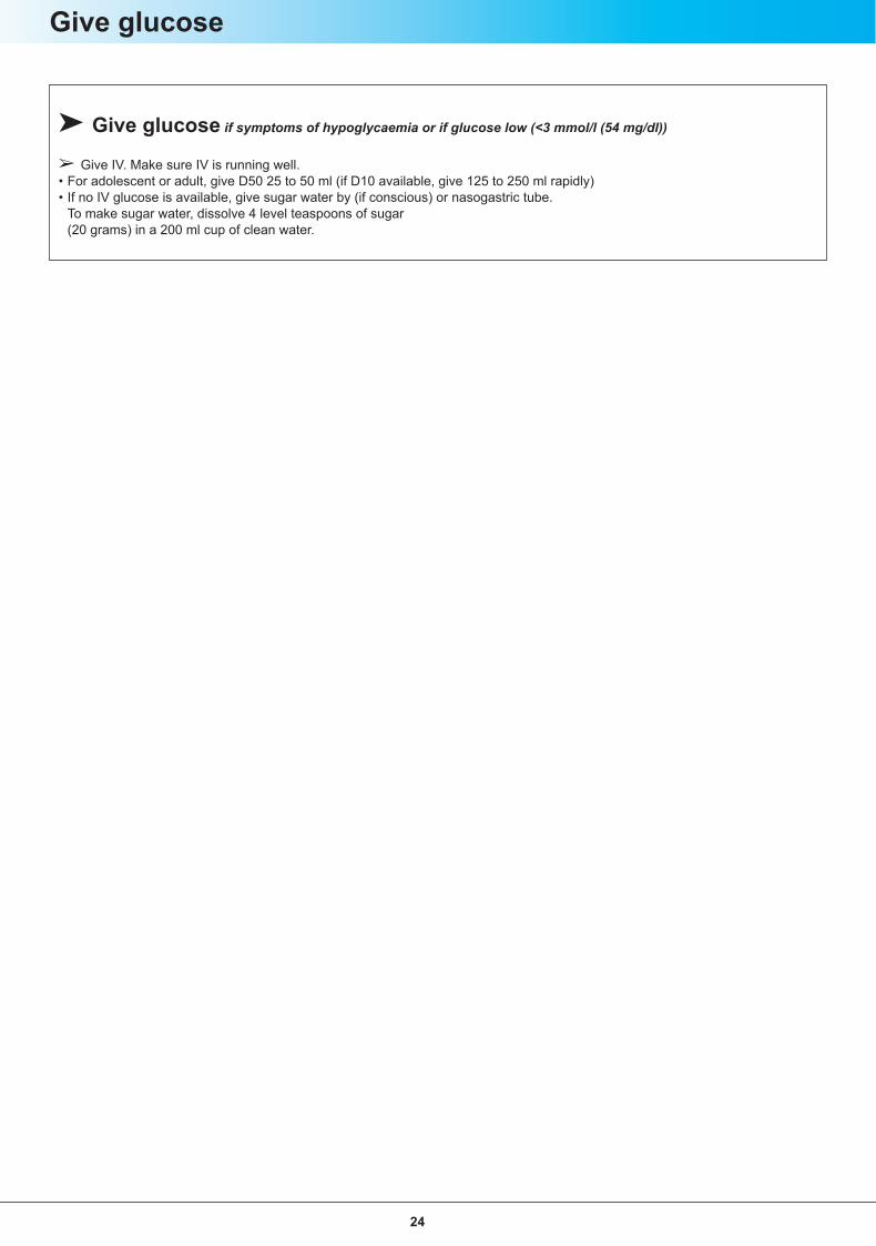
➤ Give glucose if symptoms of hypoglycaemia or if glucose low (<3 mmol/l (54 mg/dl))
➢ Give IV. Make sure IV is running well.• For adolescent or adult, give D50 25 to 50 ml (if D10 available, give 125 to 250 ml rapidly)• If no IV glucose is available, give sugar water by (if conscious) or nasogastric tube. To make sugar water, dissolve 4 level teaspoons of sugar
(20 grams) in a 200 ml cup of clean water.
Give glucose

25
Give pressors
Patients in shock may need pressor drugs to support the circulation while other treatments (fluid resuscitation, antimicrobials etc) take effect. Pressor drugs work by causing vasoconstriction, inotropy, or a mixture of both. Vasoconstrictors narrow the blood vessels, and can therefore mask or disguise inadequate fluid resuscitation; inotropic drugs increase the force of contraction of the heart, and can cause dysrrhythmias. Both can cause an increase in blood lactate. Pressor drugs must be given diluted by infusion at a strictly controlled rate, with continuous monitoring of effect, and preferably with regular electrocardiographic monitoring (ECG). If the infusion is being administered through a peripheral vein, do not measure the blood pressure using a cuff applied to the same arm. Inspect the infusion site regularly to avoid extravasation from the vein which would cause tissue necrosis.
Discontinue the infusion immediately if there is skin blanching,or if the drip has tissued. If the patient develops an irregular pulse, reduce or discontinue the infusion and seek senior help.
The infusion rate must be strictly controlled; this is more safely achieved using an additional metal gate-clamp rather than the integral roller device which can become loose.
When preparing these drugs for infusion, you must ensure that all steps have been checked by a colleague, to ensure that the concentration and infusion rates are correct. Frequent monitoring is required. They are intended for short-term use only, to allow other supportive treatments to take effect.
Epinephrine (adrenaline) is an example of a vasoconstrictor-inotrope, while nor-adrenaline (nor-epinephrine) is predominantly a vasoconstrictor. The infusion rate depends on the dilution and the response. Both drugs may be prepared for infusion as 1mg diluted to a convenient concentration in normal saline or 5% dextrose. (1 mg = 1000 micrograms, = 1 ml of 1:1000, or 10ml of 1:10,000). The volume of fluid chosen for dilution will affect the infusion rate.
If administered by infusion through a peripheral vein, make up 1mg of adrenaline or nor-adrenaline in 500 ml of normal saline or 5% dextrose. This will provide a dilution of 2 micrograms per ml which can be administered at an initial rate of 1 ml per minute with monitoring of heart rate every minute and blood pressure every 2-5 minutes.
An alternative method for infusion for hospitals able to provide advanced care is via a central venous catheter and a 50 ml syringe in a pump (syringe driver). In this circumstance, 1 mg of epinephrine (adrenaline) or nor-adrenaline in 50 ml of normal saline or 5% dextrose will provide a dilution of 20 micrograms per ml; the initial infusion rate may be set at 3 ml per hour (60 micrograms/hour; or 1 microgram per minute), and adjusted according to effect.
Vasopressor Epinephrine (adrenaline) DopamineCommonly Available Concentrations
1 mg in 1 ml ampule * 200 mg in 5 mL ampule *
Target Infusion Concentration
10 micrograms (mcg) per ml ** 1000 micrograms (mcg) per ml**
Mixing procedure to create target infusion concentration
MIX: 2 mg epinephrine in 200 mL of normal saline
OR
MIX: 10 mg epinephrine in 1000 mL of normal saline
MIX: 200 mg dopamine in 200 mL of normal saline
OR
MIX: 1000 mg dopamine in 1000 mL of normal saline
Epinephrine DopamineDose rate 0.05 0.2 for refractory
hypotension10 15 20
Infusion rate (mL/hr)****Patient Weight(kg)
50 15 ml/hour 60 ml/hour 30 ml/hour 45 ml/hour 60 ml/hour60 18 ml/hour 70 ml/hour 35 ml/hour 50 ml/hour 70 ml/hour70 20 ml/hour 80 ml/hour 40 ml/hour 60 ml/hour 80 ml/hour
*1 milligram (mg) is equal to 1000 micrograms (mcg)**Read ampule label 3 times to confirm concentration before mixing. *** Desired dose rate is weight based ****Infusion rate is commonly presented per hourInfusion Rate = Desired Dose Rate / Concentration of the Infusion

26
6. General principles of care for the patient with severe, acute medical illness
A. Rapid assessment and immediate management Patients with severe acute illnesses require rapid assessment and immediate interventions to correct the abnormalities that are identified. In emergency situations, simultaneous assessment and treatment are required and must be directed at reversing any life-threatening conditions. The Quick Check should be applied immediately at the time of presentation to the facility and also for patients who deteriorate after admission. The ABC section of the Quick Check – assessment of airway, breathing, circulation and coma/convulsions – should be used repeatedly in monitoring severely ill patients. The nature and scope of the approaches to be applied in a given setting must be determined in the context of resource availability and staff competencies. It is crucial that referral systems be in place to manage patients with severe or complex illnesses that are beyond the capability of the lower level facility.
Management of the patient with severe illness should focus on providing immediate supportive measures and correcting the underlying physiological abnormalities. For example:
• The symptom of shortness of breath or the observation of rapid breathing should prompt an assessment of the patient’s airway, administration of supplemental oxygen, listening to the chest for wheezing and administration of salbutamol as required, and an assessment for fluid overload.
• A fast heart rate or low blood pressure should prompt securing intravenous access, administration of a bolus of intravenous fluid and assessment of causes that may be immediately reversible, such as anaphylaxis, bleeding or sepsis.
These supportive interventions are aimed at “buying time” and preventing organ damage, but ultimately an assessment and treatment of the underlying cause is required, for example, antibiotics for septic shock, pneumonia or meningitis, while results from ongoing assessment and other tests are pending.
B. Monitoring and evaluating responses to interventions Close supervision of critically ill patients is vitally important. This process should proceed iteratively; for example, immediately after delivering a bolus of IV fluid, check to see if the blood pressure has risen and the heart rate fallen. Failure to respond or only a transient response should prompt a check of whether there is an equipment problem (e.g. IV line extravasation or blockage), reassessment of the diagnosis (e.g. ongoing bleeding, cardiogenic shock). If there is no alternative reason for the failure to respond, another fluid bolus should be administered and the response assessed. If there has been no improvement assistance should be sought.
Similarly, administration of supplemental oxygen to a breathless and hypoxaemic patient should result in an improvement in symptoms and/or an increase in oxygen saturation. Failure to correct hypoxaemia with oxygen should prompt a check of technical factors (e.g. is the oxygen supply working?) and possible alternative diagnoses (e.g. severe asthma). If fluid overload has been treated with intravenous furosemide, there should be an improvement in shortness of breath and respiratory rate within an hour, associated with increased urine output.
Examples of monitoring forms for use with critically ill patients are shown in Section 7.
Once supportive care has been established successfully, ongoing monitoring of the critical variables is important to detect deterioration. To facilitate monitoring severely ill patients should be cared for in a common area close to the nursing station: vital signs should be

27
measured frequently (hourly or even more frequently depending on acuity), and specific criteria should be established for actions to be taken if the patient deteriorates. Frequent checking of drugs and equipment may also be required, particularly repeat doses of antibiotics, oxygen supply from cylinders, and IV fluid flow rates.
C. Managing oxygen therapy Starting oxygen therapy: Oxygen should be started immediately for all patients with emergency signs of severe respiratory distress (listed in the Quick Check) or SpO2<90% by pulse oximetry. Because of the inherent delay of 20–30 seconds in obtaining a reliable reading (waveform) on the pulse oximeter, initiation of oxygen should not be delayed until a reading is obtained.
All patients who are receiving oxygen should be observed for their response, and the oxygen flow should be adjusted in accordance with the response. Most patients will respond to oxygen with improvement in respiratory distress and/or SpO2 within a few minutes. Oxygen is a scarce resource in some countries, thus, is should not be administered unnecessarily. Clinical reassessment and measurement of the SpO2, if possible, should be used to determine if the oxygen flow should be increased or decreased.
Continuing or escalating treatment: The oxygen flow should be increased if the patient continues to have severe respiratory distress or the SpO2 remains <90%. Do not give oxygen via nasal prongs or cannula at a flow rate higher than 5 litres/minute. In patients who are obtunded, placement of an oral or nasal airway can keep the upper airway open so that oxygen can be delivered.
Oxygen flow rates >5L/minute should be provided by a facemask. High flow oxygen delivered by a facemask with a reservoir will provide the maximal oxygen flow. A facemask with a reservoir should only be used if the flow of oxygen is high enough to keep the reservoir bag full, or it may be dangerous for the patient. If, despite increasing the flow of oxygen, the patient still has signs of hypoxaemia, the oxygen saturation falls, or the respiratory rate increases, make sure the oxygen supply and equipment are working properly:
• Check that the cylinder still has sufficient oxygen. • Check that oxygen is flowing out of the prongs or face mask (hold the end close to your
hand and you will feel the airflow). • Check that there are no leaks in the connections or oxygen tubing.
If there are no problems with the set-up for administration, alternative diagnoses, such as heart failure, airways obstruction or pleural effusion to name a few, should be sought. Some conditions, such as bacterial pneumonia, causing hypoxia are slow to respond. It is important to remember that giving oxygen will not relieve airway obstruction or improve a patient who is not adequately ventilating. These problems must be emergently addressed in addition to giving oxygen.
All seriously ill patients, including those already receiving oxygen therapy, should be monitored frequently for signs or symptoms of severe respiratory distress or hypoxaemia. The frequency will depend on how ill the patient is, whether they are deteriorating or improving, stable, or in the recovery phase. All stable patients receiving oxygen should be assessed at least twice daily to determine if oxygen is still required. As a patient improves, less oxygen will be required. It is very important to observe the patient when decreasing or removing the oxygen. Each time the oxygen flow is changed , the response must be assessed at least hourly for four hours to ensure they are stable. Monitoring should include consciousness, respiratory rate, and observation for cyanosis or use of oxygen saturation monitoring.
28
D. Nursing care for severely ill patients Nursing care is a critically important component of the management of severely ill patients. Some specific nursing tasks are listed below.
• Observe the patient frequently, with immediate response and rapid alert and notification to the district clinician when clinical changes occur.
• Record observations, procedures performed, procedures planned and changes in condition in patient’s chart.
• Administer preventive and therapeutic treatments and perform actions prescribed by the district clinician that fall within the nurse's competence.
• Check the IV cannula each day and replace if local signs of inflammation/infection. Remove IV when no longer required for fluid management. Change to oral antibiotics and fluids as soon as possible.
• Integrate infection control into treatment planning and delivery of care • Assess pain and give analgesia as indicated. • Monitor temperature and provide antipyretics as indicated. • Minimize the potential for pressure sores by changing the patient’s position at
regular intervals. • Give special care for the mouth, nose, and eyes. Treat irritated or dry mucous
membranes, pressure sores behind ears or on the side of the nose, and skin irritation/breakdown from mask or nasal prongs.
• Ensure continuity of care: keep patient’s chart current and coordinate care with other clinical team members, other shifts, and other professionals.
• Inform patient and family members about the care planning, how the unit operates, and what behaviour and support is expected.
• Maintain patient comfort, in so far as possible, by positioning, providing pillows and blankets, assist patients with personal hygiene.
• Maintain respect for the patient’s wishes, privacy and basic needs.
E. Ongoing clinical evaluation of severely ill patients This section outlines an approach to the diagnosis and differential diagnosis of presenting conditions in the severely ill patient, after the initial emergency assessment and management in the Quick Check. In an emergency situation, simultaneous assessment and treatment are required and need to be directed at reversing any life-threatening conditions. After initial assessment and stabilization, using the Quick Check, has been completed, the district clinician needs to do another assessment and make additional treatment decisions.
Further clinical decision-making starts with a list of possible diseases that may account for the symptoms and signs (the differential diagnosis). Remember that patients may present with more than one symptom. It is also important to remember that severely ill patients often have more than one disease process. Diagnosis and management in severely ill patients is often more difficult, and it is important to be systematic in your approach. Other factors, including environmental exposures, travel history, socioeconomic status, vaccination, other chronic diseases and local patterns of disease, all have an impact on the differential diagnosis. In particular, the immunological status and use of antiretroviral therapies in people living with HIV changes the differential diagnosis considerably. The list should initially be broad; additional evidence may support or eliminate possibilities from the list. It should be based on the most likely diagnoses, but should include less likely, but more serious diseases. For severely ill patients, it is particularly important to consider first the diagnosis of potential life-threatening diseases that could kill a patient rapidly, and initiate treatments and investigations appropriately.

29
Additional information such as changes in symptoms and physical examination findings over time; response to initial emergency treatments; results of investigations; knowledge of other cases of disease; and the opinion of other health workers; can help rule a diagnosis in or make one less likely. Investigations (and initial treatment in a seriously ill patient) should be directed towards the most serious, treatable disease. It should be noted that few investigations are completely accurate; they may not always be positive when a disease is present (not completely sensitive), or not always reflective of disease when positive (not completely specific).
F. Involving families in caring for severely ill patients In some hospitals with limited staff and where families are accustomed to caring for their loved ones while in hospital, families can be trained to carry out simple care and monitoring tasks. This may include feeding, cleaning and washing the patient, and purchasing medications. In some cases, patient attendants may be trained to notify staff where there has been a change in clinical status or when intravenous fluids bags are empty, and in more advanced tasks such as manual ventilation. An important responsibility of the clinical staff is to provide the family with up-to-date information on the patient’s condition, management plans, and prognosis in so far as it is known.
G. Limiting therapy and palliative care Many patients with critical illness will die, and it is an essential professional duty to maintain their comfort and dignity and support the family through this period. It may become evident that treatments are futile, and it may be appropriate to discontinue active treatments and concentrate on relieving discomfort. Where possible, this decision should be made by an experienced, senior clinician after discussion with the family.
30
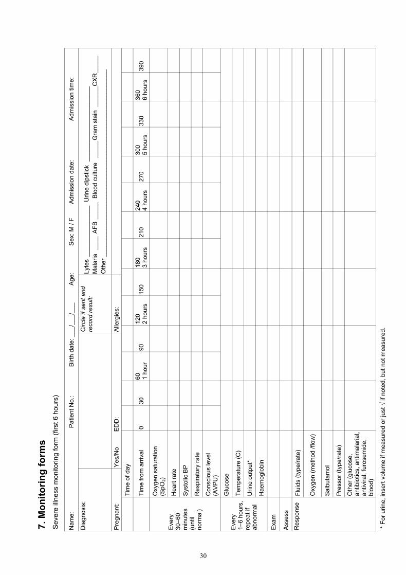
7. M
onito
ring
form
s S
ever
e illn
ess
mon
itorin
g fo
rm (f
irst 6
hou
rs)
* Fo
r urin
e, in
sert
volu
me
if m
easu
red
or ju
st √
if n
oted
, but
not
mea
sure
d.
Nam
e:
P
atie
nt N
o.:
B
irth
date
: ___
/___
/___
A
ge:
Sex
: M /
F
A
dmis
sion
dat
e:
Adm
issi
on ti
me:
Dia
gnos
is:
C
ircle
if s
ent a
nd
reco
rd re
sult:
Ly
tes
____
____
____
___
Urin
e di
pstic
k __
____
____
____
____
____
M
alar
ia _
___
AFB
____
_ B
lood
cul
ture
__
___G
ram
sta
in _
____
_CXR
____
_
Oth
er _
____
____
____
____
____
____
____
____
____
____
____
____
____
__
Pre
gnan
t: Y
es/N
o
E
DD
: A
llerg
ies:
Ti
me
of d
ay
Ti
me
from
arri
val
0 30
60
1
hour
90
12
0 2
hour
s 15
0 18
0 3
hour
s 21
0 24
0 4
hour
s 27
0 30
0 5
hour
s 33
0 36
0 6
hour
s 39
0
Ever
y 30
–60
min
utes
(u
ntil
norm
al)
Oxy
gen
satu
ratio
n(S
pO2)
Hea
rt ra
te
Sys
tolic
BP
Res
pira
tory
rate
Con
scio
us le
vel
(AV
PU
)
Ever
y 1–
6 ho
urs,
re
peat
if
abno
rmal
Glu
cose
Tem
pera
ture
(C)
Urin
e ou
tput
*
Hae
mog
lobi
n
Exa
m
Ass
ess
Res
pons
e Fl
uids
(typ
e/ra
te)
Oxy
gen
(met
hod
/flow
)
Salb
utam
ol
Pre
ssor
(typ
e/ra
te)
Oth
er (g
luco
se,
antib
iotic
s, a
ntim
alar
ial,
antiv
iral,
furo
sem
ide,
bl
ood)

31
AVP
U –
con
scio
us le
vel –
A–A
lert
V–R
espo
nds
to V
oice
P–R
espo
nds
to P
ain
U–U
nres
pons
ive
Add
ition
al n
otes
(ple
ase
note
any
cha
nges
from
sta
ndar
d pr
otoc
ol).
BEN
CH
MA
RK
S- c
heck
if a
chie
ved
If se
vere
resp
irato
ry d
istr
ess,
with
in 3
0 m
inut
es:
Oxy
gen
star
ted
Oxy
gen
satu
ratio
n m
easu
red
IV s
tarte
d A
ntib
iotic
s If
whe
ezin
g, s
albu
tam
ol g
iven
If
mal
ario
us a
rea,
ant
imal
aria
l A
ppro
pria
te in
fect
ion
cont
rol
If sh
ock,
with
in 3
0 m
inut
es
IV li
ne a
nd ra
pid
fluid
s st
arte
d 1
litre
flui
d bo
lus
Ant
ibio
tics
and
antim
alar
ials
(if a
ppro
pria
te)
If sh
ock,
with
in fi
rst 2
hou
rs
3 lit
res
appr
opria
te IV
flui
ds w
ithin
2 h
ours
of a
rriv
al
Dis
trict
clin
icia
n co
ntin
ues
initi
al re
susc
itatio
n

32
Seve
re il
lnes
s m
onito
ring
form
: hou
rs 7
–24
*For
urin
e ou
tput
, ins
ert v
olum
e if
mea
sure
d or
just
√ if
not
ed, b
ut n
ot m
easu
red.
Nam
e:
Pat
ient
No.
:
Birt
h da
te: _
__/_
__/_
__
Age
:
S
ex: M
/ F
Adm
issi
on d
ate:
A
dmis
sion
tim
e:
Dia
gnos
is:
C
ircle
if s
ent
and
reco
rd
resu
lt:
Lyte
s__
____
____
____
_ U
rine
dips
tick
____
____
____
____
____
____
__
Mal
aria
__
____
AFB
____
_ B
lood
cul
ture
____
__ G
ram
sta
in__
____
CXR
____
____
O
ther
___
____
____
____
____
____
____
____
____
____
____
____
____
____
____
____
____
Pr
egna
nt:
Yes
/No
ED
D:
Alle
rgie
s:
Tim
e of
day
Ti
me
from
arr
ival
7 hrs
8 hrs
9 hrs
10
hrs
11
hrs
12
hrs
13
hrs
14
hrs
15
hrs
16
hrs
17
hrs
18
hrs
19
hrs
20
hrs
21
hrs
22
hrs
23
hrs
24
hrs
Ever
y 1
hour
if
SB
P<9
0 or
if
on p
ress
ors
othe
rwis
e Q
2
hour
s
Oxy
gen
tatu
ratio
n(S
pO2)
Hea
rt ra
te
Sys
tolic
BP
Res
pira
tory
rate
Con
scio
us le
vel
(AV
PU
)
Ever
y 6
hour
s U
rine
outp
ut*
Te
mpe
ratu
re (C
)
Rep
eat i
f ini
-tia
l val
ue a
b-no
rmal
Glu
cose
Hae
mog
lobi
n
Exam
Ass
ess
Res
pons
e
Flui
ds (t
ype/
rate
)
Oxy
gen
(met
hod/
flo
w)
Salb
utam
ol
P
ress
or (t
ype/
rate
)
Oth
er (g
luco
se,
antib
iotic
s, a
nti-
mal
aria
l, an
tivira
l, fu
rose
mid
e,
bloo
d)

33
Add
ition
al n
otes
(ple
ase
note
any
cha
nges
from
sta
ndar
d pr
otoc
ol)

34
Annex: Background/scientific basis A novel influenza A (H1N1) virus emerged in April 2009 in Mexico and the United States, quickly spreading throughout the world and prompting the World Health Organization to declare the existence of the first influenza pandemic since 1968. To date, although the overwhelming majority of infected persons have experienced only mild, self-limited disease, and have recovered uneventfully, a small proportion have progressed to acute lung injury (ALI), acute respiratory distress syndrome (ARDS), and septic shock and multi-organ dysfunction. A significant proportion of these severely ill patients present rapidly and without pre-existing comorbidities or other indicators of their risk for progressive disease. Even a small increase in the number of such cases can threaten to overload health care systems already straining to accommodate increased case loads because of the pandemic or other co-existent health crises. The problem is most acute in resource-limited areas, where access to care is frequently delayed and trained personnel, equipment, diagnostic tests, and therapies are often in short supply. Current evidence-based medical practice supports the use of defined clinical care pathways or "bundles" to improve patient care systematically. Implementation of Surviving Sepsis Campaign treatment "bundles" has been associated in prospective randomized studies with over 50% reductions in mortality,1 while implementation of pneumonia care protocols in adults has reduced mortality by 30%.2 Although these data are derived from developed-world settings, it has also been demonstrated in limited-resource settings that childhood pneumonia mortality can be substantially reduced by using simple protocols. In the context of a global pandemic, efforts to standardize care around pragmatic, evidenced-based (or evidence-informed) interventions become even more urgent. Since March 2009 (i.e. before the beginning of the pandemic), working groups at the World Health Organization have been reviewing the current evidence based guidelines for the management of septic shock in limited resource settings. These guidelines have been incorporated into the IMAI (Integrated Management of Adolescent and Adult Illness) guidelines for district clinicians. In meetings in April, June and September, a WHO Working Group on Critical Care in Limited-Resource Settings, co-hosted by the GAR and IMAI programmes, worked on a combined or unified approach for the management of patients with septic shock and those with severe respiratory distress related to acute lung injury or severe pneumonia without shock. These guidelines define the critical physiological principles that guide their application and develop modified pathways that are less dependent on invasive monitoring and advanced technology that remain unavailable in much of the developing world. As part of this process, WHO solicited the input from clinical experts in infectious disease, critical care, emergency medicine, tropical disease, and other disciplines, from both developed and developing world settings, and representing a variety of governmental, academic, and nongovernmental (NGO) groups interested in improving the clinical care of severely ill patients. Although the urgency of this collaboration was heightened by the outbreak of pandemic (H1N1) 2009 influenza, this work was from the outset driven by the realization that its work had relevance across the spectrum of acute infectious illness. Likewise, while the identified need for such a collaboration was most apparent in outbreak or resource-limited environments, it also soon became clear that even resource-rich areas could experience shortfalls in staffing, equipment, or preparedness; in such cases, treatment pathways designed to require a minimum of advanced training and technology could prove highly useful.
1 Gao et al. The impact of compliance with 6-hour and 24-hour sepsis bundles on hospital mortality in patients with severe sepsis: a prospective observational study. Critical Care 2005, 9:R764-R770. 2 McCabe et al. Guideline-Concordant Therapy and Reduced Mortality and Length of Stay in Adults With Community-Acquired Pneumonia. Arch Intern Med. 2009;169(16):1525-1531.

35
Review of the treatment of acute lung injury and septic shock: As noted above, two of the most common presentations of severely ill patients with pandemic (H1N1) 2009 influenza disease are pneumonitis-induced acute lung injury and septic shock. The treatment of such patients is complex because of the need to reconcile two competing concerns. On the one hand, it is well established in the treatment of septic shock that failure to administer fluids and antimicrobials early (within hours) is associated with increased risk of organ failure and mortality.3 On the other, the FACTT trial clearly linked an "aggressive" fluid strategy with increased ventilator dependency and worse outcome in patients with acute lung injury.4 Given that many pandemic (H1N1) 2009 influenza patients with progressive disease may present with both severe sepsis/septic shock and evidence of acute lung injury, how can these two management philosophies be reconciled? The problem is compounded by the nonspecific presentation of many patients (e.g. the patient in septic shock with "occult pneumonia"), and is likewise intensified by the diagnostic challenges common in developing countries, where a patient with fever and cough might have influenza, malaria, typhoid, community acquired pneumonia, PCP pneumonia complicating HIV immunosuppression, or any of several other diagnoses.
Description of the clinical pathways: After intensive review of existing data and discussion about their applicability to outbreak and resource-limited settings, including environments where pandemic (H1N1) 2009 influenza was common, clinical pathways for septic shock and for severe respiratory distress without shock were developed (see Section 4). These guidelines highlight several important clinical observations and principles, which are useful in both the pandemic setting but also in the care of any patient presenting with an acute, presumably infectious cause of respiratory distress with or without hemodynamic compromise:
ο The key to improved outcomes is early recognition and early response on the part of the treating team. Failure to address unstable physiology in a timely fashion leads to decompensation, and loss of efficacy of future interventions.
ο Clinical observation and repeated assessment are of primary importance. ο The best outcomes in the treatment of severe sepsis are associated with timely and
appropriate reversal of tissue hypoperfusion, whether through intravenous fluids and (when necessary and available) vasopressor agents.
ο The best outcomes in the treatment of acute lung injury and ARDS are associated with avoiding over-hydration and "excessive" fluid administration. In this regard, it is pertinent to remember that the FACTT trial, demonstrating worse outcome among patients with ARDS who received aggressive fluid rehydration, excluded patients who were already in septic shock. Thus, the goal of treating patients presenting with evidence of acute lung injury and hemodynamic instability should be the early restoration of adequate tissue perfusion, followed by a cautious fluid replacement strategy with attentive regard to signs and symptoms of fluid overload (including pulmonary oedema).
Notable features of the clinical pathways include: ο The use of standardized intravenous fluid rates, tailored to perceived physiological
state during the course of resuscitation. ο The deliberate minimization of advanced monitoring technology, in order to improve
the pathways usefulness in limited-resource settings. ο The applicability of the pathway to any patient presenting with the syndrome of
presumed infection and acute lung injury, with or without hemodynamic compromise; a specific diagnosis is not necessary before instituting therapy.
3 Kumar A et al. Duration of hypotension before initiation of effective antimicrobial therapy is the critical determinant of survival in human septic shock. Critical Care Med 2006 Jun;34(6):1589–96. 4 Wiedemann HP et al. Comparison of two fluid-management strategies in acute lung injury. N Engl J Med. 2006 Jun 15;354(24):2564–75.

36
ο The transition between acute resuscitation of the patient and ongoing "maintenance care" post-resuscitation.
ο The standardization of physical examination and re-examination at specified intervals, to improve patient safety and simplify implementation of the pathways.
ο Empirical antimicrobials for both pandemic (H1N1) 2009 influenza pneumonia and community-acquired bacterial pneumonia (and malaria if in regions endemic for P. falciparum).
Conclusion: The acute lung injury and septic shock pathways for limited- resource settings are an attempt to synthesize, based on available evidence and expert clinician input, a practical and simple care approach for these severely ill patients. These are intended to be useful in the resource-limited setting during the H1N1 pandemic but also in non-pandemic, non-crisis environments for the management of patients with severe illness of other cause. They are designed to accentuate key physiological principles and targeted interventions in settings with minimal technology or human resources. They use available data from prospective, randomized trials to develop an "integrated approach" to care for severely ill patients. Importantly, they are written for an audience of physicians as well as senior nurses and other health care personnel who may be in charge of clinical care of severely ill patients. As more data are generated about the clinical course of H1N1 disease and other syndromes, the guidelines will be revised and improved. Process:
The WHO departments of HIV/AIDS (IMAI team), GAR, and GIP formed groups of experts who have been combined to form a WHO Working Group on Critical Care in Limited-Resource Settings to develop emergency guidance on management of septic shock and severe respiratory distress (and other severe illnesses). Expert meetings were held in March (Geneva, Switzerland), April (Addis Ababa, Ethiopia), June (Geneva) and September (Florence, Italy) 2009. This guideline group was composed of highly qualified professionals who have expertise and experience in the areas of acute lung injury, acute respiratory distress syndrome, sepsis and septic shock, pneumonia, influenza, and the treatment of critically ill patients in general. The recommendations have been developed to provide guidance to countries experiencing outbreaks of febrile disease causing critical illness (including, but not limited to, pandemic influenza), where local resources are not able to provide mechanical ventilation for medical patients and the full spectrum of "ICU-level care." This is a specific extract of those guidelines released for management of severe complications of influenza H1N1.
The guidance was developed based on existing evidence and expert advice; specifically, the sections on septic shock are based on the Surviving Sepsis Campaign consensus recommendations (based on a published GRADE systematic review) and the acute respiratory distress syndrome guidance is based on data from the ARDSnet multicountry trial (previously referenced in WHO guidance on the management of H5N1 influenza). Some management recommendations are based on Clinical management of human infection with pandemic (H1N1) 2009: revised guidance issued by WHO November 2009 and accessible at http://www.who.int/csr/resources/publications/swineflu/clinical_management/en/index.html
The oseltamivir recommendations are based on the published WHO Guidelines for Pharmacological Management of Pandemic Influenza A (H1N1) 2009 and other Influenza Viruses Revised February 2010. http://www.who.int/csr/resources/publications/swineflu/h1n1_guidelines_pharmaceutical_mngt.pdf
37
For the other recommendations, such as use of oral fluids and methods of administration of oxygen, reviews of high quality and highly relevant evidence were presented at the meetings by experts assigned to each topic. The final recommendations for limited-resource settings were developed through consideration and discussion of expert opinion at these meetings. The recommendations were reviewed and finalized at the meeting on 2–3 September 2009 in Florence, Italy. Subsequent editorial changes, which have not materially changed the prior recommendations, have been made via e-mail discussion. WHO Working Group on Critical Care in Limited-Resource Settings includes: Hillary Cohen Emergency Department, Maimonides Medical Center, New York, NY, USA Allen Cheng Monash University and Infectious Diseases Unit, Menzies, Australia Alain Vuylsteke Anaesthesia & Intensive Care, Cambridge University Teaching Hospital, UK John Kennedy New South Wales Medical Retrieval Services, Australia Walter Kloeck University of Witwatersrand, South Africa Mark Blaylock Ghana Gene Bukham Partners In Health, Rwanda Elizabeth Molyneux Queen Elizabeth Hospital, Blantyre, Malawi Eric Simoes Paediatrics, University of Colorado, Denver, CO, USA Jeremy Farrar Oxford University Clinical Research Unit, Ho Chi Minh City, Viet Name Julian Bion University Dept Anaesthesia & Intensive Care Medicine, Birmingham, UK Satish Bhagwanjee Anaesthesiology, University of Witwatersrand, South Africa Phil Hopewell SFGH/ University of California, San Francisco, CA, USA Stephen Gordon, Jamie Rylance Liverpool School of Tropical Medicine, UK Len Hudson, Eoin West, Pulmonary and CCM University of Washington, Seattle, Washington, USA Shevin Jacob Christopher Moore Infectious Diseases & International Health, University of Virginia,
Charlottesville, VA, USA Neill Adhikari Critical Care Medicine, Sunnybrook Health Sciences Centre, Toronto,
Canada Natalie Van Meerbeeck MSF Belgium Patrick Banura Masaka Regional Hospital, Uganda WHO: Sandy Gove,1 Matthew Lim,2 Cathy Roth,2 Wilson Were,3 WHO consultant: Ed Zuroweste4
1 HIV/AIDS Department; 2 Global Alert and Response; 3 Child and Adolescent Health; 4 Global Influenza Programme. Declarations of interest were received from all participants. Three people declared an interest, one of which was not relevant to the meeting. Drs Moore and Jacob both had conflicts of interest related to the meeting; these are disclosed below. Drs Moore and Jacob received funding from Pfizer; these were grants to their institutions for a research training exchange programme for students and fellows and for a study of fluid resuscitation, PRISM-U2. These recommendations have been produced under emergency procedures and will remain valid until 31 December 2010.

Cl in ical management of adul t pat ients with compl icat ions of pandemic inf luenza A (H1N1) 2009 inf luenza:
Emergency guidel ines for the management of pat ients with severe respiratory d istress and shock in d istr ict hospi ta ls in l imited-resource set t ings
Integrated Management of Adolescent and Adult Illness (IMAI)
Global Influenza Programme (GIP)
Global Alert and Response (GAR)
For further information please contact:IMAI Team Department of HIV/AIDS World Health Organization Avenue Appia, 20CH–1211 Geneva 27Switzerland
[email protected]/hiv/capacity/en

Table of Contents1. Introduction 3
2. Preparing district hospitals and clinicians to manage severe complications
of pandemic influenza A (H1N1) in limited-resource settings 3
3. Quick check and emergency treatments 5
4. Summary of management of septic shock and severe respiratory distress 10
First 2 hours 11
2-6 hours 12
6-24 hours 13
Post resuscitation 14
5. Treatment instructions 15
Manage airway 16
Give oxygen 17
Give antimicrobials: antivirals, antibiotics (and antimalarials if appropriate)1 21
Treat wheezing 23
Give glucose 24
Give pressors 25
6. General principles in caring for the severely ill patient 26
7. Monitoring forms for severely ill patients:
First 6 hours 30
7- 24 hours 32
Annex: Background/scientific basis 34
Process of development 36
1 These are generic guidelines for country adaptation.
WHO Library Cataloguing-in-Publication Data:
Clinical management of adult patients with complications of pandemic influenza A (H1N1) 2009 influenza: emergency guidelines for the management of patients with severe respiratory distress and shock in district hospitals in limited- resource settings.
1.Influenza, Human - complications. 2. Influenza A Virus, H1N1 subtype - complications. 3.Respiratory insufficiency - therapy. 4.Emergency medical services. 5.Adult. 6.Guidelines. I.World Health Organization.
ISBN 978 92 4 159961 0 (NLM classification: WC 515)
© World Health Organization 2010All rights reserved. Publications of the World Health Organization can be obtained from WHO Press, World Health Organization, 20 Avenue Appia, 1211 Geneva 27, Switzerland (tel.: +41 22 791 3264; fax: +41 22 791 4857; e-mail: [email protected]). Requests for permission to reproduce or translate WHO publications – whether for sale or for noncommercial distribution – should be addressed to WHO Press, at the above address (fax: +41 22 791 4806; e-mail: [email protected]). The designations employed and the presentation of the material in this publication do not imply the expression of any opinion whatsoever on the part of the World Health Organization concerning the legal status of any country, territory, city or area or of its authorities, or concerning the delimitation of its frontiers or boundaries. Dotted lines on maps represent approximate border lines for which there may not yet be full agreement.
The mention of specific companies or of certain manufacturers’ products does not imply that they are endorsed or recommended by the World Health Organization in preference to others of a similar nature that are not mentioned. Errors and omissions excepted, the names of proprietary products are distinguished by initial capital letters.
All reasonable precautions have been taken by the World Health Organization to verify the information contained in this publication. However, the published material is being distributed without warranty of any kind, either expressed or implied. The responsibility for the interpretation and use of the material lies with the reader. In no event shall the World Health Organization be liable for damages arising from its use.
Printed in Switzerland