clinical implementation of nipt -- technical and ... · (acog) and the royal college of...
TRANSCRIPT

Clin Genet 2016: 89: 523–530Printed in Singapore. All rights reserved
© 2015 John Wiley & Sons A/S.Published by John Wiley & Sons Ltd
CLINICAL GENETICSdoi: 10.1111/cge.12598
Review
Clinical implementation of NIPT – technicaland biological challenges
Brady P., Brison N., Van Den Bogaert K., de Ravel T., Peeters H., Van EschH., Devriendt K., Legius E., Vermeesch J.R. Clinical implementation ofNIPT – technical and biological challenges.Clin Genet 2016: 89: 523–530. © John Wiley & Sons A/S. Published byJohn Wiley & Sons Ltd, 2015
Non-invasive prenatal testing (NIPT) for fetal aneuploidy detection isincreasingly being offered in the clinical setting. Whereas the majority oftests only report fetal trisomies 21, 18 and 13, genome-wide analyses havethe potential to detect other fetal, as well as maternal, aneuploidies. In thisreview, we discuss the technical and clinical advantages and challengesassociated with genome-wide cell-free fetal DNA profiling.
Conflict of interest
JRV is founder and stockholder of Cartagenia.
P. Brady, N. Brison, K. Van DenBogaert, T. de Ravel,H. Peeters, H. Van Esch,K. Devriendt, E. Legius andJ.R. Vermeesch
Centre for Human Genetics, KU Leuven,University Hospital Leuven, Leuven,BelgiumKey words: cell-free DNA, (cfDNA) –cell-free fetal DNA, (cffDNA) – copynumber variation, (CNV) – genomesequencing – mosaicism –non-invasive prenatal testing, (NIPT) –prenatal diagnosis
Corresponding author: Prof. JorisVermeesch, Centre for HumanGenetics, KU Leuven, UniversityHospital Leuven, Leuven 3000,Belgium.Tel.: +32 (0)16 345941/345904;fax: +32 (0)16 346060;e-mail: [email protected]
Received 5 January 2015, revised andaccepted for publication 9 April 2015
During the past 3 years, non-invasive prenatal testing(NIPT) for fetal aneuploidy detection has become aclinical reality. The clinical introduction succeeded themany clinical validation studies which have applied mas-sively parallel sequencing of maternal plasma cell-freeDNA (cfDNA) using either whole-genome sequencing ortargeted-sequencing methods (either chromosome selec-tive or Single nucleotide polymorphism (SNP) based)(1–27). A meta-analysis has been undertaken on recentclinical validation and implementation of NIPT stud-ies (28), which found high sensitivity and specificityfor trisomies 21, 18 and 13: 99% sensitivity and of99.92% specificity for trisomy 21; 96.8% sensitivity and99.85% specificity for trisomy 18; and 92.1% sensitiv-ity and 99.80% specificity for trisomy 13. False-positiverates for trisomy 21, 18 and 13 were 0.08%, 0.15% and0.20%, respectively. A number of clinical studies wereexcluded from this analysis because pregnancy outcomedata were unavailable (29–33), and other studies havebeen published since (19, 26), all report high rates ofsensitivity and specificity.
A number of professional societies including theInternational Society for Prenatal Diagnosis (ISPD), the
American College of Obstetricians and Gynecologists(ACOG) and the Royal College of Obstetricians andGynecologists (RCOG) have issued position statementsprimarily not only on the use of NIPT for trisomy 21detection but also for the other common autosomal aneu-ploidies (trisomy 18 and 13) (34–38). The general opin-ion is that NIPT is a promising new technology whichhas great potential as a screening tool for those pregnantwomen with an increased risk of fetal aneuploidy. How-ever, it is important to stress that NIPT is not a diagnostictest for fetal aneuploidy, and therefore, a positive NIPTresult requires an invasive test to confirm the findings.Furthermore, these societies considered there to be insuf-ficient evidence to support the use of NIPT for screeningin the general population.
Several recent clinical studies have now demonstratedsimilar sensitivity and specificity for aneuploidy detec-tion (primarily trisomy 21 detection) between patientswith prior high or low risks of aneuploidy (16, 21, 23,31, 33, 39). Furthermore, NIPT also resulted in less falsepositives than with conventional methods of aneuploidyrisk assessment. The fetal fraction, i.e. the percentageof cell-free fetal DNA (cffDNA) within the total cfDNA
523

Brady et al.
sample obtained from maternal plasma, has been shownto be similar in high- and low-risk groups (21, 40). How-ever, the positive predictive values are expected to belower in the group of women at low risk compared withthose at increased risk of aneuploidy. The positive predic-tive values for the general population will also be depen-dent upon the specific test characteristics and furtherstudies are warranted before NIPT is routinely offeredto all women.
While the implementation of NIPT was warrantedbecause of the high accuracy for fetal trisomy 21, 18 and13 detection, several studies have now demonstrated thatgenome-wide analysis enables the detection of other fetaland also possibly maternal (mosaic) aneuploidies (27,41–47). Such genome-wide analysis has the potential toimprove overall pregnancy management and reduce tech-nical and biological factors which could adversely affecttest accuracies. In this review, we discuss the clinicaladvantages and challenges associated with genome-widecirculating free fetal DNA profiling.
Beyond common aneuploidy screening
Several groups have demonstrated the feasibility todetect all fetal chromosomal aneuploidies, segmentalimbalances, and even submicroscopic copy number vari-ations (CNVs) by sequencing cffDNA from maternalplasma (4, 6, 26, 24, 27, 41, 43–45, 48–52). Despitethe promise of detecting other fetal aneuploidies whichmight aid in the interpretation of fetal development, clin-ical implementation of genome-wide NIPT analysis inthe routine clinical setting remains limited (26, 49, 50).
In their prospective series of 1982 clinical cases, Lauet al. detected seven cases of full aneuploidy for chro-mosomes other than 21, 18, 13, X or Y (50). Follow-uptesting and pregnancy outcome were provided for fiveof these, and confined placental mosaicism (CPM) wasconfirmed in four of the five cases. Two pregnancies(including the case unconfirmed) were complicated byfetal growth restriction and delivered at 33–34 weeksgestation. In the two cases without any follow-up inves-tigations, ultrasound was normal, the pregnancies werecontinued, and no abnormalities were reported dur-ing birth. Another recent clinical study of 100,000patients acknowledged that these types of events weredetected, but did not give specific numbers or details offollow-up/outcome for individual cases (19).
Table 1 provides an overview of the different aneu-ploidies we have detected from genome-wide NIPT ina series of 4000 pregnancies [updated from 1350 preg-nancies reported in (26)].
The clinical utility of reporting all aneuploidiesdetected using NIPT, particularly in low-risk preg-nancies, is controversial because this could lead to anincrease in invasive procedures in women for whomthis would not normally be considered. Already, thereporting of trisomy 13 following NIPT in the groupof low-risk women has been questioned because of thelow-positive predicted values expected and a possibleincrease in invasive procedures because of false-positive
NIPT results (53). In an important subset of fetal ane-uploidies detected using NIPT, the aneuploidy may beconfined to the placenta (see below), and therefore, maynot adversely affect fetal development. However, thereis the risk of placental insufficiency and fetal growthrestriction because of the abnormal placental karyotype(54–56), as well as a risk of mosaic fetal aneuploidyand/or fetal uniparental disomy (UPD) because of atrisomy rescue event (57).
Similarly, routine screening for sex chromosome ane-uploidies (SCA) may be of questionable clinical util-ity, given the variability in phenotype of, for example,monosomy X (Turners), XXY (Klinefelter), XYY andXXX individuals, with some only identified as adultsdue to fertility problems (58–61). Moreover, the accu-racy of SCA detection is substantially lower for trisomy21 detection (6, 16, 25). It has been suggested that thephysical anomalies, developmental delay and/or infer-tility warrant analysis of SCA (62, 63). National poli-cies relating to screening for SCA differ from countryto country, and how these are subsequently counseledfor and managed also differ. Cultural and societal factorsplay a role in parental decision to continue or to terminatea pregnancy because of a SCA for which the phenotypicoutcome cannot be predicted (64).
We believe that a genome-wide analysis can lead to abetter clinical management. However, careful considera-tion of reporting policy is essential in order to minimizeincreases in invasive tests because of aneuploidies whichare confined to the placenta. We envision that the detec-tion of an aneuploidy would trigger careful follow-up byboth the obstetricians and the clinical geneticists. In thecase of normal development upon US examination, fur-ther invasive actions may not be warranted. In case ofabnormal growth patterns, those can be evaluated in lightof the NIPT results and the US findings. Further studiesare warranted to evaluate the outcomes of such pregnan-cies which will eventually allow the development of rulesfor the best clinical follow-up actions to be taken.
Fetal CNV detection
Currently, invasive prenatal genetic diagnosis using con-ventional karyotyping is increasingly being replaced bychromosomal microarray analysis enabling detection ofsubmicroscopic CNVs. Several prenatal chromosomalmicroarray studies have estimated the residual risk for apathogenic CNV in the absence of any major ultrasoundanomalies at between 0.5% and 1.7% (65–67). Sub-microscopic CNVs can be a cause of severe childhooddevelopmental disorders which may not present anyfeatures upon the routine US examination. The currentmainstream approaches to NIPT focusing only uponthe detection of common autosomal and SCA willnot detect other imbalances present on the remainingchromosomes. The frequency of chromosome abnor-malities, which would be missed if invasive prenatalchromosomal microarray-based analysis would bereplaced by targeted NIPT (68–70), has been estimatedto be 16.9%, including 2% of those pregnancies deemed
524

Clinical implementation of NIPT
Table 1. Different aneuploidies detected during 4000 NIPT
Risk group Follow-up invasive testing
Aneuploidy type Total number observed High risk Low risk Confirmed Not confirmed No follow-up
Trisomy 21 38 22 16 29 9Trisomy 18 9 4 5 6 a 1 (AF) 2Trisomy 13 3 1 2 1 b 2Trisomy 1 1 1 1Trisomy 7 3 2 1 2 (AF) 1Trisomy 8 1 1 1 (AF)Trisomy 9 1 1 1 (miscarriage)Trisomy 10 1 1 1Trisomy 15 1 1 1 c
Trisomy 16 1 1 1 (fetal mosaicism)Trisomy 20 1 1 1Trisomy 22 1 1 1 (AF)Partial trisomy 18 2 2 1 1 (AF)Monosomy 20 1 1 1 (AF)Monosomy 22 1 1 1 (AF)Total (21,18,13) 50 27 (54%) 23 (46%) 36 1 13Total (other chromosomes) 15 4 (27%) 11 (73%) 3 7 5Total 65 31 (48%) 34 (52%) 39 8 18
AF, amniotic fluid; NIPT, non-invasive prenatal testing.High risk is classified as those with an increased risk of aneuploidy from the combined test (<1/300) (21%). Low risk (total 79%)includes those with an increased risk of aneuploidy because of advanced maternal age alone (31%) and those referrals because ofmaternal anxiety (46%) or prior family history (2%).aOne of those six samples was shown to be normal on AF and 77% mosaic on CVS analysis for trisomy 18.bA total of 70% mosaic on CVS analysis.cMosaicism for trisomy 15 and UPD 15.
at high risk of aneuploidy based upon abnormal serumscreening results.
Because of the clinical importance of segmentalaneuploidies, several groups are exploring analyticalmethods to increase the resolution achievable usingNIPT for which proof-of-concept has been provided(27, 71). Some commercial NIPT providers [includ-ing Sequenom (San Diego, CA, USA) and Natera(San Carlos, CA, USA)] have started offering theoption of additional testing for a small number ofmicrodeletion syndromes and other autosomal ane-uploidies. One commercial provider (Natera) hasrecently demonstrated proof-of-principle (24) andsubsequent validation study (71) of their targetedSNP-based NIPT assay to include a small number ofmicrodeletion syndromes (including 1p36, Cri-du-Chat,DiGeorge, Wolf–Hirschhorn, Prader–Willi, Angel-man, Miller–Dieker and Phelan–McDermid). Another(Sequenom) has published validation of genome-widefetal CNV detection (27). However, prospective clinicalresults have not yet been published and studies on testaccuracy, specificity and sensitivity are lacking; hence,their commercial offerings are premature.
Biological sources of false-positiveand false-negative NIPT results
Placental mosaicism
The source of cffDNA has been shown to be placentalin origin (72). It is well documented from conventionalcytogenetic examination of Chorion villus sampling
(CVS) tissue that confined placental mosaicism occursin which the placental tissue contains an abnormal cellline which is not present upon subsequent examinationof amniocentesis or other fetal material. This CPM isobserved in around 1% of invasive tests (73). One wouldtherefore expect the rate of discordant results because ofCPM observed from conventional cytogenetic analysisof CVS and amniocentesis to be similar for NIPT andamniocentesis results. The origins and types of CPMare detailed further below because this is an importantbiological source of ‘false-positive’ NIPT results whichcannot be overcome by technical improvements [we referto Chromosome Abnormalities and Genetic Counselingby Gardner & Sutherland, for more information on CPM(73)].
Mitotic and meiotic CPMMitotic CPM arises from a normal diploid zygote, fol-lowing a post-zygotic error in a placental cell lineage.This generally leads to localized regions of placentaltrisomy and low levels of mosaicism upon cytogeneticinvestigation. In contrast, meiotic CPM arises from atrisomic zygote in which a trisomy rescue event hasoccurred during early fetal development. Generally, thefetus is diploid, and the placenta shows high levels ofmosaicism or full aneuploidy. However, there is a riskof mosaicism in the fetus dependent upon when loss ofthe trisomic chromosome occurs and in which embry-onic cell lineage. Additionally, there is a risk for fetalUPD following a trisomy rescue event dependent upon
525

Brady et al.
which chromosome is lost. The adverse effects of UPDon fetal development may be because of the presence ofimprinted genes or homozygous mutations for the chro-mosome of interest.
Different types of CPM: types I, II and IIICPM is further categorized into one of three typesaccording to the specific cell lineage affected by theabnormal cell line. The abnormal cell line may beconfined to the trophoblast (type I, observed upondirect CVS and short-term culture), the chorionicstroma/mesenchymal core (type II, observed uponlong-term culture), or both these placental lineages(type III). Types I and II are generally of mitotic origin,whereas, type III is primarily meiotic in origin. CPMtype I is associated with an increased risk of spontaneousabortion, intrauterine fetal death, intra uterine growthretardation (IUGR) and perinatal morbidity. CPM typeII is generally associated with a normal pregnancy out-come, with IUGR and pregnancy loss being infrequent.CPM type III is commonly associated with intrauterinefetal death or IUGR, with a large proportion being dueto mosaic trisomy 16 (56, 74–78). Note that cffDNA isderived from trophoblasts, and hence, only types I andIII errors are expected upon NIPT. Large-scale Europeanand US studies of CVS have shown that the chromo-somes involved in different types of CPM observed arenot randomly associated (79–81).
False-positive and false-negative NIPT results becauseof CPM
Unsurprisingly, numerous cases are now reported of‘false-positive’ NIPT results because of CPM (26,39, 50, 82–90), and these findings may be of clinicalrelevance being associated with IUGR for example(50). There is also the possibility of UPD in the fetusfollowing a trisomy rescue event early in embryonicdevelopment. The risk of UPD following prenatal detec-tion of mosaicism for trisomy 15 has been estimated at11–25% (57). We have reported a case in which the fetuswas affected with UPD15 (26). A case of uniparentaldisomy 21 because of trisomic rescue has also beenreported following discordant NIPT and fetal karyotyp-ing results (89). Given the potential for UPD, it may beadvisable to follow up discordant NIPT and invasive testresults with UPD testing, in particular for those chro-mosomes with well characterized imprinted syndromes,including: patUPD(6) – transient neonatal diabetes(TND; OMIM #601410); matUPD(7) – Silver–Russelsyndrome (SRS; OMIM#180860); segmentalpatUPD(11) – Beckwith–Wiedemann syndrome(BWS; OMIM #130650); matUPD(14) – Templesyndrome (TS; OMIM *605636 and #176270);patUPD(14) – paternal UPD(14) syndrome (OMIM#608149); matUPD(15) – Prader–Willi syndrome(PWS; OMIM #176270); and patUPD(15) – Angelmansyndrome (AS; OMIM #105830) (57).
The possibility for CPM means that an amniocentesisis the preferred sampling method for a follow-up con-firmatory invasive test after a positive NIPT result in
order to exclude CPM. As circulating cffDNA is derivedfrom the placental tissue, NIPT results are expected tobe highly concordant with CVS. However, an amniocen-tesis is more reflective of the true fetal genotype. Thisemphasizes that it is essential for an invasive follow-uptest in order to confirm a positive NIPT test. Any deci-sion for termination of pregnancy should not be basedupon positive NIPT results alone.
Multiple pregnancies
It has been demonstrated that NIPT can be used for detec-tion of aneuploidy in twin pregnancies (19, 91–94). Thequantity of cffDNA has been shown to be higher in twinpregnancies compared with singleton pregnancies (95).Zygosity can be determined from maternal plasma DNAsequencing and thus the fetal fraction from each twin canbe estimated in dizygotic pregnancies (96). However, theaccuracy may not be as high as for singleton pregnancies(94).
One possible biological cause of inaccurate NIPTresults may be the presence of a ‘vanishing twin’. Casesof false-positive NIPT results in which the presence of avanishing twin could be confirmed have been reported(25, 26, 32, 50, 97, 98). Furthermore, Curnow et al.estimated the theoretical incidence of a vanishing twinwith a chromosome abnormality to be around 0.11%,which is in line with the false-positive rates reportedin a meta-analysis of NIPT (28). This emphasizes theimportance of detailed ultrasound examination, particu-larly following discordant NIPT results.
Maternal CNVs
NIPT relies upon the analysis of cfDNA derived from thematernal plasma, and the majority of cfDNA is maternalin origin which can complicate the analysis and interpre-tation of NIPT results. The counting statistics of conven-tional Z-score chromosome-wide analysis methods canbe affected by maternal CNVs leading to false-positiveand false-negative NIPT results. The development andclinical application of analysis pipelines which allows fordifferentiation between localized and chromosome-wideevents, including for example maternal CNVs may avoidsuch errors (26). Wang et al. reported 2 of 25 sex chro-mosome anomalies detected by NIPT were determinedto be false positives because of maternal X chromosomeCNVs (99). Another recent study found that maternalCNVs were a major factor contributing to false-positiveand false-negative NIPT results, as well as fetal/placentalmosaicism (39). These findings demonstrate the impor-tance of differentiating between whole chromosome andsub-chromosomal events as well as those which are ofmaternal origin.
Maternal mosaicism
Maternal mosaicism can also be a source of false-positiveresults. Cytogenetic investigations have shown that lossof an X chromosome in blood cells occurs with increas-ing frequency as female age increases (100). This can
526

Clinical implementation of NIPT
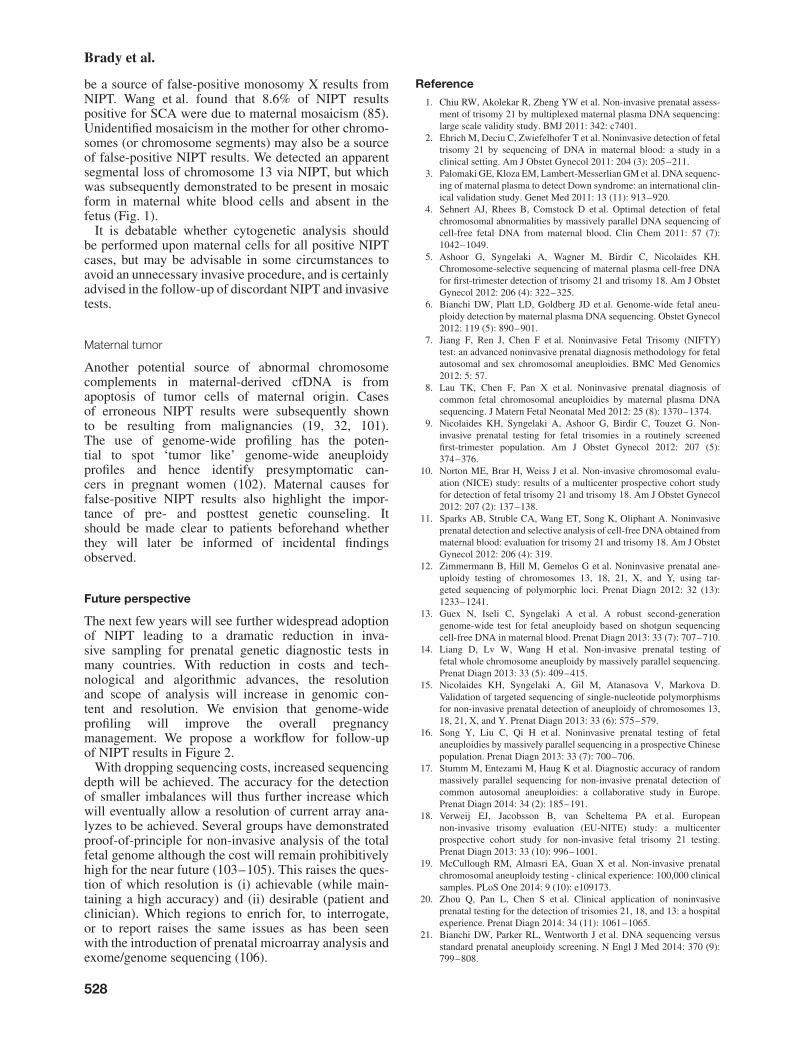
Fig. 1. Mosaic segmental maternal aneuploidy of chromosome 13. (a) Non-invasive prenatal testing (NIPT) for chromosome 13. Using conventionalZ-score analysis, this would be called as a monosomy 13. Genome-wide analysis identifies this as a partial imbalance. The read depth analysis suggeststhis imbalance to be a maternal event. Refer to our previous manuscript for full details of our analysis pipeline. (b) Chromosome 13 array CGH plotderived from DNA extracted from maternal white blood cells. Chromosome 13 is shown at the top, the array result below plotted using genomic locationon the x-axis and signal log ratio on the y-axis, and the deleted region highlighted. The average deviation of signal intensity ratio observed is −0.17,which is consistent with mosaicism at a level of ∼17%. Microarray analysis of DNA from amniotic fluid revealed the fetus to be normal (data notshown). (c) Shows a metaphase fluorescence in situ hybridization (FISH) result using probes for 13q telomere in red [TelVysion 13q (Vysis)] and13q14 in green [LSI 13q14 (Vysis)] showing an abnormal cell which confirms the presence of the deletion in the mother. This deletion was observed in22% of cells analyzed (66/300 cells) confirming mosaicism for the deleted region. FISH analysis of amniotic fluid cells in the fetus was normal (datanot shown).
Fig. 2. Proposed clinical workflow following non-invasive prenatal testing (NIPT). For those women undergoing NIPT because of increased risk ofaneuploidy, in the absence of US anomalies, a normal NIPT result implies that an invasive test is not necessary to exclude fetal aneuploidy. WhereUS anomalies indicative of a chromosomal disorder are observed, invasive testing and microarray analysis (or mutation analysis if appropriate)is offered/recommended even after a normal NIPT result. Following an abnormal NIPT result (for chr 21, 18, 13) invasive testing (preferably byamniocentesis) is required to confirm the finding. A concordant abnormal result on amniocentesis will confirm the fetal abnormality in most cases.However, in some cases the amniocentesis will return a normal fetal result after an abnormal NIPT result, and this may be suspected to be as a result ofconfined placental mosaicism (CPM). In such cases, a maternal karyotype or microarray should be performed to exclude a maternal cause, e.g. maternalcopy number variation (CNV), maternal mosaicism, or in rare cases a maternal tumor. In order to resolve such discordant cases, the sampling of placentaltissue at birth can aid follow-up. Where other autosomal aneuploidies are observed by genome-wide analysis, a detailed US examination along withgenetic counseling will support the patient in deciding to continue with the pregnancy or opt for invasive testing. Furthermore, where CPM is suspected,there is a risk for UPD because of trisomy rescue and it is therefore advisable to undertake UPD analysis, in particular where chromosomes 6, 7, 11,14, 15 or 20 are involved because of the presence of known imprinting disorders. Again, the sampling of placental tissue at birth can aid follow-up ofdiscordant cases or those not undergoing invasive testing. Finally, in the case of (segmental) aneuploidies on >2 chromosomes and identical genomerepresentation profiles upon repeat sampling a maternal tumor may be suspected as a biological cause and referral to oncology for whole body magneticresonance imaging (MRI) is warranted.
527
Brady et al.
be a source of false-positive monosomy X results fromNIPT. Wang et al. found that 8.6% of NIPT resultspositive for SCA were due to maternal mosaicism (85).Unidentified mosaicism in the mother for other chromo-somes (or chromosome segments) may also be a sourceof false-positive NIPT results. We detected an apparentsegmental loss of chromosome 13 via NIPT, but whichwas subsequently demonstrated to be present in mosaicform in maternal white blood cells and absent in thefetus (Fig. 1).
It is debatable whether cytogenetic analysis shouldbe performed upon maternal cells for all positive NIPTcases, but may be advisable in some circumstances toavoid an unnecessary invasive procedure, and is certainlyadvised in the follow-up of discordant NIPT and invasivetests.
Maternal tumor
Another potential source of abnormal chromosomecomplements in maternal-derived cfDNA is fromapoptosis of tumor cells of maternal origin. Casesof erroneous NIPT results were subsequently shownto be resulting from malignancies (19, 32, 101).The use of genome-wide profiling has the poten-tial to spot ‘tumor like’ genome-wide aneuploidyprofiles and hence identify presymptomatic can-cers in pregnant women (102). Maternal causes forfalse-positive NIPT results also highlight the impor-tance of pre- and posttest genetic counseling. Itshould be made clear to patients beforehand whetherthey will later be informed of incidental findingsobserved.
Future perspective
The next few years will see further widespread adoptionof NIPT leading to a dramatic reduction in inva-sive sampling for prenatal genetic diagnostic tests inmany countries. With reduction in costs and tech-nological and algorithmic advances, the resolutionand scope of analysis will increase in genomic con-tent and resolution. We envision that genome-wideprofiling will improve the overall pregnancymanagement. We propose a workflow for follow-upof NIPT results in Figure 2.
With dropping sequencing costs, increased sequencingdepth will be achieved. The accuracy for the detectionof smaller imbalances will thus further increase whichwill eventually allow a resolution of current array ana-lyzes to be achieved. Several groups have demonstratedproof-of-principle for non-invasive analysis of the totalfetal genome although the cost will remain prohibitivelyhigh for the near future (103–105). This raises the ques-tion of which resolution is (i) achievable (while main-taining a high accuracy) and (ii) desirable (patient andclinician). Which regions to enrich for, to interrogate,or to report raises the same issues as has been seenwith the introduction of prenatal microarray analysis andexome/genome sequencing (106).
Reference1. Chiu RW, Akolekar R, Zheng YW et al. Non-invasive prenatal assess-
ment of trisomy 21 by multiplexed maternal plasma DNA sequencing:large scale validity study. BMJ 2011: 342: c7401.
2. Ehrich M, Deciu C, Zwiefelhofer T et al. Noninvasive detection of fetaltrisomy 21 by sequencing of DNA in maternal blood: a study in aclinical setting. Am J Obstet Gynecol 2011: 204 (3): 205–211.
3. Palomaki GE, Kloza EM, Lambert-Messerlian GM et al. DNA sequenc-ing of maternal plasma to detect Down syndrome: an international clin-ical validation study. Genet Med 2011: 13 (11): 913–920.
4. Sehnert AJ, Rhees B, Comstock D et al. Optimal detection of fetalchromosomal abnormalities by massively parallel DNA sequencing ofcell-free fetal DNA from maternal blood. Clin Chem 2011: 57 (7):1042–1049.
5. Ashoor G, Syngelaki A, Wagner M, Birdir C, Nicolaides KH.Chromosome-selective sequencing of maternal plasma cell-free DNAfor first-trimester detection of trisomy 21 and trisomy 18. Am J ObstetGynecol 2012: 206 (4): 322–325.
6. Bianchi DW, Platt LD, Goldberg JD et al. Genome-wide fetal aneu-ploidy detection by maternal plasma DNA sequencing. Obstet Gynecol2012: 119 (5): 890–901.
7. Jiang F, Ren J, Chen F et al. Noninvasive Fetal Trisomy (NIFTY)test: an advanced noninvasive prenatal diagnosis methodology for fetalautosomal and sex chromosomal aneuploidies. BMC Med Genomics2012: 5: 57.
8. Lau TK, Chen F, Pan X et al. Noninvasive prenatal diagnosis ofcommon fetal chromosomal aneuploidies by maternal plasma DNAsequencing. J Matern Fetal Neonatal Med 2012: 25 (8): 1370–1374.
9. Nicolaides KH, Syngelaki A, Ashoor G, Birdir C, Touzet G. Non-invasive prenatal testing for fetal trisomies in a routinely screenedfirst-trimester population. Am J Obstet Gynecol 2012: 207 (5):374–376.
10. Norton ME, Brar H, Weiss J et al. Non-invasive chromosomal evalu-ation (NICE) study: results of a multicenter prospective cohort studyfor detection of fetal trisomy 21 and trisomy 18. Am J Obstet Gynecol2012: 207 (2): 137–138.
11. Sparks AB, Struble CA, Wang ET, Song K, Oliphant A. Noninvasiveprenatal detection and selective analysis of cell-free DNA obtained frommaternal blood: evaluation for trisomy 21 and trisomy 18. Am J ObstetGynecol 2012: 206 (4): 319.
12. Zimmermann B, Hill M, Gemelos G et al. Noninvasive prenatal ane-uploidy testing of chromosomes 13, 18, 21, X, and Y, using tar-geted sequencing of polymorphic loci. Prenat Diagn 2012: 32 (13):1233–1241.
13. Guex N, Iseli C, Syngelaki A et al. A robust second-generationgenome-wide test for fetal aneuploidy based on shotgun sequencingcell-free DNA in maternal blood. Prenat Diagn 2013: 33 (7): 707–710.
14. Liang D, Lv W, Wang H et al. Non-invasive prenatal testing offetal whole chromosome aneuploidy by massively parallel sequencing.Prenat Diagn 2013: 33 (5): 409–415.
15. Nicolaides KH, Syngelaki A, Gil M, Atanasova V, Markova D.Validation of targeted sequencing of single-nucleotide polymorphismsfor non-invasive prenatal detection of aneuploidy of chromosomes 13,18, 21, X, and Y. Prenat Diagn 2013: 33 (6): 575–579.
16. Song Y, Liu C, Qi H et al. Noninvasive prenatal testing of fetalaneuploidies by massively parallel sequencing in a prospective Chinesepopulation. Prenat Diagn 2013: 33 (7): 700–706.
17. Stumm M, Entezami M, Haug K et al. Diagnostic accuracy of randommassively parallel sequencing for non-invasive prenatal detection ofcommon autosomal aneuploidies: a collaborative study in Europe.Prenat Diagn 2014: 34 (2): 185–191.
18. Verweij EJ, Jacobsson B, van Scheltema PA et al. Europeannon-invasive trisomy evaluation (EU-NITE) study: a multicenterprospective cohort study for non-invasive fetal trisomy 21 testing.Prenat Diagn 2013: 33 (10): 996–1001.
19. McCullough RM, Almasri EA, Guan X et al. Non-invasive prenatalchromosomal aneuploidy testing - clinical experience: 100,000 clinicalsamples. PLoS One 2014: 9 (10): e109173.
20. Zhou Q, Pan L, Chen S et al. Clinical application of noninvasiveprenatal testing for the detection of trisomies 21, 18, and 13: a hospitalexperience. Prenat Diagn 2014: 34 (11): 1061–1065.
21. Bianchi DW, Parker RL, Wentworth J et al. DNA sequencing versusstandard prenatal aneuploidy screening. N Engl J Med 2014: 370 (9):799–808.
528

Clinical implementation of NIPT
22. Dar P, Curnow KJ, Gross SJ et al. Clinical experience and follow-upwith large scale single-nucleotide polymorphism-based noninvasiveprenatal aneuploidy testing. Am J Obstet Gynecol 2014: 211 (5): 527.
23. Pergament E, Cuckle H, Zimmermann B et al. Single-nucleotidepolymorphism-based noninvasive prenatal screening in a high-risk andlow-risk cohort. Obstet Gynecol 2014: 124 (2 Pt 1): 210–218.
24. Rabinowitz M, Valenti E, Pettersen B et al. Noninvasive aneuploidydetection by multiplexed amplification and sequencing of polymorphicloci. Obstet Gynecol 2014: 123 (Suppl 1): 167S.
25. Porreco RP, Garite TJ, Maurel K et al. Noninvasive prenatal screeningfor fetal trisomies 21, 18, 13 and the common sex chromosomeaneuploidies from maternal blood using massively parallel genomicsequencing of DNA. Am J Obstet Gynecol 2014: 211 (4): 365.
26. Bayindir B, Dehaspe L, Brison N, et al. Non-invasive prenatal testing(NIPT) using a novel analysis pipeline to screen for all autosomal fetalaneuploidies improves pregnancy management. Eur J Hum Genet. Epub14 January 2015.
27. Zhao C, Tynan J, Ehrich M et al. Detection of fetal subchromosomalabnormalities by sequencing circulating cell-free DNA from maternalplasma. Clin Chem 2015: 61 (4): 608–616.
28. Gil MM, Akolekar R, Quezada MS, Bregant B, Nicolaides KH. Anal-ysis of cell-free DNA in maternal blood in screening for aneuploidies:meta-analysis. Fetal Diagn Ther 2014: 35 (3): 156–173.
29. Dan S, Wang W, Ren J et al. Clinical application of massively parallelsequencing-based prenatal noninvasive fetal trisomy test for trisomies21 and 18 in 11,105 pregnancies with mixed risk factors. Prenat Diagn2012: 32 (13): 1225–1232.
30. Lau TK, Chan MK, Lo PS et al. Clinical utility of noninvasive fetaltrisomy (NIFTY) test–early experience. J Matern Fetal Neonatal Med2012: 25 (10): 1856–1859.
31. Fairbrother G, Johnson S, Musci TJ, Song K. Clinical experience ofnoninvasive prenatal testing with cell-free DNA for fetal trisomies 21,18, and 13, in a general screening population. Prenat Diagn 2013: 33(6): 580–583.
32. Futch T, Spinosa J, Bhatt S et al. Initial clinical laboratory experience innoninvasive prenatal testing for fetal aneuploidy from maternal plasmaDNA samples. Prenat Diagn 2013: 33 (6): 569–574.
33. Gil MM, Quezada MS, Bregant B, Ferraro M, Nicolaides KH. Imple-mentation of maternal blood cell-free DNA testing in early screeningfor aneuploidies. Ultrasound Obstet Gynecol 2013: 42 (1): 34–40.
34. Benn P, Borell A, Chiu R et al. Position statement from the aneuploidyscreening committee on behalf of the board of the international societyfor prenatal diagnosis. Prenat Diagn 2013: 33 (7): 622–629.
35. Gregg AR, Gross SJ, Best RG et al. ACMG statement on noninvasiveprenatal screening for fetal aneuploidy. Genet Med 2013: 15 (5):395–398.
36. Langlois S, Brock JA, Wilson RD et al. Current status in non-invasiveprenatal detection of Down syndrome, trisomy 18, and trisomy 13 usingcell-free DNA in maternal plasma. J Obstet Gynaecol Can 2013: 35 (2):177–183.
37. Royal College of Obstetricians and Gynaecologists. Non-invasiveprenatal testing for chromosomal abnormality using maternal plasmaDNA. Scientific Impact Paper No 15, 2014, https://www.rcog.org.uk/en/guidelines-research-services/guidelines/sip15/.
38. American College of Obstetricians and Gynecologists. CommitteeOpinion No. 545: Noninvasive prenatal testing for fetal aneuploidy.Obstet Gynecol 2012: 120 (6): 1532–1534.
39. Zhang H, Gao Y, Jiang F, et al. Noninvasive prenatal testing fortrisomy 21, 18 and 13 – clinical experience from 146,958 pregnancies.Ultrasound Obstet Gynecol. Epub 19 January 2015
40. Hudecova I, Sahota D, Heung MM et al. Maternal plasma fetal DNAfractions in pregnancies with low and high risks for fetal chromosomalaneuploidies. PLoS One 2014: 9 (2): e88484.
41. Jensen TJ, Dzakula Z, Deciu C, van den Boom D, Ehrich M. Detectionof microdeletion 22q11.2 in a fetus by next-generation sequencing ofmaternal plasma. Clin Chem 2012: 58 (7): 1148–1151.
42. Srinivasan A, Bianchi DW, Huang H, Sehnert AJ, Rava RP. Noninva-sive detection of fetal subchromosome abnormalities via deep sequenc-ing of maternal plasma. Am J Hum Genet 2013: 92 (2): 167–176.
43. Yu SC, Jiang P, Choy KW et al. Noninvasive prenatal molecularkaryotyping from maternal plasma. PLoS One 2013: 8 (4): e60968.
44. Straver R, Sistermans EA, Holstege H et al. WISECONDOR: detec-tion of fetal aberrations from shallow sequencing maternal plasmabased on a within-sample comparison scheme. Nucleic Acids Res2014: 42 (5): e31.
45. Chen S, Lau TK, Zhang C et al. A method for noninvasive detectionof fetal large deletions/duplications by low coverage massively parallelsequencing. Prenat Diagn 2013: 33 (6): 584–590.
46. Rampasek L, Arbabi A, Brudno M. Probabilistic method for detectingcopy number variation in a fetal genome using maternal plasmasequencing. Bioinformatics 2014: 30 (12): i212–i218.
47. Bayindir B, Dehaspe L, Brison N, et al. Noninvasive prenatal testingusing a novel analysis pipeline to screen for all autosomal fetalaneuploidies improves pregnancy management. Eur J Hum Genet. Epub14 January 2015
48. Peters D, Chu T, Yatsenko SA et al. Noninvasive prenatal diagnosisof a fetal microdeletion syndrome. N Engl J Med 2011: 365 (19):1847–1848.
49. Lau TK, Jiang FM, Stevenson RJ et al. Secondary findings fromnon-invasive prenatal testing for common fetal aneuploidies by wholegenome sequencing as a clinical service. Prenat Diagn 2013: 33 (6):602–608.
50. Lau TK, Cheung SW, Lo PS et al. Non-invasive prenatal testingfor fetal chromosomal abnormalities by low-coverage whole-genomesequencing of maternal plasma DNA: review of 1982 consecutive casesin a single center. Ultrasound Obstet Gynecol 2014: 43 (3): 254–264.
51. Chu T, Yeniterzi S, Rajkovic A et al. High resolution non-invasivedetection of a fetal microdeletion using the GCREM algorithm. PrenatDiagn 2014: 34 (5): 469–477.
52. Rabinowitz M, Savage M, Pettersen B, et al. Noninvasive Cell-FreeDNA-Based Prenatal Detection of Microdeletions Using SingleNucleotide Polymorphism-Targeted Sequencing. Obstet Gynecol2014: 123 (Suppl 1): 167S.
53. Verweij EJ, de Boer MA, Oepkes D. Non-invasive prenatal testing forTrisomy 13: more harm than good? Ultrasound Obstet Gynecol 2014:44 (1): 112–114.
54. Lestou VS, Kalousek DK. Confined placental mosaicism and intrauter-ine fetal growth. Arch Dis Child Fetal Neonatal Ed 1998: 79 (3):F223–F226.
55. Wilkins-Haug L, Quade B, Morton CC. Confined placental mosaicismas a risk factor among newborns with fetal growth restriction. PrenatDiagn 2006: 26 (5): 428–432.
56. Wolstenholme J, Rooney DE, Davison EV. Confined placentalmosaicism, IUGR, and adverse pregnancy outcome: a controlledretrospective U.K. collaborative survey. Prenat Diagn 1994: 14 (5):345–361.
57. Eggermann T, Soellner L, Buiting K, Kotzot D. Mosaicism anduniparental disomy in prenatal diagnosis. Trends Mol Med 2015: 21(2): 77–87.
58. Visootsak J, Graham JM Jr. Klinefelter syndrome and other sexchromosomal aneuploidies. Orphanet J Rare Dis 2006: 1: 42.
59. Visootsak J, Graham JM Jr. Social function in multiple X and Ychromosome disorders: XXY, XYY, XXYY, XXXY. Dev Disabil ResRev 2009: 15 (4): 328–332.
60. Visootsak J, Rosner B, Dykens E, Tartaglia N, Graham JM Jr.Behavioral phenotype of sex chromosome aneuploidies: 48,XXYY,48,XXXY, and 49,XXXXY. Am J Med Genet A 2007: 143A (11):1198–1203.
61. Boyd PA, Loane M, Garne E, Khoshnood B, Dolk H. Sex chromosometrisomies in Europe: prevalence, prenatal detection and outcome ofpregnancy. Eur J Hum Genet 2011: 19 (2): 231–234.
62. Nicolaides KH, Musci TJ, Struble CA, Syngelaki A, Gil MM. Assess-ment of fetal sex chromosome aneuploidy using directed cell-free DNAanalysis. Fetal Diagn Ther 2014: 35 (1): 1–6.
63. Samango-Sprouse C, Banjevic M, Ryan A et al. SNP-basednon-invasive prenatal testing detects sex chromosome aneuploidieswith high accuracy. Prenat Diagn 2013: 33 (7): 643–649.
64. Jeon KC, Chen LS, Goodson P. Decision to abort after a prenataldiagnosis of sex chromosome abnormality: a systematic review of theliterature. Genet Med 2012: 14 (1): 27–38.
65. Wapner RJ, Martin CL, Levy B et al. Chromosomal microarray versuskaryotyping for prenatal diagnosis. N Engl J Med 2012: 367 (23):2175–2184.
66. Armengol L, Nevado J, Serra-Juhe C et al. Clinical utility of chromo-somal microarray analysis in invasive prenatal diagnosis. Hum Genet2012: 131 (3): 513–523.
67. Lee CN, Lin SY, Lin CH et al. Clinical utility of array comparativegenomic hybridisation for prenatal diagnosis: a cohort study of 3171pregnancies. BJOG 2012: 119: 614–625.
529

Brady et al.
68. Petersen OB, Vogel I, Ekelund C, Hyett J, Tabor A. Potentialdiagnostic consequences of applying non-invasive prenatal testing:population-based study from a country with existing first-trimesterscreening. Ultrasound Obstet Gynecol 2014: 43 (3): 265–271.
69. Syngelaki A, Pergament E, Homfray T, Akolekar R, Nicolaides KH.Replacing the combined test by cell-free DNA testing in screening fortrisomies 21, 18 and 13: impact on the diagnosis of other chromosomalabnormalities. Fetal Diagn Ther 2014: 35 (3): 174–184.
70. Norton ME, Jelliffe-Pawlowski LL, Currier RJ. Chromosome abnor-malities detected by current prenatal screening and noninvasive prenataltesting. Obstet Gynecol 2014: 124 (5): 979–986.
71. Wapner RJ, Babiarz JE, Levy B et al. Expanding the scope ofnon-invasive prenatal testing: detection of fetal microdeletion syn-dromes. Am J Obstet Gynecol 2015: 212 (3): 332.e1–332.e9.
72. Alberry M, Maddocks D, Jones M et al. Free fetal DNA in maternalplasma in anembryonic pregnancies: confirmation that the origin is thetrophoblast. Prenat Diagn 2007: 27 (5): 415–418.
73. Gardner RJM, Sutherland GR. Chromosome abnormalities and geneticcounseling, 3rd edn. UK: Oxford University Press, 2004.
74. Johnson A, Wapner RJ, Davis GH, Jackson LG. Mosaicism in chorionicvillus sampling: an association with poor perinatal outcome. ObstetGynecol 1990: 75 (4): 573–577.
75. Johnson A, Wapner RJ. Mosaicism: implications for postnatal outcome.Curr Opin Obstet Gynecol 1997: 9 (2): 126–135.
76. Kalousek DK, Langlois S, Barrett I et al. Uniparental disomy forchromosome 16 in humans. Am J Hum Genet 1993: 52 (1): 8–16.
77. Langlois S, Yong PJ, Yong SL et al. Postnatal follow-up of prenatallydiagnosed trisomy 16 mosaicism. Prenat Diagn 2006: 26 (6): 548–558.
78. Robinson WP, Barrett IJ, Bernard L et al. Meiotic origin of trisomyin confined placental mosaicism is correlated with presence of fetaluniparental disomy, high levels of trisomy in trophoblast, and increasedrisk of fetal intrauterine growth restriction. Am J Hum Genet 1997: 60(4): 917–927.
79. Ledbetter DH, Zachary JM, Simpson JL et al. Cytogenetic results fromthe U.S. collaborative study on CVS. Prenat Diagn 1992: 12 (5):317–345.
80. Hahnemann JM, Vejerslev LO. Accuracy of cytogenetic findings onchorionic villus sampling (CVS)--diagnostic consequences of CVSmosaicism and non-mosaic discrepancy in centres contributing toEUCROMIC 1986–1992. Prenat Diagn 1997: 17 (9): 801–820.
81. Hahnemann JM, Vejerslev LO. European collaborative research onmosaicism in CVS (EUCROMIC)--fetal and extrafetal cell lineagesin 192 gestations with CVS mosaicism involving single autosomaltrisomy. Am J Med Genet 1997: 70 (2): 179–187.
82. Srebniak MI, Diderich KE, Noomen P et al. Abnormal NIPT resultsconcordant with the karyotype of the cytotrophoblast, but not reflectingthe abnormal fetal karyotype. Ultrasound Obstet Gynecol 2014: 44 (1):109–111.
83. Faas BH, de Ligt J, Janssen I et al. Non-invasive prenatal diagnosisof fetal aneuploidies using massively parallel sequencing-by-ligationand evidence that cell-free fetal DNA in the maternal plasma originatesfrom cytotrophoblastic cells. Expert Opin Biol Ther 2012: 12 (Suppl 1):S19–S26.
84. Gao Y, Stejskal D, Jiang F, Wang W. False-negative trisomy 18non-invasive prenatal test result due to 48,XXX,+18 placentalmosaicism. Ultrasound Obstet Gynecol 2014: 43 (4): 477–478.
85. Wang Y, Chen Y, Tian F et al. Maternal mosaicism is a significantcontributor to discordant sex chromosomal aneuploidies associated withnoninvasive prenatal testing. Clin Chem 2014: 60 (1): 251–259.
86. Wang Y, Zhu J, Chen Y et al. Two cases of placental T21 mosaicism:challenging the detection limits of non-invasive prenatal testing. PrenatDiagn 2013: 33 (12): 1207–1210.
87. Mennuti MT, Cherry AM, Morrissette JJ, Dugoff L. Is it time to soundan alarm about false-positive cell-free DNA testing for fetal aneuploidy?Am J Obstet Gynecol 2013: 209 (5): 415–419.
88. Hall AL, Drendel HM, Verbrugge JL et al. Positive cell-free fetal DNAtesting for trisomy 13 reveals confined placental mosaicism. Genet Med2013: 15 (9): 729–732.
89. Pan M, Li FT, Li Y et al. Discordant results between fetal karyotypingand non-invasive prenatal testing by maternal plasma sequencing in acase of uniparental disomy 21 due to trisomic rescue. Prenat Diagn2013: 33 (6): 598–601.
90. Choi H, Lau TK, Jiang FM et al. Fetal aneuploidy screening by maternalplasma DNA sequencing: ’false positive’ due to confined placentalmosaicism. Prenat Diagn 2013: 33 (2): 198–200.
91. Canick JA, Kloza EM, Lambert-Messerlian GM et al. DNA sequencingof maternal plasma to identify Down syndrome and other trisomies inmultiple gestations. Prenat Diagn 2012: 32 (8): 730–734.
92. Huang X, Zheng J, Chen M et al. Noninvasive prenatal testing oftrisomies 21 and 18 by massively parallel sequencing of maternalplasma DNA in twin pregnancies. Prenat Diagn 2014: 34 (4): 335–340.
93. Lau TK, Jiang F, Chan MK et al. Non-invasive prenatal screening offetal Down syndrome by maternal plasma DNA sequencing in twinpregnancies. J Matern Fetal Neonatal Med 2013: 26 (4): 434–437.
94. Bevilacqua E, Gil MM, Nicolaides KH et al. Performance of screeningfor aneuploidies by cell-free DNA analysis of maternal blood in twinpregnancies. Ultrasound Obstet Gynecol 2015: 45 (1): 61–66.
95. Attilakos G, Maddocks DG, Davies T et al. Quantification of free fetalDNA in multiple pregnancies and relationship with chorionicity. PrenatDiagn 2011: 31 (10): 967–972.
96. Qu JZ, Leung TY, Jiang P et al. Noninvasive prenatal determination oftwin zygosity by maternal plasma DNA analysis. Clin Chem 2013: 59(2): 427–435.
97. Gromminger S, Yagmur E, Erkan S et al. Fetal aneuploidy detection bycell-free DNA sequencing for multiple pregnancies and quality issueswith vanishing twins. J Clin Med 2014: 3 (3): 679–692.
98. Curnow KJ, Wilkins-Haug L, Ryan A et al. Detection of triploid,molar, and vanishing twin pregnancies by a single-nucleotidepolymorphism–based noninvasive prenatal test. Am J Obstet Gynecol2015: 212 (1): 79.e1–79.e9.
99. Wang S, Huang S, Ma L et al. Maternal X chromosome copy numbervariations are associated with discordant fetal sex chromosome aneu-ploidies detected by noninvasive prenatal testing. Clin Chim Acta 2015:444: 113–116.
100. Russell LM, Strike P, Browne CE, Jacobs PA. X chromosome loss andageing. Cytogenet Genome Res 2007: 116 (3): 181–185.
101. Osborne CM, Hardisty E, Devers P et al. Discordant noninvasive pre-natal testing results in a patient subsequently diagnosed with metastaticdisease. Prenat Diagn 2013: 33 (6): 609–611.
102. Vandenberghe P, Wlodarska I, Tousseyn T et al. Non-invasive detectionof genomic imbalances in Hodgkin/Reed-Sternberg cells in earlyand advanced stage Hodgkin lymphoma by sequencing of circulatingcell-free DNA. Lancet Haematol 2014: 2 (2): e55–e65.
103. Fan HC, Gu W, Wang J et al. Non-invasive prenatal measurement of thefetal genome. Nature 2012: 487 (7407): 320–324.
104. Lo YM, Chan KC, Sun H et al. Maternal plasma DNA sequencingreveals the genome-wide genetic and mutational profile of the fetus.Sci Transl Med 2010: 2 (61): 61ra91.
105. Kitzman JO, Snyder MW, Ventura M et al. Noninvasive whole-genomesequencing of a human fetus. Sci Transl Med 2012: 4 (137): 137ra76.
106. Vanakker O, Vilain C, Janssens K et al. Implementation of genomicarrays in prenatal diagnosis: the Belgian approach to meet the chal-lenges. Eur J Med Genet 2014: 57 (4): 151–156.
530