clinical evaluation of systemic · 2019-12-10 · the overall bvas v3 score reflects the low-grade...
TRANSCRIPT

i
Vasculitis Assessment Training Manual for BVAS and VDI
BVAS VDI Training & Certification
CLINICAL EVALUATION OF SYSTEMIC VASCULITIS
A Practical Guide to Using BVAS and VDI
Prepared by Professor R A Luqmani, Ms Denise Brown, Mrs. Cathy Hall, Mrs. Jennifer O’Donoghue, Dr Joanna Robson, Dr Cristina Ponte, Dr Ana Agueda and Dr
Surjeet Singh
Nuffield Department of Orthopaedics, Rheumatology and Musculoskeletal Science (NDORMS), University of Oxford and Rheumatology Department, Lothian University
Hospitals NHS Trust, Edinburgh
Version 8.1
Effective Date: 27th July 2018 Copyright University of Oxford 2018©

Page | 2
Section 1 – Introduction
Welcome to the Birmingham Vasculitis Assessment Score (BVAS) and Vasculitis Damage Index (VDI) vasculitis training manual. We hope that this will be a useful practical guide to enable you to evaluate patients with systemic vasculitis using standardized clinical tools. Most of the assessments are straight forward and reflect every day practice in managing patients with systemic vasculitis; those of you who are familiar with seeing these patients should have little difficulty with the evaluation process. However it is still important to standardize how we interpret the information we obtain from evaluating the patient and document their clinical status using agreed definitions of disease activity and disease damage. This manual is designed to help you to understand the concepts of BVAS and VDI, the main evaluation tools which you will be using, as well as providing you with some simple practical exercises in which you can complete BVAS and VDI assessments based on some case vignettes, prior to your use of BVAS and VDI in patients with vasculitis. Some of you may already have started using BVAS and VDI in the evaluation of your patients, but we still think you might benefit from reviewing the material in the manual to help make the assessments more straightforward to perform.
We would recommend that you make yourself familiar with the BVAS and VDI forms and glossaries which are found in section 4 of the manual, prior to reading through the relevant sections on how to assess BVAS and VDI. This will make the manual much easier to understand. Please note that BVAS has undergone a number of revisions since its original inception and this manual will discuss both the original BVAS concepts, assessment tool and the most up to date revision (BVAS Version 3.0). The assessment exercises will be based on version 3 (BVAS Version 3.0).
The systemic vasculitides are a group of heterogeneous conditions with overlapping patterns of clinical and laboratory manifestations. Moreover, clinical features can be non-specific and seemingly disparate. They are now viewed as chronic diseases rather than a fatal conditions, due to very significant improvement in survival as a result of effective immunosuppression. A major factor in defining optimal therapy and measuring treatment response is careful disease assessment targeting four main domains: activity, damage, prognosis and quality of life/function. Assessment tools such as the Birmingham Activity Score and the Vasculitis Damage Index have become a core feature of clinical trials in ANCA-associated vasculitis (AAV) and form the basis for sound clinical management of these complex conditions. We are still lacking accurate definitions of disease activity and damage progression in large vessel vasculitis. There is increasing interest in the role of patient reported outcomes as a measure of disease impact; a disease specific measure for use in AAV has recently been validated.
The use of conventional acute phase markers (CRP and ESR) to evaluate disease activity in small and medium vessel vasculitis is limited mainly by their lack of specificity. In AAV, measurement of ANCA is potentially predictive of poor prognosis in high-risk groups (e.g. PR3 positive patients with nephritis) but in general its use as a biomarker to assess disease activity has been disappointing. In eosinophilic granulomatosis with polyangiitis (EGPA), the absolute eosinophil count is not an adequate marker to assess disease activity. In cryoglobulinaemic vasculitis the reduction of circulating cryoglobulin levels and/or the increase of complement C4 have been used as indicators of disease improvement but with only modest results. Given the uncertainty over the use of serological measurements of disease status, the most widely used assessments to document disease status in small and medium vessel vasculitis are based primarily on clinical evaluation of the patient.

Page | 3
1.1 Aims and Objectives
We have developed measurement tools to assess the outcomes following a diagnosis of vasculitis. These concentrate on disease activity, flare, remission and the chronic effects of disease. The methods are practical and clinically based to enable standardized measurements of morbidity, which consist of:
Current and recent disease activity
Damage; this consists of non-healing scars which have developed as a consequence of being diagnosed with the initial disease e.g. renal failure, hypertension.
Two assessment tools are used as outcome measures in clinical trials of vasculitis and will be discussed in further detail.
1. BVAS - used to record current disease activity 2. VDI - used to record damage as a result of vasculitis and its treatment Additional measures such as the short form 36 (SF-36) patient questionnaires can be used to measure physical and mental function. Patient reported outcome measures are being developed. A new measure for patients with ANCA associated vasculitis was published in 2018. The aims of this manual are to familiarize you with the use of BVAS and VDI in the evaluation of patients with vasculitis. The objective are as follows: 1. To describe the Birmingham Vasculitis Activity Score 2. To provide an understanding of the principles underlying BVAS 3. To describe the Vasculitis Damage Index 4. To provide an understanding of the principles underlying VDI 5. To provide example case scenarios for BVAS and VDI 6. To provide an understanding of how to calculate BVAS and VDI
1.2 Methods
This training package is designed to introduce you to BVAS and VDI so that we can standardize the way in which we collect data in clinical trials as well as in clinical practice. BVAS and VDI can be completed by physicians or nurse practitioners. However, there are some differences reflecting available expertise. It is possible that nurse practitioners may lack sufficient expertise to evaluate some aspects of the cardiovascular and nervous system required by BVAS and VDI. In these circumstances it would probably be most practical to have a nurse practitioner do the majority of the evaluation but leave certain parts for a physician to complete. Wherever this is the case we have tried to mention this in the text.
The pack is composed of an explanation of BVAS and VDI, followed by the glossary for each (which is also printed behind the scoring sheets, if you are using paper versions). Footnotes are provided to add further information on a few items that may cause confusion.

Page | 4
1.3 Assessment of Vasculitis Disease Activity BVAS (Birmingham Vasculitis Activity Score)
The Birmingham Vasculitis Activity Score (BVAS) is the most effective validated tool to document disease activity. It provides valid definitions for remission and response to therapy as well as flare. Since its initial development in 1994 it has been modified with the removal of uncommon items and inclusion of additional items reflecting its use. Other versions of BVAS have been validated for use in individual forms of vasculitis, such as the BVAS/Wegener’s granulomatosis for patients with GPA. BVAS has been modified for use in children with vasculitis (the Paediatric Vasculitis Activity Score - PVAS). BVAS v3 consists of a list of items from nine organ systems, which are typical features of active systemic vasculitis. Each item should be recorded as present only if the clinician judges it to be due to active vasculitis and intends to give immunosuppressive therapy or escalate immunosuppressive therapy. This concept underpins the use of BVAS v3 because many items in BVAS could be present as result of other conditions (including damage, infection, drug toxicity or comorbidity). It is essential that the items recorded in BVAS v3 should only be recorded after consideration of the cause. This is semi-subjective because some of the evidence is derived from the patient history and physical examination and cannot always be confirmed with more objective testing. However, the BVAS v3 is valid, reliable and has been widely used in clinical trials in vasculitis to define the responsiveness to various agents including cyclophosphamide, methotrexate, mycophenolate, intravenous immunoglobulin and rituximab. BVAS v3 provides you with a checklist of potential features that typically occur in active vasculitis and could be present at any time in any patient. If these items are recorded, you should be aware that by recording their presence you are indicating your opinion that the items represent active vasculitis and would therefore lead to a significant escalation of the patient’s immunosuppressive therapy. Each item is weighted and a maximum score is applied to each system. When the scores from each system are summed, the total score ranges from 0-63 (or 0-33 for persistent disease items), providing an overall quantitative measure of disease activity. However, the checklist also provides you with a qualitative description of the pattern of disease activity. At the first evaluation of vasculitis, all features due to active vasculitis are scored irrespective of how long they have been present. However, for subsequent evaluations it is necessary to consider the duration that the features have been present before completing BVAS. Only those items which are newly present or worsening over the preceding four weeks should be recorded. This is on the basis that these features are likely to require escalation of immunosuppressive therapy. Unlike the original BVAS, BVAS version 3 includes one box to describe all the features recorded as “persistent”. The definition of “persistent disease” can be difficult to understand in BVAS v3. It represents ongoing active vasculitis that, whilst still requiring immunosuppression, does not require escalation of immunosuppression. We record items as “persistent” if they last up to three months, based on trials of vasculitis showing that most patients no longer have any active disease features following three months of induction therapy. Persistent disease is different from damage; items present longer than three months are considered as constituting damage and should be scored separately in the vasculitis damage index (VDI). If a patient only has some persistent items and no new or worse items, the “persistent disease” box is ticked at the bottom

Page | 5
right hand corner of the BVAS v3 form. Persistent items have a lower weighting attached so that the overall BVAS v3 score reflects the low-grade nature of persistent disease, which usually would not require a change in treatment. By contrast, items that have been present at a previous assessment but which have deteriorated and have been present for less than three months are considered to represent active disease and should be scored as new/worse. In some patients, a mixture of new/worse and persistent features may be recorded. In this case all items are scored as representing active (new/worse) disease. The rationale for this is that the emergence of new or worsening disease features is likely to lead to an escalation of immunosuppressive treatment, and the BVAS v3 scores should reflect this. The weighting of items used to calculate a quantitative value for BVAS is based on the perceived clinical importance of each system as well as each item. By consensus, items within critical organ systems have been given higher values than those in non-critical organ systems; in addition, each organ system has a maximum score, which is higher for critical organ systems than non-critical organ systems. For example, the renal system has a maximum value of 12 points for new/worse features; individual items such as a rise in creatinine >30% from baseline is given 6 points. By contrast, the cutaneous system has a maximum value of 6 points for new/worse features; individual items such as purpura are given 2 points. If all items are persistent then the maximum values for each system and for each item are approximately halved. These values are all preset- the individual items recorded for each patient will be scored according to these weights. A full list of the weights used to calculate BVAS for new/worse and persistent items is given in the glossary in section 4. In practice, we have developed online calculators to score BVAS automatically for you, but we have provide the information on how to calculate BVAS for your understanding. In general, by following the guidance above, you can use BVAS to record an accurate reflection of real life practice in managing patients with vasculitis, especially because their disease manifestations are likely to change over time. Using BVAS, we can create robust definitions of disease status for clinical trials and to reflect eligibility for or response to therapy. It can be applied to set a minimum level of disease activity to be eligible for a study (typically a minimum score on BVAS, or the presence of a minimum number of items, or the presence of at least one major item). In terms of response, this can be defined as the absence of any active BVAS items or no new or worse items appearing after a period of treatment; or a fall in BVAS from baseline of least 50%. Flares or relapses can be defined as a rise in BVAS or the appearance or worsening of at least one major item. Most of the systems on the BVAS form can be performed by a nurse practitioner but there are some assessments that will need to be performed by medically qualified practitioner (e.g. cardiovascular system and chest examination) and where an item may be found to be present and a clinical examination becomes necessary.
In summary, it is designed to record features that are attributable to current vasculitis activity. For new patients who have not previously been diagnosed with vasculitis, the features may have occurred at any time prior to presentation and can be recorded in BVAS. For follow up patients (who may or may not be new to you), record all features representing active vasculitis that have occurred within the previous three months (within the previous 4 weeks for new worse items, and up to the previous 2 months for persistent items), after exclusion of other causes such as infection, hypertension, etc. It reflects the need for therapy and is based on an

Page | 6
intention to treat with immunosuppressive therapy or to heighten clinical monitoring for any individual patient. By contrast it is not designed to record problems that the patient suffers which are not due to active vasculitis. These problems can be described elsewhere in the clinical records, but you should not use BVAS for them.
1.4 Practical Notes on how to Complete BVAS (V. 3)
In essence, many of the principles that apply to BVAS also apply to BVAS (V. 3). The differences are that we want to allow you to record the items reflecting persistent features as well as new worse items attributable to the vasculitis (see above). There is a box at the bottom right hand corner of the form, which can be used to characterize all the items recorded as being due to persistent disease. The change we have made is because we want to be able to accurately score the presence of active disease.
Patients will therefore be divided into 4 states:
1. No disease activity at all; by definition they will have a BVAS value of 0 (and they cannot be recorded as having any “persistent” items)
2. New or worse disease activity items only; their BVAS score will be based on the new/worse weighting for all items recorded as present
3. A mixture of new/worse and persistent disease items; their BVAS score will be based on the new/worse weighting for all items recorded as present (including those that are persistent- in other words their “persistent” items will be given the same value as would apply if the items was new/worse)*
4. Persistent disease items; their BVAS score will be based on the persistent weighting.
Section 4 gives full details on how to calculate BVAS. The end result will be that for patients with any new/worse features, the BVAS scores in BVAS (V. 3) will be higher than those that would be scored for patients in the previous versions of BVAS. If there are no features that represent new or worse disease and all of the items are persistent then you tick the persistent box at the bottom right hand corner, after you have filled in all the relevant individual items. The calculated score for BVAS will contain only values representing persistent disease (which will give a value about half that achieved if you used the new/worse scale). By contrast if even one item amongst the item list is due to either new or worse disease then you do not tick the ‘’persistent only’’ disease box. This will mean that all items recorded (whether they are new/worse or persistent) will be calculated as if they are new/worse. Therefore patients with a mixture of persistent and new vasculitis features will be recorded as having new/worse features for the purposes of the score calculation. This reflects practice because if you have someone for instance who has got persistent grumbling sinusitis but who has developed new onset of haematuria and a raised creatinine due to active vasculitis you are still going to respond to the worst aspect of their disease by adding in more aggressive therapy. Therefore BVAS (V. 3) retains the principles of intention to treat with immunosuppressive therapy/heighten clinical monitoring.
*This might cause confusion at first, but consider the clinical situation that you are describing: you have a patient in whom some

Page | 7
disease features are stable or improving (i.e. persistent) whilst other features are deteriorating or new features are appearing. In other words the patient has active vasculitis that is not entirely responding to your current therapy and your therapy decisi on is likely to reflect the worst feature recorded (i.e. the new/worse item or items). In version 2 of BVAS we included a persistent disease box for every BVAS item, so that it was possible to document a quantitative score that would represent the mixture of new/worse or persistent items more accurately. Unfortunately this added considerably to the complexity of the form and made it much harder for the clinician to complete the form accurately, reducing the validity of the data. The compromise was therefo re to allow one single box to represent those patients in whom their vasculitis was not currently worsening or new, but nor was it inactive.
Table 1. Examples of BVAS scores for patients with different clinical states of disease activity in BVAS version 2 and BVAS version 3.
Disease Status BVAS items BVAS (V.2) BVAS (V. 3) Case A Remission New/worse items:
None Persistent items: None
BVAS.1=0 BVAS.2=0
BVAS new/worse = 0 BVAS persistent = 0
Case B Active Disease, No persistent items
New/worse items: Malaise
Myalgia Arthralgia/arthritis Headache/meningitis Fever ≥ 38.50C Weight loss ≥ 2kg Nasal obstruction Bloody nasal discharge Nasal crusting Persistent cough Infiltrate
Proteinuria
Haematuria Creatinine 125-249µmol/L Rise in creatinine >30% Persistent items:
None
BVAS.1=27 BVAS.2=0
BVAS new/worse = 29 BVAS persistent = 0
Case C Persistent Disease, 1 new item
New/worse items: Stroke
Persistent items: Fever ≥ 38.50C Weight loss ≥ 2kg Haematuria
BVAS.1=9 BVAS.2=5
BVAS new/worse = 18 BVAS persistent = 0
Case D Persistent Disease, No new/worse items
New/worse items: None Persistent items: Arthralgia/arthritis Other skin vasculitis Pericardial pain/rub
BVAS.1=0 BVAS.2=5
BVAS new/worse = 0 BVAS persistent = 3
Three questions should be asked of each item on the scoring sheet on completion.
1. Is the abnormality present?
2. Can it be attributed to active vasculitis?
For example, a patient may present with a wheeze – on further questioning it is apparent that the patient has suffered from chronic asthma and it is obviously not attributed to vasculitis.

Page | 8
3. If it can be attributed to active vasculitis, is it newly present or worse or does it reflect low grade grumbling disease activity?
Scoring a new patient presenting for the first time
Score all items that are present and attributable to vasculitis as New/Worse. The items may be of recent onset or may be present for over 3 months or even longer.
Scoring a patient for review with known disease
If any item is newly present or worse, then the “persistent disease only box” is not ticked; all the items will be calculated as if they were New/Worse.
If all the ticked items have been present within the last 3 months and are not new or worse, the “persistent disease only box” is ticked and the calculated score for BVAS will contain only values representing persistent disease.
If none of the items are present, an item has been present for over 3 months or, a patient reports having had a symptom but it is not present at the time of scoring on questioning or examination, the none box is ticked for each section and the “persistent disease only box” is not ticked.
Scoring a patient with some new items and some persistent items
This patient represents the biggest change for BVAS (V. 3). Whereas in version 2 of BVAS you would be scoring new/worse items according to the new/worse weighting and items that were persistent according to the persistent weighting, in version 3 of BVAS, all disease items are weighted as if they are New/Worse, if there is at least one item that is new/worse. This is to reflect the fact that if patients have had a flare of the disease we treat them for that flare. It is possible for patients to have some symptoms which are resolving whilst at the same time others that are deteriorating. We would regard that as poor control of disease and initiate a higher level of therapy and BVAS (V. 3) reflects this. This will result in a higher overall score for such patients compared to the scores that they would achieve in version 2 of BVAS but is closer to reality in terms of management decisions for treatment. Therefore if any item is ticked as being representative of active vasculitis and at least one item is new or worse then all of the items are scored using the new/worse weighting and have a higher inherent score value. Therefore the “persistent disease only box” should not be ticked in these circumstances.
If an item on the BVAS list is present it is important to establish from the patient when it first appeared and whether or not it is likely to be due to another cause (e.g. infection or side effect).
If you score the items as New/Worse there should be an intention to treat with immunosuppressive therapy / heighten clinical monitoring or to act on at least one of the ticked items.
If, for instance a patient complains of new symptoms of joint pains but on examination there is no obvious joint swelling or tenderness and treatment is not to be changed or commenced, the item should not be ticked. However if on examination there is obvious joint inflammation, or the pattern of joint involvement suggests that the disease is worsening and you

Page | 9
intend to increase the dose of immunosuppressive treatment, the item should be ticked and all the items present will be weighted as New/Worse.
Persistent items are only ticked as stated above, if the item has been present and persistent within the last 3 months. There are however occasions when an item has been persistent for longer than 3 months but is obviously of concern and may indicate ongoing active disease that needs addressing. e.g. Nose bleeds or bloody nasal discharge which are still occurring on a regular basis. In this instance the item should not be ticked because it has lasted longer than three months. However, the clinician will need to take this into account when deciding on the treatment. The clinical decision might be to carry on or even escalate therapy even though there are no active BVAS items, according to the rules of BVAS.
Some items will need to be assessed by a physician rather than a nurse specialist. For instance if an item is found to be present, an examination might be indicated to see whether any treatment or action is necessary. For example if the patient has blurred vision or a history of visual loss, the eyes will need to be examined with an ophthalmoscope and the patient most likely referred for an ophthalmological opinion.
Systems that can usually be completed by a Nurse specialist:
1 General
2 Cutaneous
3 Mucous membranes / Eyes – If any item present the doctor will need to assess.
4 ENT
5 Chest – Recording of symptoms, chest should be examined by a doctor
7 Abdominal – If any item present, refer to a doctor
8 Renal
9 Nervous System – If any item present refer to a doctor
Systems to be completed by Physician:
6 Cardiovascular
If the patient appears to have problems that might be on the BVAS list, or if there are any unusual features, these usually require further questioning and physical examination in order to determine whether or not they are due to active vasculitis requiring immunosuppressive therapy.
1.5 Overview of Glossary for BVAS (Version 3.0)
The full glossary for BVAS (V. 3) is found in Section 4
GENERAL RULE: disease features are scored only when they are due to active vasculitis, after excluding other causes (e.g. infection, hypertension, etc.). If the feature is due to active disease, it is recorded in the boxes. It is essential to apply these principles to each item below. Items have been weighted according to the severity which each symptom or sign is thought to represent.

Page | 10
Tick the "Persistent Disease" box only if all the abnormalities are due to active (but not new or worse) vasculitis. If any of the abnormalities are due to new/worse disease, DO NOT tick the "Persistent Disease" box. For some features, further information (from specialist opinion or further tests) is required if the abnormality is newly present or worse. Remember that in most instances, you will be able to complete the whole record when you see the patient. However, you may need further information before entering some items. Please leave these items blank, until the information is available, and then fill them in. For example, if the patient has new onset of stridor, you would usually ask an otolaryngologist to investigate this further to determine whether or not it is due to active granulomatosis with polyangiitis (GPA) (Wegener's).
Footnote for BVAS (Version 3.0) glossary
Please refer to the glossary for BVAS (V. 3); retaining the principles of new/worse and persistent scoring but remembering that only patients who have persistent items throughout should have the persistent box ticked. If there is any question of any new/worse activity, do not tick the persistent box in the lower right hand corner.
Section 1 General- Fever
This is a reported feature; Temperature does not need to be taken unless patient is unwell.
Section 2 Cutaneous - Infarct Look for haemorrhages under the nails, or small black spots in the nail beds or on the fingers/ hands or toes/ feet.
Section 2 Cutaneous - Purpura Purpura or ecchymoses are characterized as a blue or purplish irregular patch.
Section 2 Cutaneous - Other skin vasculitis Livedo reticularis is a discoloured spot or patch. Erythema nodosum presents with red lumps usually on the legs but sometimes also on the forearms. When recording other skin rashes exclude other causes such as steroid acne or an allergic reaction.
Section 5 Chest – Endobronchial involvement The feature is usually only detected on bronchoscopy. Patients may present with a cough or haemoptysis and this may be the ultimate finding on further investigation.
Section 7 Abdominal - Peritonism
The clinical description of a patient you suspect with peritonitis. Peritonism is a description of the clinical findings, whereas peritonitis is the pathological finding. They really mean the same thing in the context of assessing these patients.
Section 8 Renal - Hypertension Do not tick unless you think that it is due to active vasculitis with renal involvement. It is likely to be active vasculitis if there is also proteinuria and haematuria, indicating kidney inflammation.
Section 9 Nervous system - Seizures Do not record an isolated seizure unless there is a history of status epilepticus and it is thought to be due to vasculitis. If several seizures occur in a patient who is not epileptic, this can be scored. If the patient is a known epileptic and the condition deteriorates with worse seizures it should not be scored unless you think it is due to vasculitis.

Page | 11
Section 9 Nervous System - Sensory Peripheral neuropathy Change in sensation. Symptoms of pins and needles or numbness in arms, hands, legs or feet. If the symptoms are present, the patient should be examined by a doctor to confirm.
Section 9 Nervous System - Cranial Nerve palsy e.g symptoms of pins and needles or numbness or weakness in the face or double vision. If symptoms are present, the patient should be examined by a doctor to confirm.
Section 9 Nervous System - Motor mononeuritis multiplex Weakness to a limb or part of a limb due to interruption of the blood supply e.g. foot drop. If symptoms are present, the patient should be examined by the doctor to confirm.
1.6 Practical Notes on how to Calculate BVAS (Version 3.0)
The value for each item is shown in the glossary
When calculating the score for patients according to the new/worse weighting, each section limit is exactly the same as for the original BVAS. Remember that any patients with the persistent box ticked should have all their BVAS items scored as representing ongoing disease activity but without any items of new/worse activity (the weighting for persistent items gives a calculated score of about half the value for BVAS, compared to the new/worse weighting).
If all of the “none” boxes are ticked, the patient must have a BVAS score of 0. For patients with at least one active item, there are 2 separate scales for calculating BVAS (V. 3) depending on whether the “persistent disease only box” has or has not been ticked. If the box has not been ticked this indicates that there is at least one new/worse item being recorded in which case you should use the BVAS new/worse weighting which has a maximum disease score of 63. If the “persistent disease only box” has been ticked, this implies that none of the items that you have recorded represent new/worse disease. You must calculate the BVAS using the persistent weighting. At the end you should achieve one score for BVAS (V. 3) which may reach a maximum of 63 if there are new/worse items recorded but can only reach a maximum of 33 if there are no new/worse items recorded.
The scoring sheet is divided into nine systems. Each organ system has a maximum score, which is shown on the glossary.
Using the correct BVAS weighting i.e. either the “new/worse weighting” or the “persistent weighting" add up the score for each ticked item in each system. If the scores reaches or exceeds the maximum number in any system, they only score the maximum value for that system.*
Add the scores for each system together to get the final score (The higher the scores the more active the disease)
Please note that items in the “Other” section are not scored.**
*For example in the cardiovascular system, if you record new items as follows: valvular heart disease (4), pericarditis (3) and cardiomyopathy (6), the total score value for this system is 13, but the value you can give for the cardiovascular system is a maximum of 6 points.
** This is a very important consideration when you are first starting to record BVAS. It can be tempting to tick this section and write down what the features are in the space provided; however, most of the time you will have overlooked the fact that the item you are describing is actually already on the main BVAS list. Please look carefully through the list before concluding that the item is not already mentioned, because there is no score given to “other” items. If enough patients are recorded as having any specific “other” item we may use this information to inform

Page | 12
future versions of BVAS.
1.7 Assessment of Vasculitis Disease Damage VDI (Vasculitis Damage Index)
The measurement of damage relates to chronic changes or scarring that has occurred since the onset of vasculitis, either as a result of the disease itself, the side effects of treatment, or any other comorbidity occurring after the diagnosis of vasculitis has been made. The cause of damage is not always possible to determine and attribution of damage to cause is not required when completing the VDI. However, recognizing the presence of damage is crucial because it represents problems that may require specific intervention unrelated to immunosuppression.
The Vasculitis Damage Index (VDI) is the most widely used and validated method to document damage irrespective of disease activity. It consists of a list of findings grouped into 10 systems: musculoskeletal; skin/mucous membranes; ocular; ENT; pulmonary; cardiovascular; peripheral vascular disease; gastrointestinal; renal; neuropsychiatric and an additional 11th section for additional damage items. The “other” section includes gonadal failure, marrow failure, diabetes, chemical cystitis, malignancy, as well as the opportunity to document any other unlisted feature that can be considered as damage.
Regardless of whether the features are attributable directly to vasculitis or not, if they occur following the onset of vasculitis and last for at least three months (even if they subsequently resolve), then they should be recorded in the VDI. Some VDI items are individual events, such as a stroke or a myocardial infarction, or a surgical procedure. In order to provide an equivalent way of recording these individual events, they are recorded in VDI at a time point at least 3 months after they occur.
Since VDI is a cumulative measure of damage, the score never improves; it either remains the same or gets worse. When completing the VDI, it is important to refer to previous VDI assessments and patient records, in order to ensure that all the items previously recorded in former damage evaluations are carried forward and recorded in the current record, in order to avoid underestimating the amount of damage. In practice, we use online recording of the VDI, which can automatically record previous items in the current VDI record.
It is important to consider that some items of damage may develop independently of disease activity, such as hypertension. Although this is a common feature in patients with renal involvement, it may also develop in patients without renal involvement (due in part to glucocorticoid therapy). When recording the VDI, attention should be given to features that are a direct consequence of other lesions; for example, muscle wasting can be a direct consequence of peripheral neuropathy. When scoring the VDI, only the peripheral neuropathy should be recorded.
Some items are always preceded by other items as is the case for the following: “Estimated/measured GFR ≤ 50%” prior to “End stage renal failure”; “Subglottic stenosis (no surgery)” prior to subglottic stenosis (with surgery); “Visual impairment/diplopia” precedes “blindness”; “Blindness in one eye” precedes “Blindness in second eye”. This is in part to recognize the severity of the subsequent lesion being recorded and provides a degree of weighting of the VDI (each item contributes 1 point in the VDI).
Damage is defined as the presence of non-healing scars, which develop as a consequence of the initial disease. The damage items are often but not always the direct result of previous disease activity. The VDI value can be used in one of two ways. The VDI could be regarded as a cumulative measure to define the effectiveness of therapy by limiting or preventing the

Page | 13
accumulation of damage items. If there are items scored on VDI, it means that treatment was not sufficient to prevent damage, or indeed that the treatment was actually responsible for it. Although the current treatment strategies focus on controlling disease activity, in the long term, limiting the amount of damage experienced by a patient is likely to result in an improved overall outcome. Secondly, as a prediction of mortality, a total VDI score of >4 significantly increases the risk of mortality at 2 years.
VDI is separate from BVAS and gives no indication of current disease activity.
1.8 Practical Notes on how to Complete VDI
The Vasculitis Damage Index deals with features that have occurred since the onset of vasculitis, regardless of whether or not they are attributable to vasculitis. It is very simple to calculate; each item contributes one point to the total score. However it is important to remember that it is cumulative, so that each time VDI is evaluated, you should include all the items from the previous assessment as well as adding any additional ones (i.e. it never gets better).
This is where VDI differs from BVAS.
VDI is used to record any condition that has occurred and lasted for at least 3 months since the start of vasculitis and refers to chronic damage whether or not it has anything to do with vasculitis.
BVAS is used to score conditions that are directly attributable to vasculitis within the last 3 months and refers to current disease activity.
For the VDI, damage is defined as having been present or currently present for at least 3 months, even if this has completely disappeared after this time. It is therefore possible for abnormalities which have occurred in the past (since the onset of vasculitis) but which are not currently present, to still count as damage. For example, if a patient with vasculitis developed cutaneous ulcers which have now completely healed, but were present for at least 3 months, they should be recorded. Damage is defined as irreversible. Therefore the list can only deteriorate or be stable over time.
If an item is ticked on a BVAS form such as a myocardial infarction or stroke, this is also counted as an item of damage; however it is not scored on the VDI form unless it occurred over 3 months ago. Therefore, if it occurred very recently, you will need to record it at a subsequent VDI assessment at least 3 months after the event. Similarly if the stroke or MI occurred independently of the vasculitis but after its onset, they are recorded in the VDI, three months after they occurred.
All systems can be completed by a Nurse Practitioner. All items are scored in retrospect and it is helpful to refer to past BVAS and VDI assessments and the patient’s hospital records. It is important to complete with the patient to ensure there is no condition or item missing from the records.
For example, a patient may have had an abnormal glucose tolerance test at the GP’s (primary care physician’s) or other physician’s office and forgets to mention this when attending the hospital. Before completing the form, refer to the previous VDI assessment and tick all the boxes that were present at that time. Remember the score will either be the same or worse.
In the VDI glossary below, it states that the same lesion cannot be scored more than once. This applies to an item like peripheral neuropathy where there may be related muscle wasting of the

Page | 14
limb involved which is a direct cause of the peripheral neuropathy. In this case the peripheral neuropathy is scored but not the muscle atrophy. However if a patient has hypertension and then develops valvular heart disease both items are scored as damage because they are not directly connected with each other.
In the case of breathlessness the following will apply: If a patient complains of breathlessness that has been present and persistent for at least three months then it should count as damage. If this patient has lung/cardiac pathology lasting at least 3 months, but the patient does not complain of breathlessness, then only the pathology should be scored. If there is pathology lasting at least 3 months (or occurred at least 3 months ago) and the patient complains of breathlessness, then both can be scored because the patient has defined pathology as well as symptoms.
If a patient presents for the first time with Eosinophilic granulomatosis with polyangiitis (EGPA) (Churg Strauss Syndrome) with a chronic history of asthma that was present prior to evidence of multi system involvement, this cannot be ticked as damage. In contrast the chronic ENT features of granulomatosis with polyangiitis (Wegener's) are evidence of damage and can be scored as damage at the time of diagnosis if they have been present for over three months.
The following items in the VDI form are always ticked if specific items are present:
“Visual impairment/diplopia” precedes “blindness”
“Blindness in one eye” precedes “Blindness in second eye”
“Subglottic stenosis (no surgery)” prior to subglottic stenosis (with surgery)
“Absent pulses in one limb” prior to “Second episode of absent pulses in one limb”
“Major tissue loss” prior to “Second episode of major tissue”
“Myocardial infarction” prior to “Second myocardial infarction”
“Estimated/measured GFR ≤ 50%” prior to “End stage renal failure”
“Cerebrovascular accident” prior to “2nd cerebrovascular accident”
1.9 Overview of Glossary for VDI
(The full glossary for VDI is found in Section 4)
Damage: This is a score of damage due to non-healing scars and is not a score of active vasculitis. Damage is defined as having ever been present for at least three months and has occurred since the onset of vasculitis. Please note that damage does not have to be currently present. This is a cumulative assessment of organ dysfunction, damage or scarring. Damage is ascertained by clinical assessment. As time progresses the damage index value can only remain stable or deteriorate. Although many of the features of the VDI may reflect drug related damage, this is different from but overlaps with recording of adverse drug reaction (ADR) required for regulatory or trial purposes. For any type of ADR this must be described separately. Please make sure that you carefully record a detailed description of the ADR where required.
In the case of blindness, myocardial infarction, loss of pulses, major tissue loss, or stroke, repeat episodes may be recorded but these must be at least 3 months apart to score. Please note that the same pathological lesion cannot be scored more than once in the index even though it may occur in different parts of the index (e.g. if you score optic atrophy in the ocular section, this does not score as a cranial nerve lesion).

Page | 15
Footnote for VDI Glossary
(Please refer to enclosed VDI score sheet in Section 4)
Section 1 Musculoskeletal - Avascular necrosis Necrotic areas of bone due to lack of blood supply, usually shown on x-ray or MRI.
Section 10 Neuropsychiatric - Cranial nerve lesion Functional disturbance to the cranial nerves affecting the head
Section 10 Neuropsychiatric - Peripheral neuropathy Functional disturbance to the peripheral nerves affecting arms, hands, legs or feet.
1.10 Practical Notes on how to Calculate VDI
The scoring sheet is divided into 10 systems plus an 11th section for other items, mainly related to the effects of drugs. However, there is one space left for an “other” item thought to be representative of damage, which has not already been recorded elsewhere on the VDI form. Each item scores 1 point. We can calculate 3 possible scores for each patient:
Total VDI score The total number of items scored:
Min score 0
Max score 64 System score The extent of disease defined by the number of separate systems with
at least one item scored Critical damage score The number of items of damage consistent with organ failure (defined
in the glossary)
Add up the number of positive items. The VDI score can increase or remain the same over time. When patients present for the first time and their symptoms have only been present for less than three months, the VDI score is automatically zero. However if patients have suffered a specific item of damage within that three month period or have continued to suffer for more than three months, then at the next VDI assessment you will need to record that as a positive item. If you are seeing a patient for the first time, with an onset of features of vasculitis lasting more than three months, it is possible that some items of damage should be recorded at baseline.
1.11 Assessment of Functional Outcome
SF36 Questionnaire
Short Form 36 Patient to complete
This questionnaire is designed to assess the patient’s perspective on the impact of the disease on their lives. This is well validated and used widely in research studies in rheumatic diseases.
The questionnaire consists of 36 questions divided into physical and mental function components. Patients with active vasculitis have significantly worse scores than those with

Page | 16
inactive disease and it has been shown that some components of the VDI score are associated with lower SF-36 scores.
Patients should complete the questionnaires at the appropriate visits with guidance if necessary. The assessment of functional outcomes is an important part of the assessment of patients with vasculitis but these measures are beyond the scope of this manual.
Specific patient reported outcome measures are being developed and may be included in future versions of this manual.

Page | 17
1.12 Suggested Question list for BVAS (Version 3.0)
Remember; if an item is present, please think about whether or not in your considered opinion it is due to vasculitis or not. Remember; tick the item if there is an intention to treat with immunosuppressive therapy/heighten clinical monitoring. Has it been present for more than 3 months? If so do not tick. Only tick the “persistent disease only box” if all the ticked items are persistent.
1. General
Myalgia Have you been getting pain in your muscles in the past four weeks?
Arthralgia/arthritis Do you have any pain in your joints at the moment?
Headache Have you been having any headaches over the last few weeks?
Fever Have you had any fevers in the last few weeks? If so have you taken your temperature?
Weight Loss Have you lost any weight, without dieting, in the last few weeks?
2. Cutaneous
Infarct Have you got any black spots around your nails or marks or lines under your nails of your hands or feet?
Purpura / Rashes / ulcer Have you got any rashes, spots or ulcers on your skin?
Gangrene Do you have any discoloration of your fingers or toes?
3. Mucous Membranes/Eyes
Mouth/genital ulcers Do you have any ulcers in your mouth or down below? (especially vulval or scrotal ulcers in Behçet’s disease)
Adnexal inflammation (This means swelling of the salivary and/or lacrimal glands.)
Have you had any swelling or pain in the glands around your mouth, face and eyes? (exclude lymphadenopathy as cause of swelling)
Eyes Have you been suffering from redness in the eyes, any blurred vision or sudden visual loss?
4. ENT
Bloody nasal discharge/nasal crusts
Have you recently noticed any blood staining when blowing your nose or any nose bleeds? Or have you noticed any golden crusts coming out of your nose recently?
Sinus Involvement Have you had any pain in your sinuses or over the bridge of your nose? If yes, check in notes for evidence in CT, MR or X-ray.
Hearing Are you having any difficulty in hearing?
5. Chest

Page | 18
General enquiry Have you had any symptoms in your chest recently like a cough, breathlessness or coughing up blood?
Wheeze Have you been wheezy recently? Has it been going on for a couple of weeks or more?
6. Cardiovascular
General enquiry Have you been suffering from any pain in your chest?
7. Abdominal
Abdominal Pain Have you had any pain in your "tummy"?
Bloody diarrhoea Have you recently had an upset stomach and diarrhoea or passed any blood?
8. Renal
General enquiry Have you been passing any blood in your urine or noticed any change in the colour of your urine?
9. Nervous system
Confusion/dementia Have you noticed any episodes of confusion or times when you do not know where you are?
Seizures Have you had any episodes of fitting?
Stroke Have you had any episodes of weakness of your arms or legs?
Sensory peripheral Do you have any tingling sensations, pins and needles or numbness in your arms, hands, legs or feet?
Cranial nerve palsy Have you noticed any pins and needles, tingling or numbness in your face? Have you experienced any double vision?
Motor mononeuritis Have there been any times when your legs have "given way"?
Page | 19
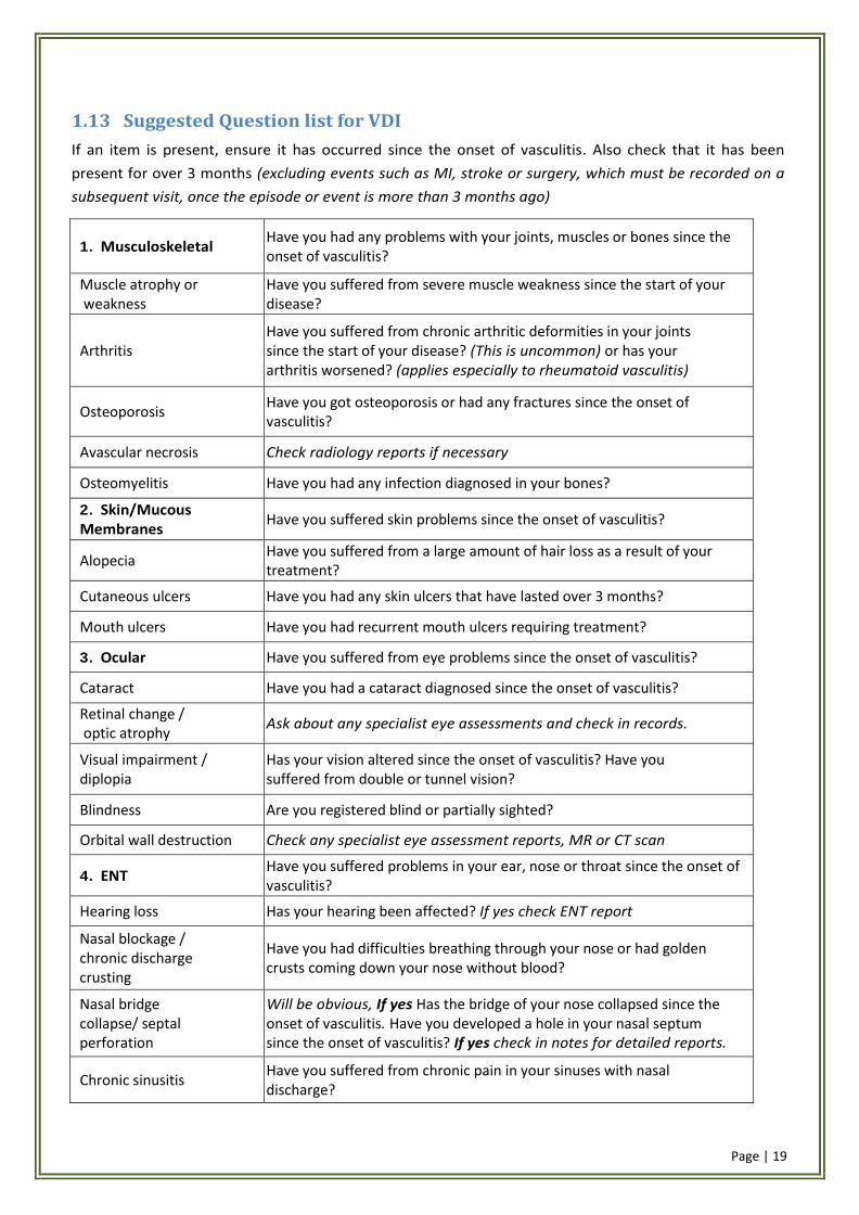
1.13 Suggested Question list for VDI
If an item is present, ensure it has occurred since the onset of vasculitis. Also check that it has been
present for over 3 months (excluding events such as MI, stroke or surgery, which must be recorded on a
subsequent visit, once the episode or event is more than 3 months ago)
1. Musculoskeletal Have you had any problems with your joints, muscles or bones since the onset of vasculitis?
Muscle atrophy or weakness
Have you suffered from severe muscle weakness since the start of your disease?
Arthritis Have you suffered from chronic arthritic deformities in your joints since the start of your disease? (This is uncommon) or has your arthritis worsened? (applies especially to rheumatoid vasculitis)
Osteoporosis Have you got osteoporosis or had any fractures since the onset of vasculitis?
Avascular necrosis Check radiology reports if necessary
Osteomyelitis Have you had any infection diagnosed in your bones?
2. Skin/Mucous Membranes
Have you suffered skin problems since the onset of vasculitis?
Alopecia Have you suffered from a large amount of hair loss as a result of your treatment?
Cutaneous ulcers Have you had any skin ulcers that have lasted over 3 months?
Mouth ulcers Have you had recurrent mouth ulcers requiring treatment?
3. Ocular Have you suffered from eye problems since the onset of vasculitis?
Cataract Have you had a cataract diagnosed since the onset of vasculitis?
Retinal change / optic atrophy
Ask about any specialist eye assessments and check in records.
Visual impairment / diplopia
Has your vision altered since the onset of vasculitis? Have you suffered from double or tunnel vision?
Blindness Are you registered blind or partially sighted?
Orbital wall destruction Check any specialist eye assessment reports, MR or CT scan
4. ENT Have you suffered problems in your ear, nose or throat since the onset of vasculitis?
Hearing loss Has your hearing been affected? If yes check ENT report
Nasal blockage / chronic discharge crusting
Have you had difficulties breathing through your nose or had golden crusts coming down your nose without blood?
Nasal bridge collapse/ septal perforation
Will be obvious, If yes Has the bridge of your nose collapsed since the onset of vasculitis. Have you developed a hole in your nasal septum since the onset of vasculitis? If yes check in notes for detailed reports.
Chronic sinusitis Have you suffered from chronic pain in your sinuses with nasal discharge?
Page | 20
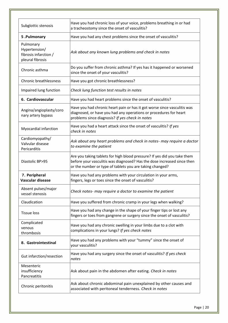
Subglottic stenosis Have you had chronic loss of your voice, problems breathing in or had a tracheostomy since the onset of vasculitis?
5 .Pulmonary Have you had any chest problems since the onset of vasculitis?
Pulmonary Hypertension/ fibrosis infarction / pleural fibrosis
Ask about any known lung problems and check in notes
Chronic asthma Do you suffer from chronic asthma? If yes has it happened or worsened since the onset of your vasculitis?
Chronic breathlessness Have you got chronic breathlessness?
Impaired lung function Check lung function test results in notes
6. Cardiovascular Have you had heart problems since the onset of vasculitis?
Angina/angioplasty/coronary artery bypass
Have you had chronic heart pain or has it got worse since vasculitis was diagnosed, or have you had any operations or procedures for heart problems since diagnosis? If yes check in notes
Myocardial infarction Have you had a heart attack since the onset of vasculitis? If yes check in notes
Cardiomyopathy/ Valvular disease Pericarditis
Ask about any heart problems and check in notes- may require a doctor to examine the patient
Diastolic BP>95 Are you taking tablets for high blood pressure? If yes did you take them before your vasculitis was diagnosed? Has the dose increased since then or the number or type of tablets you are taking changed?
7. Peripheral Vascular disease
Have you had any problems with your circulation in your arms, fingers, legs or toes since the onset of vasculitis?
Absent pulses/major vessel stenosis
Check notes- may require a doctor to examine the patient
Claudication Have you suffered from chronic cramp in your legs when walking?
Tissue loss Have you had any change in the shape of your finger tips or lost any fingers or toes from gangrene or surgery since the onset of vasculitis?
Complicated venous thrombosis
Have you had any chronic swelling in your limbs due to a clot with complications in your lungs? If yes check notes
8. Gastrointestinal Have you had any problems with your “tummy” since the onset of your vasculitis?
Gut infarction/resection Have you had any surgery since the onset of vasculitis? If yes check notes
Mesenteric insufficiency Pancreatitis
Ask about pain in the abdomen after eating. Check in notes
Chronic peritonitis Ask about chronic abdominal pain unexplained by other causes and associated with peritoneal tenderness. Check in notes
Page | 21
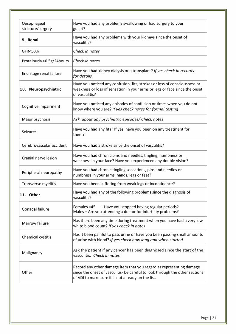
Oesophageal stricture/surgery
Have you had any problems swallowing or had surgery to your gullet?
9. Renal Have you had any problems with your kidneys since the onset of vasculitis?
GFR<50% Check in notes
Proteinuria >0.5g/24hours Check in notes
End stage renal failure Have you had kidney dialysis or a transplant? If yes check in records for details.
10. Neuropsychiatric Have you noticed any confusion, fits, strokes or loss of consciousness or weakness or loss of sensation in your arms or legs or face since the onset of vasculitis?
Cognitive impairment Have you noticed any episodes of confusion or times when you do not know where you are? If yes check notes for formal testing
Major psychosis Ask about any psychiatric episodes/ Check notes
Seizures Have you had any fits? If yes, have you been on any treatment for them?
Cerebrovascular accident Have you had a stroke since the onset of vasculitis?
Cranial nerve lesion Have you had chronic pins and needles, tingling, numbness or weakness in your face? Have you experienced any double vision?
Peripheral neuropathy Have you had chronic tingling sensations, pins and needles or numbness in your arms, hands, legs or feet?
Transverse myelitis Have you been suffering from weak legs or incontinence?
11. Other Have you had any of the following problems since the diagnosis of vasculitis?
Gonadal failure Females <45 - Have you stopped having regular periods? Males – Are you attending a doctor for infertility problems?
Marrow failure Has there been any time during treatment when you have had a very low white blood count? If yes check in notes
Chemical cystitis Has it been painful to pass urine or have you been passing small amounts of urine with blood? If yes check how long and when started
Malignancy Ask the patient if any cancer has been diagnosed since the start of the vasculitis. Check in notes
Other Record any other damage item that you regard as representing damage since the onset of vasculitis- be careful to look through the other sections of VDI to make sure it is not already on the list.

Page | 22
Section 4 Glossaries & Forms
4.1 The BVAS & VDI forms

Page | 23

Page | 24
4.2 Glossary
Glossary and scoring for BVAS (V. 3). GENERAL RULE: disease features are scored only when they are due to active vasculitis, after excluding other causes (e.g. infection, hypertension, etc.). If the feature is due to active disease, it is scored in the boxes. It is essential to apply these principles to each item below. Scores have been weighted according to the severity which each symptom or sign is thought to represent. Tick "Persistent Disease" box if all the abnormalities are due to active (but not new or worse) vasculitis. If any of the abnormalities are due to new/worse disease, DO NOT tick the "Persistent Disease" box. For some features, further information (from specialist opinion or further tests) is required if abnormality is newly present or worse. Remember that in most instances, you will be able to complete the whole record when you see the patient. However, you may need further information before entering some items. Please leave these items blank, until the information is available, and then fill them in. For example, if the patient has new onset of stridor, you would usually ask an otolaryngologist to investigate this further to determine whether or not it is due to active granulomatosis with polyangiitis (Wegener's).
System/item Description
Ne
w/W
orse
score
Pe
rsisten
t score
1. General 3 2
Myalgia Pain in the muscles 1 1
Arthralgia or arthritis Pain in the joints or joint inflammation 1 1
Fever ≥ 38.0 0C Documented oral/axillary temperature elevation. Rectal temps are 0.5 0C higher 2 2
Weight Loss At least 2kg loss of body weight (not fluid) having occurred since last assessment or in the 4 weeks not as a consequence of dieting
2 2
2. Cutaneous 6 3
Infarct Area of tissue necrosis or splinter haemorrhages 2 1
Purpura Petechiae (small red spots), palpable purpura, or ecchymoses (large plaques) in skin or oozing (in the absence of trauma) in the mucous membranes.
2 1
Ulcer Open sore in a skin surface. 4 1
Gangrene Extensive tissue necrosis (e.g. digit) 6 2
Other skin vasculitis Livedo reticularis, subcutaneous nodules, erythema nodosum, etc 2 1
3. Mucous membranes/eyes
6 3
Mouth ulcers/granulomata
Aphthous stomatitis, deep ulcers and/or “strawberry” gingival hyperplasia, excluding lupus erythematosus, and infection
2 1
Genital ulcers Ulcers localized in the genitalia or perineum, excluding infections. 1 1
Adnexal inflammation Salivary (diffuse, tender swelling unrelated to meals) or lacrimal gland inflammation. Exclude other causes (infection). Specialist opinion may be required.
4 2
Significant proptosis
Protrusion of the eyeball due to significant amounts of inflammatory in the orbit; if unilateral, there should be a difference of 2 mm between one eye and the other. This may be associated with diplopia due to infiltration of extra-ocular muscles. Developing myopia (measured on best visual acuity, see later) can also be a manifestation of proptosis
4 2

Page | 25
Red eye (Epi)scleritis Inflammation of the sclerae (specialist opinion usually required). Can be heralded by photophobia.
2 1
Red eye conjunctivitis Inflammation of the conjunctivae (exclude infectious causes and excluding uveitis as cause of red eye, also exclude conjunctivitis sicca which should not be scored as this is not a feature of active vasculitis); (specialist opinion not usually required).
1 1
Blepharitis Inflammation of eyelids. Exclude other causes (trauma, infection). Usually no specialist opinion is required
Keratitis Inflammation of central or peripheral cornea as evaluated by specialist
Blurred vision Altered measurement of best visual acuity from previous or baseline, requiring specialist opinion for further evaluation.
3 2
Sudden visual loss Sudden loss of vision requiring ophthalmological assessment. 6 *
Uveitis Inflammation of the uvea (iris, ciliary body, choroid) confirmed by ophthalmologist. 6 2
Retinal vasculitis Retinal vessel sheathing on examination by specialist or confirmed by retinal fluorescein angiography
6 2
Retinal vessel thrombosis
Arterial or venous retinal blood vessel occlusion
Retinal exudates Any area of soft retinal exudates (exclude hard exudates) seen on ophthalmoscopic examination.
Retinal haemorrhages Any area of retinal haemorrhage seen on ophthalmoscopic examination.
4. ENT 6 3
Bloody nasal discharge/ nasal crusts/ulcers and/or granulomata
Bloody, mucopurulent, nasal secretion, light or dark brown crusts frequently obstructing the nose, nasal ulcers and/or granulomatous lesions observed by rhinoscopy
4 2
Paranasal sinus involvement
Tenderness or pain over paranasal sinuses with pathologic imaging (CT, MR, x-ray).
2 1
Subglottic stenosis Stridor and hoarseness due to inflammation and narrowing of the subglottic area observed by laryngoscopy
6 3
Conductive hearing loss
Hearing loss due to middle ear involvement confirmed by otoscopy and/or tuning fork examination and/or audiometry
3 1
Sensorineural hearing loss
Hearing loss due to auditory nerve or cochlear damage confirmed by audiometry 6 2
5. Chest 6 3
Wheeze Wheeze on clinical examination 2 1
Nodules or cavities New lesions, detected by imaging. 3 *
Pleural effusion/pleurisy
Pleural pain and/or friction rub on clinical assessment or new onset of radiologically confirmed pleural effusion. Other causes (e.g. infection, malignancy) should be excluded
4 2
Infiltrate Detected by CXR or CT scan. Other causes (infection) should be excluded 4 2

Page | 26
Endobronchial involvement
Endobronchial pseudotumor or ulcerative lesions. Other causes such as infection or malignancy should be excluded. NB: smooth stenotic lesions to be included in VDI; subglottic lesions to be recorded in the ENT section.
4 2
Massive haemoptysis/ alveolar haemorrhage
Major pulmonary bleeding, with extensive pulmonary infiltrates; other causes of bleeding should be excluded if possible. Patients are usually in extremis. Do not record minor episodes of haemoptysis.
6 4
Respiratory failure Dyspnoea which is sufficiently severe as to require artificial ventilation 6 4
6. Cardiovascular 6 3
Loss of pulses Loss of pulses in any vessel detected clinically; this may include loss of pulses leading to threatened loss of limb
4 1
Valvular heart disease
Significant valve abnormalities in the aortic mitral or pulmonary valves detected clinically or echocardiographically.
4 2
Pericarditis Pericardial pain &/or friction rub on clinical assessment. 3 1
Ischaemic cardiac pain
Typical clinical history of cardiac pain leading to myocardial infarction or angina. Consider the possibility of more common causes (e.g. atherosclerosis)
4 2
Cardiomyopathy Significant impairment of cardiac function due to poor ventricular wall motion confirmed on echocardiography
6 3
Congestive cardiac failure
Heart failure by history or clinical examination 6 3
7. Abdominal 9 4
Peritonism Acute abdominal pain with peritonism/peritonitis due to perforation/infarction of small bowel, appendix or gallbladder etc., or acute pancreatitis confirmed by radiology/surgery/elevated amylase
9 3
Bloody diarrhoea Of recent onset; inflammatory bowel disease and infectious causes excluded. 9 3
Ischaemic abdominal pain
Severe abdominal pain with typical features of ischaemia confirmed by imaging or at surgery, with typical appearances of aneurysms or abnormal vasculature characteristic of vasculitis.
6 2
8. Renal 12 6
Hypertension Systolic BP>140 or Diastolic BP>95, accelerated or not, with or without retinal changes. 4 1
Proteinuria >1+ on urinalysis; >0.2g/24 hours Infection should be excluded. 4 2
Haematuria 10 or more RBC per hpf ( high power field ), excluding urinary infection and urinary lithiasis (stone)or drug side effects (e.g. cyclophosphamide)
6 3
Creatinine 125-249 Serum creatinine values 125-249 μmol/l (1.41-2.82 mg/dL); only used at first assessment. 4 *
Creatinine 250-499 Serum creatinine values 250-499 μmol/l (2.83-5.64 mg/dL); only used at first assessment. 6 *

Page | 27
Creatinine ≥ 500 Serum creatinine values 500 μmol/l(≥5.66mg/dL) or greater; only used at first assessment. 8 *
Rise in creatinine> 30% or creatinine clearance fall > 25%
Significant deterioration in renal function attributable to active vasculitis.
6 *
9. Nervous system 9 6
Headache New, unaccustomed & persistent headache 1 1
Meningitis Severe headache with neck stiffness ascribed to inflammatory meningitis after excluding infection/bleeding
3 1
Organic confusion Impaired orientation, memory or other intellectual function in the absence of metabolic, psychiatric, pharmacological or toxic causes.
3 1
Seizures (not hypertensive)
Paroxysmal electrical discharges in the brain & producing characteristic physical changes including tonic & clonic movements & certain behavioural changes.
9 3
Stroke Cerebrovascular accident resulting in focal neurological signs such as paresis, weakness, etc. A stroke due to other causes (e.g. atherosclerosis) should be considered & appropriate neurological advice is recommended
9 3
Cord lesion Transverse myelitis with lower extremity weakness or sensory loss (usually with a detectable sensory level) with loss of sphincter control (rectal & urinary bladder).
9 3
Cranial nerve palsy Facial nerve palsy, recurrent nerve palsy, oculomotor nerve palsy etc. excluding sensorineural hearing loss and ophthalmic symptoms due to inflammation
6 3
Sensory peripheral neuropathy
Sensory neuropathy resulting in glove &/or stocking distribution of sensory loss. Other causes should be excluded (e.g. idiopathic, metabolic, vitamin deficiencies, infectious, toxic, hereditary).
6 3
Motor mononeuritis multiplex
Simultaneous neuritis of peripheral nerves, only scored if motor involvement. Other causes should be excluded (diabetes, sarcoidosis, carcinoma, amyloidosis).
9 3
10. Other
Other features of active vasculitis-please describe. Remember that you should review the rest of the BVAS item list before writing the feature in this section because it may already have been described elsewhere on the form. Do not use this section to record laboratory, imaging or pathology findings. Items recorded in this section will not carry any value when calculating the BVAS score
0 0
*For some BVAS items, we do not provide recording or scoring of persistent items, either because the item is by defi nition new/worse, or for renal disease, can only reliably be recorded as new/worse on the first visit

Page | 28
Vasculitis Damage Index An Introduction and Glossary of Terms Purpose of assessment
Standard therapy for systemic vasculitis has markedly improved the acute mortality but relapse remains a problem. Mortality is no longer an acceptable end point for studies. Long term studies require detailed assessment of disease status in order to estimate the degree of improvement achieved together with any accumulated scars or damage. Both contribute to the long term outcome and are needed for proper comparison of new treatment regimens. In this document, we have already described the use of an activity assessment for systemic vasculitis. In a companion document (Vasculitis Damage Index), we describe a measurement of damage in patients with systemic vasculitis.
VDI is designed to document those features which are due to persistent damage. Damage is defined as the presence of non-healing scars and does not give any indication of current disease activity. Damage items in the Vasculitis Damage Index are often the direct result of previous disease activity
Recording Damage
The Vasculitis Damage Index deals with features which have occurred since the onset of vasculitis regardless of whether or not they are attributable to vasculitis. Damage is defined as having been present or currently present for at least 3 months. It is therefore possible for abnormalities to have occurred in the past, not be currently present, but to still count as damage. The VDI item list can only deteriorate or be stable over time (damage is defined as irreversible in this scoring system). For each item in turn, record all features which have occurred since the onset of vasculitis, regardless of the cause. For specific events, such as GI surgery, damage can be scored as present if the procedure was undertaken at least three months prior to the assessment (and also must have occurred after the onset of the disease). The same time frame is applied to all the damage items. If you are seeing the patient for the first time, and their vasculitis onset date is within three months of your assessment, then by definition, the patient cannot be recorded as having any damage. However, any features which you observe can be recorded as ascribable to damage after three months has elapsed.
Recording disease activity
The vasculitis activity score (BVAS) is designed to record features which are attributable to current active vasculitis, after exclusion of other obvious causes such as infection, hypertension, etc. It may vary rapidly and thus reflects the need for therapy. It is based on an intention to treat with immunosuppressive therapy or heighten clinical monitoring for any individual patient (see BVAS).
VASCULITIS DAMAGE INDEX (VDI)
Damage: This is a score of damage due to non-healing scars and is not a score of active vasculitis. Damage is defined as having ever been present for at least 3 months and has occurred since the onset of vasculitis. Please note that damage does not have to be currently present. This is a cumulative assessment of organ dysfunction or scarring. Damage is ascertained by clinical assessment. As time progresses the damage index score can only remain stable or deteriorate. Although many of the features of the VDI may reflect drug related damage, adverse drug reactions should be recorded separately. Please make sure that you carefully record a detailed description of the ADR.
In the case of blindness, myocardial infarction, loss of pulses, major tissue loss, or stroke, repeat episodes may be recorded but these must be at least 3 months apart to score. Please note that the same pathological lesion cannot be scored more than once in the index even though it may occur in different parts of the index ( e.g. if you score optic atrophy in the ocular section, this does not score as a cranial nerve lesion).

Page | 29
Damage system/item Description (critical items are asterisked)
1. Musculoskeletal
Significant muscle atrophy or weakness:
Demonstrated on clinical examination (not attributable to CVA which should already have been scored)
Deforming or erosive arthritis:
Deformities on clinical examination confirmed by imaging (excluding avascular necrosis), on clinical examination and imaging.
Osteoporosis with fractures or vertebral collapse:
By history confirmed on imaging (excluding avascular necrosis).
Avascular necrosis: Demonstrated by appropriate radiological techniques.
Osteomyelitis: Documented clinically, confirmed by imaging and/or culture.
2. Skin/mucous membranes
Alopecia: Major (e.g. requiring wig) chronic hair loss with or without scars, documented clinically.
Mouth ulceration: Recurrent crops or persistent mouth ulcers requiring therapy.
Cutaneous ulcers: Open sore on skin surface, excluding that caused by venous thrombosis.
3. Ocular
Cataract: A lens opacity (cataract) in either eye documented by ophthalmoscopy.
Retinal change: Any significant change documented by ophthalmoscopic examination; may result in field defect, legal blindness.
Optic atrophy: Documented by ophthalmoscopic examination.
Visual impairment/diplopia: Restricted eye movements (not due to nerve palsies), reduced visual acuity, double vision or tunnel vision.
Blindness in one eye: Complete loss of vision in one eye.
Blindness in second eye: Complete loss of vision in other eye *.
Orbital wall destruction: Significant bone destruction as documented on imaging
4. ENT
Hearing loss: Any hearing loss due to middle ear involvement or to auditory nerve/ cochlear damage, preferably confirmed by audiometry.
Nasal blockage/chronic discharge/crusting:
Difficulties with breathing through the nose and/or with purulent discharge and/or with crust formation usually requiring nasal lavage
Nasal bridge collapse/septal perforation:
Saddle nose deformity and/or perforation of nasal septum
Chronic sinusitis/radiological damage:
Chronic purulent nasal discharge with sinus pain and/or imaging evidence of sinusitis with or without bone destruction
Subglottal stenosis with or without surgery:
Persistent hoarseness and/or stridor preferably confirmed by endoscopy and/or imaging*

Page | 30
5. Pulmonary
Pulmonary hypertension: Right ventricular prominence or loud P2 (confirmed by cardiological investigation if appropriate).
Pulmonary fibrosis: According to physical signs and imaging (confirmed by relevant tests if necessary); this may include patients who require pulmonary resection.
Pulmonary infarction: According to imaging.
Pleural fibrosis: According to imaging.
Chronic asthma: Significant reversible airways obstruction.
Chronic breathlessness:
Significant symptomatic breathing difficulties and/or shortness of breath with or without hard signs on imaging or lung function tests. If the pathology and the breathlessness have been present and persistent for more than 3 months, then both can be recorded*
Impaired lung function: Significant abnormality of pulmonary function.*
6. Cardiovascular
Angina/angioplasty/coronary artery bypass:
on history confirmed at least by ECG changes
Myocardial infarction: on history confirmed at least by ECG changes or cardiac enzyme elevation
Second myocardial infarction Second MI *
Cardiomyopathy: Chronic ventricular dysfunction documented clinically or on appropriate investigation*
Valvular disease: Significant diastolic or systolic murmur confirmed by cardiological tests if appropriate
Pericarditis>3mths or pericardectomy:
Symptomatic pericardial inflammation or constriction for at least 3 months or pericardectomy
Diastolic BP>95 or requiring anti-hypertensives: With a diastolic BP >95 mm Hg or requiring anti-hypertensive drugs
7. Peripheral vascular disease
Absent pulses in one limb: In 1 limb, detected clinically.
Second episode of absent pulses in one limb:
In another limb detected clinically.
Major vessel stenosis: e.g. carotid or renal stenosis, documented on Doppler or other imaging
Claudication: Exercise related ischaemic pain in peripheral large vessel present for at least 3 months
Minor tissue loss: e.g. loss of finger tip pulp space

Page | 31
Major tissue loss: e.g. the loss of digit(s) or limb(s), including by surgical resection
Second episode of major tissue loss
Second episode*
Complicated venous thrombosis:
with persistent swelling, ulceration, or clinical evidence of venous stasis
8. Gastrointestinal
Gut infarction/resection: Infarction or resection of bowel below duodenum; or of gall bladder, spleen, or liver*
Mesenteric insufficiency/ pancreatitis:
Typical abdominal pain confirmed on angiography or enzyme changes
Chronic peritonitis: Typical abdominal pain and peritoneal irritation on clinical examination
Oesophageal stricture/or surgery: On history or by further appropriate tests
9. Renal
Estimated or measured GFR<50%:
By any locally used method
Proteinuria of >0.5g/24 hours: By any locally used method
End stage renal failure Failure of native kidneys regardless of subsequent dialysis or transplantation*
10. Neuropsychiatric
Cognitive impairment:
Memory deficit, difficulty with calculation, poor concentration, difficulty in spoken or written language, impaired performance level, documented on clinical examination (e.g. short mental test score) or by formal neuro-cognitive testing.
Major psychosis:
Altered ability to function in normal activity due to psychiatric reasons. Severe disturbance of the perception of reality characterized by the following features: delusions, hallucinations (auditory, visual), incoherence, marked loose associations, impoverished thought content, marked illogical thinking, bizarre, disorganized or catatonic behaviour.*
Seizures:
Paroxysmal electrical discharge occurring in the brain and producing characteristic physical changes including tonic and clonic movements, and certain behavioral disorders. Only seizures requiring therapy for more than 3 months are counted as damage.
Cerebrovascular accident:
CVA resulting in focal findings such as paresis, weakness, etc., OR surgical resection for causes other than malignancy.
Second Stroke Second episode*
Cranial nerve lesion: Cranial neuropathy, excluding optic nerve or sensorineural deafness
Peripheral neuropathy: Peripheral neuropathy resulting in either motor or sensory dysfunction.
Transverse myelitis: Lower extremity weakness or sensory loss with loss of sphincter control (rectal and urinary bladder).*
11. Other
Gonadal failure: Premature secondary amenorrhoea or azoospermia*

Page | 32
Marrow failure: Leucopenia (WCC <4000 per microlitre), or thrombocytopenia (platelets<140) or anaemia (haemoglobin<10), preferably confirmed by marrow aspiration (marrow abnormality to persist for 3 months to score even if G-CSF used)*
Diabetes: Requiring any type of therapy
Chemical cystitis: Persistent haematuria or shrunken bladder. This does not include acute haemorrhagic cystitis which should be scored in the ADR.
Malignancy: Documented by pathology, excluding dysplasias.*
Other: Any feature considered by patient or doctor to be an important scar or consequence which has arisen since onset of disease

Page | 33
4.3 BVAS Flow chart

Page | 34
Section 5 Further Information
5.1 Contact Details
The contact details for Raashid Luqmani are as follows:
Professor Raashid A Luqmani Professor of Rheumatology NDORMS, University of Oxford Nuffield Orthopaedic Centre Windmill Road, Headington Oxford OX3 7LD
Tel +44 (0) 1865 227357 Fax +44 (0) 1865 738048 E-mail [email protected]

Page | 35
5.2 Further Reading
Exley AR, Bacon PA, Luqmani RA, Kitas GD, Carruthers DM, Moots R Examination of disease severity in systemic vasculitis from the novel perspective of damage using the vasculitis damage index (VDI). British J Rheumatol 1998 37:57- 63.
Exley AR, Bacon PA, Luqmani RA, Kitas GD, Gordon C, Savage COS, Adu D Development and Initial Validation of the Vasculitis Damage Index (VDI) for the Standardised Clinical Assessment of Damage in the Systemic Vasculitides. Arthritis and Rheumatism 1997 40: 37 1-380.
Exley A, Carruthers DM, Luqmani RA, Kitas GD, Gordon C, Janssen BA, Savage COS, Bacon PA Damage occurs early in systemic vasculitis and is an index of outcome. Quart J Med 1997 90:39 1-399.
Luqmani RA, Exley AR, Kitas GD, Bacon PA Disease assessment and management of the vasculitides. Baillieres Clinical Rheumatology 1997. 11:423-446.
Luqmani RA, Bacon PA, Moots RJ, Janssen BA, Pall A, Emery P, Savage C, Adu D Birmingham vasculitis activity score (BVAS) in systemic necrotizing vasculitis. Quart J Med 1994 87:671-678.
Luqmani RA. Disease assessment in systemic vasculitis. Nephrol Dial Transplant. 2015 Apr;30 Suppl 1:i76-82
Merkel PA, Aydin SZ, Boers M, Direskeneli H, Herlyn K, Seo P, Suppiah R, Tomasson G, Luqmani RA. The OMERACT Core Set of Outcome Measures for Use in Clinical Trials of ANCA-associated Vasculitis. J Rheumatol. 2011 Jul;38(7):1480-6.
Miller A, Basu N, Luqmani R. Assessment of systemic vasculitis. Autoimmun Rev. 2008 Dec;8(2):170-5.
Mukhtyar C, Lee R, Brown D, Carruthers D, Dasgupta B, Dubey S, Flossmann O, Hall C, Hollywood J, Jayne D, Jones R, Lanyon P, Muir A, Scott D, Young L, Luqmani RA. Modification and validation of the Birmingham Vasculitis Activity Score (version 3). Ann Rheum Dis. 2009 Dec;68(12):1827-32.
Ntatsaki E, Carruthers D, Chakravarty K, D'Cruz D, Harper L, Jayne D, Luqmani R, Mills J, Mooney J, Venning M, Watts RA; BSR and BHPR Standards, Guidelines and Audit Working Group. BSR and BHPR guideline for the management of adults with ANCA-associated vasculitis. Rheumatology (Oxford). 2014 Dec;53(12):2306-9
Ponte C, Sznajd J, O'Neill L, Luqmani RA. Optimisation of vasculitis disease assessments in clinical trials, clinical care and long-term databases. Clin Exp Rheumatol. 2014 Sep-Oct;32(5 Suppl 85):S-118-25.
Robson J, Doll H, Suppiah R, Flossmann O, Harper L, Höglund P, Jayne D, Mahr A, Westman K, Luqmani R. Damage in the anca-associated vasculitides: long-term data from the European Vasculitis Study group (EUVAS) therapeutic trials. Ann Rheum Dis. 2013 Nov 15. doi: 10.1136/annrheumdis-2013-203927. [Epub ahead of print]
Stone JH, Hoffman GS, Merkel PA, Min Y-I., Uhlfelder ML, Hellmann DB, Specks U, Allen NB, Davis JC, Spiera RF, Calabrese LH, Wigley FM, Maiden N, Valente RM, Niles JL, Fye KH, McCune JW, St. Clair EW, Luqmani RA (2001). A Disease-Specific Activity Index for Wegener’s Granulomatosis: Modification of the Birmingham Vasculitis Activity Score. Arthritis & Rheumatism:44(4):912- 920.
Suppiah R, Mukhtyar C, Flossmann O, Alberici F, Baslund B, Batra R, Brown D, Holle J, Hruskova Z, Jayne DR, Judge A, Little MA, Palmisano A, Stegeman C, Tesar V, Vaglio A, Westman K, Luqmani R. A cross-sectional study of the Birmingham Vasculitis Activity Score version 3 in systemic vasculitis. Rheumatology (Oxford). 2011 May; 50(5):899-905.

Page | 36
Suppiah R, Flossman O, Mukhtyar C, Alberici F, Baslund B, Brown D, Hasan N, Holle J, Hruskova Z, Jayne D, Judge A, Little MA, Merkel PA, Palmisano A, Seo P, Stegeman C, Tesar V, Vaglio A, Westman K, Luqmani R. Measurement of damage in systemic vasculitis: a comparison of the Vasculitis Damage Index with the Combined Damage Assessment Index. Ann Rheum Dis. 2011 Jan; 70(1):80-5
Walsh M, Mukhtyar C, Mahr A, Herlyn K, Luqmani R, Merkel PA, Jayne DR. Health-related quality of life in patients with newly diagnosed antineutrophil cytoplasmic antibody-associated vasculitis. Arthritis Care Res (Hoboken). 2011 Jul;63(7):1055-61. doi: 10.1002/acr.20471. PMID: 21452254 [PubMed - indexed for MEDLINE]
Yates M, Watts RA, Bajema IM, Cid MC, Crestani B, Hauser T, Hellmich B, Holle JU, Laudien M, Little MA, Luqmani RA, Mahr A, Merkel PA, Mills J, Mooney J, Segelmark M, Tesar V, Westman K, Vaglio A, Yalçındağ N, Jayne DR, Mukhtyar C. EULAR/ERA-EDTA recommendations for the management of ANCA-associated vasculitis. Ann Rheum Dis. 2016 Sep;75(9):1583-94.
Useful websites
www.vasculitis.org (information about EUVAS activities)
bvasvdi.ndorms.ox.ac.uk (Training Website)
[email protected] (Training Assistance)
Training Signup Instructions for a new Trainee*
Visit https://bvasvdi.ndorms.ox.ac.uk & click ‘register’
Enter your personal details
New Trainees: Use invite code: “bvasvditrain”
You will now be logged in with access to a comprehensive training set of 60 cases (30 BVAS , 30 VDI) & access to ‘learning resources’ ; over 2 hours of instructional video
For future visits, use ‘login’ and enter details you registered
Contact [email protected] if you have any questions, need to redo any cases, or have completed the assessment (to request your certification signed by Prof Luqmani).
*Experienced Trainees: Contact [email protected] for access to the shortened ‘refresher’ set.