clinical, etiological, and therapeutic features of jaw-opening and
TRANSCRIPT
Brief Reports
Clinical, Etiological, and Therapeutic Features of Jaw-opening and Jaw-closingOromandibular Dystonias: A Decade of Experience at a Single Treatment
Center
Pedro Gonzalez-Alegre1*
, Robert L. Schneider2
& Henry Hoffman3
1 Department of Neurology, Carver College of Medicine, The University of Iowa, Iowa City, Iowa, United States of America, 2 Department of Oral & Maxillofacial
Surgery, College of Dentistry, The University of Iowa, Iowa City, Iowa, United States of America, 3 Department of Otolaryngology, Carver College of Medicine,
The University of Iowa, Iowa City, Iowa, United States of America
Abstract
Background: Dystonia is a heterogeneous hyperkinetic disorder. The anatomical location of the dystonia helps clinicians guide their evaluation and treatment
plan. When dystonia involves masticatory, lingual, and pharyngeal muscles, it is referred to as oromandibular dystonia (OMD).
Methods: We identified patients diagnosed with OMD in a Movement Disorders Clinic and Laryngeal Movement Disorders Clinic from a single institution.
Demographic, etiological, clinical, and therapeutic information was retrospectively reviewed for patients with jaw-opening (O-OMD) and jaw-closing (C-OMD)
OMD.
Results: Twenty-seven patients were included. Their average age of onset was in the sixth decade of life and there was a female predominance. Etiological factors
linked in this study to OMD included a family history of dystonia or essential tremor, occupation, cerebellar disease, a dental disorder, and tardive syndrome.
Clinically, patients with C-OMD presented with more prominent feeding difficulties, but seemed to respond better to therapy than those with O-OMD. In addition
to the known benefits of botulinum toxin therapy, patients who described sensory tricks obtained benefit from the use of customized dental prosthesis.
Discussion: This works provides useful information on potential etiological factors for OMD and its response to therapy, and highlights the potential benefit of
dental prosthesis for the treatment of OMD.
Keywords: Oromandibular dystonia, botulinum toxin, sensory trick, etiology
Citation: Gonzalez-Alegre P, Schneider RL, Hoffman H. Clinical, etiological, and therapeutic features of jaw-opening and jaw-closing oromandibular dystonias: a
decade of experience at a single treatment center. Tremor Other Hyperkinet Mov. 2014; 4. doi: 10.7916/D8TH8JSM
* To whom correspondence should be addressed. E-mail: [email protected]
Editor: Elan D. Louis, Columbia University, United States of America
Received: March 10, 2014 Accepted: April 2, 2014 Published: April 30, 2014
Copyright: ’ 2014 Gonzalez-Alegre et al. This is an open-access article distributed under the terms of the Creative Commons Attribution–Noncommercial–No Derivatives License,
which permits the user to copy, distribute, and transmit the work provided that the original author(s) and source are credited; that no commercial use is made of the work; and that the
work is not altered or transformed.
Funding: None.
Financial Disclosures: Dr. Gonzalez-Alegre has received Honoraria from Lundbeck for his participation in the Xenazine Advisory Board.
Conflict of Interest: The authors report no conflict of interest.
Introduction
Dystonia is a movement disorder characterized by sustained or
intermittent muscle contractions that cause abnormal, often repetitive,
movements, postures, or both.1 Dystonia is a very heterogeneous
phenomenon, and its clinical variability, together with the low
frequency of some of its forms, presents diagnostic and therapeutic
challenges.2,3
A recent consensus update proposed classifying patients with
dystonia according to clinical features and etiology.1 The classification
along the clinical axis aims to identify clinically defined forms of
dystonia to aid in their diagnosis and treatment. Dystonia can present
as a wide range of clinical syndromes that share the presence of this
hyperkinetic movement disorder in different body parts. Due to this
variability, there is no uniform clinical approach that is applicable to
all.3 For instance, adult-onset focal leg dystonia requires a diagnostic
investigation, as it is likely to be acquired.4,5 On the other hand, adult-
onset cervical dystonia is usually idiopathic and rarely requires
diagnostic testing beyond a good clinical examination.6
While some forms of dystonia are relatively common, such as adult-
onset cervical dystonia,6 others are less frequent and some clinicians
don’t have enough clinical experience to guide their practice.
Therefore, it is important to share the experience of centers that
Freely available online
Tremor and Other Hyperkinetic Movementshttp://www.tremorjournal.org
The Center for Digital Research and ScholarshipColumbia University Libraries/Information Services1
evaluate uncommon dystonia syndromes. This will help us to gain a
better understanding of features that are specifically observed in these
forms of dystonia, aiding clinicians and researchers in their quest to
improve the quality of life of those suffering from this hyperkinetic
disorder.
In this work, we retrospectively analyzed the etiological, clinical, and
therapeutic features of a series of consecutive patients diagnosed over a
decade with jaw-opening or jaw-closing oromandibular dystonia (O-
OMD and C-OMD, respectively).
Methods
We performed a retrospective chart review of patients evaluated for
a diagnosis of O-OMD and C-OMD at the University of Iowa
Hospitals and Clinics in the period between 2004 and 2013. We first
identified patients who had been evaluated in both the Movement
Disorders Clinic (Department of Neurology) and the Laryngeal
Movement Disorders Clinic (Department of Otolaryngology) through
our electronic medical records. Of those, we searched their diagnostic
coding information for the term dystonia. We used these criteria
because in our institution, botulinum toxin treatment for patients with
OMD is performed at the Laryngeal Movement Disorders Clinic.
However, most of these patients are also evaluated in the Movement
Disorders Clinic for the possibility of presence of dystonia outside of
the oromandibular area and for consideration of additional diagnostic
testing or therapeutic interventions.
Once identified, their medical records were reviewed, and only
those diagnosed with C-OMD and O-OMD by one of our movement
disorders experts and an otolaryngologist with expertise on oroman-
dibular movement disorders (H.H.) were included in the study. We
collected demographic, clinical, and therapeutic information including
age at onset and at diagnosis, clinical diagnosis, presumed etiology,
factors potentially linked to the development of dystonia as indicated
by the patient or clinician (such as occupation, injury, etc.), family
history of dystonia or other movement disorders, presence of a geste-
antagoniste or sensory trick,2 reported dysphagia, weight loss and body
mass index (BMI), medical treatments attempted and the results,
therapeutic use of dental prosthesis, and application of botulinum toxin
treatments (including dose and muscles selected for the treatment of O-
OMD and C-OMD). This protocol was reviewed and approved by the
Institutional Review Board of our institution and, as a retrospective
record review, the requirement to obtain informed consent from the
patients was waived.
Results
A total of 67 patients who had been evaluated in the Movement
Disorders and Laryngeal Movement Disorders Clinics during the
defined period and had the term ‘‘dystonia’’ included in their
diagnostic coding information. A review of their chart confirmed the
diagnosis of O-OMD or C-OMD in 27 of them. The 40 excluded
patients had a diagnosis of laryngeal dystonia (12), voice tremor (eight),
classical tardive dyskinesias (eight), dystonia outside the oromandibular
and laryngeal areas (six), or other unrelated diagnoses (six).
The demographic, clinical and etiological characteristics of the 27
patients are shown in Table 1. Four (15%) patients had a family
history of dystonia or ET. Among potential etiological factors, three
patients with idiopathic O-OMD related this disorder to their frequent
use of loud speech due to their occupation (a teacher, an auctioneer,
and a preacher). Furthermore, two patients developed their dystonia as
a presumed consequence of cerebellar disease (a primary cerebellar
degenerative disease and a cerebellar stroke). From a clinical point of
view, we found that self-reported weight loss (though not objectively
documented) was commonly seen in our patients with OMD, more
prominently for the C-OMD group.
As shown in Table 2, most patients received botulinum toxin
injections as a treatment for their OMD. Of the 11 with O-OMD who
received at least one treatment, only eight had repeated injections, but
the eight patients with C-OMD who initiated this treatment at our
institution had repeated injections. For O-OMD, we targeted the
anterior belly of the digastric (ABD) with or without additional
injections into the lateral pterygoid muscles bilaterally, and for C-
OMD the masseters and/or temporalis. Patients with additional
anatomical involvement received injections in other muscles (not
shown). The average dose in their more recent treatment was higher
for all targeted muscles than in the initial application, although, for
instance, the lateral pterygoid was abandoned as a target in all but two
patients with O-OMD.
At our institution, patients who present sensory tricks that relieve
their OMD symptoms are offered an evaluation at the Maxillofacial
Prosthodontics Clinic for fitting with prosthetic dental prosthesis. Four
out of eight patients evaluated in that clinic reported significant benefit
and, in two cases, botulinum toxin treatments were not pursued at all
due to the efficacy of these devices (Figure 1).
Discussion
In this study, we report a series of consecutive patients with C-OMD
and O-OMD seen at a single institution over a decade. In addition to
adding important information to the literature on the clinical features
of these relatively rare forms of dystonia, this work highlights
important features of OMD. First, consistent with previous reports,
some of our OMD patients reported family members diagnosed with
dystonia suggesting a genetic predisposition. Second, cerebellar disease
and occupational public speaking could play a role in the pathogenesis
of OMD. Third, while the small sample size precluded us from
performing a reliable statistical comparison between C-OMD and O-
OMD, our data suggested potential differences. Self-reports of weight
loss seem to be more frequent in C-OMD, although patients with C-
OMD responded better than those with O-OMD to botulinum toxin
treatment. Finally, a sizable proportion of patients with OMD exhibit
sensory tricks that can be used for the development of therapeutic
prosthetic devices.
The retrospective nature of this study adds different sources of
potential bias, such as clinician preference for specific treatment
approaches or self-reported symptoms by patients that could not be
confirmed (i.e., weight loss). In addition, the description of the
Gonzalez-Alegre P, Schneider RL, Hoffman H Oromandibular Dystonia
Tremor and Other Hyperkinetic Movementshttp://www.tremorjournal.org
The Center for Digital Research and ScholarshipColumbia University Libraries/Information Services2

Table 1. Demographic and Clinical Characteristics of Patients with OMD.
O-OMD C-OMD Total
Demographics
N 16 (59%) 11 (41%) 27
Women 56% 64% 59%
Caucasian 94% 82% 89%
Etiology
Idiopathic/primary 14 (88%) 6 (55%) 20 (74%)
Secondary 2 (12%) 5 (45%) 7 (26%)
Tardive 0 2 2
Peripheral1 1 1 2
Degenerative 12 0 1
Other 0 23 2
Family history
Dystonia 1 (6%) 1 (9%) 2 (7%)
PD 0 0 0
Essential tremor 2 (12%) 0 2 (7%)
Clinical
Age onset (SD) 56 (14.8) 52.6 (16.4) 54.8 (15)
DD (>1 year:,1 year) 5:8 3:5 8:13
Phenomenology
Sensory trick 10 (63%) 2 (18%) 12 (44%)
Task-specific 1 (6%) 1 (9%) 2 (7%)
Focal 11 (69%) 4 (36%) 15 (56%)
+Cervical 0 3 (27%) 3 (11%)
+Cranial 3 (19%) 1 (9%) 4 (15%)
+Craniocervical 2 (12%) 2 (18%) 4 (15%)
+Other 0 1 (9%) 1 (4%)
Weight loss 2 (12%) 6 (55%) 8 (30%)
BMI (SD)4 27.5 (7.4) 22.5 (7.8) 26.4 (6.3)
BMI, Body Mass Index; C-, Jaw Closing; DD, Diagnostic Delay; O-, Jaw Opening; OMD, Oromandibular Dystonia; PD, Parkinson’s Disease; SD, Standard Deviation.1Onset after dental procedures.2Progressive cerebellar degeneration of unknown etiology.3Cerebellar stroke and cerebral palsy.4Taken from most recent visit (non-significant trend).
Oromandibular Dystonia Gonzalez-Alegre P, Schneider RL, Hoffman H
Tremor and Other Hyperkinetic Movementshttp://www.tremorjournal.org
The Center for Digital Research and ScholarshipColumbia University Libraries/Information Services3

phenomenology is limited. In most patients, the more prominent
component of the OMD was either jaw-opening or jaw-closing.
However, a component of lateral deviation or tongue involvement
could also have been present. We did not include those in this
description because their presence or documentation was more
variable. None of the patients with OMD had comorbid laryngeal
dystonia. As a result, the data reported here have to be interpreted
with caution and should be followed by prospective studies.
The demographic and clinical characteristics of our patients, such as
female predominance, are comparable to and consistent with the few
previously reported series of patients with OMD.7–11 Similar to ours,
Singer and Papapetropoulos7 included only patients with C-OMD and
O-OMD and compared their clinical features, whereas most other
articles report all forms of OMD in aggregate. Therefore, we will
briefly compare our findings to theirs. In contrast to our study, they
found more males in their C-OMD group, and the reported mean age
at symptom onset is higher in our group of patients than theirs (56 vs.
48 years). The retrospective nature of both studies and potential recall
bias could explain these differences. Taking all reports in aggregate,
and consistent with other forms of primary dystonia,12,13 it is
reasonable to conclude that OMD is more prevalent in women.
Interestingly, both studies describe several consistent findings. A family
history of dystonia was found in ,10% of subjects in both studies, a
more frequent cervical involvement in subjects with O-OMD (41%
and 27% in Singer and Papapetropoulos and this report, respectively)
than C-OMD (18% and 0%), and a more frequent presence of sensory
tricks in O-OMD (33% and 63%) than in C-OMD (0% and 18%).
Furthermore, Singer and Papapetropoulos reported a moderate or
marked improvement to botulinum toxin in 90% of patients with C-
OMD but only in 43% of those with O-OMD. In our series, this
intervention was more frequently abandoned in patients with O-
OMD. Overall, these findings suggest that O-OMD and C-OMD
have different clinical and therapeutic features, perhaps reflecting
differences in the underlying pathophysiology. Replication of these
findings in future prospective studies would be important for dystonia
researchers. For instance, if the clinical and therapeutic differences
between both forms of OMD are confirmed, patients should not be
clustered together in clinical trials under the umbrella of OMD, but
stratified as O-OMD or C-OMD.
We were interested in the identification of potential etiological
factors in patients with OMD. Dystonia is thought to result from a
combination of genetic and environmental factors. Our findings are
Table 2. Response to Treatment of Patients with OMD.
O-OMD C-OMD
Botulinum toxin1
N 11 8
Muscles (N/mean initial dose)2 ABD (11/6.1 U) Temporalis (5/9.5 U)
Lat. pterygoid (5/7 U) Masseter (7/12.9 U)
Muscles (N/mean final dose)2 ABD (8/9.4 U) Temporalis (5/14 U)
Lat. pterygoid (2/8.8 U) Masseter (7/34.3 U)
Other medical therapies
(total/benefit3)
Anticholinergics 4/2 2/0
Dopaminergic 3/0 2/0
Benzodiazepines 4/1 1/0
Baclofen 1/0 2/1
Tizanidine 0/0 1/1
Reserpine 1/1 0/0
Amantadine 1/1 0/0
ABD, anterior belly of digastric; C-, Jaw Closing; lat., lateral; O-, Jaw Opening; OMD, Oromandibular Dystonia; U, units.1All patients received initially botulinum toxin type A (Botox). One patient in the O-OMD group changed to botulinum toxin type B (Myobloc, dose not included in the
analysis).2Dose injected unilaterally. However, all injections were bilateral, with the same dose in both sides for all patients.3Benefit was always reported as mild or transient. No significant and sustained benefit was described with medical treatment.
Gonzalez-Alegre P, Schneider RL, Hoffman H Oromandibular Dystonia
Tremor and Other Hyperkinetic Movementshttp://www.tremorjournal.org
The Center for Digital Research and ScholarshipColumbia University Libraries/Information Services4
consistent with a potential genetic predisposition and a role of specific
environmental factors in the development of OMD. When analyzing
potential genetic susceptibility, two of our patients reported having at
least another family member diagnosed with dystonia. This lends some
support to the role of genetic factors in the pathogenesis of different
forms of dystonia. Multiple studies have identified dystonia in some
family members of subjects with different forms of presumably
sporadic dystonia, including musician’s dystonia.14 This led to the
hypothesis that ‘‘sporadic’’ dystonia is a dominantly inherited disorder
with very low penetrance.15 Environmental factors could be important
determinants of penetrance in genetically susceptible individuals. In
addition to two patients that developed OMD following an invasive
dental procedure (diagnosed as ‘‘acquired’’), we identified three
patients with presumed idiopathic OMD that linked their disease to
their occupation (school teacher, auctioneer, and preacher). OMD has
been previously reported in an auctioneer,16 a bingo caller,17 and a
telephone operator,18 suggesting a real association. Future prospective
studies should study the potential role of loud and frequent voicing as
an environmental risk factor for OMD.
The neuroanatomical substrate underlying dystonia remains
unclear. Traditionally, the basal ganglia have been considered to be
the primary dysfunctional site in dystonia. Recent studies, however,
have highlighted a potential role of the cerebellum.19 Interestingly, two
of our patients were diagnosed with acquired forms of OMD as a
consequence of an idiopathic cerebellar degenerative process and a
cerebellar stroke. This association, previously reported in another
OMD patient,20 lends additional support to the proposed role of the
cerebellum in the pathogenesis of this movement disorder.
Furthermore, two of our subjects had family members diagnosed with
essential tremor, another hyperkinetic movement disorder that
involves the cerebellum and is frequently comorbid with dystonia.21
In terms of treatment, all C-OMD, but not O-OMD, were followed
up for repeated injections. It is unclear whether this is a consequence of
lack of benefit or other reasons, as this was a retrospective study.
Nevertheless, it is in agreement with a larger study of patients with
OMD, where those with C-OMD had better functional improvement
after botulinum toxin treatments than those with O-OMD.8,11
An interesting finding in our sample is the common presence of
sensory tricks, more often in patients with O-OMD than C-OMD.
This is particularly intriguing because, as indicated above, O-OMD
seems to be more resistant to botulinum toxin therapy, and other
therapeutic options are often needed. The use of dental prosthetic
devices for the treatment of OMD has been described previously.22,23
The protocol followed in our institution for the evaluation of this
intervention has been particularly helpful22 and will be described in
some detail. Patients who find relief of their dystonia by a sensory trick
are evaluated by a prosthodontics expert (R.L.S.). A prosthesis is
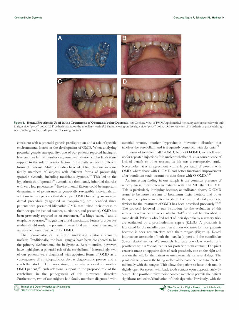
fabricated for the maxillary arch, as it is less obtrusive for most patients
because it does not interfere with their tongue (Figure 1). Dental
impressions are made of both the maxilla (upper) and the mandibular
(lower) dental arches. We routinely fabricate two clear acrylic resin
prostheses with a ‘‘pivot’’ center for posterior tooth contact. The pivot
center is made on opposite sides of each prosthesis, one on the right and
one on the left, for the patient to use alternately for several days. The
prosthesis only covers the biting surface of the back teeth so as to interfere
minimally with the tongue. This allows the patient to have their mouth
slightly open for speech with back tooth contact open approximately 3–
5 mm. The prosthesis pivot point contact somehow permits the patient
significant reduction/elimination of their dystonia. Previously, with the
Figure 1. Dental Prosthesis Used in the Treatment of Oromandibular Dystonia. (A) Occlusal view of PMMA (polymethyl methacrylate) prosthesis with built
in right side ‘‘pivot’’ point. (B) Prosthesis seated on the maxillary teeth. (C) Patient closing on the right side ‘‘pivot’’ point. (D) Frontal view of prosthesis in place with right
side touching and left side just out of closing contact.
Oromandibular Dystonia Gonzalez-Alegre P, Schneider RL, Hoffman H
Tremor and Other Hyperkinetic Movementshttp://www.tremorjournal.org
The Center for Digital Research and ScholarshipColumbia University Libraries/Information Services5

fabrication of one prosthesis with a pivot point, it was noted the dystonia
returned after several months of wear. The positive effect of the prosthesis
is extended with alternating the pivot point periodically as previously
described.22 We propose that this type of intervention should be pursued
in patients with OMD who benefit from a sensory trick, and certainly in
those for whom botulinum toxin does to provide enough benefit.
In summary, we describe a series of patients with O-OMD and C-
OMD seen in a single institution over a decade. Collectively with
previous reports, this study helps us better understand important
features of OMD. Furthermore, we identified several interesting
etiological, clinical, and therapeutic aspects of C-OMD and O-OMD
that should be prospectively evaluated in future, larger studies.
References
1. Albanese A, Bhatia K, Bressman SB, et al. Phenomenology and
classification of dystonia: A consensus update. Mov Disord 2013;28:863–873,
doi: http://dx.doi.org/10.1002/mds.25475.
2. Frucht SJ. The definition of dystonia: Current concepts and controversies.
Mov Disord 2013;28:884–888, doi: http://dx.doi.org/10.1002/mds.25529.
3. Fung VS, Jinnah HA, Bhatia K, Vidailhet M. Assessment of patients with
isolated or combined dystonia: An update on dystonia syndromes. Mov Disord
2013;28:889–898, doi: http://dx.doi.org/10.1002/mds.25549.
4. Pont-Sunyer C, Marti MJ, Tolosa E. Focal limb dystonia. Eur J Neurol
2010;17(Suppl 1):22–27, doi: http://dx.doi.org/10.1111/j.1468-1331.2010.
03046.x.
5. Strader S, Rodnitzky RL, Gonzalez-Alegre P. Secondary dystonia in a
botulinum toxin clinic: Clinical characteristics, neuroanatomical substrate and
comparison with idiopathic dystonia. Parkinsonism Relat Disord 2011;17:749–752,
doi: http://dx.doi.org/10.1016/j.parkreldis.2011.07.013.
6. Dauer WT, Burke RE, Greene P, Fahn S. Current concepts on the clinical
features, aetiology and management of idiopathic cervical dystonia. Brain 1998;
121(Pt 4):547–560, doi: http://dx.doi.org/10.1093/brain/121.4.547.
7. Singer C, Papapetropoulos S. A comparison of jaw-closing and jaw-
opening idiopathic oromandibular dystonia. Parkinsonism Relat Disord 2006;12:
115–118, doi: http://dx.doi.org/10.1016/j.parkreldis.2005.07.007.
8. Tan EK, Jankovic J. Botulinum toxin A in patients with oromandibular
dystonia: Long-term follow-up. Neurology 1999;53:2102–2107, doi: http://dx.
doi.org/10.1212/WNL.53.9.2102.
9. Bakke M, Larsen BM, Dalager T, Moller E. Oromandibular dystonia--
functional and clinical characteristics: A report on 21 cases. Oral Surg Oral Med
Oral Pathol Oral Radiol 2013;115:e21–26, doi: http://dx.doi.org/10.1016/j.oooo.
2012.04.023.
10. Merz RI, Deakin J, Hawthorne MR. Oromandibular dystonia
questionnaire (OMDQ-25): A valid and reliable instrument for measuring
health-related quality of life. Clin Otolaryngol 2010;35:390–396, doi: http://dx.
doi.org/10.1111/j.1749-4486.2010.02194.x.
11. Sinclair CF, Gurey LE, Blitzer A. Oromandibular dystonia: Long-term
management with botulinum toxin. Laryngoscope 2013 Dec;123(12):3078–83.
12. Defazio G. The epidemiology of primary dystonia: Current evidence and
perspectives. Eur J Neurol 2010;17(Suppl 1):9–14, doi: http://dx.doi.org/10.
1111/j.1468-1331.2010.03053.x.
13. Steeves TD, Day L, Dykeman J, Jette N, Pringsheim T. The prevalence
of primary dystonia: A systematic review and meta-analysis. Mov Disord 2012;27:
1789–1796, doi: http://dx.doi.org/10.1002/mds.25244.
14. Schmidt A, Jabusch HC, Altenmuller E, et al. Etiology of musician’s
dystonia: Familial or environmental? Neurology 2009;72:1248–1254, doi: http://
dx.doi.org/10.1212/01.wnl.0000345670.63363.d1.
15. Lohmann K, Klein C. Genetics of dystonia: What’s known? What’s new?
What’s next? Mov Disord 2013;28:899–905, doi: http://dx.doi.org/10.1002/mds.25536.
16. Scolding NJ, Smith SM, Sturman S, Brookes GB, Lees AJ. Auctioneer’s
jaw: A case of occupational oromandibular hemidystonia. Mov Disord 1995;10:
508–509, doi: http://dx.doi.org/10.1002/mds.870100418.
17. Diaz-Sanchez M, Martinez-Castrillo JC. Botulinum toxin in a task-
specific oromandibular dystonia in a bingo caller. J Neurol 2008;255:942–943,
doi: http://dx.doi.org/10.1007/s00415-008-0716-y.
18. Kang SY, Kim H, Ma HI, et al. Highly task-specific oromandibular
dystonia in a telephone operator. Eur J Neurol 2011;18:e136, doi: http://dx.doi.
org/10.1111/j.1468-1331.2011.03468.x.
19. Jinnah HA, Hess EJ. A new twist on the anatomy of dystonia: The basal
ganglia and the cerebellum? Neurology 2006;67:1740–1741, doi: http://dx.doi.
org/10.1212/01.wnl.0000246112.19504.61.
20. Waln O, LeDoux MS. Delayed-onset oromandibular dystonia after a
cerebellar hemorrhagic stroke. Parkinsonism Relat Disord 2010;16:623–625, doi:
http://dx.doi.org/10.1016/j.parkreldis.2010.07.010.
21. Hedera P, Phibbs FT, Fang JY, Cooper MK, Charles PD, Davis TL.
Clustering of dystonia in some pedigrees with autosomal dominant
essential tremor suggests the existence of a distinct subtype of essential
tremor. BMC Neurol 2010;10:66, doi: http://dx.doi.org/10.1186/1471-
2377-10-66.
22. Schneider R, Hoffman HT. Oromandibular dystonia: A clinical report.
J Prosthet Dent 2011;106:355–358, doi: http://dx.doi.org/10.1016/S0022-
3913(11)60145-5.
23. Maestre-Ferrin L, Burguera JA, Penarrocha-Diago M. Oromandibular
dystonia: A dental approach. Med Oral Patol Oral Cir Bucal 2010;15:e25–27.
Gonzalez-Alegre P, Schneider RL, Hoffman H Oromandibular Dystonia
Tremor and Other Hyperkinetic Movementshttp://www.tremorjournal.org
The Center for Digital Research and ScholarshipColumbia University Libraries/Information Services6