clinical anatomy of the skull of the barbados black belly...
TRANSCRIPT

Int. J. Curr. Res. Med. Sci. (2016). 2(8): 8-19
8
International Journal of Current Research inMedical Sciences
ISSN: 2454-5716www.ijcrims.com
Volume 2, Issue 8 -2016
Original Research Article SOI: http://s-o-i.org/1.15/ijcrms-2016-2-8-2
Clinical Anatomy of the skull of the Barbados Black BellySheep in Trinidad
Reda Mohamed1*, Marc Driscoll2 and Natasha Mootoo3
1Department of Basic Veterinary Sciences, Faculty of Medical Sciences, University of the West Indies,Trinidad and Tobago &Anatomy and Embryology Department, Faculty of Veterinary Medicine,
Beni Suef University, Egypt.2Department of Clinical Veterinary Sciences, Faculty of Medical Sciences, University of the West Indies,
Trinidad and Tobago.3Department of Clinical Veterinary Sciences, Faculty of Medical Sciences, University of the West Indies,
Trinidad and Tobago.*Corresponding Author: Reda Mohamed. E-mail: [email protected]
Abstract
The aim of this study is to determine the clinically important anatomical landmarks useful for regional anesthesia inthe head region of six adult Barbados Black Belly sheep in Trinidad. The applied anatomical measurements for 21parts of the skull were made for this study. The results were of clinical importance and are useful in regional nerveblocks of the supra-orbital, infra-orbital, mandibular and mental nerves during surgical operations in head region andin dental extraction. The application of local anesthetic agent was easier for the maxillary and mandibular nerveblocks through the injection of local anesthetic agent at the infra-orbital foramen and mental foramen respectively.These data were discussed with regard their application to clinical maneuvers around the head of sheep and goat.
Keywords: Clinical anatomy, Skull, sheep, Trinidad.
Introduction
The Barbados Black Belly is a breed of domesticsheep in the Caribbean island of Trinidad. Bothmale and female have no horns .This breed israised primarily for meat. It combines the rareattributes of adaption to the environment and highreproductive efficiency, which account for theiraverage of two lambs per litter and an averagelambing interval of eight to nine months. Thecranial nerves and their passages from differentforamina in the skull have clinical importance inregional anesthesia around the head (Hall et al.,2001). Studies of the clinical anatomy of the skull
has been done in the Mehraban sheep (Karimi etal., 2012), in the Iranian Native sheep (Monfared,2013), in the West African Dwarf goat (Olopadeand Onwuka, 2005), in the Black Bengal goat(Uddin et al., 2009), in the Iranian Native goat(Monfared et al., 2013), in the Makhoz goat(Goodarzi and Hoseini, 2013), in the Rasqueragoat (Pares-Casanova, 2014) and in the GwembeValley Dwarf goat (Kataba, 2014). This studywas done to provide information regarding theclinically important landmarks around the skull ofthe Barbados Black Belly sheep in Trinidad which

Int. J. Curr. Res. Med. Sci. (2016). 2(8): 8-19
9
can be used by the veterinary surgeons forregional anaesthesia as there is no documentedinformation.
Materials and Methods
I- Preparation of the skull
Six adult sheep of both sexes were collected fordissection purposes in gross anatomy lab, Schoolof Veterinary Medicine, West Indies University,Trinidad. The heads were without any skeletalabnormalities and were cut at the occipitoatlantaljoint. Skinning and defleshing of the heads weredone by using dissecting equipments such asscalpel, knives, forceps and scissors The cheekmuscles, eyes, tongue and nasal cartilages wereremoved. Also, the brain was removed by fillingthe cranial cavity with water and cleaning using along forceps with vigorous shaking and pouringoff through the foramen magnum. The processwas repeated several times until the brain wascompletely removed. The skulls were boiled in asuitable sized metal container for two hours thenthe soft tissue was removed with tooth brush thenwashed with tap water. The skull was maceratedby bacterial maceration by keeping it in a clothmesh immersed in a hot water and kept in aplastic container and stored in a warm place forsix weeks. The skull was taken out and scrapedusing a tooth brush and knife for cleaning. Theskull was left to dry for two weeks then bleachedby soaking in 3% hydrogen peroxide for four daysin a sealed container until the bones appearedclean and whitish in color. Finally, the skull wasthoroughly rinsed with water and left to dry fortwo weeks (Hildebrand, 1968 and Merai, 2012).
II- Anatomical landmarks
The following measurements and appropriateindices were calculated using measuring tapeaccording to Monfared (2013) in Iranian Nativesheep, Kataba (2014) in goat and Allouch (2014)in bovine.
1. Skull length: from the rostral end of thealveolar process of the incisive bone to theoccipital crest and divided into cranial andnasal skull lengths.
2. Nasal skull length: from the cranial edge ofthe maxillary bone cranially at the level
incisor tooth to cranial border of the frontalbone caudally.
3. Cranial skull length: from the cranial borderof the frontal bone cranially to occipital crest.
4. Infraorbital foramina distance: Width betweenthe infraorbital foramina.
5. Supra-orbital foramina distance: Greatestwidth between the two supra-orbital foramina.
6. Distance from the medial canthus to thesupraorbital foramen.
7. Distance from the medial canthus to theinfraorbital foramen.
8. Facial tuberosity to infra orbital foramen.9. Midpoint of the first upper premolar at its
alveolar border to infraorbital foramen10. Nasal process of the incisive bone to
infraorbital foramen11. Nasoincisive notch to infraorbital foramen.12. Mandibular length: from the level of alveolar
border of the incisive bone to the caudalborder of the mandible.
13. Lateral alveolar border of the first premolartooth to the mental foramen.
14. Caudal mandibular border to the mentalforamen.
15. Lateral alveolar border to mental foramen:Distance from the mental foramen to thelateral extent of the alveolar root of lowerincisor.
16. Ventral border of the mandible to mentalforamen.
17. Maximum mandibular height: from thehighest point of the coronoid process to thebasal level of the mandible.
18. Condyloid fossa to the base of the mandible.19. Caudal border of mandible to mandibular
foramen: the vertical line from the mandibularforamen to the caudal border of the mandible.
20. Base of mandible to mandibular foramen.21. Mandibular angle to mandibular foramen.
III- Statistical Analysis
All the measurements were expressed as meanmeasurements with the standard deviation (Mean± SD).
IV- Radiography of the sheep skull
Int. J. Curr. Res. Med. Sci. (2016). 2(8): 8-19
10
Results
The anatomical landmarks were of clinicalimportance and aid in regional nerve blocks of
mandibular, mental and infraorbital nerves.No differences between male and female skulls.
Table. I -Morphometry of the cranium of the Barbados Black Belly sheep (Figs. 2, 3 & 4)
Item Parameters (cm) Mean ± SD Figs1 Skull length 24.65±2.16 2/12 Nasal skull length 7.77±0.93 2/23 Cranial skull length 16.09±1.23 2/34 Infraorbital foramina distance 9.56±0.79 3/15 Supra orbital foramina distance 5.64±0.84 3/26 Medial canthus to supraorbital foramen 3.87±1.30 4/17 Medial canthus to infraorbital foramen 7.68±0.59 4/28 Facial tubercle to infra orbital foramen 3.16±0.70 4/39 Midpoint of the first upper premolar to infraorbital foramen 1.70±0.24 4/410 Nasal process of incisive bone to infra orbital foramen 6.51±0.63 4/511 Nasoincisive notch to infra orbital foramen 3.58±0.75 4/6
Table. II -Morphometric of the mandible of sheep (Figs 6, 7 & 8)
Item Parameters (cm) Mean ± SD Figs12 Mandibular length 18.16±1.53 6/113 lateral alveolar border of the first upper premolar tooth to
mental foramen2.25±0.38 6/2
14 caudal mandibular border to mental foramen 15.23±1.46 6/315 Lateral alveolar border to mental foramen 2.25±0.31 6/416 ventral border of the mandible to mental foramen 0.70±0.18 6/517 Maximum mandibular height 10.79±0.64 6/618 Condyloid fossa to base of the mandible 7.08±0.73 6/719 Caudal border of the mandible to mandibular foramen 2.3±0.25 7/120 Base of mandible to mandibular foramen 3.75±0.22 7/321 Mandibular angle to the mandibular foramen 3.41±0.25 7/2
The supraorbital foramen:
The supraorbital foramen was located dorsal tothe orbital cavity and it was detected easily bymeasuring the distance between the supraorbitalforamina (Fig.3/A) or measuring the distancedorsal to the medial canthus of the eye (Fig. 4/A).The supraorbital foramen was the pathway of thetrochlear nerve (Fig. 11/1) of the ophthalmicbranch of the trigeminal nerve which supplied theforehead and the middle two-thirds of the uppereyelid. To block the nerve, a needle was insertedinto the supraorbital foramen (Figs. 5/A, 11/A)which causes lacrimation from the eye,
desensitization of the area supplying and uppereye lid dropping.
The infraorbital foramen:
The infraorbital foramen (Figs. 1/A, 3/B & 4/C)was easily palpated under the levator labiimaxillaries and levator nasolabilais muscles, sothese muscles should be moved dorsally, theforamen was detected by using the distancebetween it and either medial canthus of the eye(Fig. 4/2) , facial tubercle (Fig. 4/3) which was avital and prominent, midpoint of the first upperpremolar (Fig. 4/4), nasal process of the incisivebone (Fig. 4/5) or nasoincisive notch (Fig. 4/6).

Int. J. Curr. Res. Med. Sci. (2016). 2(8): 8-19
11
The infraorbital foramen was the pathway of themaxillary nerve of the trigeminal nerve. Themaxillary nerve entered the maxilla through themaxillary foramen. After entrance into theinfraorbital canal, the nerve gave off alveolarbranches to the cheek teeth and branches tomaxillary sinuses then exited from the infraorbitalforamen as infraorbital nerve (Fig. 12/5) whichgave external nasal, internal nasal and maxillarylabial branches respectively to the skin of thelateral nasal region, mucosa and skin of the nasalvestibule and skin of the maxillary lip.
To block this nerve, a needle was inserted intoeither the maxillary foramen (Figs. 5. 11/B) or theinfraorbital foramen (Fig. 5, 11/C). it was easierto desensitize the nerve within the infraorbitalcanal and its exit. It was not necessary to insertthe needle to the entire distance of the canal for acomplete block because the local anesthetic drugprogressed caudally in the canal by pressure.
The mandibular foramen
The mandibular foramen was located on themedial surface of the mandible (Figs. 1/G, 7/A &8/D). It was detected by using the distancebetween the mandibular foramen and the caudalborder of the mandible (Fig. 7/1), mandibularangle (Fig. 7/2) or base of the mandible (Fig. 7/3).The inferior alveolar nerve was a branch of themandibular branch of the trigeminal nerve. Itpassed through the mandibular foramen supplying
the mandibular teeth, alveoli, gingiva as well asthe skin and mucosa of the lips and chin. Theinferior alveolar nerve was blocked difficulty byentering a needle medially and vertically into themandibular foramen, so the lower teeth and lowerlip on that side were desensitized. A potentialcomplication of haemorhage can occur in doingthe mandibular nerve block as the needle isinserted into the mandibular foramen (Figs. 9/A,10, 11/D) where the arteries and veins lie and theymay be injured during injection.
The mental foramen
The site of the mental foramen (Figs. 1/F, 6/A)was detected by using the distance between it andlateral alveolar border of the first premolar tooth(Fig. 6/2), caudal mandibular border (Fig. 6/3),lateral alveolar border of the mandible (Fig. 6/4)or ventral border of the mandible (Fig. 6/5). Tofeel the foramen, the depressor labii mandibulaismuscle was displaced dorsally. The mental nerve(Fig. 12/4) was the rostral continuation of theinferior alveolar branch after its emerging fromthe mental foramen supplying the chin and lips.The mental nerve can be injected close to its exitfrom the mental foramen on the lateral side of themandible rostral to the first check tooth. Theneedle was directed in a rostro-caudal directiontowards the foramen (Figs. 9/B, 11/E). Ipsilateraldropping and desensitization of the lower lip andincisor teeth was occurred.
A BFig.1 Gross (A) and Radiograph (B) of the Skull of the Black Belly sheep; Lateral view
A: Infraorbital foramen; B: Facial tubercle; C: First upper premolar tooth; D: Nasal process of incisivebone; E: Medial canthus of the eye; F: Mental foramen; G: Mandible; H: Condylar process of the mandible;I: Coronoid process of the mandible.
Int. J. Curr. Res. Med. Sci. (2016). 2(8): 8-19
12
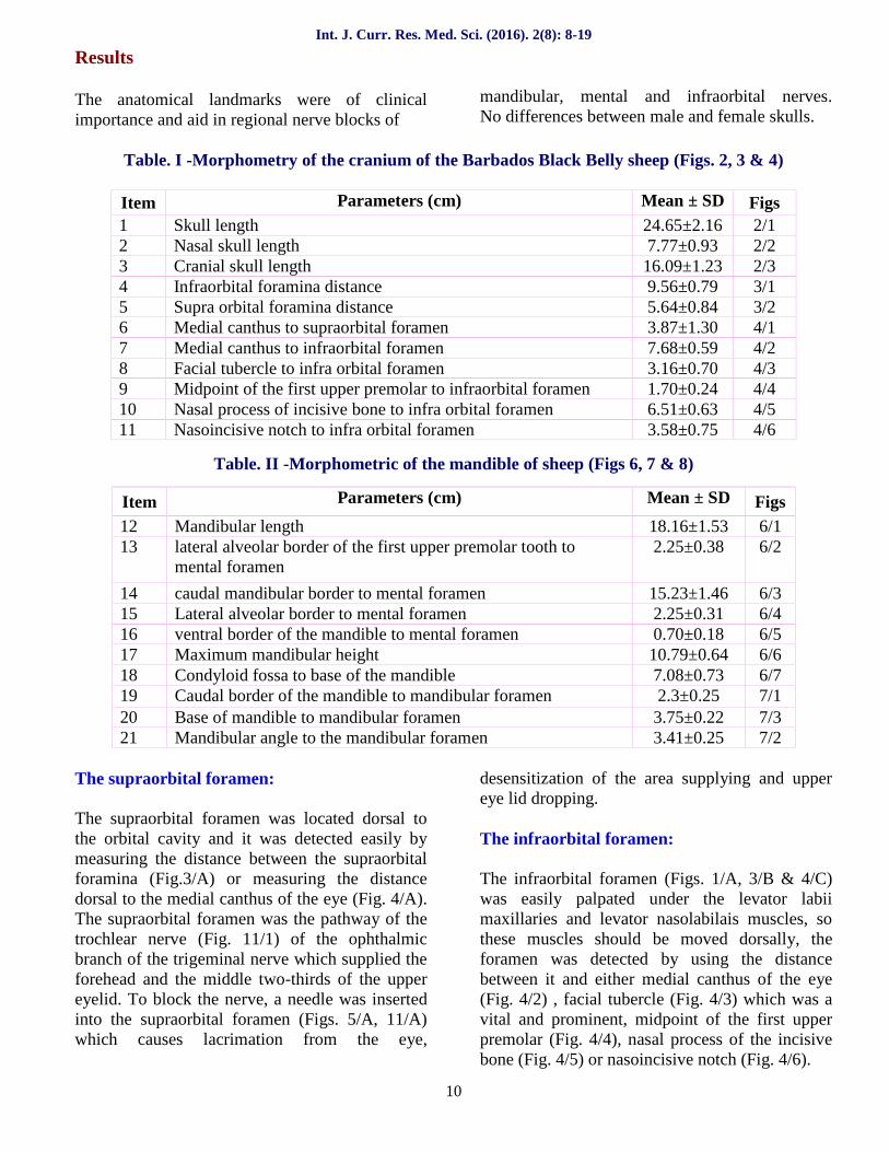
Fig. 2 Skull of the Black Belly shape
(Anatomical landmarks); Lateral view(Anatomical landmarks)A: Nasal process of the incisive boneB: Nasal bone C: Nasofrontal sutureD: Frontal bone E: Nuchal crest1: Skull length 2: Nasal length3: Cranial length
Fig. 3 Skull of the Black Belly shape
(Anatomical landmarks); Dorsal viewA: Supraorbital foraminaB: Infraorbital foramina1: Infraorbital foramina distance
2: Nasal length 2- Supraorbital foramina distance
A B
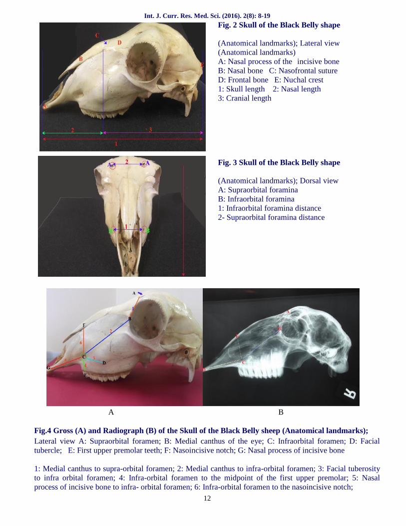
Fig.4 Gross (A) and Radiograph (B) of the Skull of the Black Belly sheep (Anatomical landmarks);Lateral view A: Supraorbital foramen; B: Medial canthus of the eye; C: Infraorbital foramen; D: Facialtubercle; E: First upper premolar teeth; F: Nasoincisive notch; G: Nasal process of incisive bone
1: Medial canthus to supra-orbital foramen; 2: Medial canthus to infra-orbital foramen; 3: Facial tuberosityto infra orbital foramen; 4: Infra-orbital foramen to the midpoint of the first upper premolar; 5: Nasalprocess of incisive bone to infra- orbital foramen; 6: Infra-orbital foramen to the nasoincisive notch;

Int. J. Curr. Res. Med. Sci. (2016). 2(8): 8-19
13
A B
Fig.5 Gross (A) and Radiograph (B) of the Skull of the Black Belly sheep; the site of needle in thesupraorbital foramina (A), maxillary foramen (B), Mental foramen (C), Lateral view. Note: 1: Skull 2:Mandible
Fig.6 Mandible of the Black Belly shape(Anatomical landmarks); Lateral viewA: Mental foramen; B: Lateral alveolar border;C: Caudal mandibular border; D: Lateralalveolar border of the first premolar tooth;
E: Ventral border of the mandible; F: Highestpoint of the coronoid process, G: Base of themandible; H: Condylar process.
1: Mandibular length; 2: Mental foramen to the lateral alveolar border of the first premolar tooth; 3: Mentalforamen to the caudal mandibular border; 4: lateral alveolar border to mental foramen; 5: Mental foramenthe ventral border of the mandible; 6: Maximum mandibular height; 7: Condyloid fossa to base of themandible
Fig.7 Mandible of the Black Belly shape(Anatomical landmarks); Medial view
A: Mandibular foramen; B: Caudal border ofthe mandible; C: Mandibular angle;D: Base of the mandible; 1: Mandibularforamen to the caudal border of themandible; 2: Mandibular foramen tothe mandibular angle; 3: Mandibularforamen to the base of the mandible
Int. J. Curr. Res. Med. Sci. (2016). 2(8): 8-19
14
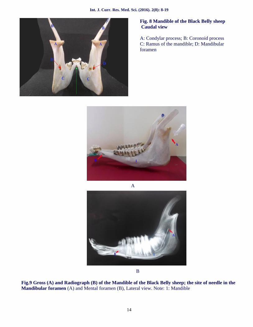
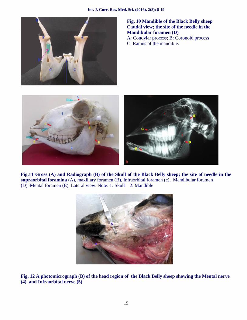
Fig. 8 Mandible of the Black Belly sheepCaudal view
A: Condylar process; B: Coronoid processC: Ramus of the mandible; D: Mandibularforamen
A
B
Fig.9 Gross (A) and Radiograph (B) of the Mandible of the Black Belly sheep; the site of needle in theMandibular foramen (A) and Mental foramen (B), Lateral view. Note: 1: Mandible
Int. J. Curr. Res. Med. Sci. (2016). 2(8): 8-19
15
Fig. 10 Mandible of the Black Belly sheepCaudal view; the site of the needle in the
V Mandibular foramen (D)A: Condylar process; B: Coronoid processC: Ramus of the mandible.
A B
Fig.11 Gross (A) and Radiograph (B) of the Skull of the Black Belly sheep; the site of needle in thesupraorbital foramina (A), maxillary foramen (B), Infraorbital foramen (c), Mandibular foramen(D), Mental foramen (E), Lateral view. Note: 1: Skull 2: Mandible
Fig. 12 A photomicrograph (B) of the head region of the Black Belly sheep showing the Mental nerve(4) and Infraorbital nerve (5)

Int. J. Curr. Res. Med. Sci. (2016). 2(8): 8-19
16
DiscussionTable. III- Comparison of the clinical anatomy landmarks of the Barbados Black Belly sheep with other sheep and goat breeds.
ParameterTI &II
BarbadosBlackBellysheep
Mehrabansheep
(Karimi etal. 2011)
IranianNativesheep
(Monfared,2013)
WestAfrican
Dwarf Goat(Olopade and
Onwuka,2005)
BlackBengal
goat(Uddin etal, 2009)
Kaganigoat
(Sarma,2006)
Red sokoto(Maradi) goat(Olopade and
Onwuka,2008)
Rasqueragoat
(Pares-Casanova,
2014)
Makhozgoat
(Goodarziand
Hoseini,2013)
IranianNativegoat
(Monfaredet al. 2013)
GwembeValleyDwarfgoat
(Kataba,2014)
1 24.65±2.16 20.06 ± 0.6 20.9 ± 4.77 16.99 ± 1.59 24.72 ± 0.93 20.3 ± 0.9 23.1 ± 1.01 15.02 ± 1.09
2 7.77±0.93 9.2 ± 0.46 5.8 ± 0.9
3 16.09±1.23 11.98 ± 0.24 11.7 ± 2.03 7.32 ± 0.93
4 9.56±0.79 6.1 ± 0.57
5 5.64±0.84 9.5 ± 1.44
6 3.87±1.30
7 7.68±0.59
8 3.16±0.70 1.87 ± 0.09 1.77 ± 0.26 1.81± 0.06 1.92 ± 0.17 2.06 ± 0-14
9 1.70±0.24 1.63 ± 0.44 1.7 ± 0.08 1.64 ± 0.11 1.13 ± 0.11
10 6.51±0.63
11 3.58±0.75
12 18.16±1.53 15.76 ± 1.59 14.08 ± 0.01 11.81 ± 1.10 14.21 ± 0.98 13.37 ± 0.67 14.1 ± 1.03 11.24 ± 0.52
13 2.25±0.38
14 15.23±1.46 13.74 ± 1.8 11.29 ± 0.47 9.75± 1.07 11.69 ± 0.4 11.42 ± 0.42 11.34± 0.96 9.26 ± 0.49
15 2.25±0.31 2.07 ± 0.45 2.76 ± 0.05 1.84± 0.76 2.11 ± 0.17 1.58 ± 0.11 2.40± 0.26 1.58 ± 0.19
16 0.70±0.18
17 10.79±0.64 9.75 ± 2.71 8.28 ± 0.48 7.11± 0.57 8.83 ± 0.57 8.94 ± 0.43 8.69± 0.18 6.64 ± 0.44
18 7.08±0.73 7.75 ± 0.96 6.26 ± 0.17 5.28± 0.57 6.38 ± 0.51 5.87 ± 0.44 4.37± 0.59 4.07 ± 0.37
19 2.3±0.25 1.35 ± 0.29 0.86 ± 0.03 0.9 ± 0.15 1.19 ± 017 1.10 ± 0.07
20 3.75±0.22 4.14 ± 0.47 2.99 ± 0.03 1.55 ± 0.25 3.64 ± 0.23 3.43 ± 0.25 2.88 ± 0.93 2.35 ± 0.26
21 3.41±0.25 2.82 ± 0.04 2.74 ± 0.17 2.18 ± 0.19

Int. J. Curr. Res. Med. Sci. (2016). 2(8): 8-19
17
The current investigation showed that the skulllength was higher that the value reported in theMehraban sheep (Karimi et al., 2011), in IranianNative sheep (Monfared, 2013), in West AfricanDwarf Goat (Olopade and Onwuka, 2005), in theRed sokoto goat (Olopade and Onwuka, 2008)and in the Gwembe Valley Dwarf goat (Kataba,2014). While, it was lower that the value reportedin the Kagani goat (Sarma, 2006).
In the present study, the nasal skull length waslonger than the value reported in the Mehrabansheep (Karimi et al., 2011) and in Kagani goat(Sarma, 2006).
The current work showed that the cranial skulllength was higher than the value mentioned byMonfared (2013) in the Iranian Native sheep.
The mean distance between the supra- orbitalforamina was lower than the value mentioned byMonfared (2013) in the Iranian Native sheep.
This study revealed that the distance between themedial canthus of the orbit and the supraorbitalforamen was 3.87 cm for Barbados Black Bellysheep. No values were given for other sheep inliterature.
The distance from the facial tuberosity to theinfra-orbital foramen was higher in BarbadosBlack Belly sheep than the value reported in theIranian Native sheep (Monfared, 2013), in theWest African Dwarf Goat (Olopade and Onwuka,2005), in the Makhoz goat (Goodarzi and Hoseini,2013), in the Iranian Native goat (Monfared et al.,2013) and in the Gwembe Valley Dwarf goat(Kataba, 2014)
The distances between the infraorbital foramenand the medial canthus and, the nasoincisivenotch and the nasal process of the incisive bonewere 7.68 cm, 3.58 cm and 6.51 cm respectively.Values are not given for other sheep.
The distance from the infra orbital foramen to themidpoint of the first upper premolar in theBarbados Black Belly sheep was similar to thatobserved in the Makhoz goat (Goodarzi andHoseini, 2013). However, it was lower for theIranian Native sheep (Monfared, 2013), the
Iranian Native goat (Monfared et al., 2013) andthe Gwembe Valley Dwarf goat (Kataba, 2014).
Our findings as well as those obtained by Hallet al. (2001) ascertained that the facial tuberosityis a prominent feature and can be used as a guidefor infra-orbital nerve block to desensitize theskin of the upper lip, nostril and face on that sideof the level of the infra-orbital foramen. Theinjection of local anesthetic drug within the infra-orbital canal leads to the analgesia and easilyextraction of the incisor, canine and first twopremolar teeth.
In the present study, the mandibular length andthe maximum mandibular height were higher thanthe values obtained in the Mehraban sheep(Karimi et al., 2011), the Iranian Native sheep(Monfared, 2013), the West African Dwarf goat(Olopade and Onwuka, 2005), the Black Bengalgoat (Uddin et al., 2009), the Makhoz goat(Goodarzi and Hoseini, 2013), the Iranian Nativegoat (Monfared et al., 2013) and the GwembeValley Dwarf goat (Kataba, 2014).
The obtained results showed that the distancefrom the condyloid fossa to the base of themandible was comparable to that reported in theMehraban sheep (Karimi et al., 2011) and washigher than in the Iranian Native sheep(Monfared, 2013), the West African Dwarf Goat(Olopade and Onwuka, 2005), the Black Bengalgoat (Uddin et al., 2009), the Makhoz goat(Goodarzi and Hoseini, 2013), the Iranian Nativegoat (Monfared et al., 2013) and the GwembeValley Dwarf goat (Kataba, 2014).
The obtained results showed that the distancefrom the caudal border of the mandible to thelevel of the mandibular foramen was higher thanthe values reported for the Mehraban sheep(Karimi et al., 2011), the Iranian Native sheep(Monfared, 2013), the Black Bengal goat (Uddinet al., 2009), the Makhoz goat (Goodarzi andHoseini, 2013) and the Gwembe Valley Dwarfgoat (Kataba, 2014).
The distance of the mandibular foramen to thebase of the mandible in the Barbados Black Bellysheep was nearly equal to what was observed inthe Black Bengal goat (Uddin et al., 2009) and inthe Makhoz goat (Goodarzi and Hoseini, 2013)
Int. J. Curr. Res. Med. Sci. (2016). 2(8): 8-19
18
but higher than was recorded in the Iranian Nativesheep (Monfared, 2013), the West African Dwarfgoat (Olopade and Onwuka, 2005), the IranianNative goat (Monfared et al., 2013) and theGwembe Valley Dwarf goat (Kataba, 2014).However Karimi et al. (2011) recorded a highervalue than the value reported in the present study.
The results showed that the distance from themandibular angle to the mandibular foramen washigher than the value reported in Iranian Nativesheep (Monfared, 2013), in the Makhoz goat(Goodarzi and Hoseini, 2013) and in the GwembeValley Dwarf goat (Kataba, 2014).
Observations of the present study confirmed thoseof Hall et al. (2001) that the parameters of themandibular foramen were of clinical importancefor attaining the regional anesthesia of themandibular foramen for desensitization of thelower jaw with its teeth and the lower lip on theside of the block.
The results showed that the distance between thelateral alveolar root and the mental foramen wascomparable to the reported value in the Mehrabansheep (Karimi et al., 2011), the Iranian Nativesheep (Monfared, 2013), the Black Bengal goat(Uddin et al., 2009) and the Iranian Native goat(Monfared et al., 2013). It was higher than in theWest African Dwarf Goat (Olopade and Onwuka,2005), the Makhoz goat (Goodarzi and Hoseini,2013), the West African Dwarf Goat (Olopadeand Onwuka, 2005), the Makhoz goat (Goodarziand Hoseini, 2013) and the Gwembe ValleyDwarf goat (Kataba, 2014).
In the present study, the distance between themental foramen to the caudal mandibular borderwas higher than the values obtained in theMehraban sheep (Karimi et al., 2011), the IranianNative sheep (Monfared, 2013), the West AfricanDwarf goat (Olopade and Onwuka, 2005), theBlack Bengal goat (Uddin et al., 2009), theMakhoz goat (Goodarzi and Hoseini, 2013), theIranian Native goat (Monfared et al., 2013) andthe Gwembe Valley Dwarf goat (Kataba, 2014).
In the present study, the distances between themental foramen to the lateral alveolar order of thefirst upper premolar tooth and to the ventralborder of the mandible were 2.25 cm and 0.70cmrespectively. No values were given for othersheep in literature.
Observations of the present study confirmed thoseof Hall et al. (2001), where the parameters of themental foramen are vital because injection oflocal anesthetic drugs can be made in the rostralaspect of the mandibular canal via the mentalforamen for blocking the infra-alveolar nerve, sothat desensitization of lower jaw with its teeth andthe lower lip will occur and this method is easierand avoids all risks of blood vessel injuries as inthe case of the infra-alveolar nerve block
The results were of clinical importance and willaid in the administration of regional nerve blocksby allowing easier methods like blocking themaxillary nerve via the injection of the localanesthetic agent through the infraorbital foramen.The mandibular nerve similarly can be blocked byinjecting the local anesthetic agent through themental foramen, in addition to the supra-orbitaland mental nerves which are useful duringsurgical operations in head region and dentalextraction or rasping. The results will also helpthe veterinarian to avoid the risks associated withthe use of general anaesthesia and the toxicity oflocal anaesthetic agents in sheep. Use Surgicalprocedures could be performed in the standingposition which would allow for shorter surgicaltime, less anesthetic equipment and low cost ofthe procedure.
Acknowledgements
I am very grateful to the technical staff and labassistants in the Departments of Basic andClinical Veterinary Sciences, School ofVeterinary Medicine, Faculty of MedicalSciences, University of the West Indies, Trinidadand Tobago for their assistance

Int. J. Curr. Res. Med. Sci. (2016). 2(8): 8-19
19
References
1. Allouch, G.M. 2014. Applied anatomy on themaxilla and mandibular regions of the bovinewith special reference to its important inregional anesthesia. Int. J. Food, Agricult.Veter. Sci.
2. Dyce, K.M., Sack, W.O., Wensing, C.J.G.2002. Textbook of veterinary anatomy, 3 rdedn. WB Saunders, Philadelphia
3. Goodarzi, N. and Hoseini, T.J., 2013.Morphometric Characteristics of theMaxillofacial and Mandibular Regions ofMarkhoz Goat Breed and its Clinical Valuefor Regional Anaesthesia in Western Iran.Global Veterinaria 11 (1): 107- 111.
4. Hall, L., Clarke, K.W., Trim, C.M.2001.Wright s Veterinary Anaesthesia andAnalgesia. 10th edn. ELBS and BaillierreTindall, London. Hildebrand, M. 1968.Anatomical preparations. University ofCalifornia Press. Berkely and Los Angeles,California.
5. Hildebrand, M. 1968. Anatomicalpreparations. University of California Press.Berkely and Los Angeles, California.
6. Karimi, I., Hadipour, M., Nikbakht, P. andMotamedi, S. 2011. The lower jawbone ofMeharaban sheep: A descriptivemorphometric approach. World’s Vet. J. 2 (4):57- 60.
7. Kataba, A. 2014. Biometric morphologiccharacteristics of the skull of the GwembeValley Dwarf goat (Capra hircus). MVSCthesis, The university of Zambia, School of
Veterinary Medicine, Biomedical SciencesDepartments, Lusaka.
8. Merai, M.K. 2012. Anatomical museumpreparations of the skeleton and respiratoryorgans of some domestic animals. M. V. Sc,Fac. of Vet. Med., Beni-Suef University.
9. Monfared, A. L. 2013. Clinical anatomy of theskull of Iranian Native sheep. GlobalVeternaria 10 (13), 271- 275.
10. Monfared, A. L., Naji, H and Sheibani, M T.2013. Applied anatomy of the head region ofthe Iranian Native goats (Capra hircus).Global Veternaria 10 (1), 60-64.
11. Olopade, J.O. and Onwuka, S.k. 2005. Anoestometric study of the skull of the WestAfrican Dwarf goat from South EasternNigeria. II- Mandibular and Maxillofacialfeatures (Short Communication). NigerianVeterinary Journal 27 (2): 66-70.
12. Olopade, J.O. and Onwuka, S.k. 2008. Acraniometric analysis of the skull of the redaokoto (Maradi) goat (Capra hircus). Eur J.Anat., 12 (1), 57-62.
13. Pares-Casanova, P.M. 2014. Osteometricstudy of the Rasquera goat. Journal of AppliedAnimal Research. 42 (2), 177-185.
14. Sarma, K.2006. Morphological andcraniometrical studies on the skull of Kaganigoat (Capra hircus) Jammu region. Int. J.Morphol., 24 (3): 449-455.
15. Uddin, M.M., Ahmed, S.S. U., Islam, K. N.and Islam, M. M., 2009. Clinical anatomy ofthe head region of the Black Bengal goat inBangladesh. International Journal ofMorpholology., 27(4):1269-1273.
How to cite this article:Reda Mohamed, Marc Driscoll and Natasha Mootoo. (2016). Clinical Anatomy of the skull of theBarbados Black Belly Sheep in Trinidad. Int. J. Curr. Res. Med. Sci. 2(8): 8-19.
Access this Article in Online
Website:www.ijcrims.com
Subject:VeterinarySciencesQuick Response Code