cleft-like formation of the aortic valve in an adult patient with a ... · aortic leaflet at both...
TRANSCRIPT
2341
□ CASE REPORT □
Cleft-Like Formation of the Aortic Valve in an AdultPatient with a Single Coronary Artery
Hiroaki Kawano 1, Tomayoshi Hayashi 2, Takako Minami 1,
Yuji Koide 1, Kiyoyuki Eishi 3 and Koji Maemura 1
Abstract
A 51-year-old woman was admitted to our hospital with heart failure due to aortic regurgitation. Examina-
tion showed severe aortic regurgitation mainly due to cleft-like deformity of the right coronary cusp and sin-
gle coronary artery. Aortic valve replacement was performed, and the deformity was seen in all aortic cusps.
Histological study showed elongation of the leaflets by myofibrotic and fibrotic hyperplasia without calcifica-
tion and inflammation in the aortic valve. This deformity likely arose from an acquired modification of a
congenitally malformed aortic valve.
Key words: aortic valve, aortic regurgitation, pathology, single coronary artery
(Intern Med 51: 2341-2345, 2012)(DOI: 10.2169/internalmedicine.51.7808)
Introduction
Aortic regurgitation (AR) can arise due to abnormalities
in the aortic leaflets (e.g., infective endocarditis, congenital
bicuspid valve, rheumatic valve, floppy valve), the aortic
root and annulus (e.g., syphilis, Marfan’s dissection), or
both (e.g., ankylosing spondylitis), and from disease affect-
ing neither the aorta nor the valve (e.g., ventricular septal
defect, systemic hypertension) (1-9). Abnormalities of the
aortic cusps leading to AR include congenital leaflet abnor-
malities, such as bicuspid, unicuspid, or quadricuspid valves,
or rupture of a congenitally fenestrated valve (9). The pres-
ent report describes a case of cleft-like formation of the tri-
cuspid aortic valve in a patient with AR and a left single
coronary artery.
Case Report
A 51-year-old woman was admitted to our hospital with
heart failure due to aortic regurgitation. She had no past his-
tory of rheumatic fever. Heart murmur was documented at
the age of 17, and she was diagnosed with asymptomatic
aortic regurgitation (Seller’s grade III) by aortography at our
hospital at the age of 22. Since then, she underwent routine
follow-up at our hospital every 3 months. She developed
chest discomfort at the age of 38, and a transthoracic echo
cardiography (TTE) showed a left ventricular ejection frac-
tion (LVEF) of 74%, LV end-diastolic dimension (LVDD) of
54 mm, aortic dimension of 32 mm, and moderate AR. At
the age of 44, aortic valve replacement was recommended
due to New York Heart Association classification status II
with LVDD of 55 mm and moderate AR by TTE, but the
patient declined. Her cardiomegaly gradually progressed,
and she ultimately agreed to undergo aortic valve replace-
ment at the age of 51.
On admission, physical examination revealed a height of
146 cm, a weight of 48.2 kg, blood pressure of 104/50
mmHg, and a regular pulse rate of 60 beats/min. A Levine
grade III diastolic murmur was heard at 3 left sternal border,
and a Levine grade II systolic murmur was heard at 2 right
sternal border, but the lung fields were clear to auscultation,
and she had no peripheral edema. Only mild cardiomegaly
was present on chest X-ray, and the electrocardiogram
(ECG) showed sinus rhythm and mild left ventricular hy-
pertrophy on admission.
1Department of Cardiovascular Medicine, Nagasaki University Graduate School of Biomedical Sciences, Japan, 2Department of Pathology, Na-
gasaki University Hospital, Japan and 3Department of Cardiovascular Surgery, Nagasaki University Graduate School of Biomedical Sciences, Ja-
pan
Received for publication March 26, 2012; Accepted for publication May 23, 2012
Correspondence to Dr. Hiroaki Kawano, [email protected]

Intern Med 51: 2341-2345, 2012 DOI: 10.2169/internalmedicine.51.7808
2342
Figure 1. Transesophageal echocardiography showed that the right coronary cusp with thickened free edge was mildly hypoplastic and that there was a gap in the center of the aortic valve orifice (A). Aortic regurgitant jet came from the central space made by three coronary cusps (B). N: non-coronary cusp, R: right coronary cusp, L: left coronary cusp
R
LN
A
B
Laboratory testing showed a white blood cell count of
6,000/mm3, blood urea nitrogen of 6 mg/dL, creatinine of
0.5 mg/dL, aspartate aminotransferase of 23 IU/L, alanine
aminotransferase of 22 IU/L, lactate dehydrogenase of 276
IU/L, creatine kinase of 139 IU/L, and C-reactive protein of
0.03 mg/dL.
A TTE demonstrated a LVEF of 63%, LVDD of 59 mm,
aortic dimension of 33 mm, and severe AR with mild mitral
regurgitation. Transesophageal echocardiography showed
that the right coronary cusp (RCC) was mildly hypoplastic
and that the AR jet came from the central space made by
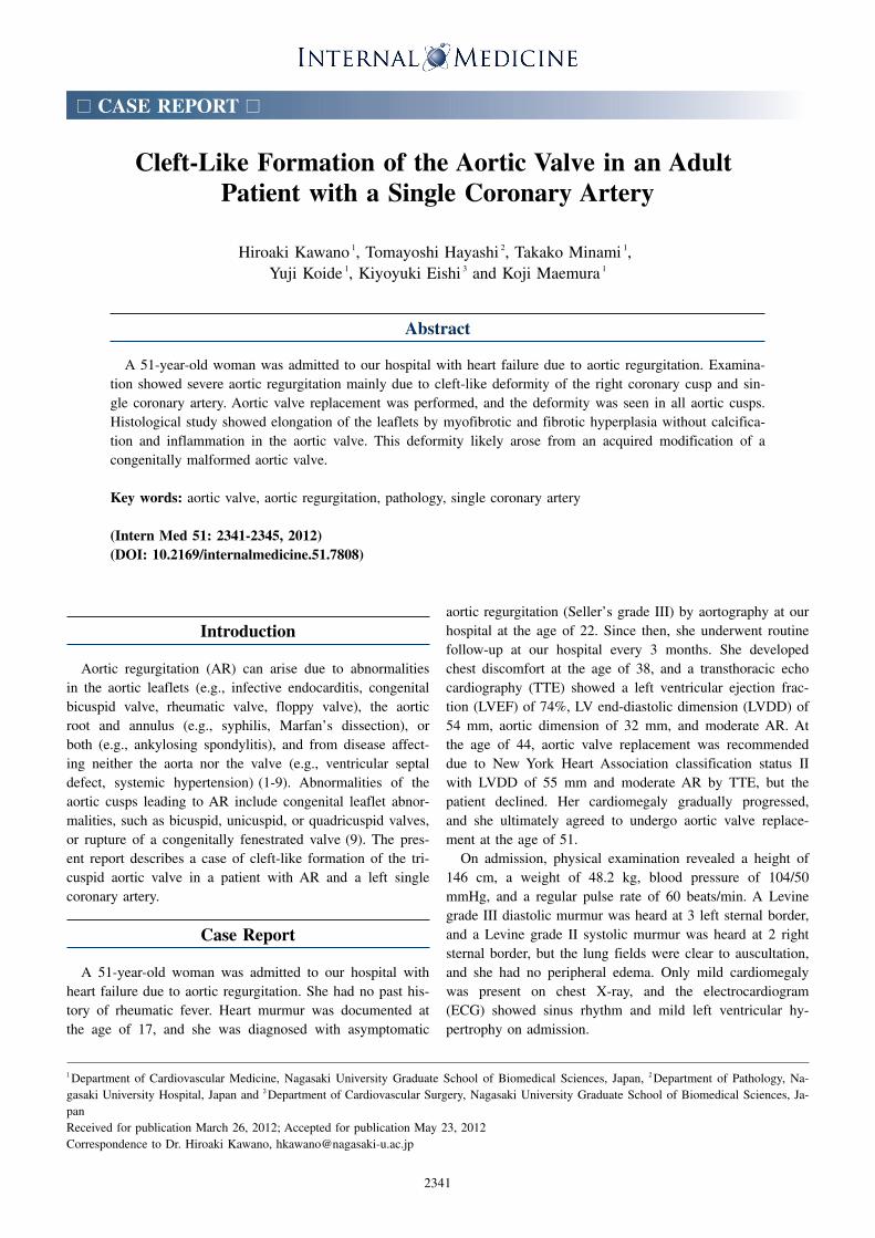
three coronary cusps (Fig. 1). Coronary angiogram (CAG)
showed a single coronary artery originating from the left
coronary cusp (LCC) and a right coronary artery branching
from the left ascending coronary artery and running to the
anterior wall of right ventricle (Fig. 2).
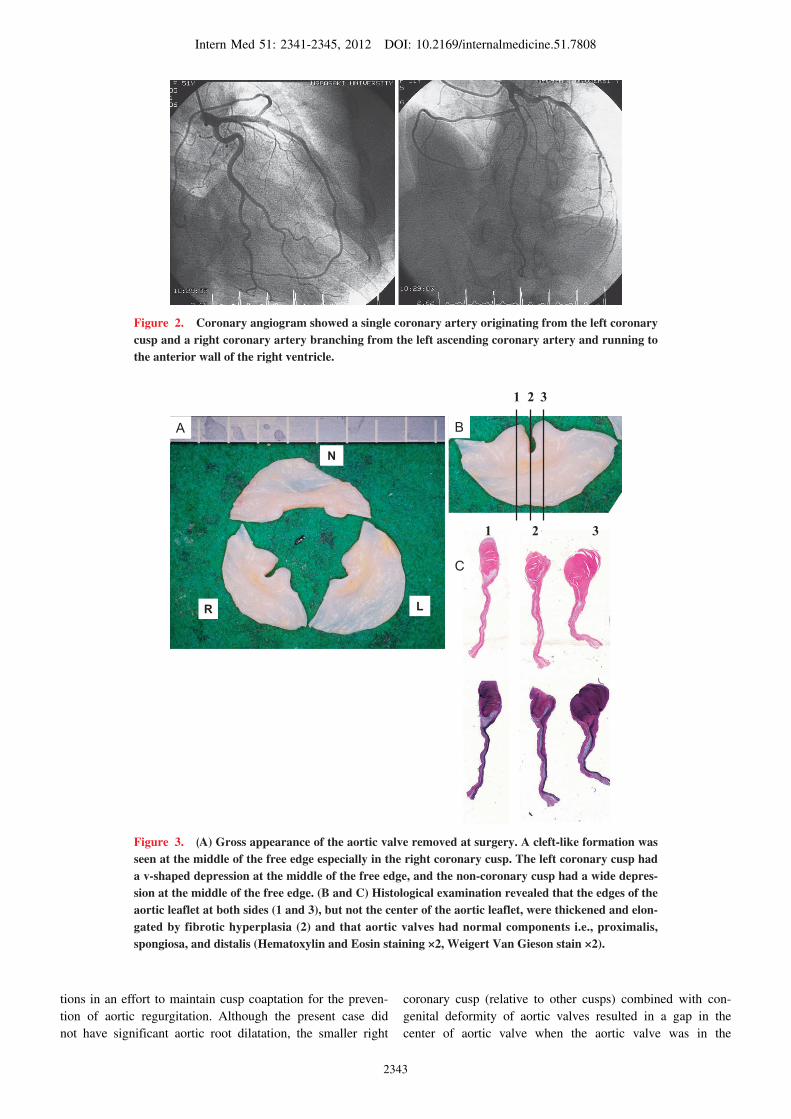
Aortic valve replacement was performed. The orifice of
the right coronary artery was not detected at operation. Mac-
roscopic examination of the aortic valve revealed that the
aortic valve was tricuspid and that the right coronary cusp
was smaller than the other cusps. Further, all the cusps were
deformed: 1) a cleft-like formation was seen at the middle
of the free edge especially in the RCC, 2) the left coronary
cusp had a v-shaped depression at the middle of the free
edge, and 3) the non-coronary cusp had a wide depression
at the middle of the free edge (Fig. 3A).
Histological examination revealed: 1) aortic valves with
normal components (i.e., proximalis, spongiosa, and distalis)
(Fig. 3C), 2) fibrous thickening and elongation of the ana-
tomical edge of these cusps except for the middle of the
anatomical edge of the cusps, which caused cleft-like forma-
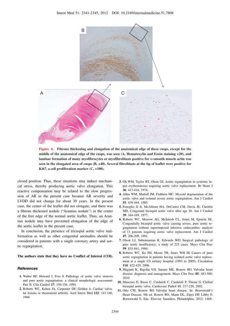
tion (Fig. 3C, 4A), and 3), laminar formation of many my-
ofibrocytes or myofibroblasts positive for α-smooth muscle
actin in the elongated area of the cusps (Fig. 4B), and sev-
eral fibroblasts at the tip of the leaflet were positive for Ki
67, a cell proliferation marker (Fig. 4C).
Discussion
One previous report describes a case of cleft of the aortic
valve (10), in which a cleft-like deformity was formed by
the fused portion of cusps in a patient with bicuspid aortic
valve. A congenital bicuspid aortic valve is present in about
1% to 2% of the population. Although it develops severe
AR, most patients with it develop severe aortic stenosis due
to calcification after 50 years of age (11). The present case
had a cleft-like formation of the tricuspid aortic valve be-
cause there was no fusion of the cusps confirmed by surgi-
cal and macroscopic inspection, and she had a single coro-
nary artery. An isolated single coronary artery is a rare con-
genital anomaly, occurring in approximately 0.024% of the
population (12, 13), and approximately one-third of cases
with a single coronary artery have other cardiovascular
anomalies (14). There are five case reports of patients with a
single coronary artery and AR (15-19), and four of these
five cases had other congenital anomalies (i.e., bicuspid aor-
tic valve, ventricular septal defect, or coronary aneu-
rysms) (16-19). Although there was only one adult case re-
port of a single coronary artery and AR without other con-
genital anomalies, the cause of AR was not determined or
described for that patient. Moreover, there have been no
published reports of a cleft-like aortic valve anomaly in a
patient with a single coronary artery in the absence of other
congenital anomalies.
In the present case, all three aortic leaflets were deformed
without calcification, and histologic evidence of inflamma-
tion was not present. This suggests that deformation of the
aortic valve was related to congenital malformation. More-
over, immunohistochemical staining showed that ongoing
proliferation of fibroblasts leading to elongation of the free
edge of the leaflet, resulted in a cleft-like formation. Taken
together, these observations suggest that this deformity
likely resulted from adaptive modification of a congenitally
malformed aortic valve.
The mechanism of elongation of the aortic valve is un-
clear. Previous studies suggested that dilatation of the aortic
root (20) or a non-compliant aortic root (21) can lead to en-
largement or elongation of leaflets, which may render leaf-
lets more susceptible to mechanical stress. Thus, aortic valve
enlargement or elongation may represent reactive compensa-
Intern Med 51: 2341-2345, 2012 DOI: 10.2169/internalmedicine.51.7808
2343
Figure 2. Coronary angiogram showed a single coronary artery originating from the left coronary cusp and a right coronary artery branching from the left ascending coronary artery and running to the anterior wall of the right ventricle.
Figure 3. (A) Gross appearance of the aortic valve removed at surgery. A cleft-like formation was seen at the middle of the free edge especially in the right coronary cusp. The left coronary cusp had a v-shaped depression at the middle of the free edge, and the non-coronary cusp had a wide depres-sion at the middle of the free edge. (B and C) Histological examination revealed that the edges of the aortic leaflet at both sides (1 and 3), but not the center of the aortic leaflet, were thickened and elon-gated by fibrotic hyperplasia (2) and that aortic valves had normal components i.e., proximalis, spongiosa, and distalis (Hematoxylin and Eosin staining ×2, Weigert Van Gieson stain ×2).
1 2 3
B
C
1 2 3
R L
N
A
tions in an effort to maintain cusp coaptation for the preven-
tion of aortic regurgitation. Although the present case did
not have significant aortic root dilatation, the smaller right
coronary cusp (relative to other cusps) combined with con-
genital deformity of aortic valves resulted in a gap in the
center of aortic valve when the aortic valve was in the
Intern Med 51: 2341-2345, 2012 DOI: 10.2169/internalmedicine.51.7808
2344
Figure 4. Fibrous thickening and elongation of the anatomical edge of these cusps, except for the middle of the anatomical edge of the cusps, was seen (A, Hematoxylin and Eosin staining ×20), and laminar formation of many myofibrocytes or myofibroblasts positive for α-smooth muscle actin was seen in the elongated area of cusps (B, ×40). Several fibroblasts at the tip of leaflet were positive for Ki67, a cell proliferation marker (C, ×100).
A
B
C
closed position. Thus, these situations may induce mechani-
cal stress, thereby producing aortic valve elongation. This
reactive compensation may be related to the slow progres-
sion of AR in the present case because AR severity and
LVDD did not change for about 30 years. In the present
case, the center of the leaflet did not elongate, and there was
a fibrous thickened nodule (“Arantius nodule”) in the center
of the free edge of the normal aortic leaflet. Thus, an Aran-
tius nodule may have prevented elongation of the edge of
the aortic leaflet in the present case.
In conclusion, the presence of tricuspid aortic valve mal-
formation as well as other congenital anomalies should be
considered in patients with a single coronary artery and aor-
tic regurgitation.
The authors state that they have no Conflict of Interest (COI).
References
1. Waller BF, Howard J, Fess S. Pathology of aortic valve stenosis
and pure aortic regurgitation: a clinical morphologic assessment:
Part II. Clin Cardiol 17: 150-156, 1994.
2. Roberts WC, Kehoe JA, Carpenter DF, Golden A. Cardiac valvu-
lar lesions in rheumatoid arthritis. Arch Intern Med 122: 141-146,
1968.
3. Oh WM, Taylor RT, Olsen GJ. Aortic regurgitation in systemic lu-
pus erythematosus requiring aortic valve replacement. Br Heart J
36: 413-416, 1974.
4. Allen WM, Matloff JM, Fishbein MC. Myxoid degeneration of the
aortic valve and isolated severe aortic regurgitation. Am J Cardiol
55: 439-444, 1985.
5. Fenoglio JJ Jr, McAllister HA, DeCastro CM, Davia JE, Cheitlin
MD. Congenital bicuspid aortic valve after age 20. Am J Cardiol
39: 164-169, 1977.
6. Roberts WC, Morrow AG, McIntosh CL, Jones M, Epstein SE.
Congenitally bicuspid aortic valve causing severe, pure aortic re-
gurgitation without superimposed infective endocarditis: analysis
of 13 patients requiring aortic valve replacement. Am J Cardiol
47: 206-209, 1981.
7. Olson LJ, Subramanian R, Edwards WD. Surgical pathology of
pure aortic insufficiency: a study of 225 cases. Mayo Clin Proc
59: 835-841, 1984.
8. Roberts WC, Ko JM, Moore TR, Jones WH III. Causes of pure
aortic regurgitation in patients having isolated aortic valve replace-
ment at a single US tertiary hospital (1993 to 2005). Circulation
114: 422-429, 2006.
9. Maganti K, Rigolin VH, Sarano ME, Bonow RO. Valvular heart
disease: diagnosis and management. Mayo Clin Proc 85: 483-500,
2010.
10. Mancuso D, Basso C, Cardaioli C, Cardaioli P, Thiene G. Clefted
bicuspid aortic valve. Cardiovasc Pathol 11: 217-220, 2002.
11. Otto CM, Bonow RO. Valvular heart disease. In: Brawnwald’s
Heart Disease. 9th ed. Bonow RO, Mann DL, Zipes DP, Libby P,
Brawnwald E, Eds. Elsevier Saunders, Philadelphia, 2012: 1468-
Intern Med 51: 2341-2345, 2012 DOI: 10.2169/internalmedicine.51.7808
2345
1539.
12. Sharbaugh AH, White RS. Single coronary artery. Analysis of the
anatomic variation, clinical importance, and report of five cases.
JAMA 230: 243-246, 1974.
13. Lipton MJ, Barry WH, Obrez I, Silverman JF, Wexler L. Isolated
single coronary artery: diagnosis, angiographic classification, and
clinical significance. Radiology 130: 39-47, 1979.
14. Ogden JA, Goodyer AV. Patterns of distribution of the single coro-
nary artery. Yale J Biol Med 43: 11-21, 1970.
15. Katsetos MC, Toce DT. Single coronary artery with aortic regurgi-
tation. Cardiovasc Intervent Radiol 26: 567-578, 2003.
16. Nanba M, Hanawa N, Mita H, Hongo M, Minemawari M. A case
of single coronary artery complicated with bicuspid aortic valve
and aortic regurgitation. Nippon Naika Gakkai Zasshi 80: 1959-
1961, 1991 (in Japanese).
17. Shore DF, Ho SY, Anderson RH, de Leval M, Lincoln C. Aorto-
pulmonary septal defect coexisting with ventricular septal defect
and pulmonary atresia. Ann Thorac Surg 35: 132-137, 1983.
18. Goto Y, Matsuno Y, Yoshikane H, et al. Ruptured aneurysm of the
sinus Valsalva with single coronary artery. Cathet Cardiovasc Di-
agn 17: 172-174, 1989.
19. Oohara K, Yamazaki T, Sakaguchi K, Nakayama M, Kobayashi A.
Acute aortic dissection, aortic insufficiency, and a single coronary
artery in a patient with Turner’s syndrome. J Cardiovasc Surg 36:
273-275, 1995.
20. Thubrikar MJ, Labrosse MR, Zehr KJ, Robicsek F, Gong GG,
Fowler BL. Aortic root dilatation may alter the dimensions of the
valve leaflets. Eur Cardiothorac Surg 28: 850-855, 2005.
21. Fokin AA, Robicsek F, Cook JW, Thubrikar MJ, Schaper J. Mor-
phological changes of the aortic valve leaflets in non-compliant
aortic roots: in-vivo experiments. J Heart Valve Dis 13: 444-451,
2004.
Ⓒ 2012 The Japanese Society of Internal Medicine
http://www.naika.or.jp/imonline/index.html