cleavable linkers
DESCRIPTION
biologyTRANSCRIPT

Bioorganic & Medicinal Chemistry 20 (2012) 571–582
Contents lists available at ScienceDirect
Bioorganic & Medicinal Chemistry
journal homepage: www.elsevier .com/locate /bmc
Cleavable linkers in chemical biology
Geoffray Leriche, Louise Chisholm, Alain Wagner ⇑Laboratory of Functional Chemo-Systems UMR 7199, 74 Route du Rhin, 67401 Illkirch-Graffenstaden, France
a r t i c l e i n f o a b s t r a c t
Article history:Available online 30 July 2011
Keywords:Cleavable linkerChemical biologyProteomicABPPBiocompatible
0968-0896/$ - see front matter � 2011 Published bydoi:10.1016/j.bmc.2011.07.048
⇑ Corresponding author. Fax: +33 368854306.E-mail address: [email protected] (A. W
Interest in cleavable linkers is growing due to the rapid development and expansion of chemical biology.The chemical constrains imposed by the biological conditions cause significant challenges for organicchemists. In this review we will present an overview of the cleavable linkers used in chemical biologyclassified according to their cleavage conditions by enzymes, nucleophilic/basic reagents, reducingagents, photo-irradiation, electrophilic/acidic reagents, organometallic and metal reagents, oxidizingreagents.
� 2011 Published by Elsevier Ltd.
1. Introduction
The ability to break chemical bonds that link two molecularentities can be an effective tool, and consequently many cleavablelinkers with this capability have been developed. Applications ofcleavable linkers have been characterized for organic synthesisand more recently chemical biology.
Progress in combinatorial chemistry, solid phase synthesis (SPS)and micro-chip chemistry in the early 1990s renewed the interestin cleavable linkers.1–4 These linkers connect the organic substrateto the solid support. They are required to be stable during the syn-thesis steps and are eventually cleaved to release the product. Thechoice of linker is crucial during the planning of the synthesis strat-egy. An array of linkers have been developed and optimized forspecific small molecule synthetic strategies; these were optimizedto be resistant to a diverse range of conditions involved in synthe-sis, while reactive towards other chemistries. Generally, these link-ers are cleaved under harsh chemical conditions. The linkerstrategies in solid-phase organic synthesis have been extensivelyreviewed.5
The recent rise of chemical biology has lead to the demand forcleavable linkers that are compatible with the biological moleculesand systems. Chemists started to reassess potential functionalgroups and cleavage reactions that could be used in biologicalchemistry, since the harsh conditions required by organic synthesislinkers are unsuitable. The new types of cleavable linkers have tosatisfy various challenging constraints, such as mild cleavageconditions, usage of bio-orthogonal reagents, high yield at low con-centrations, and the ease of eliminating excess of reagents andby-products. Additional technical requirements will depend on
Elsevier Ltd.
agner).
the specific application of the linker, for example often the objec-tive of proteomic experiments is to isolate and identify labelledproteins, which are expressed at low levels, and to determinewhere the label is incorporated in the protein.6–9 Consequently,the ideal cleavable affinity purification linkers enable the quantita-tive and specific release of proteins in low abundance from solidsupports, without generating by-products detrimental to subse-quent analysis, such as mass spectrometry (MS). The linkers mustbe resistant to the labelling and purification steps, but are also re-quired to be labile in mild conditions, so not to degrade the la-belled proteins.
In this review, we define a linker as a molecule with two func-tional heads joined together through a cleavable bond (Scheme 1).The functional heads serve to interact with or to manipulate thebiological target; they can be reactive groups for protein cross-linking, ligation or for click chemistry; fluorescent probes fordiagnostic tools, proteomic analysis or cell imaging; tags for MSanalysis or purification; targeting molecules for functional proteo-mics or activity based probe profiling (ABPP).
The flexibility in the design of the cleavable linkers has led totheir application in many different biological disciplines includingdrug development, proteomics, imaging and DNA sequencing.
Scheme 1. General representation of a cleavable probe.

572 G. Leriche et al. / Bioorg. Med. Chem. 20 (2012) 571–582
1.1. Drug development
Many drugs often cause unwanted, dose-limiting, and debilitat-ing side effects because they are untargeted, toxic compounds thatact on the normal and disease cells alike. Prodrugs and drug deliv-ery systems have been developed to try to reduce amount of off-target effects. Prodrugs are defined as chemicals which have littleor no pharmacological activity undergoing biotransformation to atherapeutically active metabolite. The objective of prodrugs is toimprove the pharmacokinetic and pharmacodynamic profile ofthe molecule.139 Prodrugs generally consist of two elements, theactive agent and a promoiety ‘‘carrier’’, which are joined togetherby a linker and are frequently activated by enzymatic hydrolysisor chemical conditions specific to the diseased tissue.
Drug delivery systems aim to administer an active drug toachieve a therapeutic effect and to improve the product’s efficacyand safety, compared to the drug alone. Many different drug deliverysystems have been developed and can be prepared from soluble andinsoluble, or biodegradable natural and synthetic polymers, micro-capsules, cells, cell ghosts, lipoproteins, liposomes, and micelles.All of the different carriers must be able to carry, target and releasethe drugs within the required timeframe, and at the desired location.Cleavable linkers used for drug delivery should be sensitive to enzy-matic hydrolysis or to chemical conditions.140,141
1.2. Proteomics
Cross-linking agents aim to form a covalent bond between twospatially adjacent residues within one or two polypeptide chains.To identify protein binding partners, the cross linking agents needto be able to detect and stabilize transient interactions. The cross-linking agents frequently form covalent links between lysine orcysteine residues in the proteins. Alternatively, the cross-linkingagent can be photoreactive. Cross-linking cleavable linkers can beused to distinguish between inter- and intra-protein interactionsof receptors, signalling cascades, and the structure of multi-proteincomplexes.142
Affinity purification is used to obtain an uncontaminated sam-ple of a protein or to determine a physical interaction between aprotein and interacting partners. The cleavable affinity purificationprobe can be used to conjugate the ligand to the solid support andit is cleaved to liberate the proteins under mild conditions. The li-
Table 1Examples of different cleavable groups and their applications
Cleavageconditions
Cleavable group
Enzymes TEV,10 trypsin,11,12 thrombin,13 cathepsin B,14 cathespin D,15 catmatrix metalloproteinase18–20 sequences, phosphodiester,21,22 pb-galactose25,26
Nucleophilic/basic reagents
Dialkyl dialkoxysilane,27–29 cyanoethyl group,30,31 sulfone,32 ethglycolyl disuccinate,33,34 2-N-acyl nitrobenzenesulfonamide,35 aunsaturated vinyl sulfide,37,38 sulfonamide after activation,39–41
indole derivative,42 levulinoyl ester,43 hydrazone,44 acylhydrazoReducing reagents Disulfide bridges,49–64 azo compounds65–79
Photo-irradiation 2-Nitrobenzyl derivatives,80–90 phenacyl ester,91,92 8-quinolinylcoumarin,94,95 phosphotriester,96 bis-arylhydrazone,97 bimane bderivative98
Electrophilic/acidic reagents
Paramethoxybenzyl derivative,99,100 tert-butylcarbamate analogudialkoxysilane,102 orthoester,103,104 acetal,103,105 aconityl,106,107 hb-thiopropionate,111–113 phosphoramidate,114 imine,115,116 trityl,polyketal,119,120 alkyl 2-(diphenylphosphino)benzoate derivative
Organometallicand metalcatalyst
Allyl ester,122,123 8-hydroxyquinoline ester,124 picolinate ester125
Oxidizingreagents
Vicinal diols,126–136 selenium compounds137,138
gand can be an inhibitor, a suicide substrate, a small molecule sub-strate (for ABPP), a metal ion chelate, or a drug. The identificationof the proteins can be determined through MS, and the purifiedproteins could be used in functional studies.143,144
1.3. Imaging
Medical imaging is used for instance to diagnose or examine dis-eases and to study anatomy and physiology. The detection and stag-ing of tumors currently relies on computed tomography, which hasthe disadvantages of being expensive and using radiation; whilemagnetic resonance imaging (MRI) scans are cheaper, they are lim-ited by the sensitivity of gadolinium chelates, or iron. Currently,25% of the MRI examinations use contrast agents to increase theclarity of the soft tissue being visualized. Recently developed MRIscan contrast agents contain cleavable linkers, which enable thedetection of enzymes involved in malignancy and metastasis.18,26
Molecular imaging can be used to characterize biological pro-cesses or conditions in vivo, using animal and cellular models.Most molecular imaging probes use inducible fluorescence to mon-itor biological reactions. Some rely on a cleavable linker to separatethe fluorophore from either a quencher or another fluorescent dye.The cleavable bonds can be responsive to specific conditions suchas enzyme activity, or the presence of thiol compounds.
1.4. DNA sequencing
Recently developed DNA sequencing by synthesis has reducedthe cost and increased the speed of sequencing. The developmentof this technology has been possible due to the conjugation ofnucleotides bases to a fluorescent probe via a cleavable linker.These linkers can be cleaved via exposure to light, palladium, orby enzymes. In DNA sequencing by synthesis, a DNA polymeraseincorporates a single fluorescently modified nucleotide, comple-mentary to the template base, and this stops the sequencing reac-tion. The remaining unincorporated nucleotides are washed away.The identity of the incorporated nucleotide is recorded and thenthe fluorescent dye is removed by a cleavage reaction. The nextincorporation step is started after washing. DNA sequencing isused in a wide range of research sectors including comparativegenomics and evolution, forensics, epidemiology, and appliedmedicine for diagnostics and therapeutics.145,146
Applications
hepsin K,16 caspase 1,17
hospholipid,23 ester,24Protein purification, imaging enzyme activity andtumor, drug delivery, DNA sequencing, metaboliteenrichment
ylene-thiophenylester,36
malondialdehyde (MDA)-ne,45 alkyl thioester46–48
Protein modification and purification, structuralbiology, imaging, synthesis of oligonucleotides
Protein modification and purification, structuralbiology, tumor-targeting, imaging, visualization ofPEG shedding, drug delivery
benzenesulfonate,93
i-thiopropionic acidProtein purification, imaging protein activity,structural biology, drug delivery, DNA sequencing,metabolite enrichment
e,101 dialkyl or diarylydrazone,55,108–110
117 vinyl ether,118
s121
Protein purification, structural biology, drugdelivery
DNA sequencing
Structural biology

G. Leriche et al. / Bioorg. Med. Chem. 20 (2012) 571–582 573
In this review we will present an overview of cleavable linkersclassified according to their cleavage conditions, along with aselection of their biological applications (Table 1).
2. Enzymatically cleavable linkers
In chemical biology, among the six different enzyme classes(oxidoreductases, transferases, hydrolases, lyases, isomerases, li-gases), hydrolases are widely used to induce chemical-bond cleav-age using water as the nucleophile. Many different types of bonds(ester, peptide, glycoside, etc.) are able to be cleaved but each onerequires a specific enzyme (esterase, protease, glycosidase, etc.).This enzyme–substrate specificity distinguishes enzymes fromother chemical catalysts and enables enzymes to be used as a cleav-age reagent. Proteases (also called peptidases) are enzymes thatcatalyze the breakdown of proteins by hydrolysis of peptide bonds.
Hydrolytic activity is restricted when one amino acid or a spe-cific sequence of amino acids is recognized by the enzyme. Thelonger the amino acid sequence required for the enzyme to recog-nize the cleavage site, the more specific the cleavage. Comparedto commonly used proteases, such as pepsin, trypsin or chymotryp-sin, virus proteases have more stringent sequence specificities.147
Speers et al. designed a tobacco etch virus protease (TEV)-sensitiveprobe, using the ENLYFQG sequence as the cleavable linker, a biotingroup for affinity purification and an azide for a click reaction.10 Theprobe was designed to be used in tandem orthogonal proteolysis(TOP) experiments. Following proteome labelling with an alkynylABPP probe, the cleavable biotin probe was attached to proteinsby a click reaction. The proteins were captured on a solid support,purified from the cell lysate and digested by trypsin. The MS anal-ysis of the tryptic peptides can be used to identify the protein.The proteins remaining on the solid support can be subsequentlycleaved with TEV and analyzed by MS to identify the probe’s label-ling site. In this study, cleavage times were overnight and 12 h fortrypsin and TEV proteases respectively. Based on this trypsin cleav-able linker approach, the same group developed a protease cleav-able probe strategy to chemically tag and enrich specific sets ofmetabolites based on shared functional-group composition.11
Since proteases have key roles in many diseases, protease-responsive linkers are widely used in drug release systems13 orin diagnostic tools.18,19,148 MRI scan contrast agents can detect spe-cific types of malignant and metastatic cancer. Activatable cellpenetrating peptides (ACPPs) are polycationic cell penetrating pep-tides conjugated to a polyanionic inhibitory domain which mini-mizes the peptides’ adsorption and cellular uptake. The release ofthe inhibitory domain by proteolytic cleavage leads to the uptakeof the ACPP by surrounding cells. ACPPs labeled with Cy5 or gado-linium(III) can be 2 and 9, which are overexpressed in a wide vari-ety of tumors.20 This can be used for disease progressionmonitoring or for fluorescence guided surgery. The use of fluores-cence imaging to visualize the tumor during surgery improved sur-vival in murine models of cancer.
Proteases are not the only enzymes that can be exploited inchemical biology; other enzyme-sensitive linkers have been devel-oped such as esterases and b-galactosidases. FRET-based probeswere designed to image esterase activities. The insertion ofphospholipid and phosphodiester groups between two fluorescentdyes or a dye/quencher pair allows the detection of phospholipaseand phosphodiesterase cleavage activity, respectively.21–23
According to our definition the linker’s cleavable group needs tobe modified on two positions; for enzymatically cleavable linkersthis requirement can affect enzyme–substrate recognition. Forexample, b-galactosidase can only tolerate the modification of itssubstrate, b-galactopyranoside, on the anomeric carbon. Komatsuet al. used quinone methide chemistry to overcome this problemand developed a FRET-based probe by conjugating a trifunctionnal
4-hydroxymandelic acid to b-galactopyranoside on the phenol po-sition, and adding two fluorescent dyes on the mandelate linkage,with a carbamate as the leaving group (Scheme 2).25 In thepresence of b-galactosidase, the sugar unit is removed to form qui-none methide. The species created reacts with a nucleophile andreleases acceptor of fluorescence to restore fluorescence ofdonor.
3. Nucleophile/base sensitive linkers
3.1. Halogen nucleophiles
Fluoridolyzable linkers are widely used in organic chemistry assilicon-based protecting groups for alcohols. The high thermody-namic affinity of fluorine for silicon allows their removal in orthog-onal and mild conditions using a fluorine source. In this reaction afluoride ion reacts with silicon as nucleophilic species and thecleavage conditions depend on the steric hindrance of the silicon’salkyl group. Since amino acid residues or DNA bases are not cova-lently altered by fluoride ions, fluoridolyzable linkers appear to bea suitable candidate for bioorthogonal and biocompatible cleavageconditions. Lin et al. were the first to describe silicon cleavage inphysiological conditions.27 The authors prepared a probe that con-tained a biotin group for affinity purification, a dialkoxy silanefunction as the cleavable group and a chloroacetyl ester to targetthe cysteine-protease papain. Once formed, the biotinylated pro-tein was purified by affinity chromatography using potassium fluo-ride as eluent. It was shown that elution was temperaturedependent, and 0.5 M potassium fluoride was required for a com-plete cleavage at 4 �C in less than 6 h. Since this work, this cleavagemethodology has been used for oligonucleotide purification ratherthan in the proteomics field.28,29
Fluoride ions can also trigger bond cleavage due to their basicproperties. Knapp et al. designed a fluoride cleavable linker basedon a 2-cyanoethyl function that is widely used as a protectinggroup in oligonucleotide synthesis.30 In basic conditions, the labileacidic vicinal proton of the nitrile function is removed to inducethe molecule dissociation by b-elimination. Associated with a re-porter molecule (like azide or NHS group), this linker was usedto reversibly label oligonucleotides. However, currently cleavageis limited to nonaqueous systems (TBAF 1 M in THF) and this linkeris only used in organic synthesis.
3.2. Oxygen nucleophiles
According to our literature review, two different oxygen nucleo-phile sensitive linkers were used with reactiveness toward hydroxylions. The first generation was developed for protein cross-linkingand is based on a sulfone linker, which could be cleaved at pH 11.6at 37 �C for 2 h.32 The second generation of linkers were the estergroups cleaved by saponification. The reactivity of the ester tocleavage can be enhanced by the use of electron-withdrawinggroups. In chemical biology, ester-based cleavable compounds wereinitially used for protein purification149 and in structural biology.The structure of protein complexes is determined by cross-linkingthe proteins and MS and MS/MS analysis; Petrotchenko et al.described a bi-reactive sulfo-NHS ester cross-linking reagent witha deuterated ethylene glycolyl disuccinate as the cleavable group.33
After cross-linking and enzyme digestion, cross-linked peptideswere incubated in a 1 M ammonium hydroxide cleavage solutionfor either 2 h or overnight at 25 �C. The cleavage cocktail was directlyanalyzed by MS and the interacting subunits of the HIV-reversetranscriptase complex were identified. For this approach, chemicalcleavable cross-linkers were used to identify the interpeptide andintrapeptide interactions within the HIV-reverse transcriptasecomplex.
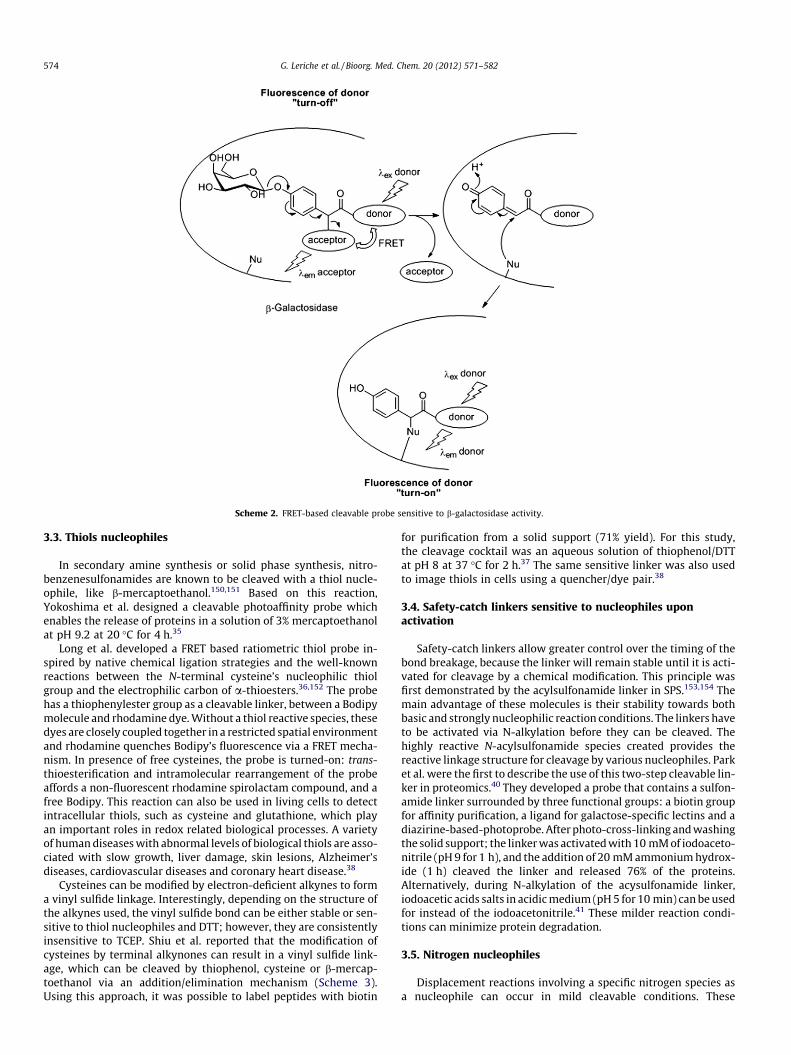
Scheme 2. FRET-based cleavable probe sensitive to b-galactosidase activity.
574 G. Leriche et al. / Bioorg. Med. Chem. 20 (2012) 571–582
3.3. Thiols nucleophiles
In secondary amine synthesis or solid phase synthesis, nitro-benzenesulfonamides are known to be cleaved with a thiol nucle-ophile, like b-mercaptoethanol.150,151 Based on this reaction,Yokoshima et al. designed a cleavable photoaffinity probe whichenables the release of proteins in a solution of 3% mercaptoethanolat pH 9.2 at 20 �C for 4 h.35
Long et al. developed a FRET based ratiometric thiol probe in-spired by native chemical ligation strategies and the well-knownreactions between the N-terminal cysteine’s nucleophilic thiolgroup and the electrophilic carbon of a-thioesters.36,152 The probehas a thiophenylester group as a cleavable linker, between a Bodipymolecule and rhodamine dye. Without a thiol reactive species, thesedyes are closely coupled together in a restricted spatial environmentand rhodamine quenches Bodipy’s fluorescence via a FRET mecha-nism. In presence of free cysteines, the probe is turned-on: trans-thioesterification and intramolecular rearrangement of the probeaffords a non-fluorescent rhodamine spirolactam compound, and afree Bodipy. This reaction can also be used in living cells to detectintracellular thiols, such as cysteine and glutathione, which playan important roles in redox related biological processes. A varietyof human diseases with abnormal levels of biological thiols are asso-ciated with slow growth, liver damage, skin lesions, Alzheimer’sdiseases, cardiovascular diseases and coronary heart disease.38
Cysteines can be modified by electron-deficient alkynes to forma vinyl sulfide linkage. Interestingly, depending on the structure ofthe alkynes used, the vinyl sulfide bond can be either stable or sen-sitive to thiol nucleophiles and DTT; however, they are consistentlyinsensitive to TCEP. Shiu et al. reported that the modification ofcysteines by terminal alkynones can result in a vinyl sulfide link-age, which can be cleaved by thiophenol, cysteine or b-mercap-toethanol via an addition/elimination mechanism (Scheme 3).Using this approach, it was possible to label peptides with biotin
for purification from a solid support (71% yield). For this study,the cleavage cocktail was an aqueous solution of thiophenol/DTTat pH 8 at 37 �C for 2 h.37 The same sensitive linker was also usedto image thiols in cells using a quencher/dye pair.38
3.4. Safety-catch linkers sensitive to nucleophiles uponactivation
Safety-catch linkers allow greater control over the timing of thebond breakage, because the linker will remain stable until it is acti-vated for cleavage by a chemical modification. This principle wasfirst demonstrated by the acylsulfonamide linker in SPS.153,154 Themain advantage of these molecules is their stability towards bothbasic and strongly nucleophilic reaction conditions. The linkers haveto be activated via N-alkylation before they can be cleaved. Thehighly reactive N-acylsulfonamide species created provides thereactive linkage structure for cleavage by various nucleophiles. Parket al. were the first to describe the use of this two-step cleavable lin-ker in proteomics.40 They developed a probe that contains a sulfon-amide linker surrounded by three functional groups: a biotin groupfor affinity purification, a ligand for galactose-specific lectins and adiazirine-based-photoprobe. After photo-cross-linking and washingthe solid support; the linker was activated with 10 mM of iodoaceto-nitrile (pH 9 for 1 h), and the addition of 20 mM ammonium hydrox-ide (1 h) cleaved the linker and released 76% of the proteins.Alternatively, during N-alkylation of the acysulfonamide linker,iodoacetic acids salts in acidic medium (pH 5 for 10 min) can be usedfor instead of the iodoacetonitrile.41 These milder reaction condi-tions can minimize protein degradation.
3.5. Nitrogen nucleophiles
Displacement reactions involving a specific nitrogen species asa nucleophile can occur in mild cleavable conditions. These

Scheme 3. Chemoselective cysteine modification by electron-deficient alkynes and cleavage of the formed vinyl sulfide linkage by thiophenol.
G. Leriche et al. / Bioorg. Med. Chem. 20 (2012) 571–582 575
reactions can be classified into two groups; cleavage by aminolysisor exchange reaction.
For aminolysis cleavage, examples include the cleavage of amalondialdehyde (MDA) indole derivative by either pyrrolidineor hydrazine,42 and the cleavage of an ester linker by hydroxyl-amine34 or hydrazine.43 In organic synthesis, a levulinoyl ester,an acid stable protecting group, can be removed either by sodiumborohydride, sulfite anion donors, or by hydrazinolysis.155 Thepresence of the c-keto ester group in levulinoyl esters allows thereaction with hydrazine to occur in mild conditions, generatingthe corresponding alcohol and dihydropyridazinone compounds.Geurink et al. fine-tuned the levulinoyl ester structure to overcomebase lability.43 An optimized affinity probe was designed to containa cleavable levulinoyl ester linked to a biotin tag and a selectiveproteasome inhibitor. After capture and pull-down, proteins werequantitatively released from the solid support using 100 mMhydrazine solution at 38 �C at pH 7.5 for 15 h.
Acylhydrazones44 and hydrazones45,156 can be used as cleavablelinkers through transimination in a mildly acidic medium. Anamine catalyst (e.g., aniline, p-anisidine or hydroxylamine) acceler-ates hydrolysis and enables the effective transition between stableand dynamic states, which is required for cleavage and exchange.This approach has the advantage of enabling the replacement ofone functional group with another, when the linker is cleaved; thisprovides additional possibilities for protein labelling. For example,authors reported purification proteins by biotin–avidin capture fol-lowed by exchange of the biotin tag for a fluorescent dye or an iso-tope-coded affinity tag (ICAT) for further analysis. To identify newbinding partners of anticoagulant protein S, Dirksen et al. modifiedprotein S with a purification probe which contained a bisarylhyd-razone cleavage site and a biotin tag (Scheme 4).44 Proteins com-plexes were eluted using a mixture of 100 mM hydroxylamineand 100 mM aniline at pH 6.0 at room temperature for overnight.Since the use of strongly nucleophilic amines could induce proteindenaturation, Dirksen et al. proposed an alternative strategy tocleave the hydrazone bond using an electrophilic reagent, like analdehyde. However, this strategy also requires aniline as a catalystfor transimination, because the aldehyde cannot directly react withthe hydrazone linkage. Although this method reduced the proteins
yield, it released aldehyde-functionalized proteins, which provideda new group for chemical modification.
4. Reduction sensitive linkers
Reduction sensitive linkages have been used in chemical biol-ogy for a long time and it is a commonly used class of cleavable lin-ker. However, there are only two types of cleavable linkerssensitive to reductive conditions: disulfide bridges and azocompounds.
4.1. Disulfide bridges
In naturally occurring proteins, disulfide bridges generally playa role in maintaining the protein structure. They are known to beefficiently and rapidly cleaved by mild reducing agents like dithio-threitol (DTT), b-mercaptoethanol or tris(2-carboxyethyl)phos-phine (TCEP). In chemical biology, disulfide bridges have beenused in a wide range of applications including functional and struc-tural proteomics,49–54 drug delivery,55–57 tumor imaging,59,60 DNAand protein–DNA complex purifications.61,62 The disulfide-basedcleavable linker is commonly used due to its straightforward syn-thesis and rapid cleavage. However, these linkers have several dis-advantages including their instability towards both electrophilicand nucleophilic polar reagents. This subjects disulfide linkers tothiol exchanges and can lead to non-specific cleavage in intracellu-lar conditions.
Pullela et al. designed a fluorescent probe to detect thiolsin vitro and in vivo.63 Fluorescein and rhodamine dyes were intro-duced on either side of a phenyl disulfide linker, and the fluores-cence was turned-on by endogenous thiols cleaving the linker inzebrafish embryos.
Based on extracellular disulfide exchange principle, Zhang et al.developed a prodrug system.56 The anticancer drug Camptothecin(CPT) is a potent topoisomerase I inhibitor but it is poorly solubleand is instable under physiological conditions. The prodrug strat-egy adopted targeted CPT to the tumors by linking the drug to neu-ropeptide substance P (SP) via a disulfide bond. SP can specificallybind to the neurokinin-1 receptor, which is overexpressed in many
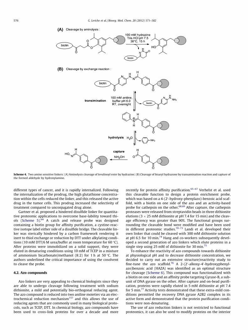
Scheme 4. Two amine sensitive linkers: (A) Aminolysis cleavage of levulinoyl ester by hydrazine; (B) Cleavage of bisaryl hydrazone by transamination reaction and capture ofthe formed aldehyde by hydroxylamine.
576 G. Leriche et al. / Bioorg. Med. Chem. 20 (2012) 571–582
different types of cancer, and it is rapidly internalized. Followingthe internalization of the prodrug, the high glutathione concentra-tion within the cells reduced the linker, and this released the activedrug in the tumor cells. This prodrug increased the selectivity oftreatment compared to unconjugated drug alone.
Gartner et al. proposed a hindered disulfide linker for quantita-tive proteomic applications to overcome base-lability toward thi-ols (Scheme 5).64 A catch and release probe was designedcontaining a biotin group for affinity purification, a cystine-reac-tive isotope label either side of a disulfide bridge. The cleavable lin-ker was sterically hindered by a carbon framework rendering itinert to thiol exchange or reduction by DTT under alkylating condi-tions (10 mM DTT/6 M urea/buffer at room temperature for 60 �C).After proteins were immobilized on a solid support, they wereeluted in denaturing conditions using 10 mM of TCEP in a mixtureof ammonium bicarbonate/methanol (8:2) for 1 h at 50 �C. Theauthors underlined the critical importance of using the cosolventto cleave the probe.
4.2. Azo compounds
Azo linkers are very appealing to chemical biologists since theyare able to undergo cleavage following treatment with sodiumdithionite, a mild and potentially bio-orthogonal reducing agent.The azo compound is reduced into two aniline moieties via an elec-trochemical reduction mechanism157 and this allows the use ofreducing agents that are commonly used in many biological proto-cols, such as TCEP, DTT. In chemical biology, azo compounds havebeen used to cross-link proteins for over a decade and more
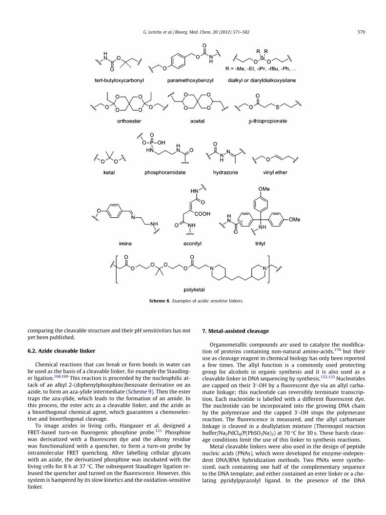
recently for protein affinity purification.65–67 Verhelst et al. usedthis cleavable function to design a protein enrichment probe,which was based on a 4-(20-hydroxy-phenylazo)-benzoic acid scaf-fold, with a biotin on one side of the azo and an activity-basedprobe for cathepsin on the other.68,69 After capture, the cathepsinproteases were released from streptavidin beads in three dithioniteelutions (3 � 25 mM dithionite at pH 7.4 for 15 min) and the cleav-age efficiency was greater than 90%. The functional groups sur-rounding the cleavable bond were modified and have been usedin different proteomic studies.70–73 Landi et al. developed theirown linker that could be cleaved with 300 mM dithionite solutionat pH 6.5 for 10 min.74 Hang and co-workers subsequently devel-oped a second generation of azo linkers which elute proteins in asingle step using 25 mM of dithionite for 30 min.75
To enhance the reactivity of azo compounds towards dithioniteat physiological pH and to decrease dithionite concentration, wedecided to carry out an extensive structure/reactivity study tofine-tune the azo scaffold.76 A 2-(20-alkoxy-40-hydroxyphenyl-azo)benzoic acid (HAZA) was identified as an optimal structurefor cleavage (Scheme 6). This compound was functionalized witha biotin on one side and an affinity probe targeting Gyrase-B, a sub-unit of DNA gyrase on the other. After capture and on-bead purifi-cation, proteins were rapidly eluted in 5 mM dithionite at pH 7.4for 5 min.77 Activity tests demonstrated that these extra-mild con-ditions permitted the recovery DNA gyrase A2B2 complex in itsactive form and demonstrated that the protein purification condi-tions were non-denaturing.
The use of azo reduction linkers is not restricted to functionalproteomics, it can also be used to modify proteins on the interior
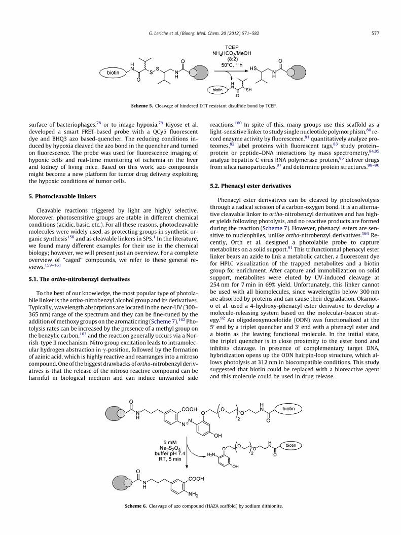
Scheme 5. Cleavage of hindered DTT resistant disulfide bond by TCEP.
G. Leriche et al. / Bioorg. Med. Chem. 20 (2012) 571–582 577
surface of bacteriophages,78 or to image hypoxia.79 Kiyose et al.developed a smart FRET-based probe with a QCy5 fluorescentdye and BHQ3 azo based-quencher. The reducing conditions in-duced by hypoxia cleaved the azo bond in the quencher and turnedon fluorescence. The probe was used for fluorescence imaging ofhypoxic cells and real-time monitoring of ischemia in the liverand kidney of living mice. Based on this work, azo compoundsmight become a new platform for tumor drug delivery exploitingthe hypoxic conditions of tumor cells.
5. Photocleavable linkers
Cleavable reactions triggered by light are highly selective.Moreover, photosensitive groups are stable in different chemicalconditions (acidic, basic, etc.). For all these reasons, photocleavablemolecules were widely used, as protecting groups in synthetic or-ganic synthesis158 and as cleavable linkers in SPS.1 In the literature,we found many different examples for their use in the chemicalbiology; however, we will present just an overview. For a completeoverview of ‘‘caged’’ compounds, we refer to these general re-views.159–161
5.1. The ortho-nitrobenzyl derivatives
To the best of our knowledge, the most popular type of photola-bile linker is the ortho-nitrobenzyl alcohol group and its derivatives.Typically, wavelength absorptions are located in the near-UV (300–365 nm) range of the spectrum and they can be fine-tuned by theaddition of methoxy groups on the aromatic ring (Scheme 7).162 Pho-tolysis rates can be increased by the presence of a methyl group onthe benzylic carbon,163 and the reaction generally occurs via a Nor-rish-type II mechanism. Nitro group excitation leads to intramolec-ular hydrogen abstraction in c-position, followed by the formationof azinic acid, which is highly reactive and rearranges into a nitrosocompound. One of the biggest drawbacks of ortho-nitrobenzyl deriv-atives is that the release of the nitroso reactive compound can beharmful in biological medium and can induce unwanted side
Scheme 6. Cleavage of azo compound (H
reactions.160 In spite of this, many groups use this scaffold as alight-sensitive linker to study single nucleotide polymorphism,80 re-cord enzyme activity by fluorescence,81 quantitatively analyze pro-teomes,82 label proteins with fluorescent tags,83 study protein–protein or peptide–DNA interactions by mass spectrometry,84,85
analyze hepatitis C virus RNA polymerase protein,86 deliver drugsfrom silica nanoparticules,87 and determine protein structures.88–90
5.2. Phenacyl ester derivatives
Phenacyl ester derivatives can be cleaved by photosolvolysisthrough a radical scission of a carbon-oxygen bond. It is an alterna-tive cleavable linker to ortho-nitrobenzyl derivatives and has high-er yields following photolysis, and no reactive products are formedduring the reaction (Scheme 7). However, phenacyl esters are sen-sitive to nucleophiles, unlike ortho-nitrobenzyl derivatives.164 Re-cently, Orth et al. designed a photolabile probe to capturemetabolites on a solid support.91 This trifunctionnal phenacyl esterlinker bears an azide to link a metabolic catcher, a fluorescent dyefor HPLC visualization of the trapped metabolites and a biotingroup for enrichment. After capture and immobilization on solidsupport, metabolites were eluted by UV-induced cleavage at254 nm for 7 min in 69% yield. Unfortunately, this linker cannotbe used with all biomolecules, since wavelengths below 300 nmare absorbed by proteins and can cause their degradation. Okamot-o et al. used a 4-hydroxy-phenacyl ester derivative to develop amolecule-releasing system based on the molecular-beacon strat-egy.92 An oligodeoxynucelotide (ODN) was functionalized at the50 end by a triplet quencher and 30 end with a phenacyl ester anda biotin as the leaving functional molecule. In the initial state,the triplet quencher is in close proximity to the ester bond andinhibits cleavage. In presence of complementary target DNA,hybridization opens up the ODN hairpin-loop structure, which al-lows photolysis at 312 nm in biocompatible conditions. This studysuggested that biotin could be replaced with a bioreactive agentand this molecule could be used in drug release.
AZA scaffold) by sodium dithionite.

Scheme 7. Examples of photocleavable linkers; (dashed line = sites of cleavage).
578 G. Leriche et al. / Bioorg. Med. Chem. 20 (2012) 571–582
5.3. Others photocleavable linkers
Other photolabile linkages were previously used to purifyligand–receptor complexes,93 to generate photocleavable peptides/proteins94 or for drug delivery (Scheme 7).95 We will limit this sec-tion to the application of photo-sensitive linkages for MS analysis.
During MS analysis, the cleavage of photo-sensitive cross-link-ing agents needs to incorporate a label into the cross-linked pep-tides, which enables the identification of cross-linked peptidesfrom other peptides. Gardner et al. presented two generations ofcross-linking agents. The first is based on a triester phosphategroup which is cleaved upon IR irradiation by a CO2 laser.96 Thesecond is based on a bis-aryl hydrazone linker and is cleaved underUV irradiation at 355 nm.97 In 2009, Petrotchenko et al. designed anew cross-linking agent: a bimane bisthiopropionic acid N-succin-imidyl ester (BiPS), which is photocleavable, fluorescent, homobi-functional, amine reactive and isotopically coded.98 This moleculeenables the identification of cross-linked proteins by fluorescenceand MS. In this application, cleavage is laser photo-induced and oc-curs directly in MALDI (Matrix-Assisted Laser Desorption/Ioniza-tion) conditions.
6. Electrophile/acid sensitive linkers
Two different modes of electrophilic cleavage are used in chem-ical biology: acidic sensitive linkers that are sensitive to proton
sources, and alkyl 2-(diphenylphosphino)benzoate derivatives sen-sitive to azide compounds.
6.1. Acid cleavable linker
Proton sensitive bonds are among the most frequently usedcleavable functions in organic chemistry; illustrated by the devel-opment of the BOC group which protects amines or the Merrifieldresin used in solid phase synthesis. In organic chemistry, the cleav-age conditions that can be tolerated are very flexible regarding theacids’ reagents, solvents, temperatures and pH. In contrast, biocom-patible acid cleavable linkers must be responsive to minor changesin pH. Strong acidic conditions can lead to the denaturation of pro-teins and DNA. Biocompatible acid cleavable linkers are chosen fortheir instability near physiological pH and are often different fromthe classical protecting groups, which are cleaved with strong acids.However, paramethoxybenzyl99,100 or tert-butylcarbamate101 scaf-folds, which are both cleaved by trifluoroacetic acid, were previ-ously used as probes for proteomic expression profiling analysis(Scheme 8). However, their use has been somewhat limited sincetrifluoroacetic acid denatures proteins and DNA and induce non-specific cleavage.
Recently Szychowski et al. designed an enrichment probe with adiphenyldialkoxysilane cleavable bond, a biotin as a purificationtag and an azide as the chemical reactive group.102 Following thelabelling of GFP, 95% of proteins were eluted from the solid supportwith 5% formic acid (at pH �2.5, incubated at room temperature,for 30 min). These elution conditions are milder than those previ-ously described, but they are still denaturing. To the best of ourknowledge, no probe has been described to be sensitive to mildacids for protein pull-downs. Only a few papers characterizereversible protein cross-linking probes sensitive to acidic condi-tions.103,106 This is due to the difficulties in designing linkers thatare stable during protein capture and purification, but are cleavedunder mild biocompatible conditions.
Acidic pH-gradients are widely used to trigger cleavage for drugdelivery systems or responsive polymeric biomaterials. It is wellknown that the local acidic conditions are correlated with variousdiseased states, such as tumors, ischemia and inflammation.165,166
To ensure that drugs accumulate in these specific areas and notnormal tissues, different rates of hydrolysis must be induced byeven smaller differences in pH. Selectivity and efficiency of cleav-age is based on its specific reaction kinetics. In contrast to classicallinkers, this is not a simple turn on/off system stimulated by achemical agent. In the literature, we found many different exam-ples of acid-sensitive linkers for drug delivery and has been previ-ously reviewed.167 For example, an acid-sensitive linker was usedas a part of a nanoparticle drug delivery system, which was basedon the natural polysaccharide pullulan, and the anticancer drug,doxorubicin (Dox).108 Dox is joined to the pullulan backbone by apH-sensitive hydrazone bond, and these conjugates self-organizeinto nanoparticles due to the hydrophibic nature of the drug inwater. The drug is released in acidic conditions of pH 5, which sur-rounds hypoxic tumor cells, but the conjugate is stable at pH 7. Incomparison to the unmodified Dox, the pullulan–Dox conjugatehas an improved biocirculation, biosafety and cardiotoxicity pro-file, while the anti-tumor effects remain comparable.
In the field of drug delivery, the most common cleavablestructures are: dialkyl and diaryldialkoxysilane,55 orthoester,104 ace-tal,105 aconityl,107 hydrazone,109,110 b-thiopropionate,111–113 phos-phoramidate,114 imine,115,116 trityl,117 vinyl ether,118 and polyketal(Scheme 8).119,120 Although a broad spectrum of groups have beenused in physiological conditions, it is difficult to choose the bestlinker for cleavage at a specific pH, because a systematic study
Scheme 8. Examples of acidic sensitive linkers.
G. Leriche et al. / Bioorg. Med. Chem. 20 (2012) 571–582 579
comparing the cleavable structure and their pH sensitivities has notyet been published.
6.2. Azide cleavable linker
Chemical reactions that can break or form bonds in water canbe used as the basis of a cleavable linker, for example the Stauding-er ligation.168,169 This reaction is proceeded by the nucleophilic at-tack of an alkyl 2-(diphenylphosphino)benzoate derivative on anazide, to form an aza-ylide intermediate (Scheme 9). Then the estertraps the aza-ylide, which leads to the formation of an amide. Inthis process, the ester acts as a cleavable linker, and the azide asa bioorthogonal chemical agent, which guarantees a chemoselec-tive and bioorthogonal cleavage.
To image azides in living cells, Hangauer et al. designed aFRET-based turn-on fluorogenic phosphine probe.121 Phosphinewas derivatized with a fluorescent dye and the alkoxy residuewas functionalized with a quencher, to form a turn-on probe byintramolecular FRET quenching. After labelling cellular glycanswith an azide, the derivatized phosphine was incubated with theliving cells for 8 h at 37 �C. The subsequent Staudinger ligation re-leased the quencher and turned on the fluorescence. However, thissystem is hampered by its slow kinetics and the oxidation-sensitivelinker.
7. Metal-assisted cleavage
Organometallic compounds are used to catalyze the modifica-tion of proteins containing non-natural amino-acids,170 but theiruse as cleavage reagent in chemical biology has only been reporteda few times. The allyl function is a commonly used protectinggroup for alcohols in organic synthesis and it is also used as acleavable linker in DNA sequencing by synthesis.122,123 Nucleotidesare capped on their 30-OH by a fluorescent dye via an allyl carba-mate linkage; this nucleotide can reversibly terminate transcrip-tion. Each nucleotide is labelled with a different fluorescent dye.The nucleotide can be incorporated into the growing DNA chainby the polymerase and the capped 30-OH stops the polymerasereaction. The fluorescence is measured, and the allyl carbamatelinkage is cleaved in a deallylation mixture (Thermopol reactionbuffer/Na2PdCl4/P(PhSO3Na)3) at 70 �C for 30 s. These harsh cleav-age conditions limit the use of this linker to synthesis reactions.
Metal cleavable linkers were also used in the design of peptidenucleic acids (PNAs), which were developed for enzyme-indepen-dent DNA/RNA hybridization methods. Two PNAs were synthe-sized, each containing one half of the complementary sequenceto the DNA template; and either contained an ester linker or a che-lating pyridylpyrazolyl ligand. In the presence of the DNA

Scheme 9. FRET-based cleavable probe triggered by a Staudinger ligation between a phosphine derivative and an azide.
580 G. Leriche et al. / Bioorg. Med. Chem. 20 (2012) 571–582
template, the two PNAs are brought into close proximity and theCu(II) complexation on the ester linkage leads to 73% cleavage in30 min.124 This technique has the disadvantage that a single mis-match within quinoline-derivated PNA/DNA duplex reduced therate of cleavage fourfold. This problem was overcome by Bollet al. who used a picolinate-ester instead of a 8-hydroxyquinolineester, and their reaction is slightly more selective than the naturalT4 DNA ligase enzyme.125
8. Oxidation sensitive linkers
Sodium periodate is undoubtedly the most frequently used bio-compatible oxidizing agent due to its ability to cleave vicinal diolsto form two aldehydes compounds (Scheme 10). One example of thistype of cleavable linker consists of a vicinal diol with a tartaric acidspacer and two functional groups at both ends. Using this structureas a base, Smith et al. designed two disuccimidyl tartarate (DST) re-agents to examine the ubiquinone cytochrome c reductase proteincomplex.126 After cross-linking, both reagents were cleaved with15 mM sodium periodate at pH 7.0 for 2 h. Since this study, water-soluble DST and DST-modified reagents have been made commer-cially available and have been used in many different proteincross-linking studies.127–133 Diepoxybutane is also a linker used instructural biology and forms, after cross-linking, the vicinal diolsstructure and can be cleaved by sodium periodate.134–136
Selenium based linkers contain cleavable bonds sensitive to oxi-dizing agents, such as sodium periodate or N-chlorobenzenesulf-onamide immobilized on polystyrene beads (iodo-beads).137,138
The trigger agent oxidizes the labile bond to selenium oxide, whichis then cleaved directly via intramolecular b-elimination or rear-rangement. Advantageously, sodium periodate does not react withDNA,171 or destroy disulfide linkages; consequently protein struc-ture and complexes are frequently maintained (Scheme 10), in con-trast to reductive cleavable agents. However, sodium periodate candamage RNA and some proteins; glycoproteins are particularlysensitive.
9. Summary and conclusions
This review outlines seven different classes of cleavable linkerused in chemical biology. Although there is no systematic study
that comprehensively compares the advantages and disadvantagesof the various cleavage strategies, some general observations canbe drawn from the literature. The use of enzyme-sensitive linkagesprovides chemo-selectivity and biocompatibility. However, thespecific peptide sequence required by the enzyme will impose cer-tain biophysical restrictions that may limit their use and can belaborious to synthesize. Nucleophile sensitive linkers are mainlyused in proteomic studies and can efficiently release proteins froma solid support. Significantly this cleavage strategy presents theopportunity to introduce an additional functionality to the probevia an exchange reaction. However, it appears that nucleophilesensitive linkers require a medium with a basic pH to enhancethe nucleophilic character of the reagent. Currently, reducing agentsensitive linkers appear to be the most prominent class of linkersin chemical biology. Disulfide bridges were historically one of thefirst cleavable linkers, although their sensitivity to nucleophiliccompounds and especially thiols results in unwanted cleavages.In the proteomics field, the cleavage of disulfide linkers might alsotrigger the reduction of proteins’ disulfide bridges, changing theprotein’s structure. Azo compounds, cleaved by sodium dithionite,are an alternative for protein pull-down assays since they are alsochemically stable in physiological conditions. Optimized azo-basedcleavable linkers were recently described to isolate endogenousprotein complexes in non-denaturing conditions. However, thecompatibility of sodium dithionite with disulfide bridges has notyet been demonstrated, also azo linkers have not been used tostudy DNA. Photocleavable linkers are widely used in chemicalbiology and many different photo labile structures permit efficientcleavage. However, near-UV light is toxic for cells below 400 nm,and wavelengths above 300 nm must be used to prevent proteindegradation. Photocleavable groups must be carefully selected ifthey are to be used in conjunction with photo-reactive groups(eg. diazirine, phenylazide and benzophenone) which are activatedin near-UV light. Acid sensitive linkages are very versatile espe-cially since they can be cleaved in mildly acidic medium. A widepanel of acid labile functions was developed to cover the completerange of pH sensitivity from 0 to 6. Using pH to influence the rate ofhydrolysis has been used in drug delivery and imaging molecules.For protein purification, acid sensitive linkers usually require ahigh pH to be cleaved, which likely to lead to non specific cleavageand protein denaturation. Linkers sensitive to oxidative conditions
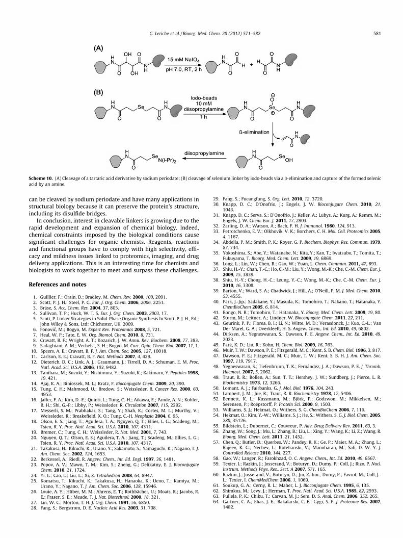
Scheme 10. (A) Cleavage of a tartaric acid derivative by sodium periodate; (B) cleavage of selenium linker by iodo-beads via a b-elimination and capture of the formed selenicacid by an amine.
G. Leriche et al. / Bioorg. Med. Chem. 20 (2012) 571–582 581
can be cleaved by sodium periodate and have many applications instructural biology because it can preserve the protein’s structure,including its disulfide bridges.
In conclusion, interest in cleavable linkers is growing due to therapid development and expansion of chemical biology. Indeed,chemical constraints imposed by the biological conditions causesignificant challenges for organic chemists. Reagents, reactionsand functional groups have to comply with high selectivity, effi-cacy and mildness issues linked to proteomics, imaging, and drugdelivery applications. This is an interesting time for chemists andbiologists to work together to meet and surpass these challenges.
References and notes
1. Guillier, F.; Orain, D.; Bradley, M. Chem. Rev. 2000, 100, 2091.2. Scott, P. J. H.; Steel, P. G. Eur. J. Org. Chem. 2006, 2006, 2251.3. Bräse, S. Acc. Chem. Res. 2004, 37, 805.4. Sullivan, T. P.; Huck, W. T. S. Eur. J. Org. Chem. 2003, 2003, 17.5. Scott, P. Linker Strategies in Solid-Phase Organic Synthesis In Scott, P. J. H., Ed.;
John Wiley & Sons, Ltd: Chichester, UK, 2009.6. Fonovic, M.; Bogyo, M. Expert Rev. Proteomics 2008, 5, 721.7. Heal, W. P.; Tate, E. W. Org. Biomol. Chem. 2010, 8, 731.8. Cravatt, B. F.; Wright, A. T.; Kozarich, J. W. Annu. Rev. Biochem. 2008, 77, 383.9. Sadaghiani, A. M.; Verhelst, S. H.; Bogyo, M. Curr. Opin. Chem. Biol. 2007, 11, 1.
10. Speers, A. E.; Cravatt, B. F. J. Am. Chem. Soc. 2005, 127, 10018.11. Carlson, E. E.; Cravatt, B. F. Nat. Methods 2007, 4, 429.12. Dieterich, D. C.; Link, A. J.; Graumann, J.; Tirrell, D. A.; Schuman, E. M. Proc.
Natl. Acad. Sci. U.S.A. 2006, 103, 9482.13. Tanihara, M.; Suzuki, Y.; Nishimura, Y.; Suzuki, K.; Kakimaru, Y. Peptides 1998,
19, 421.14. Ajaj, K. A.; Biniossek, M. L.; Kratz, F. Bioconjugate Chem. 2009, 20, 390.15. Tung, C. H.; Mahmood, U.; Bredow, S.; Weissleder, R. Cancer Res. 2000, 60,
4953.16. Jaffer, F. A.; Kim, D.-E.; Quinti, L.; Tung, C.-H.; Aikawa, E.; Pande, A. N.; Kohler,
R. H.; Shi, G.-P.; Libby, P.; Weissleder, R. Circulation 2007, 115, 2292.17. Messerli, S. M.; Prabhakar, S.; Tang, Y.; Shah, K.; Cortes, M. L.; Murthy, V.;
Weissleder, R.; Breakefield, X. O.; Tung, C.-H. Neoplasia 2004, 6, 95.18. Olson, E. S.; Jiang, T.; Aguilera, T. A.; Nguyen, Q. T.; Ellies, L. G.; Scadeng, M.;
Tsien, R. Y. Proc. Natl. Acad. Sci. U.S.A. 2010, 107, 4311.19. Bremer, C.; Tung, C. H.; Weissleder, R. Nat. Med. 2001, 7, 743.20. Nguyen, Q. T.; Olson, E. S.; Aguilera, T. A.; Jiang, T.; Scadeng, M.; Ellies, L. G.;
Tsien, R. Y. Proc. Natl. Acad. Sci. U.S.A. 2010, 107, 4317.21. Takakusa, H.; Kikuchi, K.; Urano, Y.; Sakamoto, S.; Yamaguchi, K.; Nagano, T. J.
Am. Chem. Soc. 2002, 124, 1653.22. Berkessel, A.; Riedl, R. Angew. Chem., Int. Ed. Engl. 1997, 36, 1481.23. Popov, A. V.; Mawn, T. M.; Kim, S.; Zheng, G.; Delikatny, E. J. Bioconjugate
Chem. 2010, 21, 1724.24. Yi, L.; Cao, L.; Liu, L.; Xi, Z. Tetrahedron 2008, 64, 8947.25. Komatsu, T.; Kikuchi, K.; Takakusa, H.; Hanaoka, K.; Ueno, T.; Kamiya, M.;
Urano, Y.; Nagano, T. J. Am. Chem. Soc. 2006, 128, 15946.26. Louie, A. Y.; Hüber, M. M.; Ahrens, E. T.; Rothbächer, U.; Moats, R.; Jacobs, R.
E.; Fraser, S. E.; Meade, T. J. Nat. Biotechnol. 2000, 18, 321.27. Lin, W. C.; Morton, T. H. J. Org. Chem. 1991, 56, 6850.28. Fang, S.; Bergstrom, D. E. Nucleic Acid Res. 2003, 31, 708.
29. Fang, S.; Fueangfung, S. Org. Lett. 2010, 12, 3720.30. Knapp, D. C.; D’Onofrio, J.; Engels, J. W. Bioconjugate Chem. 2010, 21,
1043.31. Knapp, D. C.; Serva, S.; D’Onofrio, J.; Keller, A.; Lubys, A.; Kurg, A.; Remm, M.;
Engels, J. W. Chem. Eur. J. 2011, 17, 2903.32. Zarling, D. A.; Watson, A.; Bach, F. H. J. Immunol. 1980, 124, 913.33. Petrotchenko, E. V.; Olkhovik, V. K.; Borchers, C. H. Mol. Cell. Proteomics 2005,
4, 1167.34. Abdella, P. M.; Smith, P. K.; Royer, G. P. Biochem. Biophys. Res. Commun. 1979,
87, 734.35. Yokoshima, S.; Abe, Y.; Watanabe, N.; Kita, Y.; Kan, T.; Iwatsubo, T.; Tomita, T.;
Fukuyama, T. Bioorg. Med. Chem. Lett. 2009, 19, 6869.36. Long, L.; Lin, W.; Chen, B.; Gao, W.; Yuan, L. Chem. Commun. 2011, 47, 893.37. Shiu, H.-Y.; Chan, T.-C.; Ho, C.-M.; Liu, Y.; Wong, M.-K.; Che, C.-M. Chem. Eur. J.
2009, 15, 3839.38. Shiu, H.-Y.; Chong, H.-C.; Leung, Y.-C.; Wong, M.-K.; Che, C.-M. Chem. Eur. J.
2010, 16, 3308.39. Barton, V.; Ward, S. A.; Chadwick, J.; Hill, A.; O’Neill, P. M. J. Med. Chem. 2010,
53, 4555.40. Park, J.-Jip.; Sadakane, Y.; Masuda, K.; Tomohiro, T.; Nakano, T.; Hatanaka, Y.
ChemBioChem 2005, 6, 814.41. Bongo, N. B.; Tomohiro, T.; Hatanaka, Y. Bioorg. Med. Chem. Lett. 2009, 19, 80.42. Sturm, M.; Leitner, A.; Lindner, W. Bioconjugate Chem. 2011, 22, 211.43. Geurink, P. P.; Florea, B. I.; Li, N.; Witte, M. D.; Verasdonck, J.; Kuo, C.-L.; Van
Der Marel, G. A.; Overkleeft, H. S. Angew. Chem., Int. Ed. 2010, 49, 6802.44. Dirksen, A.; Yegneswaran, S.; Dawson, P. E. Angew. Chem., Int. Ed. 2010, 49,
2023.45. Park, K. D.; Liu, R.; Kohn, H. Chem. Biol. 2009, 16, 763.46. Muir, T. W.; Dawson, P. E.; Fitzgerald, M. C.; Kent, S. B. Chem. Biol. 1996, 3, 817.47. Dawson, P. E.; Fitzgerald, M. C.; Muir, T. W.; Kent, S. B. H. J. Am. Chem. Soc.
1997, 119, 7917.48. Yegneswaran, S.; Tiefenbrunn, T. K.; Fernández, J. A.; Dawson, P. E. J. Thromb.
Haemost. 2007, 5, 2062.49. Traut, R. R.; Bollen, A.; Sun, T. T.; Hershey, J. W.; Sundberg, J.; Pierce, L. R.
Biochemistry 1973, 12, 3266.50. Lomant, A. J.; Fairbanks, G. J. Mol. Biol. 1976, 104, 243.51. Lambert, J. M.; Jue, R.; Traut, R. R. Biochemistry 1978, 17, 5406.52. Bennett, K. L.; Kussmann, M.; Björk, P.; Godzwon, M.; Mikkelsen, M.;
Sørensen, P.; Roepstorff, P. Protein Sci. 2000, 9, 1503.53. Williams, S. J.; Hekmat, O.; Withers, S. G. ChemBioChem 2006, 7, 116.54. Hekmat, O.; Kim, Y.-W.; Williams, S. J.; He, S.; Withers, S. G. J. Biol. Chem. 2005,
280, 35126.55. Bildstein, L.; Dubernet, C.; Couvreur, P. Adv. Drug Delivery Rev. 2011, 63, 3.56. Zhang, W.; Song, J.; Mu, L.; Zhang, B.; Liu, L.; Xing, Y.; Wang, K.; Li, Z.; Wang, R.
Bioorg. Med. Chem. Lett. 2011, 21, 1452.57. Chen, Q.; Butler, D.; Querbes, W.; Pandey, R. K.; Ge, P.; Maier, M. A.; Zhang, L.;
Rajeev, K. G.; Nechev, L.; Kotelianski, V.; Manoharan, M.; Sah, D. W. Y. J.Controlled Release 2010, 144, 227.
58. Gao, W.; Langer, R.; Farokhzad, O. C. Angew. Chem., Int. Ed. 2010, 49, 6567.59. Texier, I.; Razkin, J.; Josserand, V.; Boturyn, D.; Dumy, P.; Coll, J.; Rizo, P. Nucl.
Instrum. Methods Phys. Res., Sect. A 2007, 571, 165.60. Razkin, J.; Josserand, V.; Boturyn, D.; Jin, Z.-hui.; Dumy, P.; Favrot, M.; Coll, J.-
L.; Texier, I. ChemMedChem 2006, 1, 1069.61. Soukup, G. A.; Cerny, R. L.; Maher, L. J. Bioconjugate Chem. 1995, 6, 135.62. Shimkus, M.; Levy, J.; Herman, T. Proc. Natl. Acad. Sci. U.S.A. 1985, 82, 2593.63. Pullela, P. K.; Chiku, T.; Carvan, M. J.; Sem, D. S. Anal. Chem. 2006, 352, 265.64. Gartner, C. A.; Elias, J. E.; Bakalarski, C. E.; Gygi, S. P. J. Proteome Res. 2007,
1482.
582 G. Leriche et al. / Bioorg. Med. Chem. 20 (2012) 571–582
65. Fasold, H.; Klappenberger, J.; Meyer, C.; Remold, H. Angew. Chem., Int. Ed. Engl.1971, 10, 795.
66. Jaffe, C. L.; Lis, H.; Sharon, N. Biochemistry 1980, 19, 4423.67. Denny, J.; Blobel, G. Proc. Natl. Acad. Sci. U.S.A. 1984, 81, 5286.68. Verhelst, S. H. L.; Fonovic, M.; Bogyo, M. Angew. Chem., Int. Ed. 2007, 46, 1284.69. Fonovic, M.; Verhelst, S. H. L.; Sorum, M. T.; Bogyo, M. Mol. Cell. Proteomics
2007, 6, 1761.70. Yang, Y.-Y.; Ascano, J. M.; Hang, H. C. J. Am. Chem. Soc. 2010, 132, 3640.71. Rangan, K. J.; Yang, Y. Y.; Charron, G.; Hang, H. C. J. Am. Chem. Soc. 2010, 132,
465.72. Yount, J. S.; Moltedo, B.; Yang, Y.-Y.; Charron, G.; Moran, T. M.; López, C. B.;
Hang, H. C. Nat. Chem. Biol. 2010, 6, 610.73. Grammel, M.; Zhang, M. M.; Hang, H. C. Angew. Chem., Int. Ed. 2010, 49, 5970.74. Landi, F.; Johansson, C. M.; Campopiano, D. J.; Hulme, A. N. Org. Biomol. Chem.
2010, 8, 56.75. Yang, Y.-Y.; Grammel, M.; Raghavan, A. S.; Charron, G.; Hang, H. C. Chem. Biol.
2010, 17, 1212.76. Leriche, G.; Budin, G.; Brino, L.; Wagner, A. Eur. J. Org. Chem. 2010, 2010, 4360.77. Budin, G.; Moune-Dimala, M.; Leriche, G.; Saliou, J.-M.; Papillon, J.; Sanglier-
Cianférani, S.; Van Dorsselaer, A.; Lamour, V.; Brino, L.; Wagner, A.ChemBioChem 2010, 11, 2359.
78. Hooker, J. M.; Kovacs, E. W.; Francis, M. B. J. Am. Chem. Soc. 2004, 126, 3718.79. Kiyose, K.; Hanaoka, K.; Oushiki, D.; Nakamura, T.; Kajimura, M.; Suematsu,
M.; Nishimatsu, H.; Yamane, T.; Terai, T.; Hirata, Y.; Nagano, T. J. Am. Chem.Soc. 2010, 132, 15846.
80. Hammond, N.; Koumi, P.; Langley, G. J.; Lowe, A.; Brown, T. Org. Biomol. Chem.2007, 5, 1878.
81. Pellois, J.-P.; Hahn, M. E.; Muir, T. W. J. Am. Chem. Soc. 2004, 126, 7170.82. Zhou, H.; Ranish, J. A.; Watts, J. D.; Aebersold, R. Nat. Biotechnol. 2002, 20, 512.83. Maurel, D.; Banala, S.; Laroche, T.; Johnsson, K. ACS Chem. Biol. 2010, 5, 507.84. Chowdhury, S. M.; Munske, G. R.; Tang, X.; Bruce, J. E. Anal. Chem. 2006, 78,
8183.85. Olejnik, J.; Lüdemann, H. C.; Krzymanska-Olejnik, E.; Berkenkamp, S.;
Hillenkamp, F.; Rothschild, K. J. Nucleic Acid Res. 1999, 27, 4626.86. Cho, S.; Lee, S.-H.; Chung, W.-J.; Kim, Y.-K.; Lee, Y.-S.; Kim, B.-G. Electrophoresis
2004, 25, 3730.87. Vivero-Escoto, J. L.; Slowing, I. I.; Wu, C.-W.; Lin, V. S.-Y. J. Am. Chem. Soc. 2009,
131, 3462.88. Lemaire, R.; Stauber, J.; Wisztorski, M.; Van Camp, C.; Desmons, A.;
Deschamps, M.; Proess, G.; Rudlof, I.; Woods, A. S.; Day, R.; Salzet, M.;Fournier, I. J. Proteome Res. 2007, 6, 2057.
89. Wang, Z.; Udeshi, N. D.; O’Malley, M.; Shabanowitz, J.; Hunt, D. F.; Hart, G. W.Mol. Cell. Proteomics 2010, 9, 153.
90. Yang, L.; Tang, X.; Weisbrod, C. R.; Munske, G. R.; Eng, J. K.; Von Haller, P. D.;Kaiser, N. K.; Bruce, J. E. Anal. Chem. 2010, 82, 3556.
91. Orth, R.; Sieber, S. A. J. Org. Chem. 2009, 74, 8476.92. Okamoto, A.; Tanabe, K.; Inasaki, T.; Saito, I. Angew. Chem., Int. Ed. 2003, 42,
2502.93. Aoki, S.; Matsuo, N.; Hanaya, K.; Yamada, Y.; Kageyama, Y. Bioorg. Med. Chem.
2009, 17, 3405.94. Katayama, K.; Tsukiji, S.; Furuta, T.; Nagamune, T. Chem. Commun. 2008, 5399.95. Härtner, S.; Kim, H. C.; Hampp, N. J. Polym. Sci., Part A: Polym. Chem. 2007, 45,
2443.96. Gardner, M. W.; Vasicek, L. A.; Shabbir, S.; Anslyn, E. V.; Brodbelt, J. S. Anal.
Chem. 2008, 80, 4807.97. Gardner, M. W.; Brodbelt, J. S. Anal. Chem. 2009, 81, 4864.98. Petrotchenko, E. V.; Xiao, K.; Cable, J.; Chen, Y.; Dokholyan, N. V.; Borchers, C.
H. Mol. Cell. Proteomics 2009, 8, 273.99. Chowdhury, S. M.; Munske, G. R.; Siems, W. F.; Bruce, J. E. Rapid Commun. Mass
Spectrom. 2005, 19, 899.100. Van Der Veken, P.; Dirksen, E. H. C.; Ruijter, E.; Elgersma, R. C.; Heck, A. J. R.;
Rijkers, D. T. S.; Slijper, M.; Liskamp, R. M. J. ChemBioChem 2005, 6, 2271.101. Fauq, A. H.; Kache, R.; Khan, M. A.; Vega, I. E. Bioconjugate Chem. 2006, 17, 248.102. Szychowski, J.; Mahdavi, A.; Hodas, J. J. L.; Bagert, J. D.; Ngo, J. T.; Landgraf, P.;
Dieterich, D. C.; Schuman, E. M.; Tirrell, D. A. J. Am. Chem. Soc. 2010, 132,18351.
103. Srinivasachar, K.; Neville, D. M. Biochemistry 1989, 28, 2501.104. Parrott, M. C.; Luft, J. C.; Byrne, J. D.; Fain, J. H.; Napier, M. E.; Desimone, J. M. J.
Am. Chem. Soc. 2010, 132, 17928.105. Masson, C.; Garinot, M.; Mignet, N.; Wetzer, B.; Mailhe, P.; Scherman, D.;
Bessodes, M. J. Controlled Release 2004, 99, 423.106. Blattler, W. A.; Kuenzi, B. S.; Lambert, J. M.; Senter, P. D. Biochemistry 1985, 24,
1517.107. Shen, W. C.; Ryser, H. J. P. Biochem. Biophys. Res. Commun. 1981, 102, 1048.108. Lu, D.; Liang, J.; Fan, Y.; Gu, Z.; Zhang, X. Adv. Eng. Mater. 2010, 12, B496.109. Shamay, Y.; Adar, L.; Ashkenasy, G.; David, A. Biomaterials 2011, 32, 1377.110. Di Stefano, G.; Lanza, M.; Kratz, F.; Merina, L.; Fiume, L. Eur. J. Pharm. Sci. 2004,
23, 393.111. Liu, R.; Zhang, Y.; Zhao, X.; Agarwal, A.; Mueller, L. J.; Feng, P. J. Am. Chem. Soc.
2010, 132, 1500.112. Oishi, M.; Sasaki, S.; Nagasaki, Y.; Kataoka, K. Biomacromolecules 2003, 4,
1426.
113. Oishi, M.; Nagasaki, Y.; Itaka, K.; Nishiyama, N.; Kataoka, K. J. Am. Chem. Soc.2005, 127, 1624.
114. Jeong, J. H.; Kim, S. W.; Park, T. G. Bioconjugate Chem. 2003, 14, 473.115. Zhao, Y.-L.; Li, Z.; Kabehie, S.; Botros, Y. Y.; Stoddart, J. F.; Zink, J. I. J. Am. Chem.
Soc. 2010, 132, 13016.116. Jia, Y.; Fei, J.; Cui, Y.; Yang, Y.; Gao, L.; Li, J. Chem. Commun. 2011, 47, 1175.117. Patel, V. F.; Hardin, J. N.; Mastro, J. M.; Law, K. L.; Zimmermann, J. L.; Ehlhardt,
W. J.; Woodland, J. M.; Starling, J. J. Bioconjugate Chem. 1996, 7, 497.118. Shin, J.; Shum, P.; Thompson, D. H. J. Controlled Release 2003, 91, 187.119. Heffernan, M. J.; Murthy, N. Bioconjugate Chem. 2005, 16, 1340.120. Sankaranarayanan, J.; Mahmoud, E. A.; Kim, G.; Morachis, J. M.; Almutairi, A.
A. C. S. Nano 2010, 4, 5930.121. Hangauer, M. J.; Bertozzi, C. R. Angew. Chem., Int. Ed. 2008, 47, 2394.122. Bi, L.; Kim, D. H.; Ju, J. J. Am. Chem. Soc. 2006, 128, 2542.123. Ju, J.; Kim, D. H.; Bi, L.; Meng, Q.; Bai, X.; Li, Z.; Li, X.; Marma, M. S.; Shi, S.; Wu,
J.; Edwards, J. R.; Romu, A.; Turro, N. J. Proc. Natl. Acad. Sci. U.S.A. 2006, 103,19635.
124. Brunner, J.; Mokhir, A.; Kraemer, R. J. Am. Chem. Soc. 2003, 125, 12410.125. Boll, I.; Krämer, R.; Brunner, J.; Mokhir, A. J. Am. Chem. Soc. 2005, 127, 7849.126. Smith, R. J.; Capaldi, R. A.; Muchmore, D.; Dahlquist, F. Biochemistry 1978, 17,
3719.127. Jin, R. Z.; Karol, M. H. Chem. Res. Toxicol. 1988, 1, 281.128. Predescu, S. A.; Predescu, D. N.; Palade, G. E.; Jolla, L. Mol. Biol. Cell 2001, 12,
1019.129. Fang, G.; Cech, T. R. Proc. Natl. Acad. Sci. U.S.A. 1993, 90, 6056.130. Bragg, P. D.; Hou, C. Eur. J. Biochem. 1980, 106, 495.131. Jahn, O.; Eckart, K.; Brauns, O.; Tezval, H.; Spiess, J. Proc. Natl. Acad. Sci. U.S.A.
2002, 99, 12055.132. Haberland, J.; Becker, J.; Gerke, V. J. Biol. Chem. 1997, 272, 24717.133. Parry, D. A.; Marekov, L. N.; Steinert, P. M. J. Biol. Chem. 2001, 276, 39253.134. Brockmöller, J.; Kamp, R. M. Biochemistry 1988, 27, 3372.135. Bergmann, U.; Wittmann-Liebold, B. Biochemistry 1993, 32, 2880.136. Pohl, T.; Wittmann-Liebold, B. J. Biol. Chem. 1988, 263, 4293.137. Buchardt, O.; Elsner, H. I.; Nielsen, P. E.; Petersen, L. C.; Suenson, E. Anal. Chem.
1986, 158, 87.138. Koch, T.; Suenson, E.; Henriksen, U.; Buchardt, O. Bioconjugate Chem. 1990, 1,
296.139. Testa, B. Curr. Opin. Chem. Biol. 2009, 13, 338.140. Nori, A.; Kopecek, J. Adv. Drug Delivery Rev. 2005, 57, 609.141. Mahato, R.; Tai, W.; Cheng, K. Adv. Drug Delivery Rev. 2011, 63, 659.142. Leitner, A.; Walzthoeni, T.; Kahraman, A.; Herzog, F.; Rinner, O.; Beck, M.;
Aebersold, R. Mol. Cell. Proteomics 2010, 9, 1634.143. Peters, K.; Richards, F. M. Ann. Rev. Biochem. 1977, 46, 523.144. Kaake, R. M.; Wang, X.; Huang, L. Mol. Cell. Proteomics 2010, 9, 1650.145. Gao, L.; Lu, Z. Biochem. Biophys. Res. Commun. 2009, 387, 421.146. Metzker, M. L. Nat. Rev. Genet. 2009, 11, 31.147. Babé, L. M.; Craik, C. S. Cell 1997, 91, 427.148. Law, B.; Tung, C.-H. Bioconjugate Chem. 2009, 20, 1683.149. Jahng, W. J.; David, C.; Nesnas, N.; Nakanishi, K.; Rando, R. R. Biochemistry
2003, 42, 6159.150. Fukuyama, T.; Jow, C.; Cheung, M. Tetrahedron Lett. 1995, 36, 6373.151. Fukuyama, T.; Cheung, M.; Jow, C.-K.; Hidai, Y.; Kan, T. Tetrahedron Lett. 1997,
38, 5831.152. Dawson, P. E.; Muir, T. W.; Clark-Lewis, I.; Kent, S. B. Science 1994, 266, 776.153. Kenner, G.; McDermott, J.; Sheppard, R. J. Am. Chem. Soc. 1971, 636.154. Backes, B.; Ellman, J. A. J. Org. Chem. 1999, 64, 2322.155. Timokhin, B. V.; Baransky, V. A.; Eliseeva, G. D. Russ. Chem. Rev. 1999, 68, 73.156. Klement, E.; Lipinszki, Z.; Kupihár, Z.; Udvardy, A.; Medzihradszky, K. F. J.
Proteome Res. 2010, 9, 2200.157. Gemeay, A. H. Dyes and Pigments 2002, 54, 201.158. Wuts, P. G. M.; Greene, T. W. Greene’s Protective Groups in Organic Synthesis;
John Wiley & Sons, Inc.: Hoboken, NJ, USA, 2006.159. Bochet, C. G. J. Chem. Soc., Perkin Trans 1 2002, 125.160. Mayer, G.; Heckel, A. Angew. Chem., Int. Ed. 2006, 45, 4900.161. Yu, H.; Li, J.; Wu, D.; Qiu, Z.; Zhang, Y. Chem. Soc. Rev. 2010, 39, 464.162. Adams, S. R.; Kao, J. P. Y.; Tsien, R. Y. J. Am. Chem. Soc. 1989, 111, 7957.163. Holmes, C. P. J. Org. Chem. 1997, 62, 2370.164. Tjoeng, F. S.; Heavner, G. A. J. Org. Chem. 1983, 48, 355.165. Tannock, I. F.; Rotin, D. Cancer Res. 1989, 49, 4373.166. Stubbs, M.; McSheehy, P. M. J.; Griffiths, J. R.; Bashford, C. L. Mol. Med. Today
2000, 6, 15.167. Gao, W.; Chan, J. M.; Farokhzad, O. C. Mol. Pharmaceutics 2010, 7, 1913.168. Saxon, E.; Bertozzi, C. R. Science 2000, 287, 2007.169. Saxon, E.; Armstrong, J. I.; Bertozzi, C. R. Org. Lett. 2000, 2, 2141.170. Kodama, K.; Fukuzawa, S.; Nakayama, H.; Kigawa, T.; Sakamoto, K.; Yabuki, T.;
Matsuda, N.; Shirouzu, M.; Takio, K.; Tachibana, K.; Yokoyama, S.ChemBioChem 2006, 7, 134.
171. Nauwelaerts, K.; Vastmans, K.; Froeyen, M.; Kempeneers, V.; Rozenski, J.;Rosemeyer, H.; Van Aerschot, A.; Busson, R.; Lacey, J. C.; Efimtseva, E.;Mikhailov, S.; Lescrinier, E.; Herdewijn, P. Nucleic Acid Res. 2003, 31, 6758.