circulatory disorders
TRANSCRIPT

Circulatory Disorders
Done By : Dr. Rasha Khalil

• Topics included:
• Hyperemia and Congestion• Hemorrhage• Thrombosis

Hyperemia and Congestion
• Hyperemia and congestion both refer to an increase in blood volume within a tissue but they have different underlying mechanisms.


• Hyperemia:• Is an active process resulting from arteriolar
dilation and increased blood inflow, as occurs at sites of inflammation or in exercising skeletal muscle.
• Characteristics:1. Redder than normal because of engorgement
with oxygenated blood.2. Warmer than usual 3. Swollen4. Pulse may be felt readily − due to the elastic
recoil of arterioles, hyperemia is dissipated after death in skin and mucous membranes and is usually not apparent post-mortem
• Causes:
• physiologic: in response to increased demands for nutrients because of work being done .
• pathologic: in response to certain vasodilator chemicals and neurogenic stimuli associated with irritation of tissue.

• Congestion: • Is a passive process resulting from impaired outflow of venous
blood from a tissue. It can occur systemically, as in cardiac failure, or locally as a consequence of an isolated venous obstruction.
• Characteristics:1. An abnormal blue-red color (cyanosis) that stems from the
accumulation of deoxygenated hemoglobin in the affected area.2. Swollen .3. Cooler than normal 4. After death the color becomes even more dark blue and the cut
surface of such tissues oozes blood freely5. If chronic, the tissue may have a brown color
• In long-standing chronic congestion, inadequate tissue perfusion and persistent hypoxia may lead to parenchymal cell death and secondary tissue fibrosis, and the elevated intravascular pressures may cause edema or sometimes rupture capillaries, producing focal hemorrhages.

• Causes:
1) Peripherala. obstructive - e.g., thrombus or embolusb. constrictive - e.g., collapsing veins
2) Central - due to heart failure, which may be caused by: conditions associated with weakened ventricular muscle conditions causing ventricles to pump against increased
resistance (e.g., valvular stenosis, hypertension) conditions that increase volume of blood delivered to
ventricles (leaky valves, septal defects)
• MORPHOLOGY• Cut surfaces of hyperemic or congested tissues feel
wet and typically ooze blood.
• On microscopic examination,• Acute pulmonary congestion is marked by blood-
engorged alveolar capillaries and variable degrees of alveolar septal edema and intra-alveolar hemorrhage.
• Chronic pulmonary congestion, the septa become thickened and fibrotic, and the alveolar spaces contain numerous macrophages laden with hemosiderin (“heart failure cells”) derived from phagocytosed red cells.

• In acute hepatic congestion, the central vein and sinusoids are distended with blood, and there may even be central hepatocyte drop-out due to necrosis. The periportal hepatocytes, better oxygenated because of their proximity to hepatic arterioles, experience less severe hypoxia and may develop only reversible fatty change.
• In chronic congestion of the liver, the central regions of the hepatic lobules, viewed on gross examination, are red-brown and slightly depressed (owing to cell loss) and are accentuated against the surrounding zones of uncongested tan, sometimes fatty liver (nutmeg liver).
• Microscopic findings include: centrilobular hepatocyte necrosis hemorrhage hemosiderin-laden macrophages
• In long-standing, severe hepatic congestion (most commonly associated with heart failure), hepatic fibrosis (“cardiac cirrhosis”) can develop.

Hemorrhage

• Defined: as the extravasation of blood from vessels.
• Classification:
1. Site• 1. External. Bleeding Is visible as it occursa. through skin wounds b. or from a body orifice as in epistaxis or hematemesis. • 2. Internal. a. In body cavities, e.g., hemoperitoneum and
hemothorax. b. Interstitial, e.g., fracture hematoma.

2. By size and form
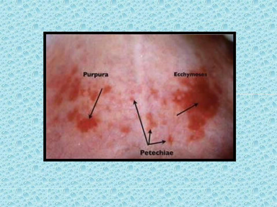
• Petechiae
• Are minute (1 to 2 mm in diameter) hemorrhages into skin, mucous membranes, or serosal surfaces
• Causes include: low platelet counts (thrombocytopenia), defective platelet function, loss of vascular wall support, as in vitamin C deficiency
• Purpura
• Are slightly larger (3 to 5 mm) hemorrhages.• Causes include : same disorders that cause petechiae, trauma, vascular inflammation (vasculitis), increased vascular fragility.
• Ecchymosis • Are larger (1 to 2 cm) subcutaneous hematomas( called bruises). • Extravasated red cells are phagocytosed and
degraded by macrophages;• The characteristic color changes of a bruise are due
to : Enzymatic conversion of hemoglobin (red-blue color)
to Bilirubin (blue-green color) And eventually hemosiderin (golden-brown).

3. Type of disrupted vessel• 1. Arterial… blood is bright red and comes in pulsatile jets. • 2. Venous... blood is dark red and comes in a steady flow.• 3. Capillary...bleeding occurs as diffuse ooze of bright red blood.
4. Timing in relation to trauma• 1. Primary hemorrhage occurs at the time of trauma.
• 2. Reactionary hemorrhage occurs within 24 hours after trauma. As the blood pressure rises due to correction of hypovolemia or secondary to post-operative pain, an insecure ligature slips or a clot is dislodged.
• 3. Secondary hemorrhage occurs one to two weeks after trauma due to infection eroding a vessel wall. It can be fatal if a large artery is Involved, (e.g., the carotid )after sloughing of the skin flaps of a radical neck dissection.
• Etiology 1. Traumatic• a. Accidental.• b. Surgical.• c. Interventional procedures.
2. Pathological • a. Atherosclerotic (ruptured aortic aneurysm).• b. Inflammatory (bleeding peptic ulcer).• c. Neoplastic (hematuria in renal cancer).
3. Bleeding diathesis can increase the amount of traumatic and pathological
bleeding, or cause bleeding with little or no trauma (spontaneous haemorrhage).
• Physiological response to hemorrhage • The physiological response to hemorrhage has two aims:1. Stop the bleeding a. Vasoconstriction and retraction of the intima of the injured vessel. b. Platelet plug. c. Blood clotting.
2. Maintaining effective circulatory volume and perfusion of critical tissues (brain and heart), at the expense of less critical tissues (skin, skeletal muscle ). This is achieved by neural and endocrine factors:
A. Neural factors. A sympathoadrenal discharge develops due to decrease in the
stimulation of arterial baroreceptors (aortic arch and carotid sinus) and atrial stretch receptors leading to reduction of the normal Inhibitory discharge in the vagus and glossopharyngeal nerves on the vasomotor center with consequent stimulation of the sympathetic system.

The effects include: • 1. Constriction of veins, which normally contain
two-thirds of the blood volume, displaces blood from the capacitance side of the circulation into the heart.
• 2. Constriction of arterioles raises the peripheral resistance but this is not uniform.
• It involves mainly the arterioles of the skin and skeletal muscle
• 3. Increased rate and strength of cardiac contraction.


B. Endocrine factors 1. Catecholamine discharge occurs from the adrenal
medulla and from the nerve endings throughout the autonomic nervous system. They increase the heart rate and myocardial contraction and cause constriction of the arterioles of the skin, kidney and viscera.
2. The metabolic hormones ACTH, cortisol, growth hormone and glucagon are increased. Insulin release is inhibited by adrenaline and noradrenalin.

3. The renin-angiotensin aldosterone system.
The juxtaglomerular cells of the afferent renal arterioles secrete renin in response to renal hypoperfusion.
Renin splits angiotensinogen to angiotensin I which Is converted to angiotensin II by a converting enzyme in the lung.
Angiotensin II is a powerful vasoconstrictor and stimulates sodium and water retention by a direct action on the kidney as well as indirectly through release of aldosterone from the zona glomerulosa of the adrenal cortex.
Angiotensin mediated vasoconstriction takes 20 minutes to occur, whereas baroreceptor-vasoconstriction occurs within seconds.

4. ADH (vasopressin).• Blood loss greater than 10% stimulates ADH release.
• ADH increases the permeability of the renal collecting tubules allowing water absorption into the hypertonic renal medullary interstitium.
• With severe hemorrhage high levels of ADH also cause vasoconstriction.
C. Transcapillary refill.• Reduction of blood volume and constriction of arterioles causes a
fall in capillary hydrostatic pressure and promotes movement of fluid from the interstitium into the capillaries.
• The resulting hemodilution increases the blood volume and lowers its viscosity, thus improving effective circulatory volume.

Effect and significance of hemorrhage:1. According to amount and rate of blood loss If rapid blood loss occurs (1/4 to 1/3 of the
total blood volume over a period of less than a few hours), hypovolemic shock and perhaps death will occur.
If slower blood loss occurs (as much as 1/2 total blood volume over weeks or months), no serious consequences may occur because the body compensates.

2. According to site of hemorrhage
Bleeding that would be trivial in the subcutaneous tissues can cause death if located in the brain .
Chronic or recurrent external blood loss (e.g., due to peptic ulcer) frequently culminates in iron deficiency anemia as a consequence of loss of iron in hemoglobin.
By contrast, iron is efficiently recycled from phagocytosed red cells, so internal bleeding (e.g., a hematoma) does not lead to iron deficiency.
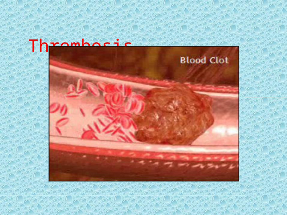
Thrombosis

• Defined as :the formation of a blood clot within the vasculature.
• Causes of thrombus formation (Virchow’s triad):
(1) Endothelial injury, (2) Stasis or turbulent blood flow, (3) Hypercoagulability of the blood

1) Endothelial Injury• Endothelial injury is an important cause of thrombosis,
particularly in the heart and the arteries, where high flow rates might otherwise impede clotting by preventing platelet adhesion or diluting coagulation factors.
• Examples of thrombosis related to endothelial damage are:
The formation of thrombi in the cardiac chambers after myocardial infarction,
Over ulcerated plaques in atherosclerotic arteries, At sites of traumatic or inflammatory vascular injury
(vasculitis).• Overt loss of endothelium exposes subendothelial ECM
(leading to platelet adhesion), releases tissue factor, and reduces local production of PGI2 and plasminogen activators.
• Any perturbation in the dynamic balance of the prothrombotic and antithrombotic effects of endothelium can influence clotting locally.
• Thus, dysfunctional endothelium elaborates greater amounts of procoagulant factors (e.g., platelet adhesion molecules, tissue factor) and synthesizes lesser amounts of anticoagulant molecules (e.g., thrombomodulin, PGI2, t-PA).
• Endothelial dysfunction can be induced by a variety of insults, including :
1. Hypertension,2. Turbulent blood flow, 3. Bacterial products4. Radiation injury,5. Metabolic abnormalities such as homocystinuria and toxins
absorbed from cigarette smoke.

2) Abnormal Blood Flow•
• Turbulence contributes to arterial and cardiac thrombosis by causing
1. endothelial injury or dysfunction,2. forming countercurrents and local pockets of stasis.
• Stasis Is a major factor in the development of venous thrombi. Under conditions of normal laminar blood flow, platelets (and
other blood cells) are found mainly in the center of the vessel lumen, separated from the endothelium by a slower-moving layer of plasma.

• Stasis and turbulent blood flow have the following effects:
1. Both promote endothelial cell activation and enhanced procoagulant activity, in part through flow-induced changes in endothelial gene expression.
2. Stasis allows platelets and leukocytes to come into contact with the endothelium when the flow is sluggish.
3. Stasis also slows the washout of activated clotting factors and impedes the inflow of clotting factor inhibitors.

• Turbulent and static blood flow contribute to thrombosis in a number of clinical settings including:
Ulcerated atherosclerotic plaques not only expose subendothelial ECM but also cause turbulence.
Abnormal aortic and arterial dilations called aneurysms create local stasis and consequently a fertile site for thrombosis.
Acute myocardial infarction results in focally noncontractile myocardium.

Ventricular remodeling after more remote infarction can lead to aneurysm formation. In both cases, cardiac mural thrombi are more easily formed due to the local blood stasis
Mitral valve stenosis (e.g., after rheumatic heart disease) results in left atrial dilation. In conjunction with atrial fibrillation, a dilated atrium is a site of profound stasis and a prime location for the development of thrombi.
Hyperviscosity syndromes (such as polycythemia) increase resistance to flow and cause small vessel stasis;
The deformed red cells in sickle cell anemia cause vascular occlusions, and the resultant stasis also predisposes to thrombosis.

3) Hypercoagulability• Hypercoagulability contributes infrequently to
arterial or intracardiac thrombosis but is an important underlying risk factor for venous thrombosis.
• Defined as any alteration of the coagulation pathways that predisposes affected persons to thrombosis,
• Divided into:1. Primary (genetic) disorders2. Secondary (acquired) disorders
1) Primary (inherited) hypercoagulability Most often is caused by mutations in the factor V and
prothrombin genes:• Approximately 2% to 15% of whites carry a specific factor V
mutation (called the Leiden mutation). • The mutation alters an amino acid residue in factor V and
renders it resistant to protein C. • Thus, an important antithrombotic counter-regulatory
mechanism is lost.• Heterozygotes carry a 5-fold increased risk for venous
thrombosis, with homozygotes having a 50-fold increased risk.• A single-nucleotide substitution (G to A) in the 3 -untranslated ′
region of the prothrombin gene is a fairly common allele • This variant results in increased prothrombin transcription and
is associated with a nearly threefold increased risk for venous thromboses.
• Less common primary hypercoagulable states include:
Inherited deficiencies of anticoagulants such as antithrombin III protein C protein S
• Affected patients typically present with venous thrombosis and recurrent thromboembolism in adolescence or early adult life.
2) Secondary (acquired) hypercoagulability
In some situations (e.g., cardiac failure or trauma), stasis or vascular injury may be the most important factor.
The hypercoagulability associated with oral contraceptive use and the hyperestrogenic state of pregnancy may be related to increased hepatic synthesis of coagulation factors and reduced synthesis of antithrombin III
In disseminated cancers, release of procoagulant tumor products (e.g., mucin from adenocarcinoma) predisposes to thrombosis.
The hypercoagulability seen with advancing age has been attributed to increased platelet aggregation and reduced release of PGI2 from endothelium.
Smoking and obesity promote hypercoagulability by unknown mechanisms.

• Two significant situations are:
1. Heparin-induced thrombocytopenic (HIT) syndrome.
This syndrome occurs in up to 5% of patients treated with unfractionated heparin (for therapeutic anticoagulation).
It is marked by the development of autoantibodies that bind complexes of heparin and platelet membrane protein (platelet factor-4)
• Although the mechanism is unclear, it appears that these antibodies may also bind similar complexes present on platelet and endothelial surfaces, resulting in platelet activation, aggregation, and consumption (hence thrombocytopenia), as well as causing endothelial cell injury.
The overall result is a prothrombotic state, even in the face of heparin administration and low platelet counts.
Newer low-molecular-weight fractionated heparin preparations induce autoantibodies less frequently but can still cause thrombosis if antibodies have already formed.

2) Antiphospholipid antibody syndrome. • This syndrome has manifestations, including :1. recurrent thrombosis2. repeated miscarriages,3. cardiac valve vegetations, 4. and thrombocytopenia;
• It is associated with autoantibodies directed against anionic phospholipids (e.g., cardiolipin) or—more accurately—plasma protein antigens that are unveiled by binding to such phospholipids (e.g., prothrombin).
• In vivo, these antibodies induce a hypercoagulable state, perhaps by inducing endothelial injury, by activating platelets or complement directly, or by interacting with the catalytic domains of certain coagulation factors.
• In vitro (in the absence of platelets and endothelium), however, the antibodies interfere with phospholipid complex assembly, thereby inhibiting coagulation (hence the designation lupus anticoagulant).
• Patients with antiphospholipid antibody syndrome fall into two categories:
1. Many have secondary antiphospholipid syndrome due to a well-defined autoimmune disease, such as systemic lupus erythematosus
2. The remainder of these patients exhibit only the manifestations of a hypercoagulable state without evidence of another autoimmune disorder (primary antiphospholipid syndrome).

• MORPHOLOGY• Thrombi can develop anywhere in the cardiovascular
system.• Arterial or cardiac thrombi typically arise at sites of
endothelial injury or turbulence;• venous thrombi characteristically occur at sites of stasis.• Thrombi are focally attached to the underlying vascular
surface and tend to propagate toward the heart; thus, arterial thrombi grow in a retrograde direction from the point of attachment,
• while venous thrombi extend in the direction of blood flow. The propagating portion of a thrombus tends to be poorly attached and therefore prone to fragmentation and migration through the blood as an embolus.
• Thrombi occurring in heart chambers or in the aortic lumen are designated mural thrombi. Abnormal myocardial contraction (arrhythmias, dilated cardiomyopathy, or myocardial infarction) or endomyocardial injury (myocarditis, catheter trauma) promote cardiac mural thrombi , while ulcerated atherosclerotic plaques and aneurysmal dilation promote aortic thrombosis
• Arterial thrombi are typically relatively rich in platelets, as the processes underlying their development (e.g., endothelial injury) lead to platelet activation. Although usually superimposed on a ruptured atherosclerotic plaque, other vascular injuries (vasculitis, trauma) can also be causal

• Venous thrombi (phlebothrombosis)• Frequently propagate some distance toward the
heart, forming a long cast within the vessel lumen that is prone to give rise to emboli.
• An increase in the activity of coagulation factors is involved in the genesis of most venous thrombi, with platelet activation playing a secondary role.
• The veins of the lower extremities are most commonly affected (90% of venous thrombosis);

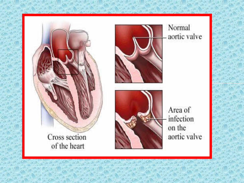
• Thrombi on heart valves are called vegetations.
• Bacterial or fungal blood-borne infections can cause valve damage, leading to the development of large thrombotic masses (infective endocarditis)
• Sterile vegetations also can develop on noninfected valves in hypercoagulable states—the lesions of so-called nonbacterial thrombotic endocarditis

Venous Thrombosis (Phlebothrombosis).
• Most venous thrombi occur in either the superficial or the deep veins of the leg.
• Superficial venous thrombi usually arise in the saphenous system, particularly in the setting of varicose viens; these rarely embolize but can be painful and can cause local congestion and swelling from impaired venous outflow, predisposing the overlying skin to development of infections and varicose ulcers

• Deep venous thrombosis (DVT)
Occur in the larger leg veins at or above the knee joint (e.g., popliteal, femoral, and iliac veins)
Are more serious because they are prone to embolize.
Although such DVTs may cause local pain and edema, the venous obstruction often is circumvented by collateral channels.
Consequently, DVTs are entirely asymptomatic in approximately 50% of patients and are recognized only after they have embolized to the lungs.

Deep venous thromboses (DVT)

• Lower-extremity DVTs are associated with stasis and hypercoagulable states
Common predisposing factors include:
1. Patient factors Age Previous DVT or PE Immobility Obesity Pregnancy Thrombophilia (eg.protein C & protein S deficiencies,
lupus anticoagulant & factor v leiden) Oral contraceptive pill

2. Factors involving the disease or surgical procedure:
Trauma and surgery, especially of the pelvis and lower limb
Malignancy, especially pelvic and abdominal MI Congestive heart failure Polycythemia Inflammatory bowel disease Nephrotic syndrome Length of operation

• Outcomes of DVT Pulmonary embolism Post_phlebitic limb Resolution without complications
• Treatment of DVT AnalgesiaGraduated compression stockingAnticoagulation with heparin (LMW) Long_term anticoagulation is undertaken with warfarinCaval filter Fibrinolytic agents (in very exrensive DVT ,but not after
major surgery)

• Fate of the Thrombus
• If a patient survives an initial thrombotic event, over the coming days to weeks the thrombus evolves through some combination of the following four processes:
1. Propagation.2. Embolization.3. Dissolution4. Organization and recanalization.

1) Propagation.• Propagation of additional platelets and fibrin, increasing the odds of
vascular occlusion or embolization.
2) Embolization.• Part or all of the thrombus is dislodged and transported elsewhere in the
vasculature.
3) Dissolution. If a thrombus is newly formed, activation of fibrinolytic factors may lead
to its rapid shrinkage and complete dissolution.
With older thrombi, extensive fibrin polymerization renders the thrombus substantially more resistant to plasmin-induced proteolysis, and lysis is ineffective..
This acquisition of resistance to lysis has clinical significance, as therapeutic administration of fibrinolytic agents (e.g., t-PA in the setting of acute coronary thrombosis) generally is not effective unless given within a few hours of thrombus formation.

4) Organization and recanalization.• Older thrombi become organized by the ingrowth of endothelial cells,
smooth muscle cells, and fibroblasts into the fibrin-rich thrombus• In time, capillary channels are formed that—to a limited extent—
create conduits along the length of the thrombus, thereby reestablishing the continuity of the original lumen.
• Further recanalization can sometimes convert a thrombus into a vascularized mass of connective tissue that is eventually incorporated into the wall of the remodeled vessel.
• Occasionally, instead of organizing, the center of a thrombus undergoes enzymatic digestion, presumably because of the release of lysosomal enzymes from entrapped leukocytes.
• If bacterial seeding occurs, the contents of degraded thrombi serve as an ideal culture medium, and the resulting infection may weaken the vessel wall, leading to formation of a mycotic aneurysm
• Clinical Correlation
• Thrombi are significant because they cause obstruction of arteries and veins and may give rise to emboli.
1. Venous thrombi can cause congestion and edema in vascular beds distal to an obstruction, they are most worrisome because of their potential to embolize to the lungs and cause death.
2. Arterial thrombi can embolize and cause tissue infarction, their tendency to obstruct vessels (e.g., in coronary and cerebral vessels) is considerably more important.
