circulation & respiration chapter 42
DESCRIPTION
Circulation & Respiration Chapter 42. Circulatory systems. Closed circulatory system Vertebrates Blood Enclosed in blood vessels & heart Lymph Lymph system Interstitial fluid. Functions. 1. Transportation Substances needed for cellular respiration A. Respiratory CO 2 and O 2 - PowerPoint PPT PresentationTRANSCRIPT
Circulation & Circulation & Respiration Respiration
Chapter 42Chapter 42

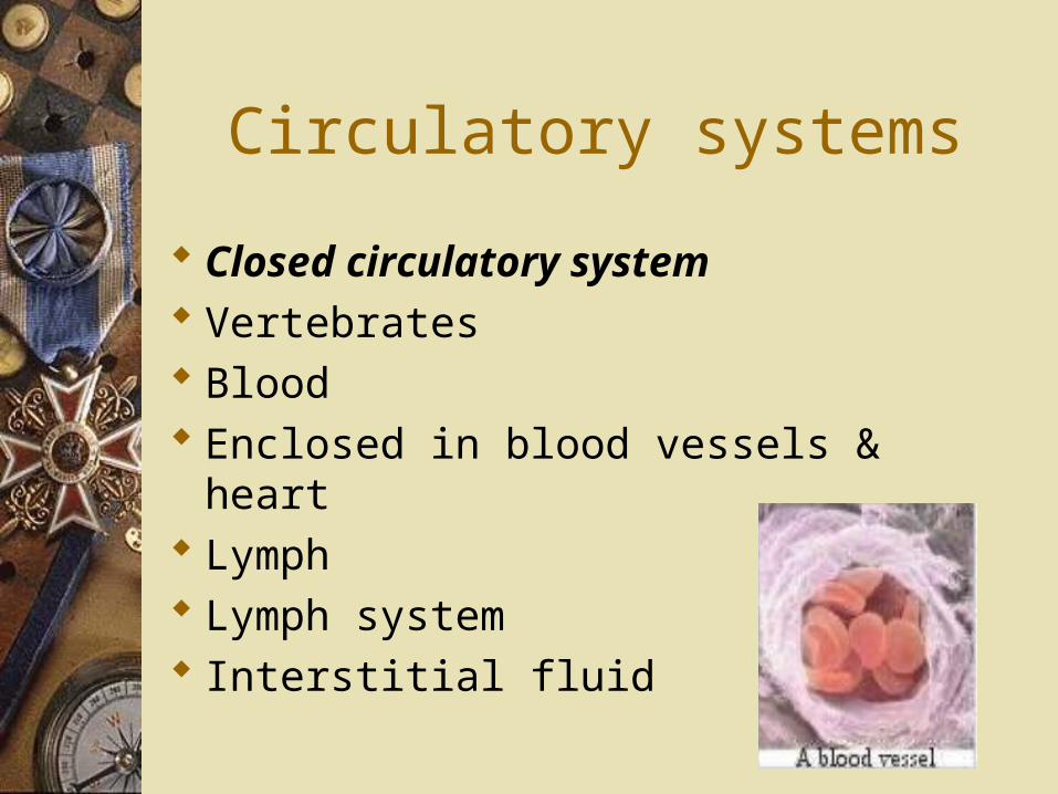
Circulatory systems
Closed circulatory system Vertebrates Blood Enclosed in blood vessels & heart Lymph Lymph system Interstitial fluid

Functions
1. Transportation Substances needed for cellular
respiration A. Respiratory
– CO2 and O2
B. Nutritive– glucose
C. Excretory– Metabolic wastes, ions, water
Functions
2. Regulation– Hormones– Temperature regulation (Endotherms)
3. Protection– Clotting
• Proteins, platelets
– Immune defense• WBC, AB
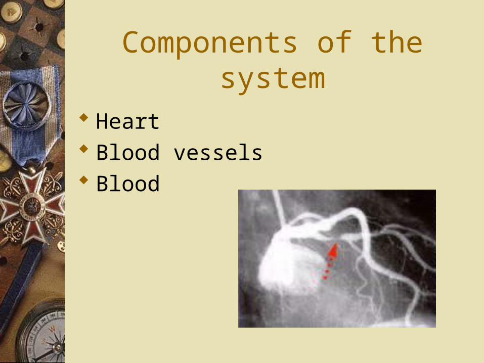
Components of the system
Heart Blood vessels Blood
Heart
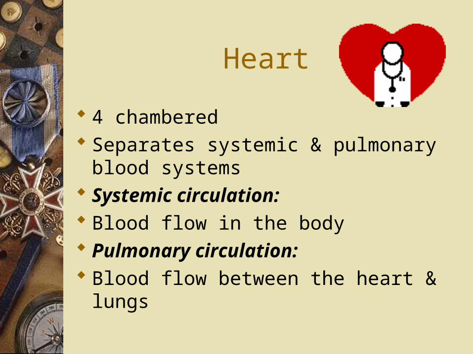
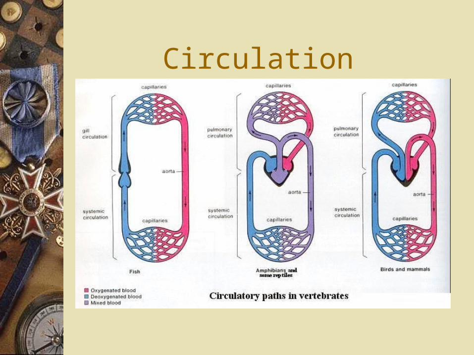
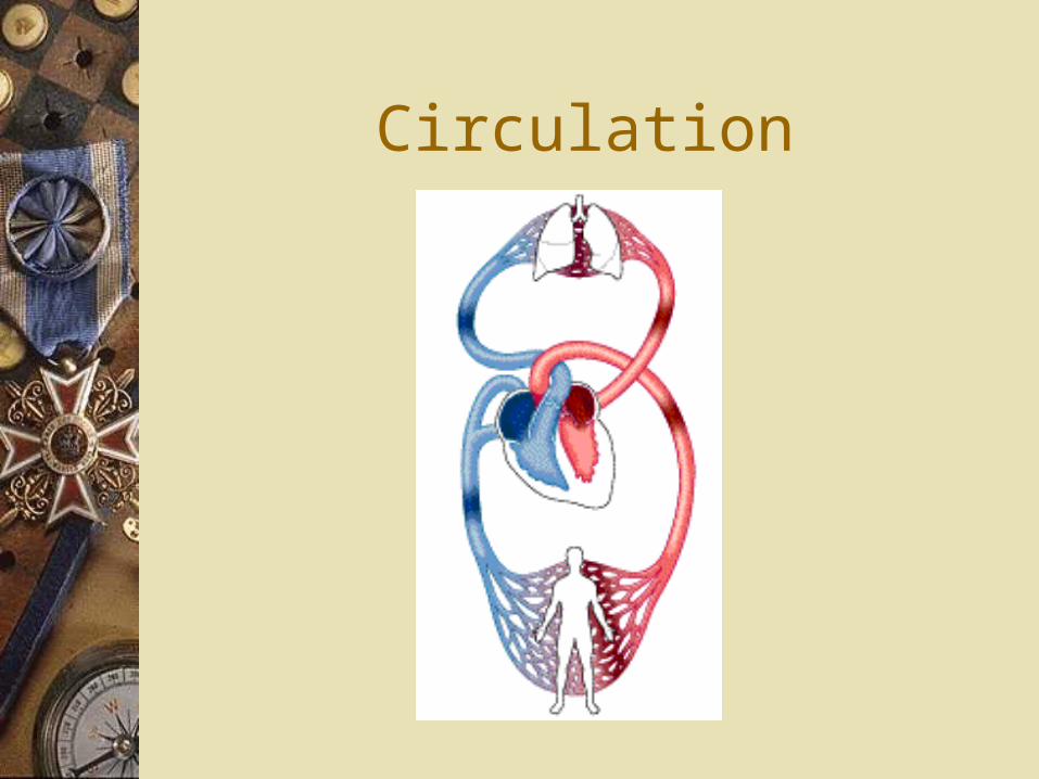
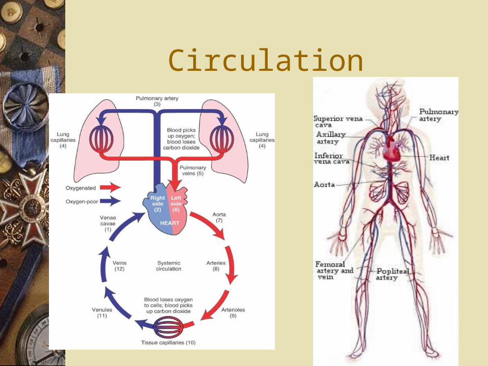
4 chambered Separates systemic & pulmonary
blood systems Systemic circulation: Blood flow in the body Pulmonary circulation: Blood flow between the heart &
lungs
Circulation
Circulation
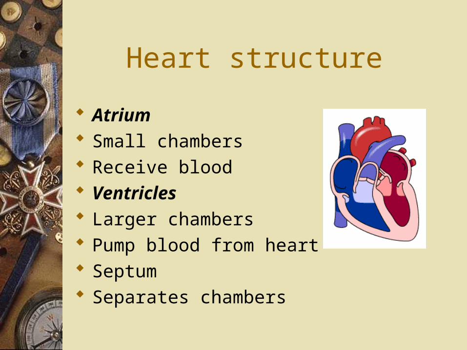
Heart structure
Atrium Small chambers Receive blood Ventricles Larger chambers Pump blood from heart Septum Separates chambers
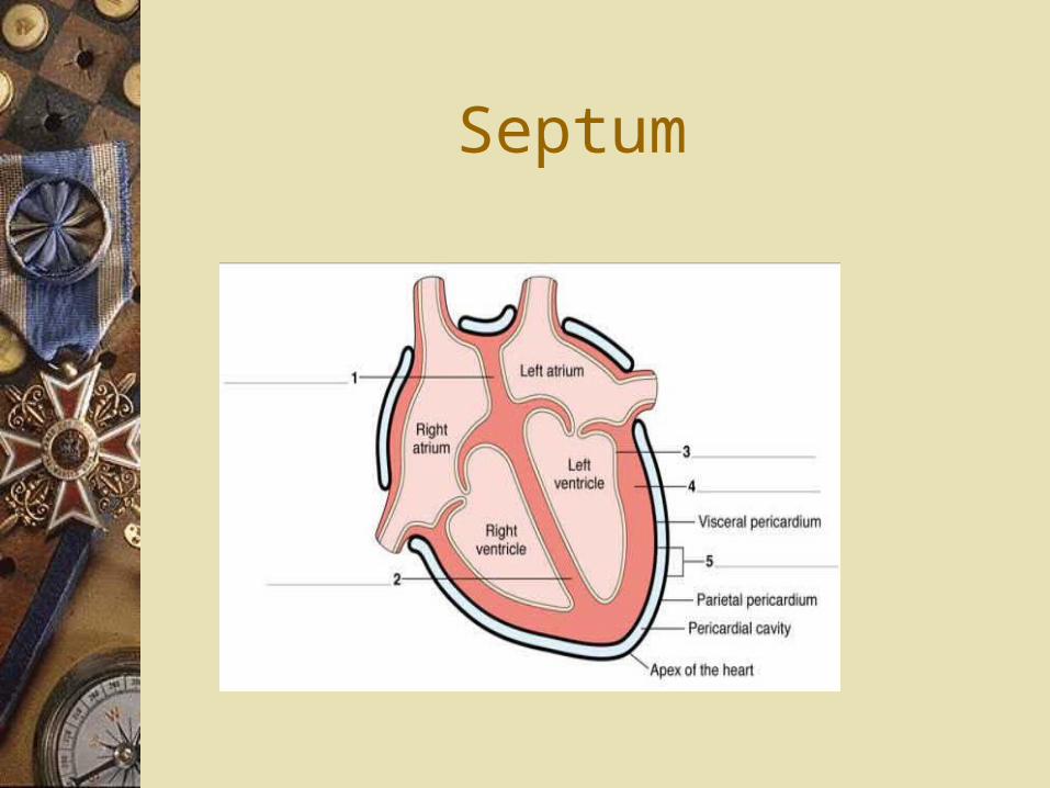
Septum
Heart structure
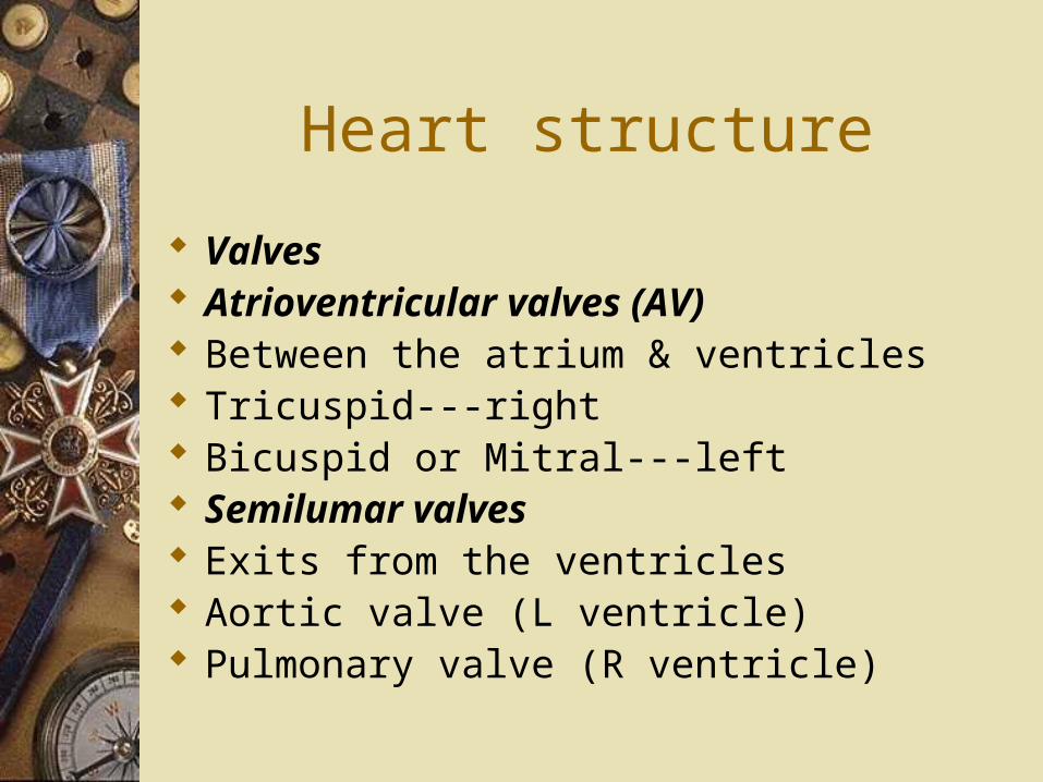
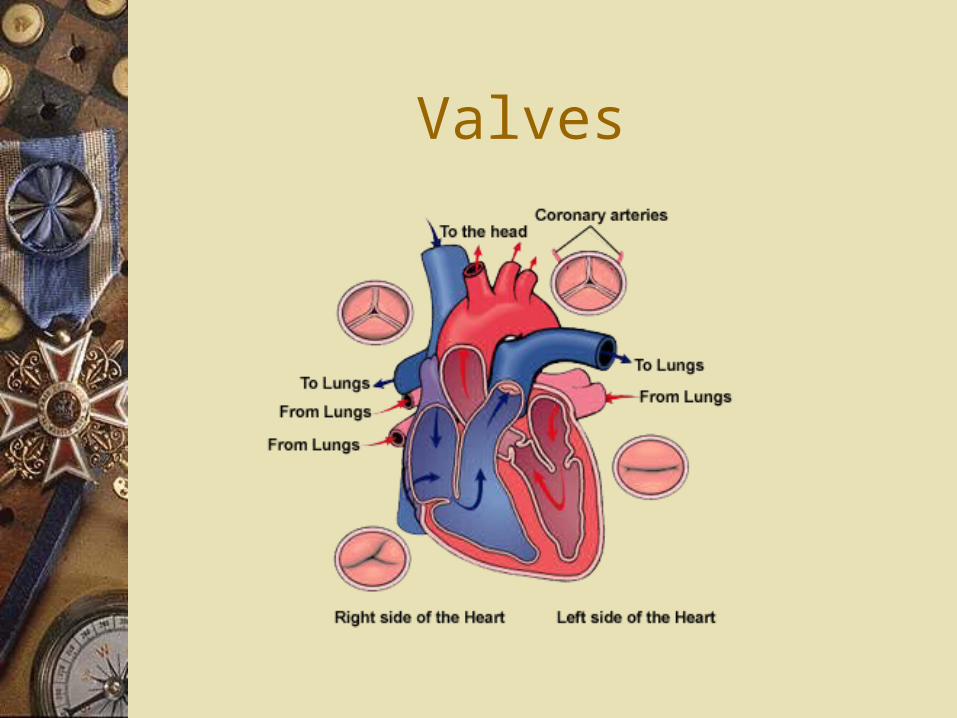
Valves Atrioventricular valves (AV) Between the atrium & ventricles Tricuspid---right Bicuspid or Mitral---left Semilumar valves Exits from the ventricles Aortic valve (L ventricle) Pulmonary valve (R ventricle)
Valves
Heart structure
Heart sounds “lub-dub” -valves closing “lub” closing of the AV valves “dub” closing of the semilunar valves Murmur: Abnormal heart sound
Heart structure
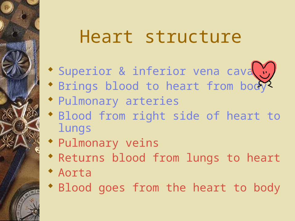
Superior & inferior vena cava Brings blood to heart from body Pulmonary arteries Blood from right side of heart to lungs Pulmonary veins Returns blood from lungs to heart Aorta Blood goes from the heart to body
Heart structure
Coronary arteries First branches off the aorta Supply blood to the heart
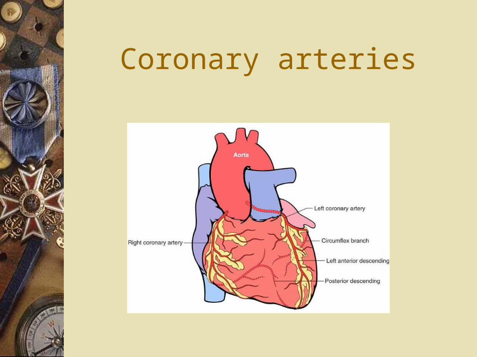
Coronary arteries
Circulation
Blood flow
E:\Chapter_42\A_PowerPoint_Lectures\42_Lecture_Presentation\42_06PathOfBloodFlow_A.html
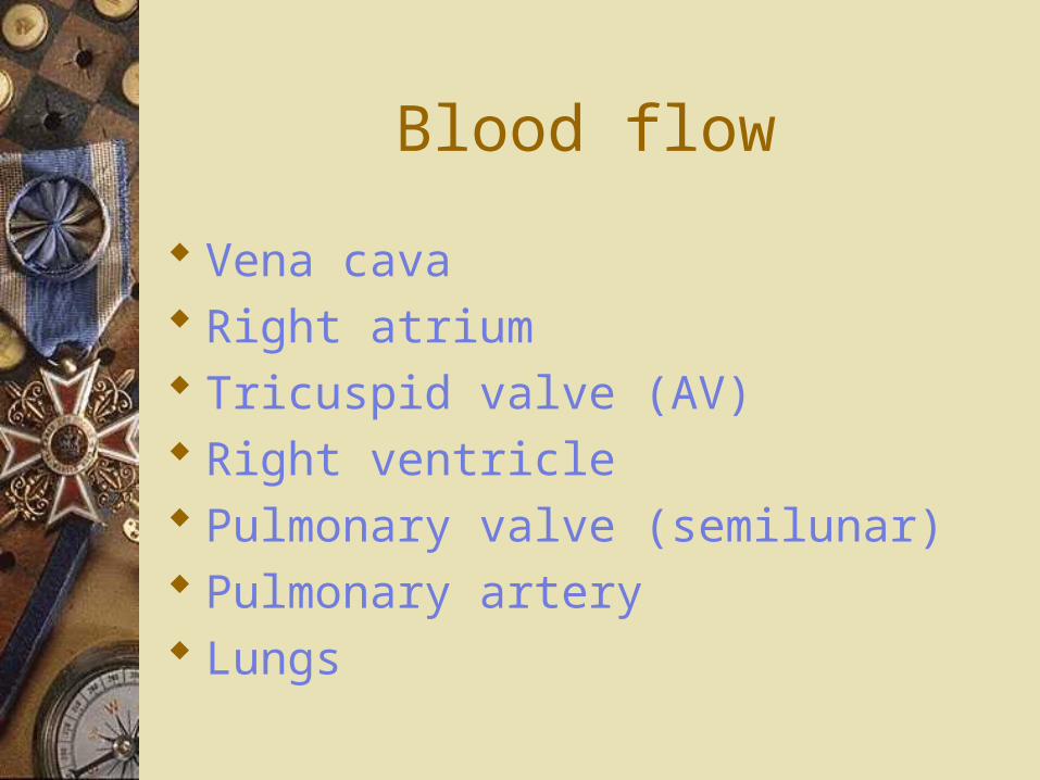
Blood flow
Vena cava Right atrium Tricuspid valve (AV) Right ventricle Pulmonary valve (semilunar) Pulmonary artery Lungs
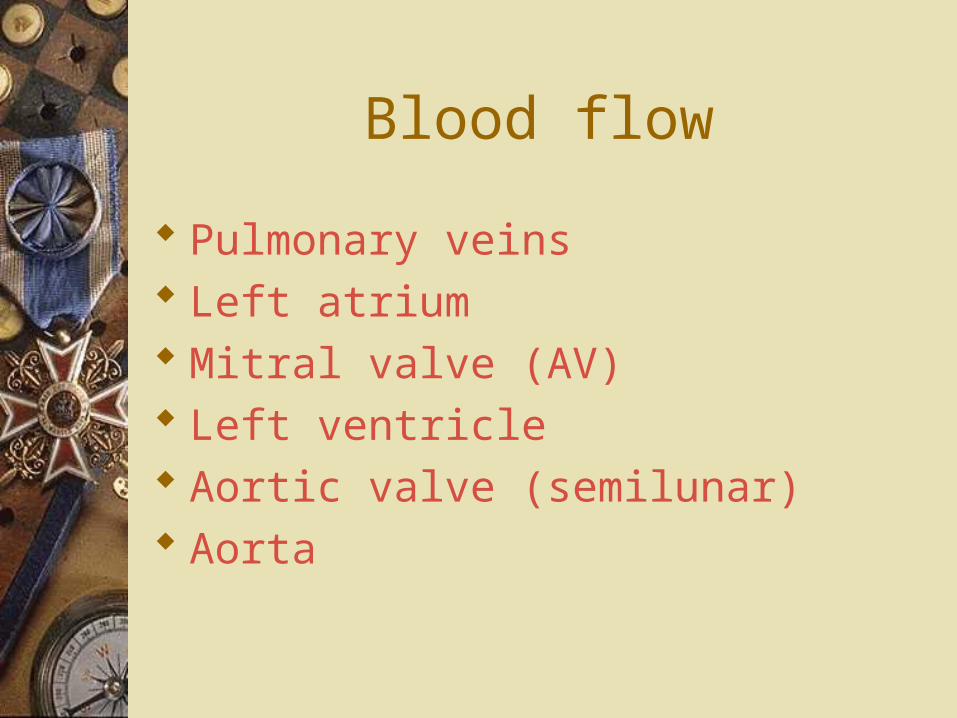
Blood flow
Pulmonary veins Left atrium Mitral valve (AV) Left ventricle Aortic valve (semilunar) Aorta
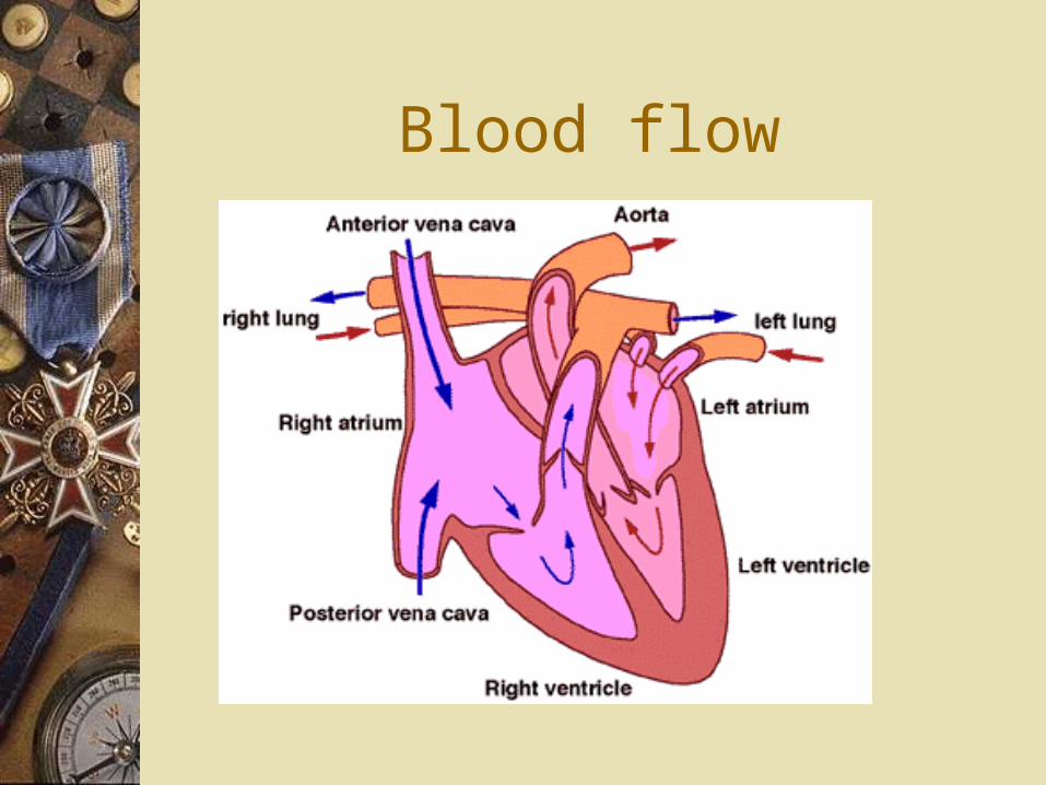
Blood flow
Blood flow
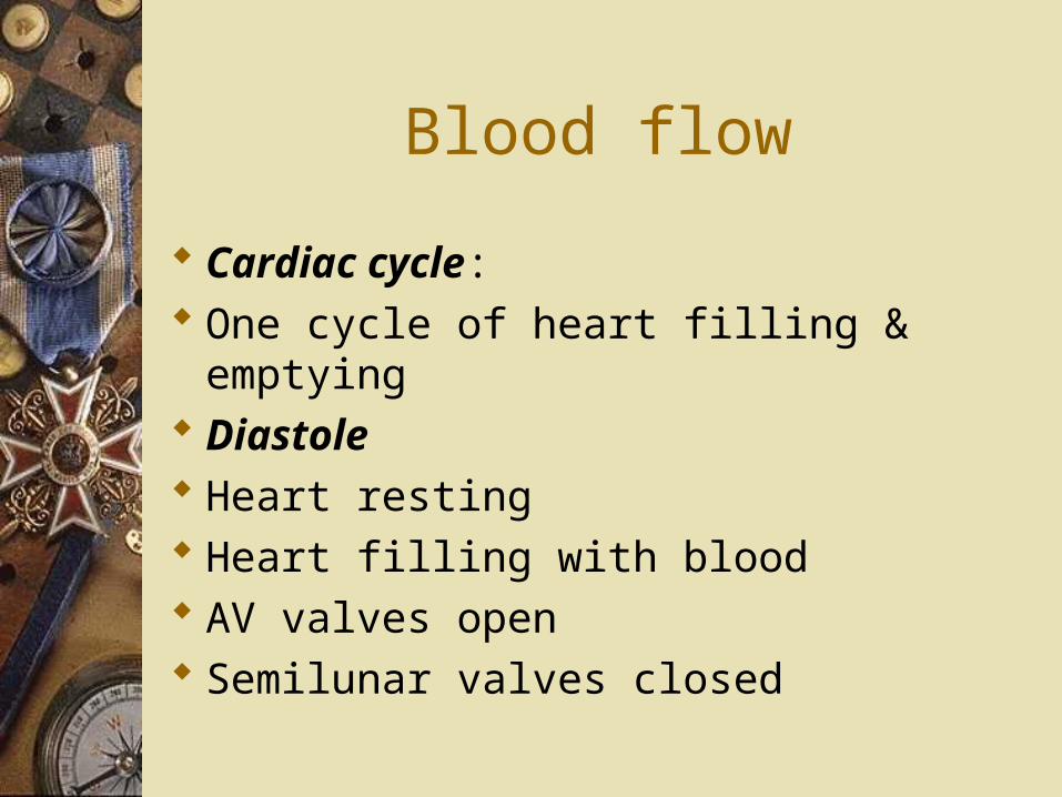
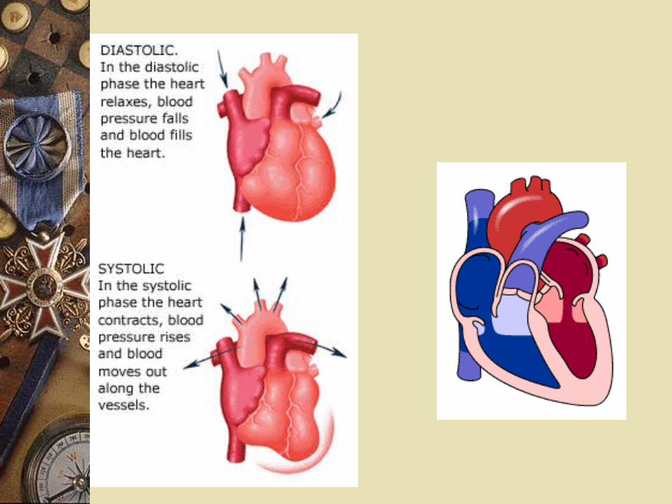
Cardiac cycle: One cycle of heart filling & emptying Diastole Heart resting Heart filling with blood AV valves open Semilunar valves closed
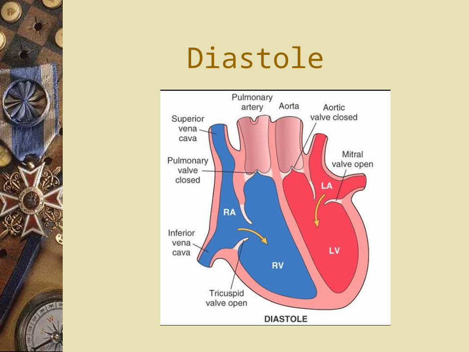
Diastole
Blood flow
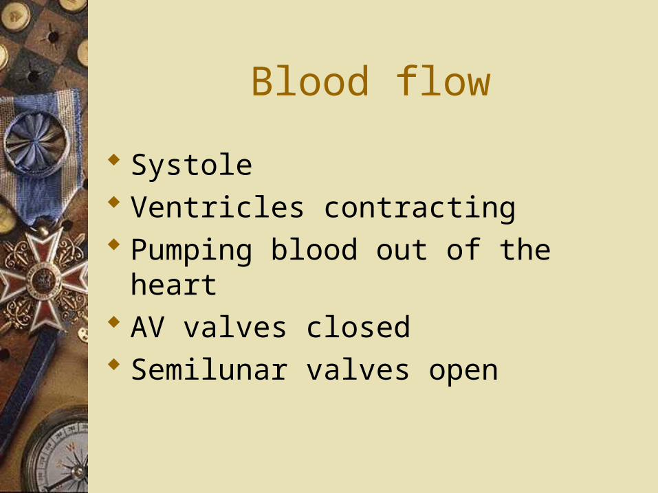
Systole Ventricles contracting Pumping blood out of the heart AV valves closed Semilunar valves open

Systole
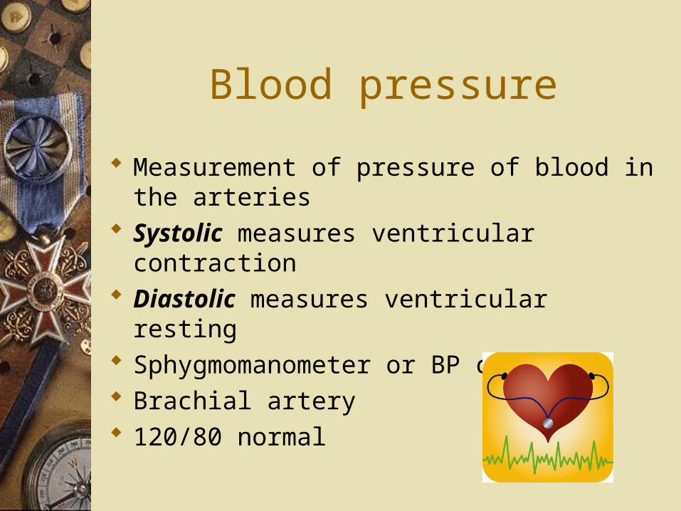
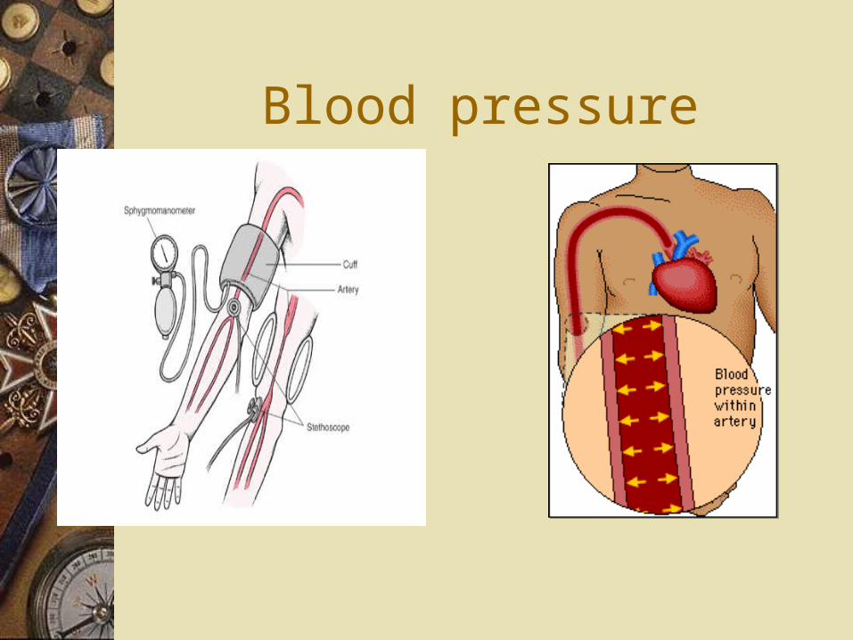
Blood pressure
Measurement of pressure of blood in the arteries
Systolic measures ventricular contraction
Diastolic measures ventricular resting Sphygmomanometer or BP cuff Brachial artery 120/80 normal
Blood pressure
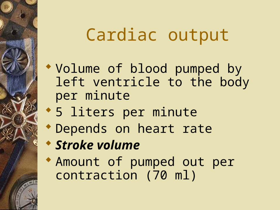
Cardiac output
Volume of blood pumped by left ventricle to the body per minute
5 liters per minute Depends on heart rate Stroke volume Amount of pumped out per
contraction (70 ml)
Cardiac output
Increases with exercise Increased HR Better stroke volume
Blood pressure
Cardiac output Flow resistance in the arteries Affect BP More constriction higher BP More dilation lower BP Baroreceptors Aorta & carotids Medulla oblongata
Blood pressure
Depends on blood volume Decreased volume Decreased cardiac output Decreased BP
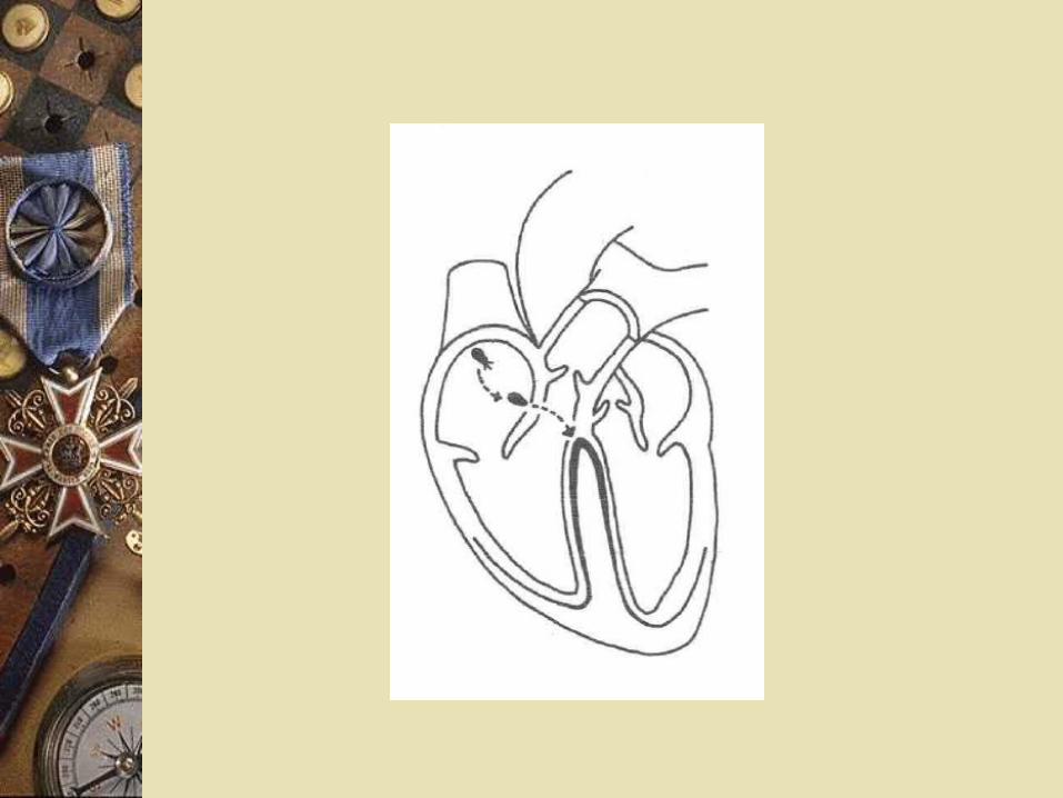
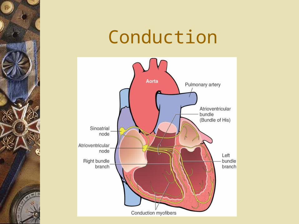
Conduction
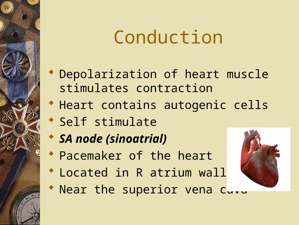
Depolarization of heart muscle stimulates contraction
Heart contains autogenic cells Self stimulate SA node (sinoatrial) Pacemaker of the heart Located in R atrium wall Near the superior vena cava
Conduction
SA node Causes atrium to contract Sends signal to the AV node AV (atrioventricular) node Located in wall between R atrium
& ventricle Sends signal to the bundle of His
Contraction
Bundle of His Sends signal to the Purkinje fibers Ventricles contract
SA⇨AV⇨Bundle of His⇨Purkinje fibers
Conduction
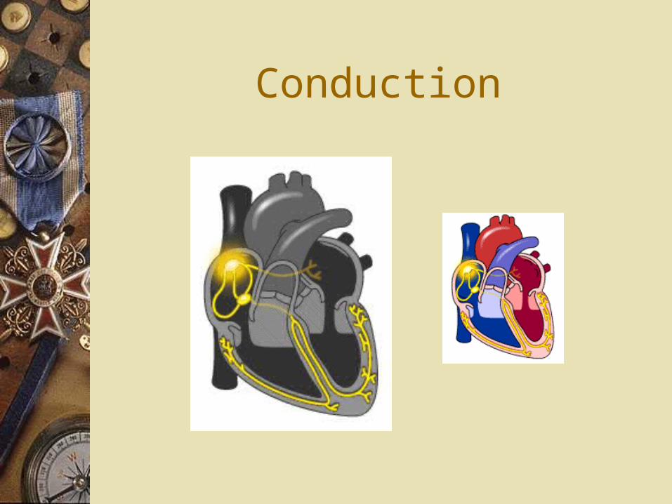
Conduction
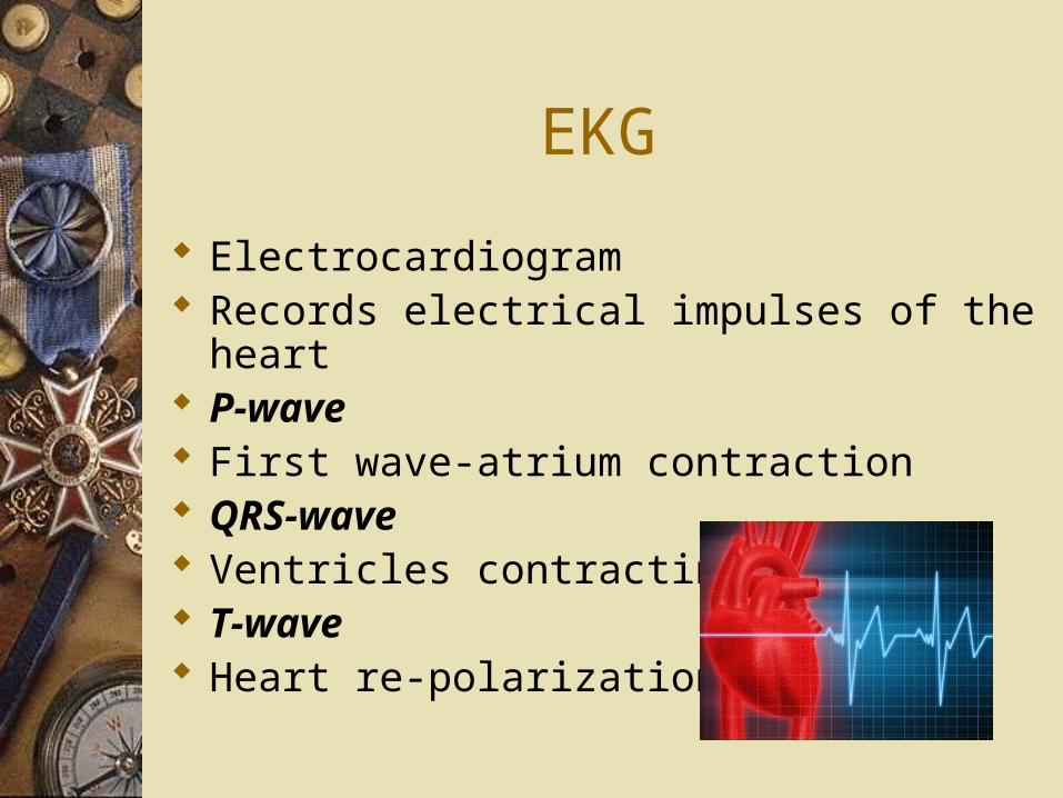
EKG
Electrocardiogram Records electrical impulses of the heart P-wave First wave-atrium contraction QRS-wave Ventricles contracting T-wave Heart re-polarization
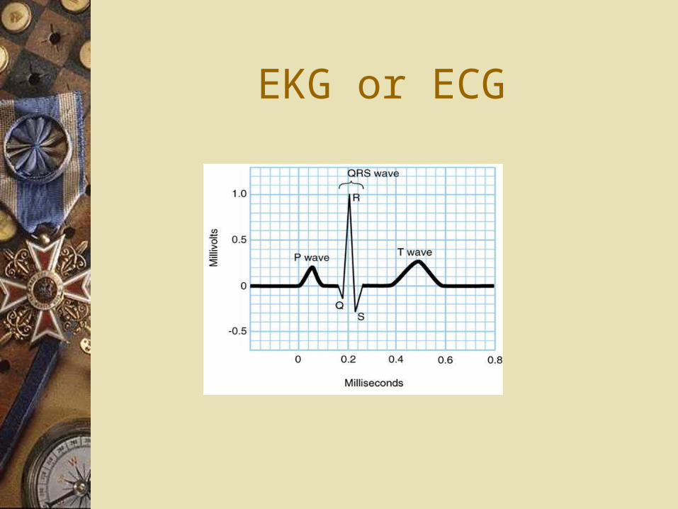
EKG or ECG
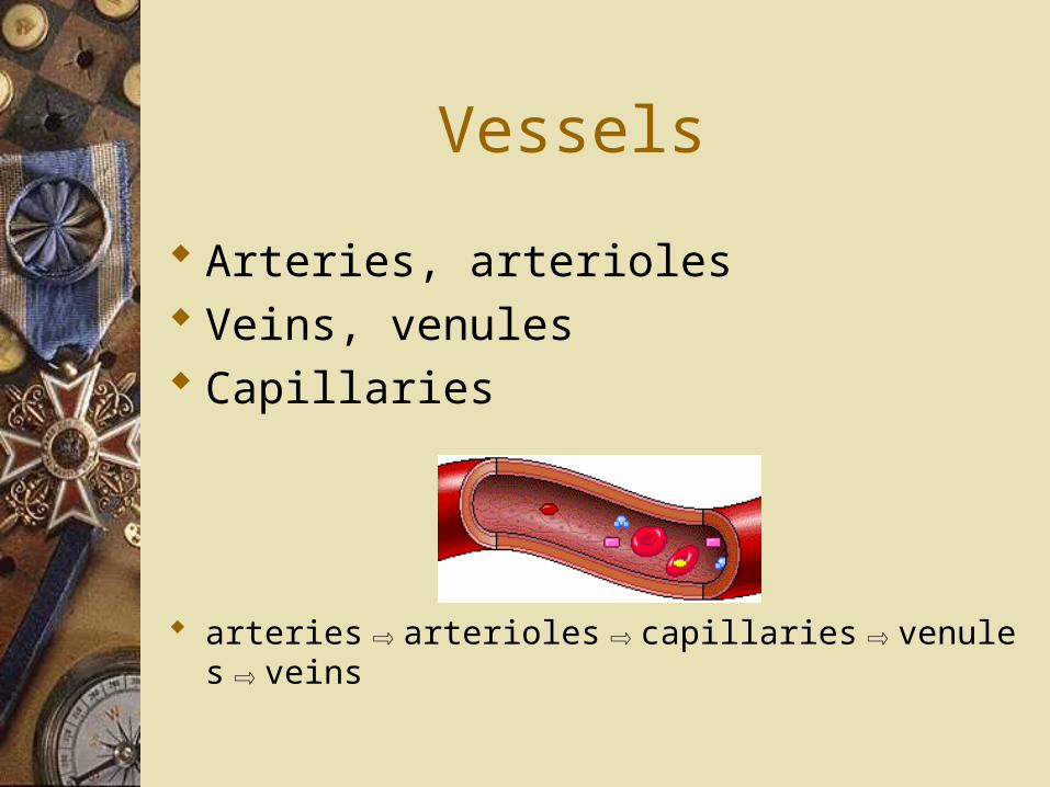
Vessels
Arteries, arterioles Veins, venules Capillaries
arteries⇨arterioles⇨capillaries⇨venules⇨veins
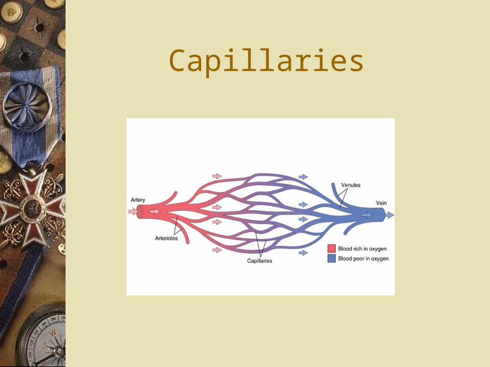
Capillaries
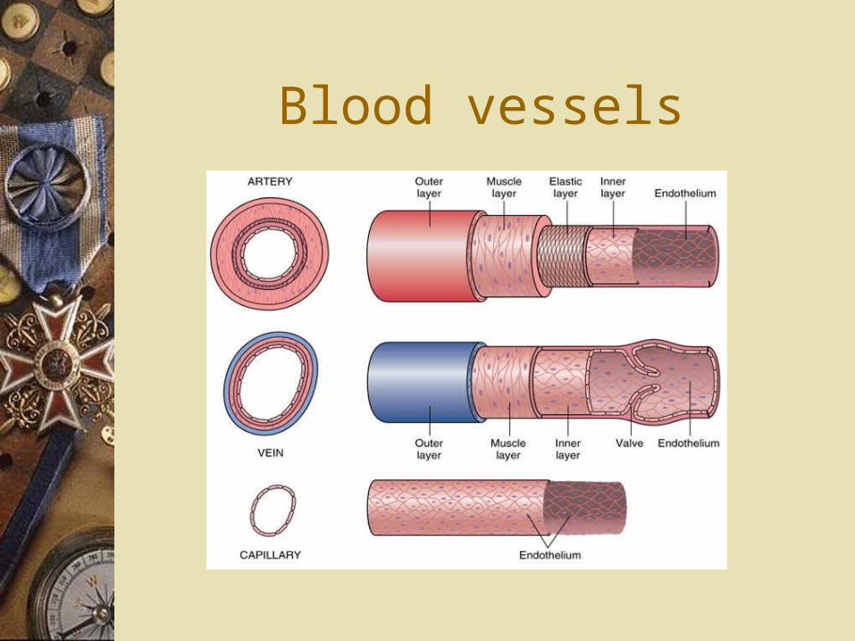
Blood vessels
Vessels
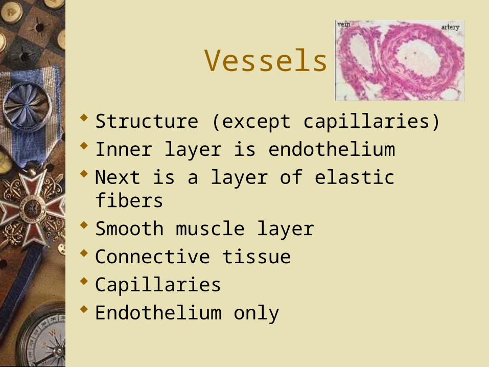
Structure (except capillaries) Inner layer is endothelium Next is a layer of elastic fibers Smooth muscle layer Connective tissue Capillaries Endothelium only
Arteries
Carry oxygen rich blood away from heart
Arterioles: Smaller arteries Larger the artery More elastic & recoil as blood is
pumped
Arteries
Vasoconstriction: Contraction of smooth muscle in
arterioles Decrease blood flow Vasodilation: Relaxation of smooth muscle Increase blood flow Precapillary sphincters: Regulate blood flow
Veins
Carry oxygen poor blood to heart Venules Smaller veins Less smooth muscle Skeletal muscles constrict Help flow of blood to heart Venous valves: Help blood flow to heart prevent backflow

Capillaries
Passage of oxygen & nutrients Into cells or extracellular fluids Passage of carbon dioxide &
wastes From cells to blood
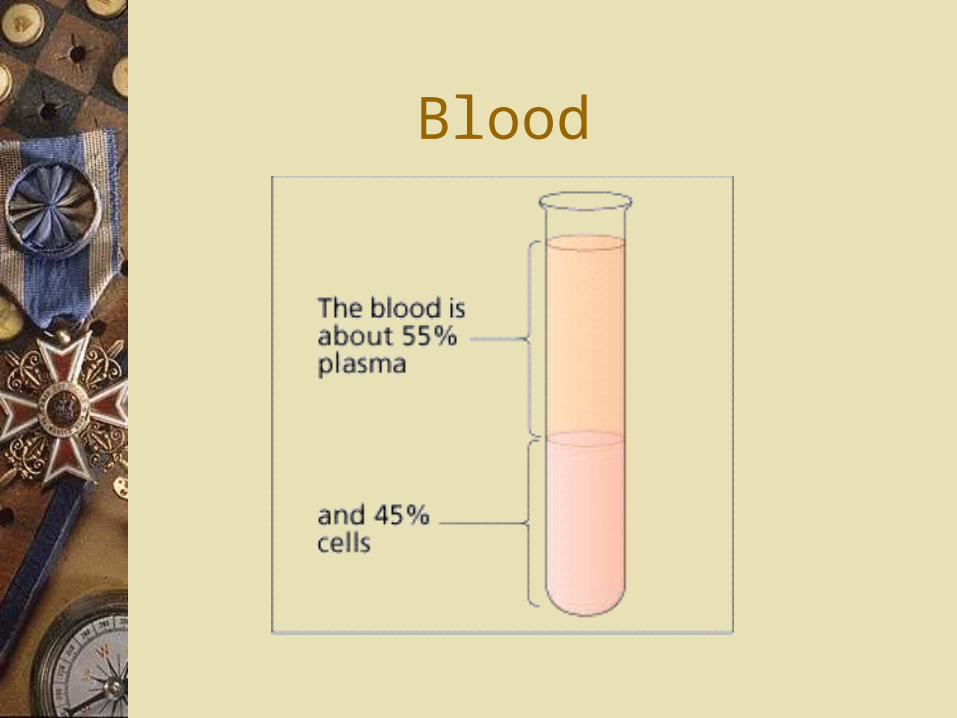
Blood
Blood
Plasma (matrix) yellow Metabolites, wastes, hormones Ions Proteins Albumin (fluids), globulins (antibodies),
fibrinogen (clots) Cells RBC, WBC, platelets
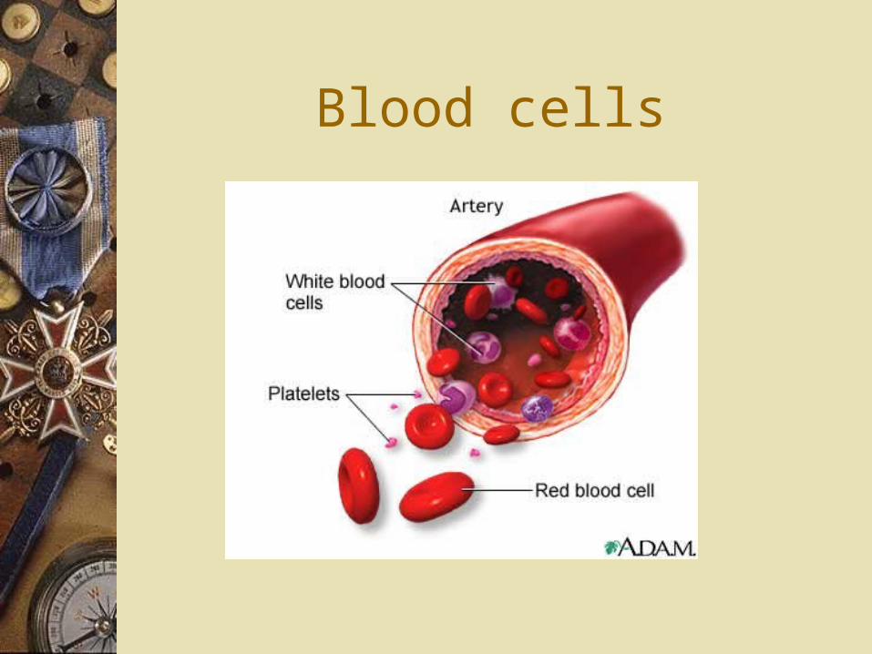
Blood cells
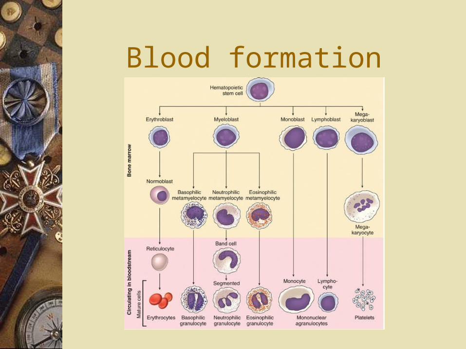
Blood formation
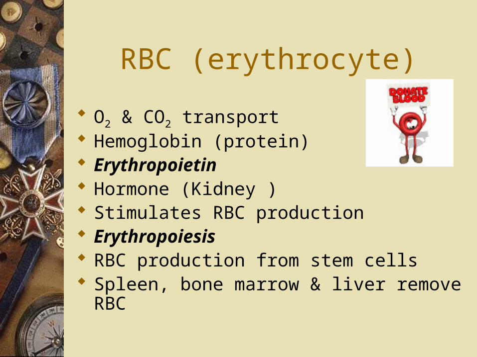
RBC (erythrocyte)
O2 & CO2 transport Hemoglobin (protein) Erythropoietin Hormone (Kidney ) Stimulates RBC production Erythropoiesis RBC production from stem cells Spleen, bone marrow & liver remove
RBC
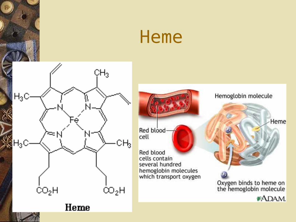
Heme
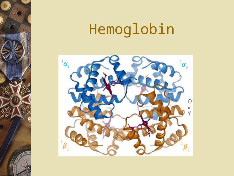
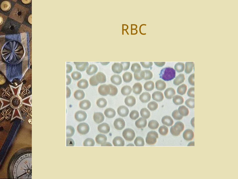
RBC
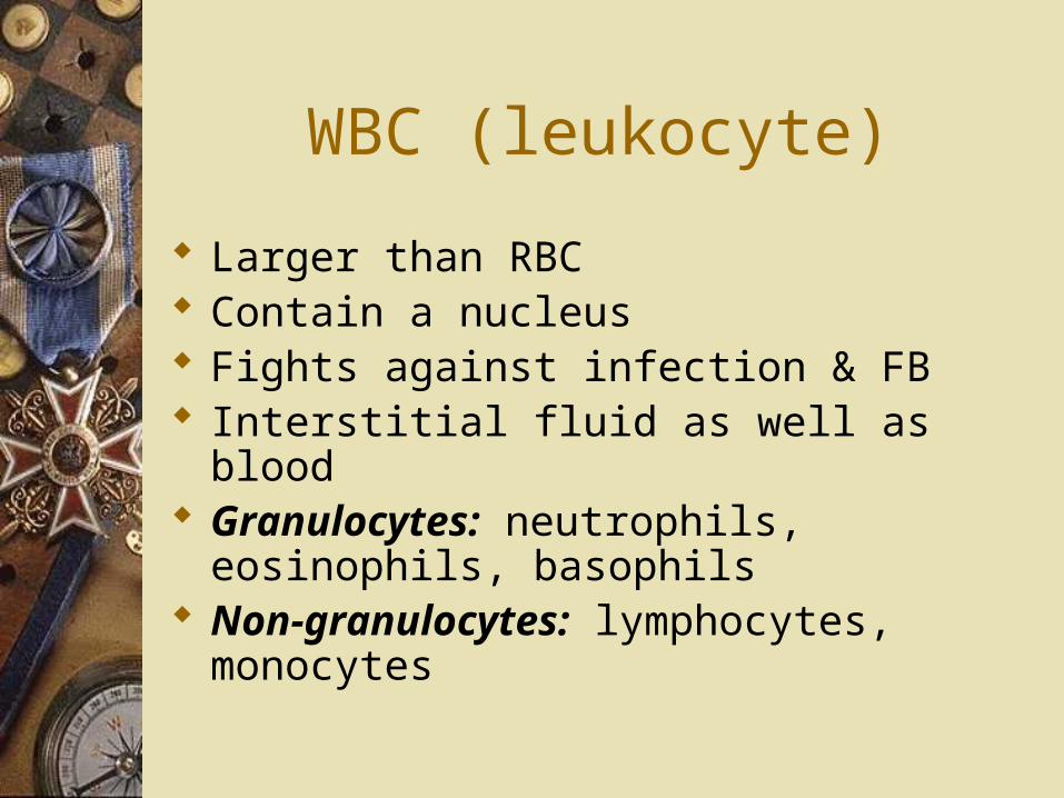
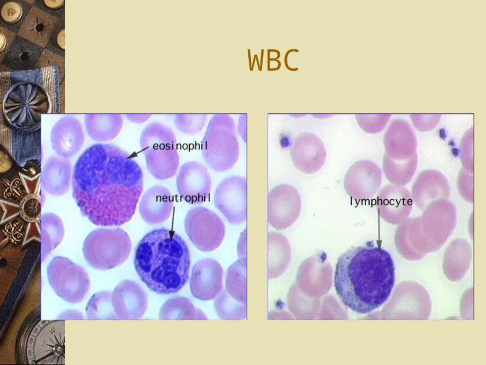
WBC (leukocyte)
Larger than RBC Contain a nucleus Fights against infection & FB Interstitial fluid as well as blood Granulocytes: neutrophils,
eosinophils, basophils Non-granulocytes: lymphocytes,
monocytes
WBC
Platelets
Megakaryocyte (bone marrow) Cell fragments Clotting First to site of injury Fibrin (protein that forms clot)

Platelets
Clotting
Tissue damage Platelets arrive Cascade reactions start Prothrombin changes to Thrombin (enzyme) Changes fibrinogen to fibrin Forms clot
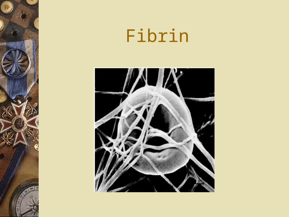
Fibrin
Blood
More RBC than WBC Hematocrit Measurement of RBC’s in the
blood RBC last about 120 days
Lymph system
Interstitial fluid Fluid from blood plasma that leaves the
capillaries Surrounds the tissues Lymph: Returns to circulation via lymph system Lymph nodes, lymph vessels & organs
(spleen & thymus)
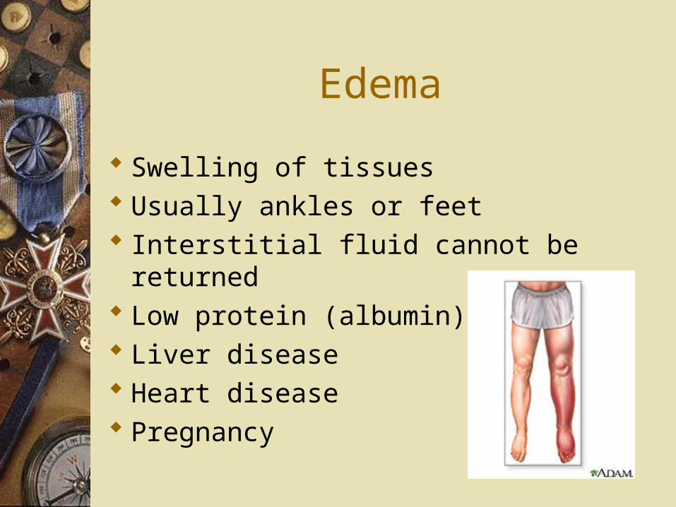
Edema
Swelling of tissues Usually ankles or feet Interstitial fluid cannot be returned Low protein (albumin) Liver disease Heart disease Pregnancy
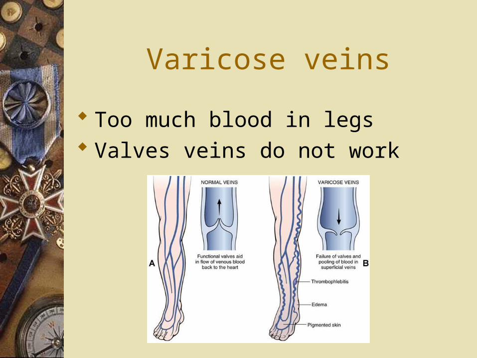
Varicose veins
Too much blood in legs Valves veins do not work
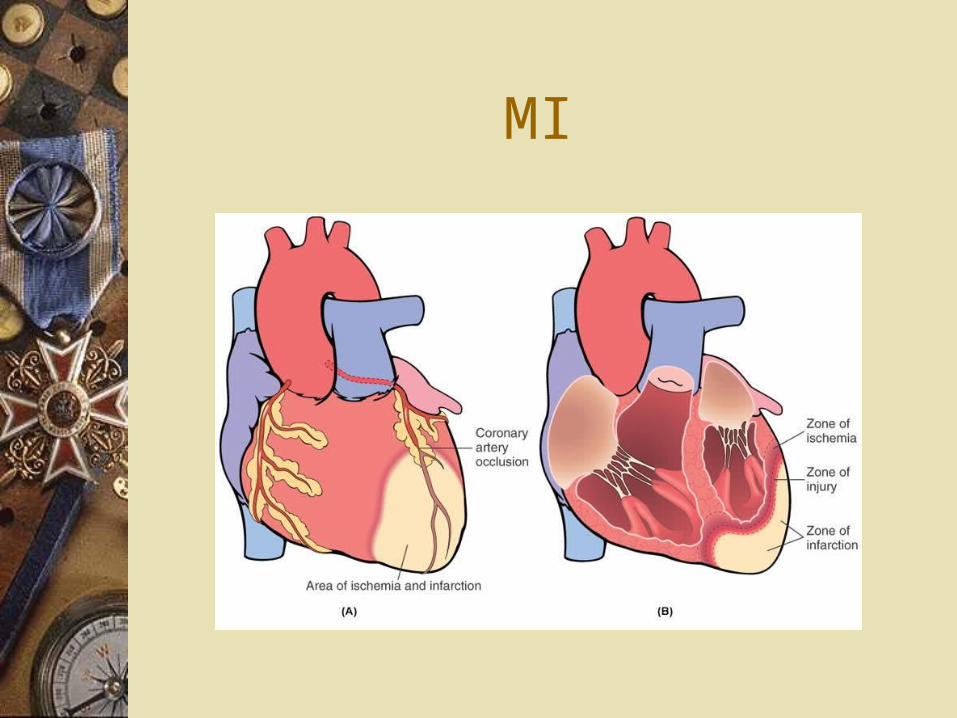
MI
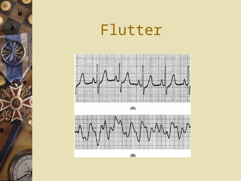
Flutter
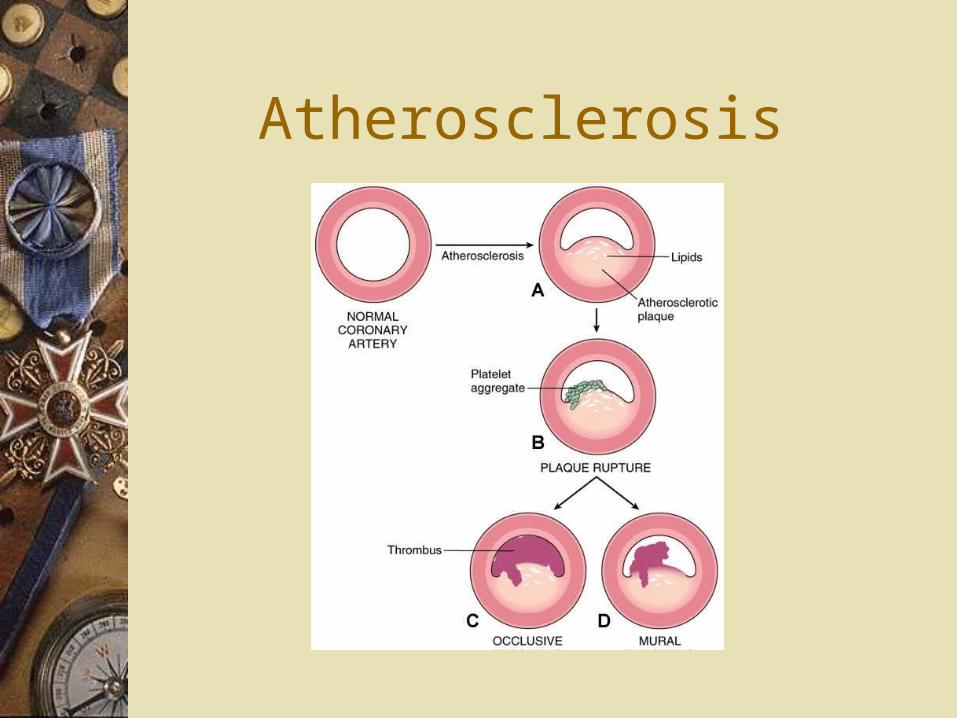
Atherosclerosis
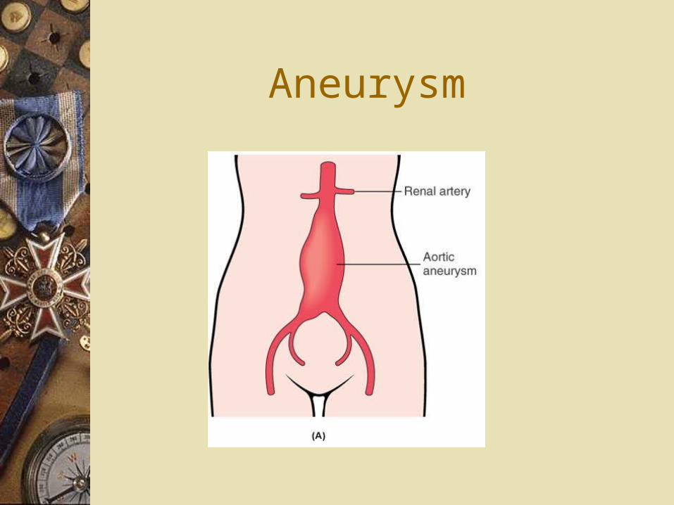
Aneurysm
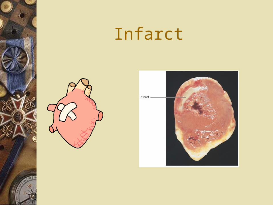
Infarct
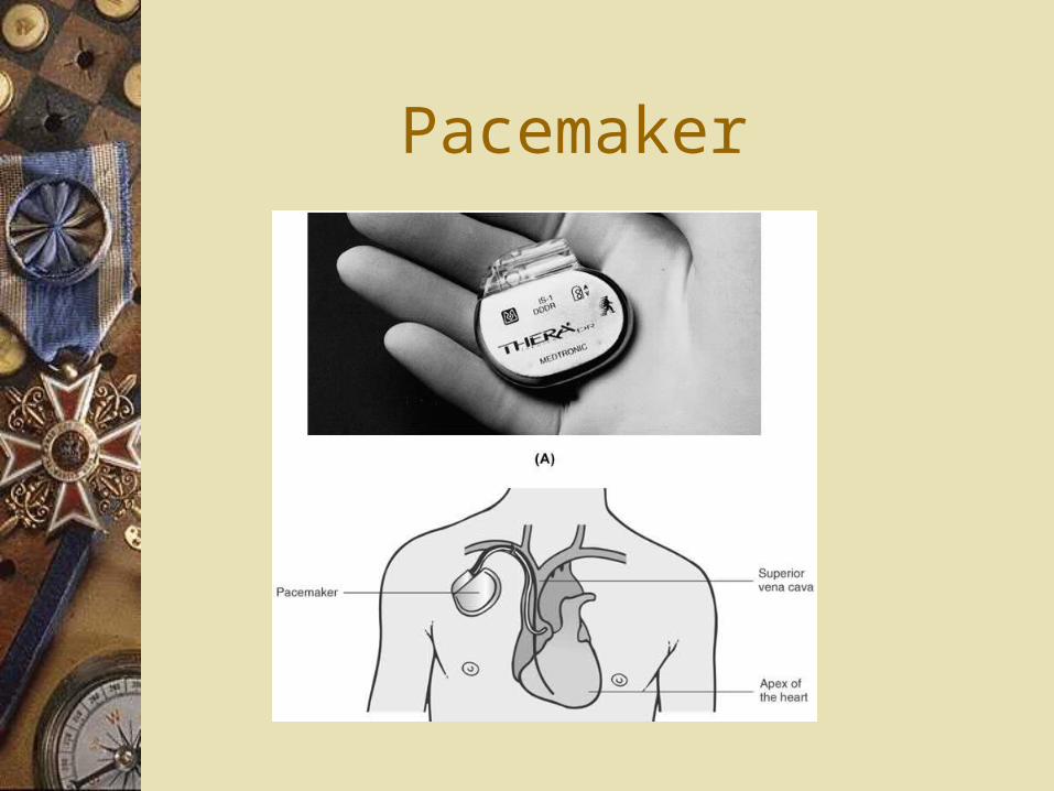
Pacemaker
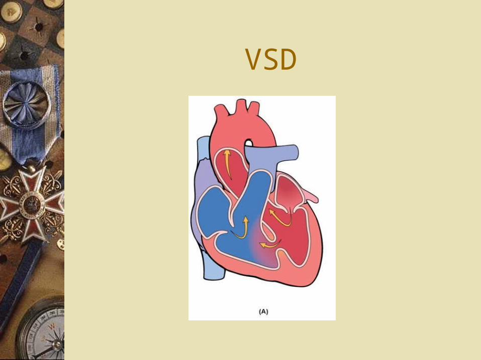
VSD
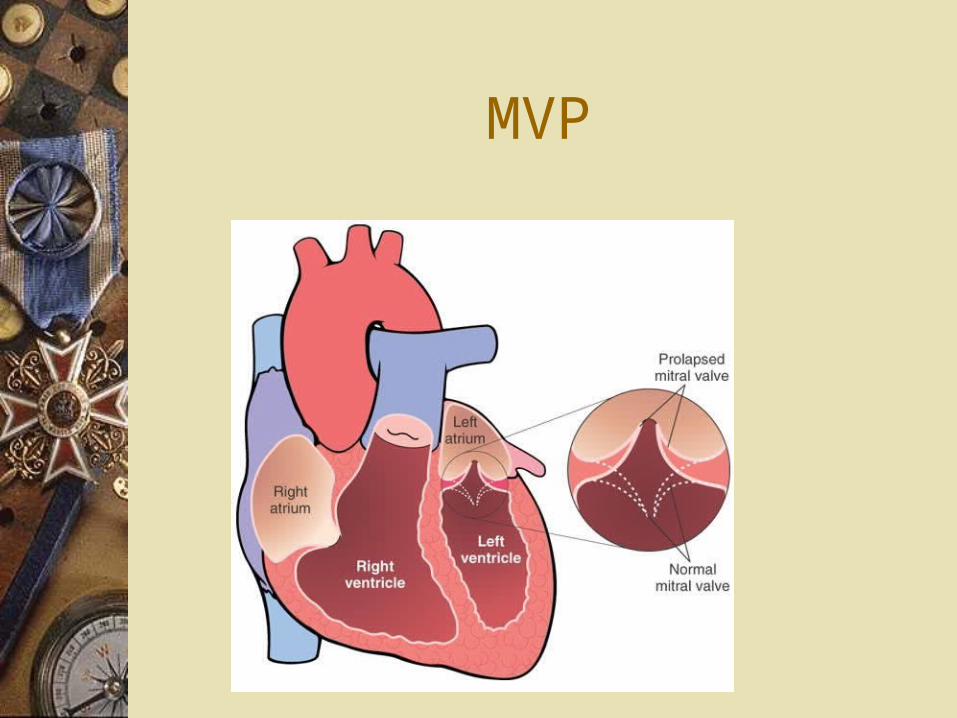
MVP