circulation and gas exchange ap biology. invertebrate open circulatory system arthropods and...
TRANSCRIPT
Circulation and Gas
Exchange
AP Biology

Invertebrate Open Circulatory System
• Arthropods and mollusks• Blood and interstitial fluid
are the same (hemolymph)• Tubular heart pumps
hemolymph through a dorsal vessel out into sinuses
• Hemolymph bathes cells and allows for exchange of nutrients
• When heart relaxes, hemolymph flows back into vessels through ostia
• Body movements squeeze sinuses to aid circulation
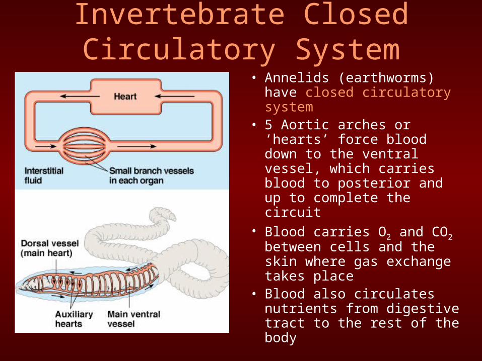
Invertebrate Closed Circulatory System
• Annelids (earthworms) have closed circulatory system
• 5 Aortic arches or ‘hearts’ force blood down to the ventral vessel, which carries blood to posterior and up to complete the circuit
• Blood carries O2 and CO2 between cells and the skin where gas exchange takes place
• Blood also circulates nutrients from digestive tract to the rest of the body

Vertebrate Circulatory System
• Closed system with a chambered heart that pumps blood through arteries that lead away from the heart to capillaries.
• Capillaries—small vessels in tissues where exchange of materials take place
• Blood is carried back to heart through veins

Fish
• 2 chamber heart– One artrium
– One ventricle
• Blood from ventricle picks up O2 in gills, then is collected into a large artery to pass directly to the rest of the body before returning to the atrium
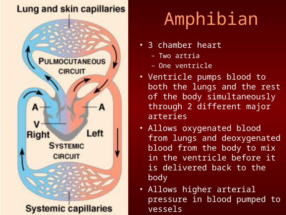
Amphibian• 3 chamber heart
– Two artria
– One ventricle
• Ventricle pumps blood to both the lungs and the rest of the body simultaneously through 2 different major arteries
• Allows oxygenated blood from lungs and deoxygenated blood from the body to mix in the ventricle before it is delivered back to the body
• Allows higher arterial pressure in blood pumped to vessels

Birds and Mammals• 4 chamber heart
– Two artria
– Two ventricle
• Higher metabolic need met by division of heart into 2 pumps
• Right atrium and ventricle pumps deoxygenated blood to lungs through pulmonary circulation
• Left atrium and ventricle pumps oxygenated blood to the rest of the body through systemic circulation
• Avoids mixing of oxygenated and deoxygenated blood
• Allows high arterial pressure required for quick delivery
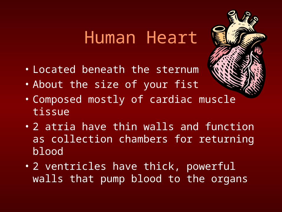
Human Heart
• Located beneath the sternum
• About the size of your fist
• Composed mostly of cardiac muscle tissue
• 2 atria have thin walls and function as collection chambers for returning blood
• 2 ventricles have thick, powerful walls that pump blood to the organs


–Atrioventricular valves • Prevent backflow
when ventricles contract
–Semilunar valves • Prevent backflow
when ventricles relax
Four valves function to prevent backflow of blood

–Systole—heart muscles contract and the chambers pump blood
–Diastole—heart muscles relax and fills with blood
–Cardiac output—volume of blood per minute that the left ventricle pumps into the systemic circuit
Cardiac Cycle

Control of Heart Rhythm• Sinoatrial (SA) node—cells are self-
excitable—generate electrical impulses
• Cardiac muscle cells are electrically coupled by intercalated discs b/w cells

Control of Heart Rhythm• Atrioventricular (AV) node—receives
signal from atria, delays 0.1 sec, and then sends signal throughout walls of ventricle via the bundle branches and Purkinje fibers

Blood Vessels
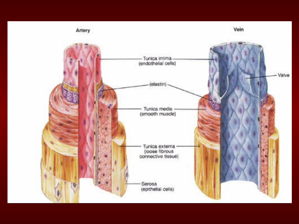
• Arteries—carry blood away from the heart to the tissues– Branch into smaller arterioles, which supply
blood to tissues via capillaries– Thick-walled, muscular (smooth muscle), and
elastic, transporting blood at high pressure– Blood is oxygenated, except the pulmonary
artery that carries deoxygenated blood from tissues to lungs through the right atrium and ventricle

• Veins—carry blood to the heart from the capillaries– Capillaries branch into larger venules, which supply blood to
veins and back to the heart– Thin-walled, little smooth muscle, transporting blood at low
pressure, and contain many valves to prevent backflow– Veins have no pulse and carry deoxygenated blood, except
the pulmonary vein which carries oxygenated blood from the lungs
– Skeletal muscle contraction aids in systemic circulation

• Capillaries—thin-walled vessels (simple squamous) • Permit exchange of
materials between blood and body cells
• Controlled by precapillary sphincters

• Capillaries• Fluid containing water
with nutrients and hormones seep from capillaries into tissues, driven by pressure
• Cells and proteins are retained in the capillaries and draw water back into the capillaries by osmosis
• Excess fluid in tissue can enter lymphatic system to be filtered and cycled back to the circulatory system
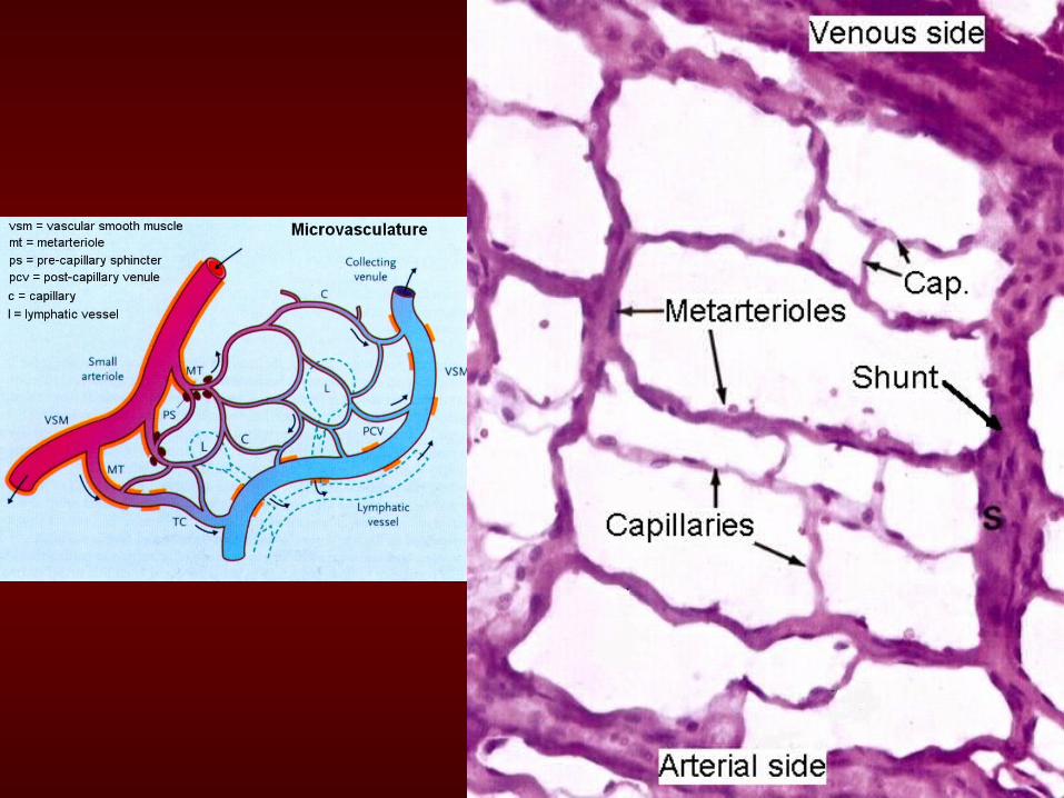

Capillary Exchange

Regulation of Blood Flow
• Regulated to match the metabolic needs
• Smooth muscle in walls of arterioles constrict to reduce blood flow to capillaries
• Smooth muscle relaxes when blood leaving capillaries is low in O2, allowing more blood to flow through capillary bed
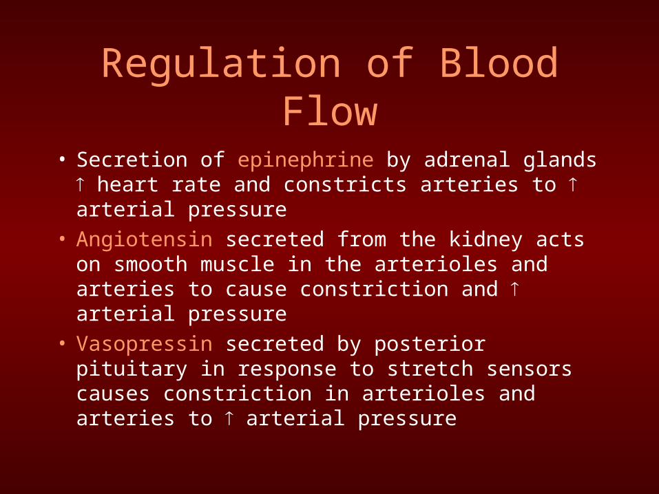
Regulation of Blood Flow
• Secretion of epinephrine by adrenal glands heart rate and constricts arteries to arterial pressure
• Angiotensin secreted from the kidney acts on smooth muscle in the arterioles and arteries to cause constriction and arterial pressure
• Vasopressin secreted by posterior pituitary in response to stretch sensors causes constriction in arterioles and arteries to arterial pressure
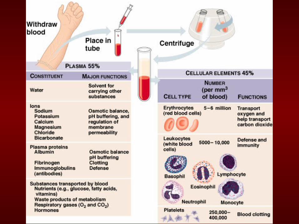

Erythrocytes: Red Blood Cells
• Primary function to carry oxygen
• Production in red bone marrow of bones stimulated by erythropoietin (produced by kidneys)
• Mature cells lack nuclei and circulate ~4mos.
• Mature cells lack mitochondria—produce ATP without oxygen through glycolysis
• Contain hemoglobin-pigment that binds oxygen

Erythrocytes: Red Blood Cells
• Red blood cells (rbc) manufacture 2 antigens, antigen A (Blood Type A) and antigen B (Blood Type B)
• Plasma carries antibodies for the antigens that are not present on the rbcs

Leukocytes: White Blood Cells
• Involved in immune functions in the body– Phagocytes—engulf
bacteria • Neutrophils—1st to arrive at
site of inflammation
• Macrophages and Monocytes
– Lymphocytes (B and T cells)—immune response
• B cells produce antibodies
• Helper T cells kill infected cells


Leukocytes: White Blood Cells• Platelets—cell
fragments produced in marrow– Involved in blood
clotting mechanism– Activation of
protease thrombin cleaves fibrinogen protein in the blood to make fibrin that polymerizes to for a net across the wound, trapping more cells and blocking the flow of blood

Cardiovascular Disease
• Heart attack—death of cardiac muscle tissue resulting from artery blockage of one or more coronary arteries which supply oxygen to the heart
• Stroke—death of nervous tissue in the brain resulting from artery blockage in the head


Cardiovascular Disease
• Atherosclerosis—plaques develop on inner walls of arteries– Forms where smooth muscle thickens
abnormally and is infiltrated by fibrous connective tissue
• Arteriosclerosis—hardening of the arteries by calcium deposits
• Hypertension—high blood pressure


Cardiovascular Disease
• Hypertension and atherosclerosis have genetic component and environmental component (smoking, lack of exercise, high fat and cholesterol diet)– Low-density lipoproteins (LDLs)—add
deposits of cholesterol in arterial plaques– High-density lipoproteins (HDLs)—may reduce
cholesterol deposition
• Exercise increases HDL concentration
• Smoking increases LDL concentration

Gas Exchange
• Involves both Respiratory system and Circulatory system

Invertebrate Gas Exchange
• Water contains less oxygen than air
• As an adaptation, most aquatic animals have gills
• Total surface area of gills is often larger than that of the rest of the body
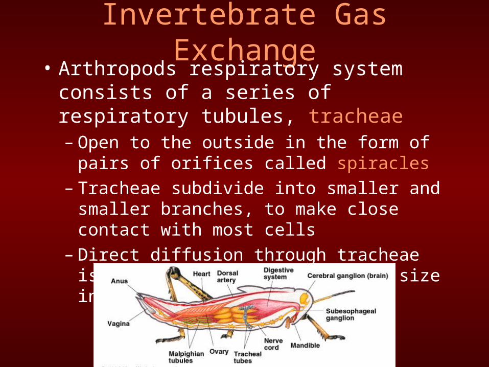
Invertebrate Gas Exchange• Arthropods respiratory system consists of a
series of respiratory tubules, tracheae– Open to the outside in the form of pairs of
orifices called spiracles– Tracheae subdivide into smaller and smaller
branches, to make close contact with most cells– Direct diffusion through tracheae is one factor
that limits body size in arthropods

Fish

Countercurrent Exchange
• Maximizes exchange of gases between blood inside the gills and the water flowing over the gills
• Blood flows through capillaries in direction opposite of water flowing across gills

Amphibians
• Simple air sac with little surface area
• Must supplement gas exchange in lungs with exchange across the thin moist skin
http://www.answersingenesis.org/home/area/magazines/images/v22frogR.jpg
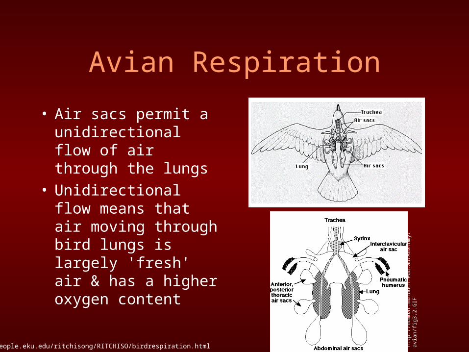
Avian Respiration
• Air sacs permit a unidirectional flow of air through the lungs
• Unidirectional flow means that air moving through bird lungs is largely 'fresh' air & has a higher oxygen content
http://people.eku.edu/ritchisong/RITCHISO/birdrespiration.html http
://n
umba
t.mur
doch
.edu
.au/
Ana
tom
y/av
ian/
fig3
.2.G
IF

Air Flow through Avian System1. On first inhalation, air flows through the trachea & bronchi & primarily into
the posterior (rear) air sacs 2. On exhalation, air moves from the posterior air sacs & into the lungs 3. With the second inhalation, air moves from the lungs & into the anterior
(front) air sacs 4. With the second exhalation, air moves from the anterior air sacs back into
the trachea & out • Air flow is driven by changes in pressure within the respiratory system: • So, it takes two respiratory cycles to move one 'packet' of air completely
through the avian respiratory system
http://people.eku.edu/ritchisong/RITCHISO/birdrespiration.html


Ventilating Lungs: Breathing

Automatic Control of Breathing
• Breathing control center in brain = medulla oblongata and pons
• Monitors CO2 levels in blood by changes in pH– CO2 + H2O Carbonic acid pH = depth and rate of
breathing
altitude = O2 levels• Sensors in aorta and
carotid arteries detect and signal control center to breathing rate

Loading and Unloading of
Respiratory Gases


Oxygen Transport
• Oxygen carried by respiratory pigments– Invertebrates utilize hemocyanin—Cu is the
oxygen-binding component– Vertebrates utilize hemoglobin—four heme
groups surrounding an Fe atom• Can carry four oxygen atoms

Oxygen Dissociation Curves for Hemoglobin
Bohr Shift:Active tissue releases CO2 CO2 reacts with H2O to form carbonic acidThis ↓ pH which induces hemoglobin to release more O2

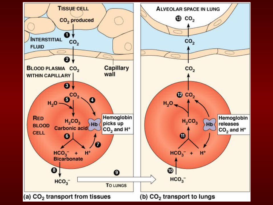
Carbon Dioxide Transport
• Hemoglobin transports CO2 and assists with buffering the blood—prevents dramatic changes in pH
• 7% CO2 released by cells transported as dissolved CO2
• 23% binds to amino group of hemoglobin• 70% transported in form of bicarbonate ions
in red blood cells