cip mitocondrias ros
TRANSCRIPT
DOI: 10.1126/scitranslmed.3006055, 192ra85 (2013);5 Sci Transl Med et al.Sameer Kalghatgi
Damage in Mammalian CellsBactericidal Antibiotics Induce Mitochondrial Dysfunction and Oxidative
Editor's Summary
made about oxidative damage to mammalian tissues.important to confirm this antibiotic effect in humans, with a broader range of antibiotics, before any conclusions can becan also be prevented by taking antioxidants or by switching to bacteriostatic antibiotics. Nevertheless, it will be
suggest that not only does this damage occur with long-term use of antibiotics, but itet al.These studies by Kalghatgi -acetyl-l-cysteine (NAC) without disrupting the bacteria-killing properties of the antibiotics.Npowerful antioxidant
in blood tests, tissue analysis, and gene expression studies. This ROS-mediated damage could be reversed by the Mice treated with clinically relevant doses of bactericidal antibiotics similarly showed signs of oxidative damage
transport chain, which would lead to a buildup of ROS. showed that bactericidal antibiotics disrupted the mitochondrial electronet al.shed light on the mechanism, Kalghatgi
DNA, protein, and lipid damage in vitro. A bacteriostatic antibiotic, tetracycline, had no effect on ROS production. Todose- and time-dependent increases in intracellular ROS in various human cell lines. Such increases in ROS led to
induced−−-lactam), and kanamycin (an aminoglycoside)βciprofloxacin (a fluoroquinolone), ampicillin (a −−antibioticsbactericidaltriggering mitochondrial release of reactive oxygen species (ROS). Indeed, in culture, three representative
antibiotics damage mammalian tissues by−−but not bacteriostatic−−The authors hypothesized that bactericidal
regimens.antibiotics may also cause damage to mammalian cells and thus pose problems for patients on long-term antibiotic
Antibiotics hurt only bacteria, right? According to a new study from Kalghatgi and colleagues, certain types of
Antibiotics Affect Mitochondria in Mammalian Cells
http://stm.sciencemag.org/content/5/192/192ra85.full.htmlcan be found at:
and other services, including high-resolution figures,A complete electronic version of this article
http://stm.sciencemag.org/content/suppl/2013/07/01/5.192.192ra85.DC1.html can be found in the online version of this article at: Supplementary Material
http://stm.sciencemag.org/content/scitransmed/4/137/137rv5.full.html http://stm.sciencemag.org/content/scitransmed/3/109/109ra115.full.html
http://stm.sciencemag.org/content/scitransmed/4/140/140sr2.full.html http://stm.sciencemag.org/content/scitransmed/5/190/190ra81.full.html
can be found online at:Related Resources for this article
http://www.sciencemag.org/about/permissions.dtl in whole or in part can be found at: article
permission to reproduce this of this article or about obtaining reprintsInformation about obtaining
is a registered trademark of AAAS. Science Translational Medicinerights reserved. The title NW, Washington, DC 20005. Copyright 2013 by the American Association for the Advancement of Science; alllast week in December, by the American Association for the Advancement of Science, 1200 New York Avenue
(print ISSN 1946-6234; online ISSN 1946-6242) is published weekly, except theScience Translational Medicine
on
July
22,
201
3pd
f.hig
hwire
.org
Dow
nloa
ded
from
R E S EARCH ART I C L E
ANT IB IOT I CS
Bactericidal Antibiotics Induce MitochondrialDysfunction and Oxidative Damage inMammalian CellsSameer Kalghatgi,1* Catherine S. Spina,1,2,3* James C. Costello,1 Marc Liesa,3
J. Ruben Morones-Ramirez,1 Shimyn Slomovic,1 Anthony Molina,3,4
Orian S. Shirihai,3 James J. Collins1,2,3†
n Ju
ly 2
2, 2
013
Prolonged antibiotic treatment can lead to detrimental side effects in patients, including ototoxicity, nephro-toxicity, and tendinopathy, yet the mechanisms underlying the effects of antibiotics in mammalian systemsremain unclear. It has been suggested that bactericidal antibiotics induce the formation of toxic reactive oxy-gen species (ROS) in bacteria. We show that clinically relevant doses of bactericidal antibiotics—quinolones,aminoglycosides, and b-lactams—cause mitochondrial dysfunction and ROS overproduction in mammaliancells. We demonstrate that these bactericidal antibiotic–induced effects lead to oxidative damage to DNA, pro-teins, and membrane lipids. Mice treated with bactericidal antibiotics exhibited elevated oxidative stress markersin the blood, oxidative tissue damage, and up-regulated expression of key genes involved in antioxidant de-fense mechanisms, which points to the potential physiological relevance of these antibiotic effects. The deleteriouseffects of bactericidal antibiotics were alleviated in cell culture and in mice by the administration of the antioxidantN-acetyl-L-cysteine or prevented by preferential use of bacteriostatic antibiotics. This work highlights the role ofantibiotics in the production of oxidative tissue damage in mammalian cells and presents strategies to mitigateor prevent the resulting damage, with the goal of improving the safety of antibiotic treatment in people.
oor
g
pdf.hig
hwire
.D
ownl
oade
d fr
om
INTRODUCTION
Antibiotics have led to an extraordinary decrease in morbidity andmortality associated with bacterial infections. Yet, despite the greatbenefits, antibiotic use has been linked to various adverse side effects,including ototoxicity (1), nephrotoxicity (2), and tendinopathy (3). Al-though antibiotic targets and modes of action have been widely studiedand well characterized in bacteria, the mechanistic effects of common-ly prescribed antibiotics on mammalian cells remain unclear. Recently,it has been demonstrated that major classes of bactericidal antibiotics,irrespective of their drug-target interactions, induce a common oxida-tive damage cellular death pathway in bacteria, leading to the produc-tion of lethal reactive oxygen species (ROS) (4–12) via disruption ofthe tricarboxylic acid (TCA) cycle and electron transport chain (ETC)(4, 6). The role of ROS in antibiotic-induced bacterial killing is cur-rently a matter of debate (13, 14) and the subject of intense experimen-tal investigation in our laboratory and other laboratories; however,the techniques critiqued in (13, 14) were not used in the present study,which focuses on mammalian systems.
Bactericidal and bacteriostatic antibiotics have been shown to targetmitochondrial components (15–20). In mammalian cells, the mito-chondrial ETC is a major source of ROS during normal metabolismbecause of leakage of electrons (21). Given the proposed bacterial or-igin of mitochondria (22), we hypothesized that bactericidal antibiot-
1Howard Hughes Medical Institute, Department of Biomedical Engineering and Center ofSynthetic Biology, Boston University, Boston, MA 02215, USA. 2Wyss Institute for Bio-logically Inspired Engineering, Harvard University, Boston, MA 02215, USA. 3Department ofMedicine, Boston University School of Medicine, Boston, MA 02118, USA. 4Department ofInternal Medicine, Section on Gerontology and Geriatric Medicine, Wake Forest UniversitySchool of Medicine, Winston-Salem, NC 27105, USA.*These authors contributed equally to this work.†Corresponding author. E-mail: [email protected]
www.S
ics commonly disrupt mitochondrial function in mammalian cells,leading to oxidative stress and oxidative damage. Previous work hasshown that mammalian cells can be damaged by antibiotic treatment,but these results were shown at concentrations considerably higherthan those applied clinically. At these high concentrations, select anti-biotics inhibited cell growth and metabolic activity, in addition to im-pairing mitochondrial function in vitro (23, 24).
Here, we focused on characterizing the mechanistic effects of clini-cally relevant levels of bactericidal antibiotics on mammalian cells,both in vitro and in vivo. We showed that bactericidal antibiotics—quinolones, aminoglycosides, and b-lactams—caused mitochondrialdysfunction and ROS overproduction in mammalian cells, ultimatelyleading to the accumulation of oxidative tissue damage. We found thatthese deleterious effects could be alleviated by administration of theFood and Drug Administration (FDA)–approved antioxidant, N-acetyl-L-cysteine (NAC), or prevented by preferential use of bacteriostaticantibiotics. These results reflect two therapeutic strategies to combatthe adverse side effects of long-term antibiotic treatment.
RESULTS
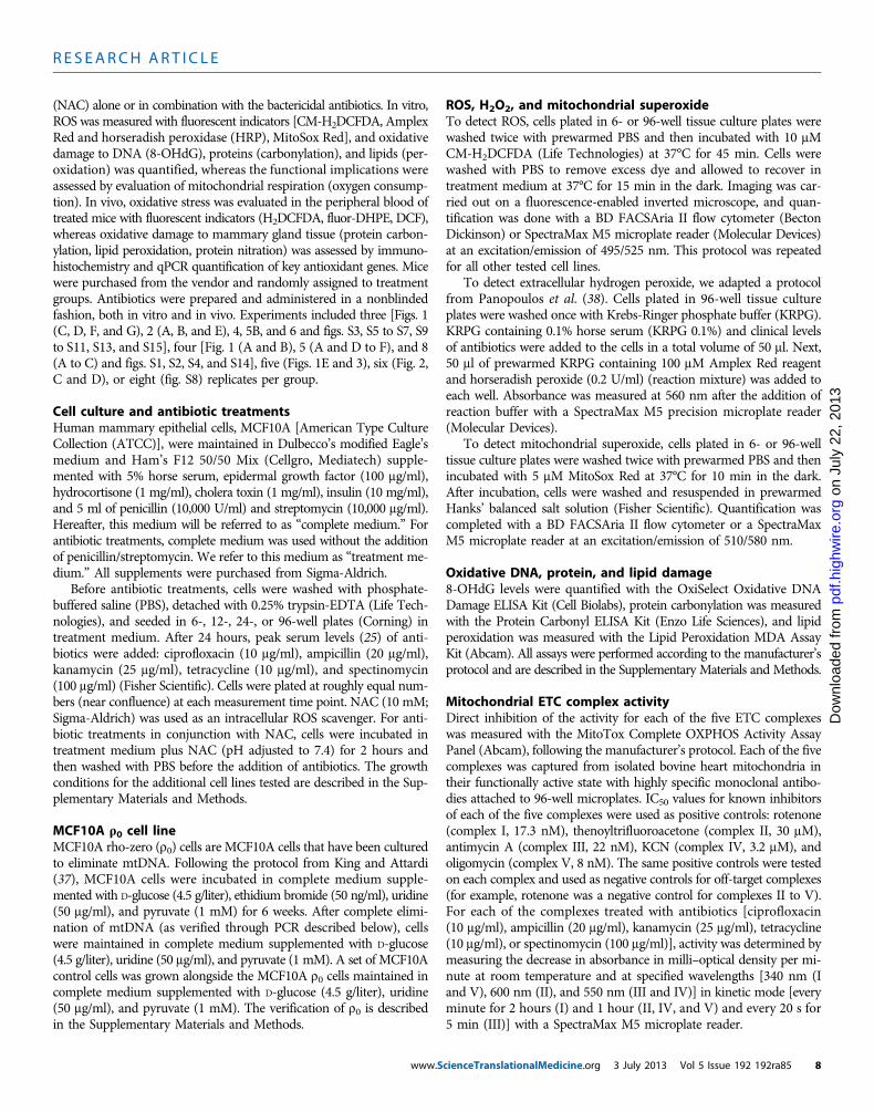
Bactericidal antibiotics induce oxidative stress and damagein mammalian cellsWe first examined whether clinically relevant doses of antibiotics in-duce the formation of ROS in mammalian cells. Here, clinically rele-vant doses are defined by peak serum levels (25). We exposed a humanmammary epithelial cell line, MCF10A, to representative bactericidalantibiotics from three different classes: ciprofloxacin (a fluoroquinolone),ampicillin (a b-lactam), and kanamycin (an aminoglycoside). All threebactericidal antibiotics induced a dose- and time-dependent increase
cienceTranslationalMedicine.org 3 July 2013 Vol 5 Issue 192 192ra85 1
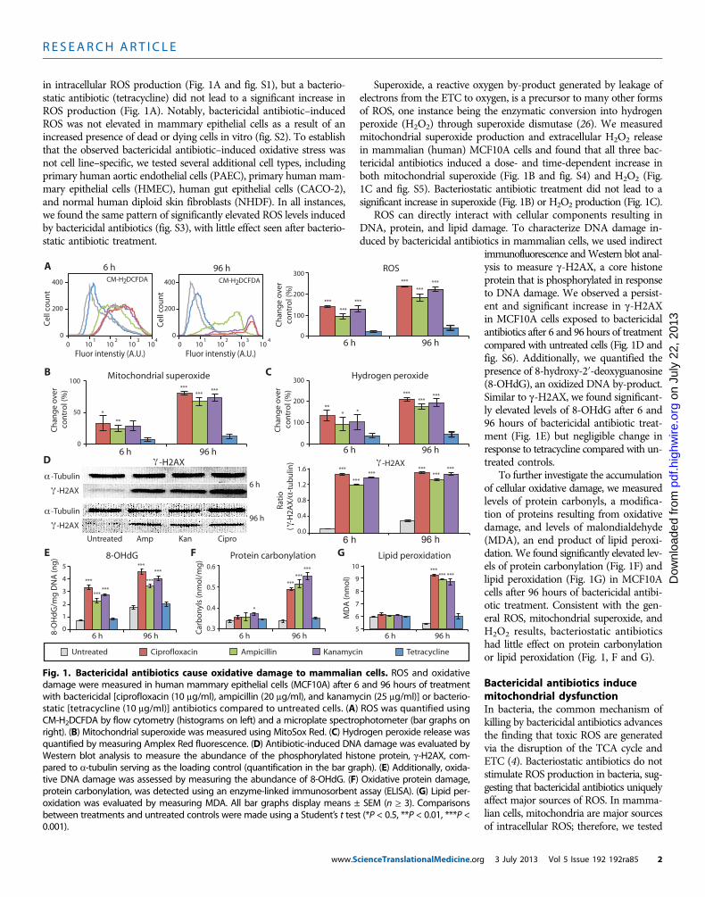
R E S EARCH ART I C L E
in intracellular ROS production (Fig. 1A and fig. S1), but a bacterio-static antibiotic (tetracycline) did not lead to a significant increase inROS production (Fig. 1A). Notably, bactericidal antibiotic–inducedROS was not elevated in mammary epithelial cells as a result of anincreased presence of dead or dying cells in vitro (fig. S2). To establishthat the observed bactericidal antibiotic–induced oxidative stress wasnot cell line–specific, we tested several additional cell types, includingprimary human aortic endothelial cells (PAEC), primary human mam-mary epithelial cells (HMEC), human gut epithelial cells (CACO-2),and normal human diploid skin fibroblasts (NHDF). In all instances,we found the same pattern of significantly elevated ROS levels inducedby bactericidal antibiotics (fig. S3), with little effect seen after bacterio-static antibiotic treatment.
www.S
Superoxide, a reactive oxygen by-product generated by leakage ofelectrons from the ETC to oxygen, is a precursor to many other formsof ROS, one instance being the enzymatic conversion into hydrogenperoxide (H2O2) through superoxide dismutase (26). We measuredmitochondrial superoxide production and extracellular H2O2 releasein mammalian (human) MCF10A cells and found that all three bac-tericidal antibiotics induced a dose- and time-dependent increase inboth mitochondrial superoxide (Fig. 1B and fig. S4) and H2O2 (Fig.1C and fig. S5). Bacteriostatic antibiotic treatment did not lead to asignificant increase in superoxide (Fig. 1B) or H2O2 production (Fig. 1C).
ROS can directly interact with cellular components resulting inDNA, protein, and lipid damage. To characterize DNA damage in-duced by bactericidal antibiotics in mammalian cells, we used indirect
cienceTranslationalMedicine.org
on
July
22,
201
3pd
f.hig
hwire
.org
Dow
nloa
ded
from
immunofluorescence andWestern blot anal-ysis to measure g-H2AX, a core histoneprotein that is phosphorylated in responseto DNA damage. We observed a persist-ent and significant increase in g-H2AXin MCF10A cells exposed to bactericidalantibiotics after 6 and 96 hours of treatmentcompared with untreated cells (Fig. 1D andfig. S6). Additionally, we quantified thepresence of 8-hydroxy-2′-deoxyguanosine(8-OHdG), an oxidized DNA by-product.Similar to g-H2AX, we found significant-ly elevated levels of 8-OHdG after 6 and96 hours of bactericidal antibiotic treat-ment (Fig. 1E) but negligible change inresponse to tetracycline compared with un-treated controls.
To further investigate the accumulationof cellular oxidative damage, we measuredlevels of protein carbonyls, a modifica-tion of proteins resulting from oxidativedamage, and levels of malondialdehyde(MDA), an end product of lipid peroxi-dation. We found significantly elevated lev-els of protein carbonylation (Fig. 1F) andlipid peroxidation (Fig. 1G) in MCF10Acells after 96 hours of bactericidal antibi-otic treatment. Consistent with the gen-eral ROS, mitochondrial superoxide, andH2O2 results, bacteriostatic antibioticshad little effect on protein carbonylationor lipid peroxidation (Fig. 1, F and G).
Bactericidal antibiotics inducemitochondrial dysfunctionIn bacteria, the common mechanism ofkilling by bactericidal antibiotics advancesthe finding that toxic ROS are generatedvia the disruption of the TCA cycle andETC (4). Bacteriostatic antibiotics do notstimulate ROS production in bacteria, sug-gesting that bactericidal antibiotics uniquelyaffect major sources of ROS. In mamma-lian cells, mitochondria are major sourcesof intracellular ROS; therefore, we tested
6 h
Cel
l co
un
t
96 hA
400
200
0
CM-H DCFDA2
B
Untreated CiproAmp Kan
-Tubulin
-H2AX
6 h
96 h
Cel
l co
un
t
400
200
01 2 3 4
Fluor intenstiy (A.U.)0 10 10 10 10
6 h 96 h
-H2AX-H2AXD
AmpicillinCiprofloxacin Kanamycin Tetracycline
C
FE
96 h6 h
8-OHdG Lipid peroxidation
96 h6 h
G
1 2 3 4
Fluor intenstiy (A.U.)0 10 10 10 10
CM-H DCFDA2
-Tubulin
-H2AX
2
3
5
1
0
4
Untreated
Protein carbonylation
96 h6 h
*
0.3
0.4
0.6
0.5
5
7
9
8
10
Car
bo
nyl
s (n
mo
l/m
g)
MD
A (n
mo
l)0
300
200
100
96 h6 h
ROSC
han
ge
ove
rco
ntr
ol (
%)
96 h6 h
Hydrogen peroxide
0
300
200
100
Ch
ang
e o
ver
co
ntr
ol (
%)
0
50
100Mitochondrial superoxide
6 h 96 h
Ch
ang
e o
ver
co
ntr
ol (
%)
8-O
Hd
G/m
g D
NA
(ng
)
6
0.0
0.4
0.8
1.2
1.6
Rat
io (
-H
2AX
/ -
tub
ulin
)
*
*** *
******
*** *********
***
******
***
***
***
***
******
******
***
*** ****** *** ***
*****
***
***
***
***
******
Fig. 1. Bactericidal antibiotics cause oxidative damage to mammalian cells. ROS and oxidativedamage were measured in human mammary epithelial cells (MCF10A) after 6 and 96 hours of treatment
with bactericidal [ciprofloxacin (10 mg/ml), ampicillin (20 mg/ml), and kanamycin (25 mg/ml)] or bacterio-static [tetracycline (10 mg/ml)] antibiotics compared to untreated cells. (A) ROS was quantified usingCM-H2DCFDA by flow cytometry (histograms on left) and a microplate spectrophotometer (bar graphs onright). (B) Mitochondrial superoxide was measured using MitoSox Red. (C) Hydrogen peroxide release wasquantified by measuring Amplex Red fluorescence. (D) Antibiotic-induced DNA damage was evaluated byWestern blot analysis to measure the abundance of the phosphorylated histone protein, g-H2AX, com-pared to a-tubulin serving as the loading control (quantification in the bar graph). (E) Additionally, oxida-tive DNA damage was assessed by measuring the abundance of 8-OHdG. (F) Oxidative protein damage,protein carbonylation, was detected using an enzyme-linked immunosorbent assay (ELISA). (G) Lipid per-oxidation was evaluated by measuring MDA. All bar graphs display means ± SEM (n ≥ 3). Comparisonsbetween treatments and untreated controls were made using a Student’s t test (*P < 0.5, **P < 0.01, ***P <0.001).3 July 2013 Vol 5 Issue 192 192ra85 2
R E S EARCH ART I C L E
on
July
22,
201
3pd
f.hig
hwire
.org
Dow
nloa
ded
from
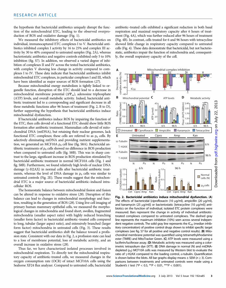
the hypothesis that bactericidal antibiotics uniquely disrupt the func-tion of the mitochondrial ETC, leading to the observed overpro-duction of ROS and oxidative damage (Fig. 1).
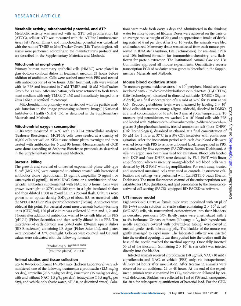
We measured the inhibitory effects of bactericidal antibiotics onindividual, immunocaptured ETC complexes I to V. Bactericidal anti-biotics inhibited complex I activity by 16 to 25% and complex III ac-tivity by 30 to 40% compared to untreated samples (Fig. 2A), whereasbacteriostatic antibiotics and negative controls exhibited only 5 to 10%inhibition (fig. S7). In addition, we observed a varied degree of inhi-bition of complexes II and IV across the tested bactericidal antibiotics,with complex V showing less change in activity compared to com-plexes I to IV. These data indicate that bactericidal antibiotics inhibitmitochondrial ETC complexes, in particular complexes I and III, whichhave been identified as major sources of ROS formation (27).
Because mitochondrial energy metabolism is tightly linked to or-ganelle function, disruption of the ETC should lead to a decrease inmitochondrial membrane potential (DYm), adenosine triphosphate(ATP) levels, and overall metabolic activity. Indeed, bactericidal anti-biotic treatment led to a corresponding and significant decrease in allthree metabolic functions after 96 hours of treatment (Fig. 2, B to D),further supporting the hypothesis that bactericidal antibiotics inducemitochondrial dysfunction.
If bactericidal antibiotics induce ROS by impairing the function ofthe ETC, then cells devoid of a functional ETC should show little ROSformation after antibiotic treatment. Mammalian cells devoid of mito-chondrial DNA (mtDNA), but retaining their nuclear genomes, lackfunctional ETC complexes; these cells are referred to as r0 cells. Byselectively eliminating mtDNA and providing nutrient supplementa-tion, we generated an MCF10A r0 cell line (fig. S8A). Bactericidal an-tibiotic treatments of r0 cells showed no difference in ROS productionwhen compared to untreated cells (fig. S8B). This was in sharp con-trast to the large, significant increase in ROS production stimulated bybactericidal antibiotic treatment in normal MCF10A cells (Fig. 1 andfig. S8B). Furthermore, we found relatively high levels of nuclear DNAdamage (g-H2AX) in normal cells after bactericidal antibiotic treat-ments, whereas the level of DNA damage in r0 cells was similar tountreated controls (Fig. 2E). These results suggest that the mitochon-drial ETC is a major source of bactericidal antibiotic–induced intra-cellular ROS.
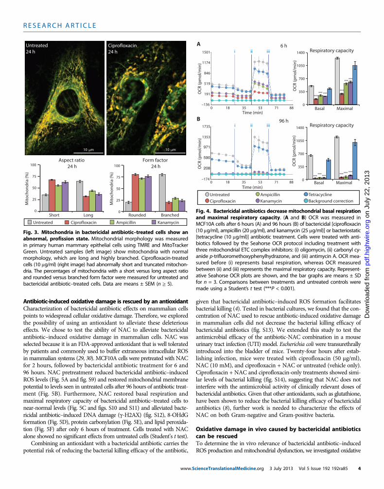
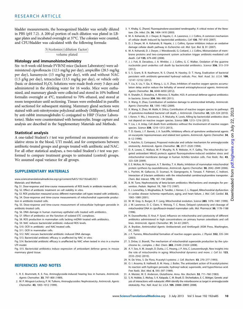
The homeostatic balance between mitochondrial fission and fusioncan be altered in response to oxidative stress (28). Disruption of thisbalance can lead to changes in mitochondrial morphology and func-tion, resulting in the generation of ROS (28). Using live-cell imaging ofprimary human mammary epithelial cells, we measured the morpho-logical changes in mitochondria and found short, swollen, fragmentedmitochondria (smaller aspect ratio) with highly reduced branching(smaller form factor) in bactericidal antibiotic–treated cells comparedto long, tubular (larger aspect ratio), and extensively branched (largerform factor) mitochondria in untreated cells (Fig. 3). These resultssuggest that bactericidal antibiotics shift the balance toward a profis-sion state. Consistent with our data in Fig. 2, a profission state can leadto a loss of membrane potential, loss of metabolic activity, and anoverall increase in oxidative stress (28).
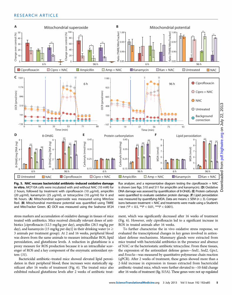
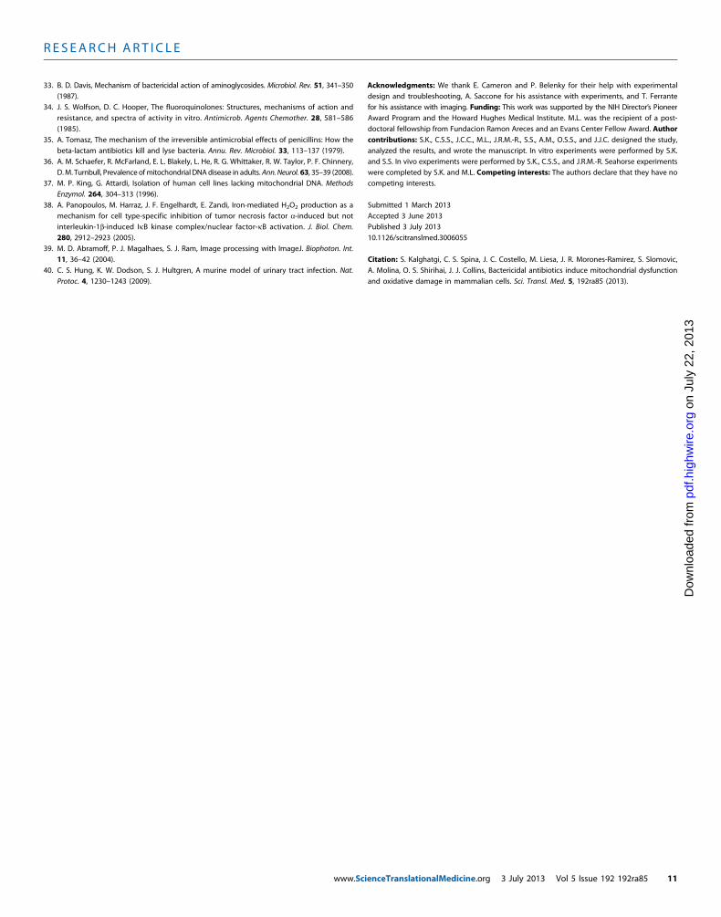
Thus far, we have characterized isolated processes involved inmitochondrial respiration. To directly test the mitochondrial respira-tory capacity of antibiotic-treated cells, we measured changes in theoxygen consumption rate (OCR) of intact MCF10A cells using theSeahorse XF24 flux analyzer. Compared to untreated cells, bactericidal
www.S
antibiotic–treated cells exhibited a significant reduction in both basalrespiration and maximal respiratory capacity after 6 hours of treat-ment (Fig. 4A), which was further reduced after 96 hours of treatment(Fig. 4B). In contrast, cells treated for 6 and 96 hours with tetracyclineshowed little change in respiratory capacity compared to untreatedcells (Fig. 4). These data demonstrate that bactericidal, but not bacterio-static, antibiotics impair the function of mitochondria and, consequent-ly, the overall respiratory capacity of the cell.
A
ATP levels
AmpicillinCiprofloxacin Kanamycin
C–60
Mitochondrial complex inhibition
–40
–20
0
D
Untreated
-H2AX
-Tubulin
0 0 0 0N N N N
Cipro Amp KanE
6 h
96 h
B
-H2AX
-Tubulin
6 h 96 h
Dec
reas
e in
co
mp
lex
acti
vity
(%)
IC50
Tetracycline
*
6 h 96 h
Normalcells (N)
cells0
0.8
0.6
0.4
1.0
6 h 96 h
Mitochondrial potential Metabolic activity
6 h 96 h
2.05
1.35
1.00
2.40
1.70
Untreated
* *
Cipro Amp KanCtrl
Flo
ur.
(A47
5-66
0nm
)
Rati
o (T
MRE
/Mit
oTr
acke
r)15
25
35
45
ATP
( m
ol)
Cipro Amp KanCtrl0.0
0.5
1.0
1.5
2.0
Rat
io (
-H
2AX
/ -
tub
ulin
)
( -
H2A
X/
-tu
bu
lin)
0.0
0.5
1.0
Rat
io
*****
****
**
**
**
**
***
***
******
***
***
**
******
***
******
***
*****
Fig. 2. Bactericidal antibiotics induce mitochondrial dysfunction. (A)The effects of bactericidal [ciprofloxacin (10 mg/ml), ampicillin (20 mg/ml),
and kanamycin (25 mg/ml)] or bacteriostatic [tetracycline (10 mg/ml)] anti-biotics on the function of individual, isolated ETC protein complexes weremeasured. Bars represent the change in activity of individual antibiotic-treated complexes compared to untreated complexes. The dashed grayline represents the maximum inhibition (10%) seen across several indepen-dent negative controls. The solid gray line represents the IC50 (median inhib-itory concentration) of positive control drugs shown to inhibit specific targetcomplexes (see fig. S7 for all positive and negative control results). (B) Mito-chondrial membrane potential was quantified using tetramethylrhodamineester (TMRE) and MitoTracker Green. (C) ATP levels were measured using aluciferin/luciferase assay. (D) Metabolic activity was measured using a color-imetric tetrazolium dye (XTT). (E) DNA damage in normal (N) and mtDNA-depleted (r0) MCF10A cells was measured by Western blot to evaluate theratio of g-H2AX compared to the loading control, a-tubulin. Quantificationis shown below the blots. All bar graphs display means ± SEM (n ≥ 3). Com-parisons between treatments and untreated controls were made using aStudent’s t test (*P < 0.5, **P < 0.01, ***P < 0.001).cienceTranslationalMedicine.org 3 July 2013 Vol 5 Issue 192 192ra85 3
R E S EARCH ART I C L E
on
July
22,
201
3pd
f.hig
hwire
.org
Dow
nloa
ded
from
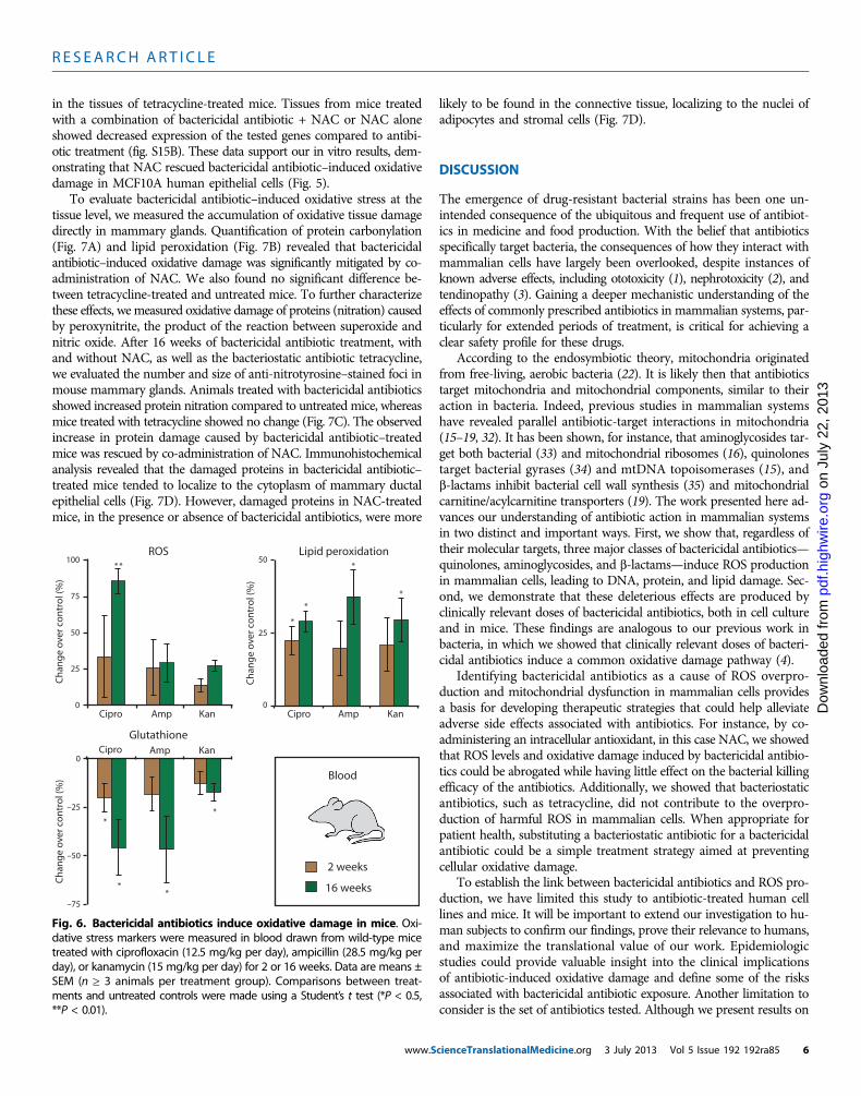
Antibiotic-induced oxidative damage is rescued by an antioxidantCharacterization of bactericidal antibiotic effects on mammalian cellspoints to widespread cellular oxidative damage. Therefore, we exploredthe possibility of using an antioxidant to alleviate these deleteriouseffects. We chose to test the ability of NAC to alleviate bactericidalantibiotic–induced oxidative damage in mammalian cells. NAC wasselected because it is an FDA-approved antioxidant that is well toleratedby patients and commonly used to buffer extraneous intracellular ROSin mammalian systems (29, 30). MCF10A cells were pretreated with NACfor 2 hours, followed by bactericidal antibiotic treatment for 6 and96 hours. NAC pretreatment reduced bactericidal antibiotic–inducedROS levels (Fig. 5A and fig. S9) and restored mitochondrial membranepotential to levels seen in untreated cells after 96 hours of antibiotic treat-ment (Fig. 5B). Furthermore, NAC restored basal respiration andmaximal respiratory capacity of bactericidal antibiotic–treated cells tonear-normal levels (Fig. 5C and figs. S10 and S11) and alleviated bacte-ricidal antibiotic–induced DNA damage (g-H2AX) (fig. S12), 8-OHdGformation (Fig. 5D), protein carbonylation (Fig. 5E), and lipid peroxida-tion (Fig. 5F) after only 6 hours of treatment. Cells treated with NACalone showed no significant effects from untreated cells (Student’s t test).
Combining an antioxidant with a bactericidal antibiotic carries thepotential risk of reducing the bacterial killing efficacy of the antibiotic,
www.S
given that bactericidal antibiotic–induced ROS formation facilitatesbacterial killing (4). Tested in bacterial cultures, we found that the con-centration of NAC used to rescue antibiotic-induced oxidative damagein mammalian cells did not decrease the bacterial killing efficacy ofbactericidal antibiotics (fig. S13). We extended this study to test theantimicrobial efficacy of the antibiotic-NAC combination in a mouseurinary tract infection (UTI) model. Escherichia coli were transurethrallyintroduced into the bladder of mice. Twenty-four hours after estab-lishing infection, mice were treated with ciprofloxacin (50 mg/ml),NAC (10 mM), and ciprofloxacin + NAC or untreated (vehicle only).Ciprofloxacin + NAC and ciprofloxacin-only treatments showed simi-lar levels of bacterial killing (fig. S14), suggesting that NAC does notinterfere with the antimicrobial activity of clinically relevant doses ofbactericidal antibiotics. Given that other antioxidants, such as glutathione,have been shown to reduce the bacterial killing efficacy of bactericidalantibiotics (8), further work is needed to characterize the effects ofNAC on both Gram-negative and Gram-positive bacteria.
Oxidative damage in vivo caused by bactericidal antibioticscan be rescuedTo determine the in vivo relevance of bactericidal antibiotic–inducedROS production and mitochondrial dysfunction, we investigated oxidative
Short Long Rounded Branched
Form factor 24 h
Aspect ratio 24 h
Mit
och
on
dri
a (%
)
AmpicillinCiprofloxacin KanamycinUntreated
100
75
50
25
0
Untreated24 h
Ciprofloxacin24 h
10 m
100
75
50
25
0
Mit
och
on
dri
a (%
)
10 m
Fig. 3. Mitochondria in bactericidal antibiotic–treated cells show anabnormal, profission state. Mitochondrial morphology was measured
in primary human mammary epithelial cells using TMRE and MitoTrackerGreen. Untreated samples (left image) show mitochondria with normalmorphology, which are long and highly branched. Ciprofloxacin-treatedcells (10 mg/ml) (right image) had abnormally short and truncated mitochon-dria. The percentages of mitochondria with a short versus long aspect ratioand rounded versus branched form factor were measured for untreated andbactericidal antibiotic–treated cells. Data are means ± SEM (n ≥ 5).Kanamycin
Ampicillin
Background correction
TetracyclineUntreated
Ciprofloxacin
0 18 35 53 71 88
Time (min)
1353
971
590
208
–174
1735
OC
R (p
mo
l/m
in)
i ii iii
0 18 35 53 71Time (min)
88
1174
846
519
191
–136
1501
OC
R (p
mo
l/m
in)
i ii iii6 h
Basal Maximal
96 h
Basal Maximal
B
A
Respiratory capacity
Respiratory capacity
OC
R (p
mo
l/m
in)
0
350
700
1050
1400
OC
R (p
mo
l/m
in)
0
350
700
1050
1400
****** ***
***
******
******
****** *** ***
Fig. 4. Bactericidal antibiotics decrease mitochondrial basal respirationand maximal respiratory capacity. (A and B) OCR was measured in
MCF10A cells after 6 hours (A) and 96 hours (B) of bactericidal [ciprofloxacin(10 mg/ml), ampicillin (20 mg/ml), and kanamycin (25 mg/ml)] or bacteriostatic[tetracycline (10 mg/ml)] antibiotic treatment. Cells were treated with anti-biotics followed by the Seahorse OCR protocol including treatment withthree mitochondrial ETC complex inhibitors: (i) oligomycin, (ii) carbonyl cy-anide p-trifluoromethoxyphenylhydrazone, and (iii) antimycin A. OCR mea-sured before (i) represents basal respiration, whereas OCR measuredbetween (ii) and (iii) represents the maximal respiratory capacity. Represent-ative Seahorse OCR plots are shown, and the bar graphs are means ± SDfor n = 3. Comparisons between treatments and untreated controls weremade using a Student’s t test (***P < 0.001).cienceTranslationalMedicine.org 3 July 2013 Vol 5 Issue 192 192ra85 4
R E S EARCH ART I C L E
on
July
22,
201
3pd
f.hig
hwire
.org
Dow
nloa
ded
from
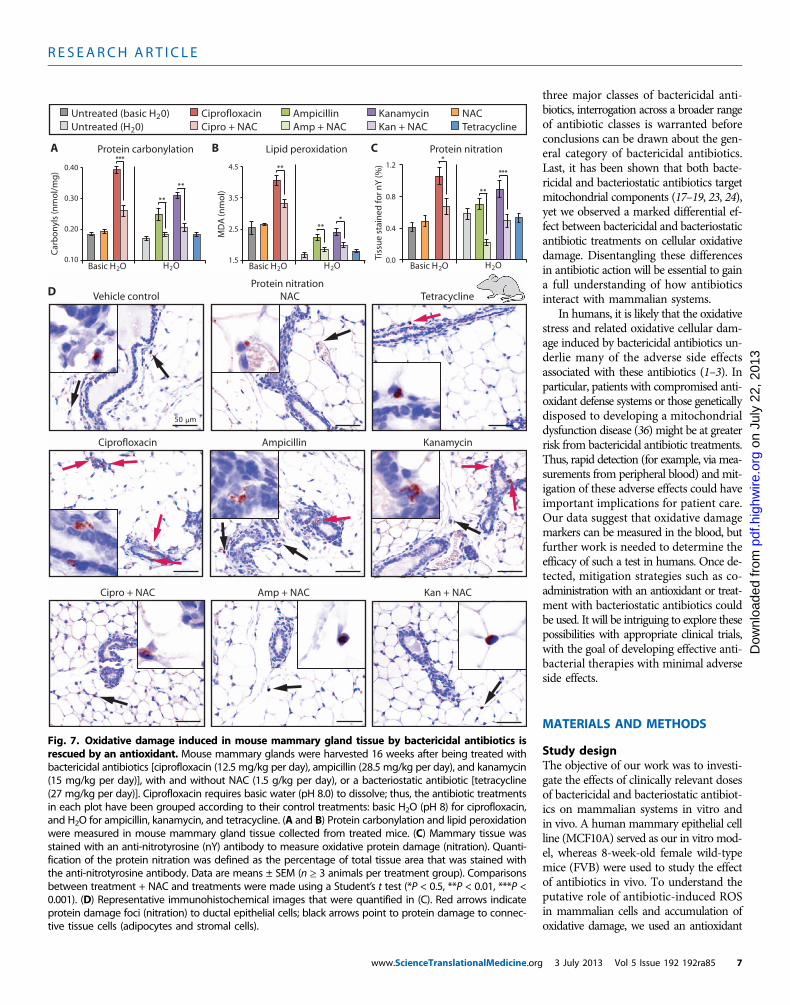
stress markers and accumulation of oxidative damage in tissues of micetreated with antibiotics. Mice received clinically relevant doses of anti-biotics [ciprofloxacin (12.5 mg/kg per day), ampicillin (28.5 mg/kg perday), and kanamycin (15 mg/kg per day)] in their drinking water (n≥3 animals per treatment group). At 2 and 16 weeks, peripheral bloodwas drawn from the same animals to measure intracellular ROS, lipidperoxidation, and glutathione levels. A reduction in glutathione is aproxy measure for ROS production because it is an intracellular scav-enger of ROS and a key component of the enzymatic antioxidant sys-tem (31).Bactericidal antibiotic–treated mice showed elevated lipid peroxi-dation in their peripheral blood; these increases were statistically sig-nificant after 16 weeks of treatment (Fig. 6). The treated mice alsoexhibited reduced glutathione levels after 2 weeks of antibiotic treat-
www.S
ment, which was significantly decreased after 16 weeks of treatment(Fig. 6). However, only ciprofloxacin led to a significant increase inROS in treated animals after 16 weeks.
To further characterize the in vivo oxidative stress response, weevaluated the transcriptional changes in key genes involved in antiox-idant defense mechanisms. Mammary glands were extracted frommice treated with bactericidal antibiotics in the presence and absenceof NAC or the bacteriostatic antibiotic tetracycline. From these tissues,the expression of the antioxidant defense genes—Sod1, Sod2, Gpx1,and Foxo3a—was measured by quantitative polymerase chain reaction(qPCR). After 2 weeks of treatment, these genes showed more than a2-fold increase in expression in tissues extracted from bactericidalantibiotic–treated mice, which were further elevated to ~10-fold changeafter 16 weeks of treatment (fig. S15A). These genes were not up-regulated
0
4
6
2
0
4
6
2
0.15
0.35
0.45
0.25
0.15
0.35
0.45
0.25
Lipid peroxidationProtein carbonylationD E8-OHdG F
C
NAC
Untreated
Ciprofloxacin
Cipro + NAC
Background correction
0 19 37 56 75 93Time (min)
923
665
408
150
–107
1180
OC
R (p
mo
l/m
in)
i ii iii
96 h
0 18 35 53 71 88
Time (min)
i ii iii
6 h1174
846
519
191
–136
OC
R (p
mo
l/m
in)
1501
A
6 h 96 h
B
96 h 6 h 6 h 96 h 6 h 96 h
0.85
0.90
0.95
1.00
Mitochondrial superoxide
0.85
0.90
0.95
1.00
Mitochondrial potential
UntreatedAmpicillin Amp + NACCiprofloxacin Cipro + NAC Kanamycin Kan + NAC NAC
Untreated Ampicillin Amp + NACCiprofloxacin Cipro + NAC Kanamycin Kan + NACNAC
Rati
o (T
MRE
/Mit
oTr
acke
r)
Rati
o (T
MRE
/Mit
oTr
acke
r)
Car
bo
nyl
s (n
mo
l/m
g)
Car
bo
nyl
s (n
mo
l/m
g)
6 h 0
10
20
30
40
50
Ch
ang
e o
ver c
on
tro
l (%
)
96 h 0
20
40
60
80
100
Ch
ang
e o
ver c
on
tro
l (%
)
*
8-O
Hd
G/m
g D
NA
(ng
)
8-O
Hd
G/m
g D
NA
(ng
)
4
7
8
5
MD
A (n
mo
l)6
9
4
7
8
5
MD
A (n
mo
l)
6
9
** **
** **
**
**
** *** * ** *
** *** *
** *** *** *
** *
** *
** *** *
Fig. 5. NAC rescues bactericidal antibiotic–induced oxidative damagein vitro.MCF10A cells were incubated with and without NAC (10 mM) for
flux analyzer, and a representative diagram testing the ciprofloxacin + NACis shown (see figs. S10 and S11 for ampicillin and kanamycin). (D) Oxidative
2 hours, followed by treatment with ciprofloxacin (10 mg/ml), ampicillin(20 mg/ml), kanamycin (25 mg/ml), or tetracycline (10 mg/ml) for 6 and96 hours. (A) Mitochondrial superoxide was measured using MitoSoxRed. (B) Mitochondrial membrane potential was quantified using TMREand MitoTracker Green. (C) OCR was measured using the Seahorse XF24
DNA damage was assessed by quantification of 8-OHdG. (E) Protein carbonylswere quantified to evaluate oxidative protein damage. (F) Lipid peroxidationwas measured by quantifying MDA. Data are means ± SEM (n ≥ 3). Compar-isons between treatment + NAC and treatments were made using a Student’st test (*P < 0.5, **P < 0.01, ***P < 0.001).
cienceTranslationalMedicine.org 3 July 2013 Vol 5 Issue 192 192ra85 5
R E S EARCH ART I C L E
on
July
22,
201
3e.
org
in the tissues of tetracycline-treated mice. Tissues from mice treatedwith a combination of bactericidal antibiotic + NAC or NAC aloneshowed decreased expression of the tested genes compared to antibi-otic treatment (fig. S15B). These data support our in vitro results, dem-onstrating that NAC rescued bactericidal antibiotic–induced oxidativedamage in MCF10A human epithelial cells (Fig. 5).
To evaluate bactericidal antibiotic–induced oxidative stress at thetissue level, we measured the accumulation of oxidative tissue damagedirectly in mammary glands. Quantification of protein carbonylation(Fig. 7A) and lipid peroxidation (Fig. 7B) revealed that bactericidalantibiotic–induced oxidative damage was significantly mitigated by co-administration of NAC. We also found no significant difference be-tween tetracycline-treated and untreated mice. To further characterizethese effects, wemeasured oxidative damage of proteins (nitration) causedby peroxynitrite, the product of the reaction between superoxide andnitric oxide. After 16 weeks of bactericidal antibiotic treatment, withand without NAC, as well as the bacteriostatic antibiotic tetracycline,we evaluated the number and size of anti-nitrotyrosine–stained foci inmouse mammary glands. Animals treated with bactericidal antibioticsshowed increased protein nitration compared to untreated mice, whereasmice treated with tetracycline showed no change (Fig. 7C). The observedincrease in protein damage caused by bactericidal antibiotic–treatedmice was rescued by co-administration of NAC. Immunohistochemicalanalysis revealed that the damaged proteins in bactericidal antibiotic–treated mice tended to localize to the cytoplasm of mammary ductalepithelial cells (Fig. 7D). However, damaged proteins in NAC-treatedmice, in the presence or absence of bactericidal antibiotics, were more
www.S
likely to be found in the connective tissue, localizing to the nuclei ofadipocytes and stromal cells (Fig. 7D).
pdf.h
ighw
irD
ownl
oade
d fr
om
DISCUSSION
The emergence of drug-resistant bacterial strains has been one un-intended consequence of the ubiquitous and frequent use of antibiot-ics in medicine and food production. With the belief that antibioticsspecifically target bacteria, the consequences of how they interact withmammalian cells have largely been overlooked, despite instances ofknown adverse effects, including ototoxicity (1), nephrotoxicity (2), andtendinopathy (3). Gaining a deeper mechanistic understanding of theeffects of commonly prescribed antibiotics in mammalian systems, par-ticularly for extended periods of treatment, is critical for achieving aclear safety profile for these drugs.
According to the endosymbiotic theory, mitochondria originatedfrom free-living, aerobic bacteria (22). It is likely then that antibioticstarget mitochondria and mitochondrial components, similar to theiraction in bacteria. Indeed, previous studies in mammalian systemshave revealed parallel antibiotic-target interactions in mitochondria(15–19, 32). It has been shown, for instance, that aminoglycosides tar-get both bacterial (33) and mitochondrial ribosomes (16), quinolonestarget bacterial gyrases (34) and mtDNA topoisomerases (15), andb-lactams inhibit bacterial cell wall synthesis (35) and mitochondrialcarnitine/acylcarnitine transporters (19). The work presented here ad-vances our understanding of antibiotic action in mammalian systemsin two distinct and important ways. First, we show that, regardless oftheir molecular targets, three major classes of bactericidal antibiotics—quinolones, aminoglycosides, and b-lactams—induce ROS productionin mammalian cells, leading to DNA, protein, and lipid damage. Sec-ond, we demonstrate that these deleterious effects are produced byclinically relevant doses of bactericidal antibiotics, both in cell cultureand in mice. These findings are analogous to our previous work inbacteria, in which we showed that clinically relevant doses of bacteri-cidal antibiotics induce a common oxidative damage pathway (4).
Identifying bactericidal antibiotics as a cause of ROS overpro-duction and mitochondrial dysfunction in mammalian cells providesa basis for developing therapeutic strategies that could help alleviateadverse side effects associated with antibiotics. For instance, by co-administering an intracellular antioxidant, in this case NAC, we showedthat ROS levels and oxidative damage induced by bactericidal antibio-tics could be abrogated while having little effect on the bacterial killingefficacy of the antibiotics. Additionally, we showed that bacteriostaticantibiotics, such as tetracycline, did not contribute to the overpro-duction of harmful ROS in mammalian cells. When appropriate forpatient health, substituting a bacteriostatic antibiotic for a bactericidalantibiotic could be a simple treatment strategy aimed at preventingcellular oxidative damage.
To establish the link between bactericidal antibiotics and ROS pro-duction, we have limited this study to antibiotic-treated human celllines and mice. It will be important to extend our investigation to hu-man subjects to confirm our findings, prove their relevance to humans,and maximize the translational value of our work. Epidemiologicstudies could provide valuable insight into the clinical implicationsof antibiotic-induced oxidative damage and define some of the risksassociated with bactericidal antibiotic exposure. Another limitation toconsider is the set of antibiotics tested. Although we present results on
16 weeks
2 weeks
Blood
Glutathione
Ch
ang
e o
ver c
on
tro
l (%
)
0
–25
–50
–75
Cipro Amp Kan
ROS
0
50
Ch
ang
e o
ver c
on
tro
l (%
)
25
75
Cipro Amp Kan
100 Lipid peroxidation
Ch
ang
e o
ver c
on
tro
l (%
)
Cipro Amp Kan
50
25
0
*
*
*
**
*
*
**
*
Fig. 6. Bactericidal antibiotics induce oxidative damage in mice. Oxi-dative stress markers were measured in blood drawn from wild-type mice
treated with ciprofloxacin (12.5 mg/kg per day), ampicillin (28.5 mg/kg perday), or kanamycin (15 mg/kg per day) for 2 or 16 weeks. Data are means ±SEM (n ≥ 3 animals per treatment group). Comparisons between treat-ments and untreated controls were made using a Student’s t test (*P < 0.5,**P < 0.01).cienceTranslationalMedicine.org 3 July 2013 Vol 5 Issue 192 192ra85 6
R E S EARCH ART I C L E
www.ScienceTranslationalMedicine.org
on
July
22,
201
3pd
f.hig
hwire
.org
Dow
nloa
ded
from
three major classes of bactericidal anti-biotics, interrogation across a broader rangeof antibiotic classes is warranted beforeconclusions can be drawn about the gen-eral category of bactericidal antibiotics.Last, it has been shown that both bacte-ricidal and bacteriostatic antibiotics targetmitochondrial components (17–19, 23, 24),yet we observed a marked differential ef-fect between bactericidal and bacteriostaticantibiotic treatments on cellular oxidativedamage. Disentangling these differencesin antibiotic action will be essential to gaina full understanding of how antibioticsinteract with mammalian systems.
In humans, it is likely that the oxidativestress and related oxidative cellular dam-age induced by bactericidal antibiotics un-derlie many of the adverse side effectsassociated with these antibiotics (1–3). Inparticular, patients with compromised anti-oxidant defense systems or those geneticallydisposed to developing a mitochondrialdysfunction disease (36) might be at greaterrisk from bactericidal antibiotic treatments.Thus, rapid detection (for example, via mea-surements from peripheral blood) and mit-igation of these adverse effects could haveimportant implications for patient care.Our data suggest that oxidative damagemarkers can be measured in the blood, butfurther work is needed to determine theefficacy of such a test in humans. Once de-tected, mitigation strategies such as co-administration with an antioxidant or treat-ment with bacteriostatic antibiotics couldbe used. It will be intriguing to explore thesepossibilities with appropriate clinical trials,with the goal of developing effective anti-bacterial therapies with minimal adverseside effects.
MATERIALS AND METHODS
Study designThe objective of our work was to investi-gate the effects of clinically relevant dosesof bactericidal and bacteriostatic antibiot-ics on mammalian systems in vitro andin vivo. A human mammary epithelial cellline (MCF10A) served as our in vitro mod-el, whereas 8-week-old female wild-typemice (FVB) were used to study the effectof antibiotics in vivo. To understand theputative role of antibiotic-induced ROSin mammalian cells and accumulation ofoxidative damage, we used an antioxidant
A
NACTetracycline
AmpicillinAmp + NAC
KanamycinKan + NAC
Protein carbonylation Lipid peroxidation Protein nitrationB C
D Vehicle control NAC Tetracycline
Ciprofloxacin Ampicillin Kanamycin
Kan + NACAmp + NACCipro + NAC
Protein nitration
1.5
2.5
3.5
4.5
MD
A (n
mo
l)
0.10
0.20
0.30
0.40
Car
bo
nyl
s (n
mo
l/m
g)
CiprofloxacinCipro + NAC
Untreated (basic H 0)Untreated (H 0)
2
2
0.0
0.4
0.8
1.2
Tiss
ue
stai
ned
for n
Y (%
)
50 m
*
** *
**
**
**
**
*
**
** *
Basic H O2 H O2 Basic H O2 H O2 Basic H O2 H O2
Fig. 7. Oxidative damage induced in mouse mammary gland tissue by bactericidal antibiotics isrescued by an antioxidant. Mouse mammary glands were harvested 16 weeks after being treated with
bactericidal antibiotics [ciprofloxacin (12.5 mg/kg per day), ampicillin (28.5 mg/kg per day), and kanamycin(15 mg/kg per day)], with and without NAC (1.5 g/kg per day), or a bacteriostatic antibiotic [tetracycline(27 mg/kg per day)]. Ciprofloxacin requires basic water (pH 8.0) to dissolve; thus, the antibiotic treatmentsin each plot have been grouped according to their control treatments: basic H2O (pH 8) for ciprofloxacin,and H2O for ampicillin, kanamycin, and tetracycline. (A and B) Protein carbonylation and lipid peroxidationwere measured in mouse mammary gland tissue collected from treated mice. (C) Mammary tissue wasstained with an anti-nitrotyrosine (nY) antibody to measure oxidative protein damage (nitration). Quanti-fication of the protein nitration was defined as the percentage of total tissue area that was stained withthe anti-nitrotyrosine antibody. Data are means ± SEM (n ≥ 3 animals per treatment group). Comparisonsbetween treatment + NAC and treatments were made using a Student’s t test (*P < 0.5, **P < 0.01, ***P <0.001). (D) Representative immunohistochemical images that were quantified in (C). Red arrows indicateprotein damage foci (nitration) to ductal epithelial cells; black arrows point to protein damage to connec-tive tissue cells (adipocytes and stromal cells).3 July 2013 Vol 5 Issue 192 192ra85 7
R E S EARCH ART I C L E
on
July
22,
201
3pd
f.hig
hwire
.org
Dow
nloa
ded
from
(NAC) alone or in combination with the bactericidal antibiotics. In vitro,ROS was measured with fluorescent indicators [CM-H2DCFDA, AmplexRed and horseradish peroxidase (HRP), MitoSox Red], and oxidativedamage to DNA (8-OHdG), proteins (carbonylation), and lipids (per-oxidation) was quantified, whereas the functional implications wereassessed by evaluation of mitochondrial respiration (oxygen consump-tion). In vivo, oxidative stress was evaluated in the peripheral blood oftreated mice with fluorescent indicators (H2DCFDA, fluor-DHPE, DCF),whereas oxidative damage to mammary gland tissue (protein carbon-ylation, lipid peroxidation, protein nitration) was assessed by immuno-histochemistry and qPCR quantification of key antioxidant genes. Micewere purchased from the vendor and randomly assigned to treatmentgroups. Antibiotics were prepared and administered in a nonblindedfashion, both in vitro and in vivo. Experiments included three [Figs. 1(C, D, F, and G), 2 (A, B, and E), 4, 5B, and 6 and figs. S3, S5 to S7, S9to S11, S13, and S15], four [Fig. 1 (A and B), 5 (A and D to F), and 8(A to C) and figs. S1, S2, S4, and S14], five (Figs. 1E and 3), six (Fig. 2,C and D), or eight (fig. S8) replicates per group.
Cell culture and antibiotic treatmentsHuman mammary epithelial cells, MCF10A [American Type CultureCollection (ATCC)], were maintained in Dulbecco’s modified Eagle’smedium and Ham’s F12 50/50 Mix (Cellgro, Mediatech) supple-mented with 5% horse serum, epidermal growth factor (100 mg/ml),hydrocortisone (1 mg/ml), cholera toxin (1 mg/ml), insulin (10 mg/ml),and 5 ml of penicillin (10,000 U/ml) and streptomycin (10,000 mg/ml).Hereafter, this medium will be referred to as “complete medium.” Forantibiotic treatments, complete medium was used without the additionof penicillin/streptomycin. We refer to this medium as “treatment me-dium.” All supplements were purchased from Sigma-Aldrich.
Before antibiotic treatments, cells were washed with phosphate-buffered saline (PBS), detached with 0.25% trypsin-EDTA (Life Tech-nologies), and seeded in 6-, 12-, 24-, or 96-well plates (Corning) intreatment medium. After 24 hours, peak serum levels (25) of anti-biotics were added: ciprofloxacin (10 mg/ml), ampicillin (20 mg/ml),kanamycin (25 mg/ml), tetracycline (10 mg/ml), and spectinomycin(100 mg/ml) (Fisher Scientific). Cells were plated at roughly equal num-bers (near confluence) at each measurement time point. NAC (10 mM;Sigma-Aldrich) was used as an intracellular ROS scavenger. For anti-biotic treatments in conjunction with NAC, cells were incubated intreatment medium plus NAC (pH adjusted to 7.4) for 2 hours andthen washed with PBS before the addition of antibiotics. The growthconditions for the additional cell lines tested are described in the Sup-plementary Materials and Methods.
MCF10A r0 cell lineMCF10A rho-zero (r0) cells are MCF10A cells that have been culturedto eliminate mtDNA. Following the protocol from King and Attardi(37), MCF10A cells were incubated in complete medium supple-mented with D-glucose (4.5 g/liter), ethidium bromide (50 ng/ml), uridine(50 mg/ml), and pyruvate (1 mM) for 6 weeks. After complete elimi-nation of mtDNA (as verified through PCR described below), cellswere maintained in complete medium supplemented with D-glucose(4.5 g/liter), uridine (50 mg/ml), and pyruvate (1 mM). A set of MCF10Acontrol cells was grown alongside the MCF10A r0 cells maintained incomplete medium supplemented with D-glucose (4.5 g/liter), uridine(50 mg/ml), and pyruvate (1 mM). The verification of r0 is describedin the Supplementary Materials and Methods.
www.S
ROS, H2O2, and mitochondrial superoxideTo detect ROS, cells plated in 6- or 96-well tissue culture plates werewashed twice with prewarmed PBS and then incubated with 10 mMCM-H2DCFDA (Life Technologies) at 37°C for 45 min. Cells werewashed with PBS to remove excess dye and allowed to recover intreatment medium at 37°C for 15 min in the dark. Imaging was car-ried out on a fluorescence-enabled inverted microscope, and quan-tification was done with a BD FACSAria II flow cytometer (BectonDickinson) or SpectraMax M5 microplate reader (Molecular Devices)at an excitation/emission of 495/525 nm. This protocol was repeatedfor all other tested cell lines.
To detect extracellular hydrogen peroxide, we adapted a protocolfrom Panopoulos et al. (38). Cells plated in 96-well tissue cultureplates were washed once with Krebs-Ringer phosphate buffer (KRPG).KRPG containing 0.1% horse serum (KRPG 0.1%) and clinical levelsof antibiotics were added to the cells in a total volume of 50 ml. Next,50 ml of prewarmed KRPG containing 100 mM Amplex Red reagentand horseradish peroxide (0.2 U/ml) (reaction mixture) was added toeach well. Absorbance was measured at 560 nm after the addition ofreaction buffer with a SpectraMax M5 precision microplate reader(Molecular Devices).
To detect mitochondrial superoxide, cells plated in 6- or 96-welltissue culture plates were washed twice with prewarmed PBS and thenincubated with 5 mM MitoSox Red at 37°C for 10 min in the dark.After incubation, cells were washed and resuspended in prewarmedHanks’ balanced salt solution (Fisher Scientific). Quantification wascompleted with a BD FACSAria II flow cytometer or a SpectraMaxM5 microplate reader at an excitation/emission of 510/580 nm.
Oxidative DNA, protein, and lipid damage8-OHdG levels were quantified with the OxiSelect Oxidative DNADamage ELISA Kit (Cell Biolabs), protein carbonylation was measuredwith the Protein Carbonyl ELISA Kit (Enzo Life Sciences), and lipidperoxidation was measured with the Lipid Peroxidation MDA AssayKit (Abcam). All assays were performed according to the manufacturer’sprotocol and are described in the Supplementary Materials and Methods.
Mitochondrial ETC complex activityDirect inhibition of the activity for each of the five ETC complexeswas measured with the MitoTox Complete OXPHOS Activity AssayPanel (Abcam), following the manufacturer’s protocol. Each of the fivecomplexes was captured from isolated bovine heart mitochondria intheir functionally active state with highly specific monoclonal antibo-dies attached to 96-well microplates. IC50 values for known inhibitorsof each of the five complexes were used as positive controls: rotenone(complex I, 17.3 nM), thenoyltrifluoroacetone (complex II, 30 mM),antimycin A (complex III, 22 nM), KCN (complex IV, 3.2 mM), andoligomycin (complex V, 8 nM). The same positive controls were testedon each complex and used as negative controls for off-target complexes(for example, rotenone was a negative control for complexes II to V).For each of the complexes treated with antibiotics [ciprofloxacin(10 mg/ml), ampicillin (20 mg/ml), kanamycin (25 mg/ml), tetracycline(10 mg/ml), or spectinomycin (100 mg/ml)], activity was determined bymeasuring the decrease in absorbance in milli–optical density per mi-nute at room temperature and at specified wavelengths [340 nm (Iand V), 600 nm (II), and 550 nm (III and IV)] in kinetic mode [everyminute for 2 hours (I) and 1 hour (II, IV, and V) and every 20 s for5 min (III)] with a SpectraMax M5 microplate reader.
cienceTranslationalMedicine.org 3 July 2013 Vol 5 Issue 192 192ra85 8
R E S EARCH ART I C L E
on
July
22,
201
3pd
f.hig
hwire
.org
Dow
nloa
ded
from
Metabolic activity, mitochondrial potential, and ATPMetabolic activity was assayed with an XTT cell proliferation kit(ATCC), cellular ATP was measured with the ATPlite LuminescenceAssay kit (Perkin Elmer), and mitochondrial potential was calculatedwith the ratio of TMRE to MitoTracker Green (Life Technologies). Allassays were performed according to the manufacturer’s protocol andare described in the Supplementary Materials and Methods.
Mitochondrial morphometryPrimary human mammary epithelial cells (HMEC) were plated inglass-bottom confocal dishes in treatment medium 24 hours beforeaddition of antibiotics. Cells were washed once with PBS and treatedwith antibiotics for 24 or 96 hours. After treatment, cells were washedwith 1× PBS and incubated in 7 nM TMRE and 10 mM MitoTrackerGreen for 30 min. After incubation, cells were returned to fresh treat-ment medium with only TMRE (7 nM) and immediately imaged on aZeiss LSM710 confocal microscope.
Mitochondrial morphometry was carried out with the particle anal-ysis function in the image processing software ImageJ [NationalInstitutes of Health (NIH)] (39), as described in the SupplementaryMaterials and Methods.
Mitochondrial oxygen consumptionOCRs were measured at 37°C with an XF24 extracellular analyzer(Seahorse Bioscience). MCF10A cells were seeded at a density of40,000 cells per well on XF24 tissue culture plates overnight and thentreated with antibiotics for 6 and 96 hours. Measurements of OCRwere done according to Seahorse Bioscience protocols as describedin the Supplementary Materials and Methods.
Bacterial killingThe growth and survival of untreated exponential-phase wild-typeE. coli (MG1655) were compared to cultures treated with bactericidalantibiotics alone [ciprofloxacin (5 mg/ml), ampicillin (5 mg/ml), orkanamycin (5 mg/ml)], 10 mM NAC alone, or a combination of bac-tericidal antibiotics supplemented with NAC for 3 hours. Cells weregrown overnight at 37°C and 300 rpm in a light-insulated shakerand then diluted 1:500 in 25-ml LB in a 250-ml flask. Cultures weregrown to an optical density (OD600) of about 0.3, as measured withthe SPECTRAFluor Plus spectrophotometer (Tecan). Antibiotics wereadded at this point. For bacterial count measurements [colony-formingunits (CFU)/ml], 100 ml of culture was collected 30 min and 1, 2, and3 hours after addition of antibiotics, washed twice with filtered 1× PBS(pH 7.2) (Fisher Scientific), and then serially diluted in 1× PBS. Tenmicroliters of each dilution was plated onto 120-mm square dishes(BD Biosciences) containing LB Agar (Fisher Scientific), and plateswere incubated at 37°C overnight. Colonies were counted, and CFU/mlvalues were calculated with the following formula:
ð#coloniesÞ � 10dilution factor
ðvolume platedÞ � 1000
Animal studies and tissue collectionSix- to 8-week-old female FVB/NJ mice (Jackson Laboratory) were ad-ministered one of the following treatments: ciprofloxacin (12.5 mg/kgper day), ampicillin (28.5 mg/kg per day), kanamycin (15 mg/kg per day),with and without NAC (1.5 g/kg per day), tetracycline (13.5 mg/kg perday), and vehicle only (basic water, pH 8.0, or deionized water). Solu-
www.S
tions were made fresh every 3 days and administered in the drinkingwater for mice to feed ad libitum. Doses were achieved on the basis ofan average mouse weight of 20 g and an approximate intake of drink-ing water of 4 ml per day. After 2 or 16 weeks, the animals were bledand euthanized. Mammary tissue was collected from each mouse, pre-served in RNAlater (Ambion, Life Technologies) for real-time qPCRand 10% buffered formalin for immunohistochemistry, and flash-frozen for protein extraction. The Institutional Animal Care and UseCommittee approved all mouse experiments. Quantitative reversetranscription PCR of oxidative stress genes is described in the Supple-mentary Materials and Methods.
Mouse blood oxidative stressTo measure general oxidative stress, 1 × 10⁷ peripheral blood cells wereincubated with 2′,7′-dichlorodihydrofluorescein diacetate (H2DCFDA)(Life Technologies), dissolved in dimethyl sulfoxide (DMSO) (Sigma-Aldrich), at a final concentration of 0.4 mM at 37°C for 15 min at 5%CO2. Reduced glutathione levels were measured by labeling 2 × 10⁷
blood cells with mercury orange (Sigma-Aldrich), dissolved in DMSO,at a final concentration of 40 mM for 3 min at room temperature. Tomeasure lipid peroxidation, we washed 2 × 10⁷ blood cells with PBSand labeled with N-(fluorescein-5-thiocarbamoyl)-1,2-dihexadecanoyl-sn-glycero-3-phosphoethanolamine, triethyl-ammonium salt (fluor-DHPE)(Life Technologies), dissolved in ethanol, at a final concentration of50 mM for 1 hour at 37°C in a 5% CO2 incubator with continuousagitation. After the incubation period with each dye, blood cells werewashed twice with PBS to remove unbound label, resuspended in PBS,and analyzed by flow cytometry (FACSFortessa, Becton Dickinson). A488-nm argon laser beam was used for excitation. Blood cells labeledwith DCF and fluor-DHPE were detected by FL-1 PMT with linearamplification, whereas mercury orange–labeled red blood cells weredetected by FL-2 PMT with log amplification. For each assay, treatedand untreated unstained cells were used as controls. Instrument cali-bration and settings were performed with CaliBRITE-3 beads (BectonDickinson). The mean fluorescence channel of the entire population wascalculated for DCF, glutathione, and lipid peroxidation by the fluorescence-activated cell sorting (FACS)–equipped BD FACSDiva software.
UTI mouse modelEight-week-old C57BL/6 female mice were inoculated with 50 ml of8% (w/v) mucin solution in sterile saline containing 2 × 10⁹ E. coli(MG1655) cells, via transurethral catheterization into their bladders,as described previously (40). Briefly, mice were anesthetized with 2to 4% isoflurane. Urinary catheters (30-gauge × 1/2-inch hypodermicneedle aseptically covered with polyethylene tubing) were coated inmedical-grade, sterile lubricating jelly. The bladder of the mouse wasgently massaged to expel urine. The lubricated catheter was insertedinto the urethral opening. It was then pushed into the urethra until thebase of the needle reached the urethral opening. Once fully inserted,50 ml of the inoculum (containing 2 × 10⁹ E. coli cells) was injecteddirectly into the bladder.
Infected animals received ciprofloxacin (50 mg/ml), NAC (10 mM),ciprofloxacin and NAC, or vehicle (PBS) only, via intraperitonealdelivery 24 hours after inoculation. After treatment, animals wereobserved for an additional 24 or 48 hours. At the end of the experi-ment, animals were euthanized by CO2 asphyxiation followed by cer-vical dislocation. Bladders were collected in 1 ml of PBS and homogenizedfor 30 s for subsequent quantification of bacterial load. For the CFU/
cienceTranslationalMedicine.org 3 July 2013 Vol 5 Issue 192 192ra85 9
R E S EARCH ART I C L E
on
July
22,
201
3.h
ighw
ire.o
rg
bladder measurements, the homogenized bladder was serially dilutedin PBS (pH 7.2). A 200-ml portion of each dilution was plated in LB-agar plates and incubated overnight at 37°C. The colonies were counted,and CFU/bladder was calculated with the following formula:
5ð#coloniesÞðdilution factorÞvolume plated
Histology and immunohistochemistrySix- to 8-week-old female FVB/NJ mice (Jackson Laboratory) were ad-ministered ciprofloxacin (12.5 mg/kg per day), ampicillin (28.5 mg/kgper day), kanamycin (15 mg/kg per day), with and without NAC(1.5 g/kg per day), tetracycline (13.5 mg/kg per day), or vehicle only(basic or deionized H2O). Solutions were made fresh every 3 days andadministered in the drinking water for 16 weeks. Mice were eutha-nized, and mammary glands were collected and stored in 10% bufferedformalin overnight at 4°C, transferred to 70% ethanol, and stored atroom temperature until sectioning. Tissues were embedded in paraffinand sectioned for subsequent staining. Mammary gland sections werestained with anti-nitrotyrosine polyclonal antibody (Millipore), followedby anti-rabbit immunoglobulin G conjugated to HRP (Vector Labora-tories). Slides were counterstained with hematoxylin. Image capture andanalysis are described in the Supplementary Materials and Methods.
Statistical analysisA one-tailed Student’s t test was performed on measurements of ox-idative stress in the blood, UTI model, and for comparisons betweenantibiotic-treated groups and groups treated with antibiotic and NAC.For all other statistical analyses, a two-tailed Student’s t test was per-formed to compare treatment groups to untreated (control) groups.We assumed equal variance for all groups.
Dow
nloa
ded
from
SUPPLEMENTARY MATERIALS
www.sciencetranslationalmedicine.org/cgi/content/full/5/192/192ra85/DC1Materials and MethodsFig. S1. Dose-response and time-course measurements of ROS levels in antibiotic-treated cells.Fig. S2. Effect of antibiotic treatment on cell viability in vitro.Fig. S3. ROS production measured across different mammalian cell types treated with antibiotics.Fig. S4. Dose-response and time-course measurements of mitochondrial superoxide produc-tion in antibiotic-treated cells.Fig. S5. Dose-response and time-course measurement of extracellular hydrogen peroxide inantibiotic-treated cells.Fig. S6. DNA damage in human mammary epithelial cells treated with antibiotics.Fig. S7. Effect of antibiotics on the function of isolated ETC complexes.Fig. S8. ROS production in mammalian cells lacking mtDNA treated with antibiotics.Fig. S9. NAC reduces bactericidal antibiotic–induced ROS levels.Fig. S10. OCR in antibiotic- and NAC-treated cells.Fig. S11. OCR in mammalian cells.Fig. S12. NAC rescues bactericidal antibiotic–induced DNA damage.Fig. S13. Bactericidal antibiotic efficacy is unaffected by NAC in vitro.Fig. S14. Bactericidal antibiotic efficacy is unaffected by NAC when tested in vivo in a murineUTI model.Fig. S15. Bactericidal antibiotics induce expression of antioxidant defense genes in mousemammary gland tissue.
REFERENCES AND NOTES
1. R. E. Brummett, K. E. Fox, Aminoglycoside-induced hearing loss in humans. Antimicrob.Agents Chemother. 33, 797–800 (1989).
2. M. P. Mingeot-Leclercq, P. M. Tulkens, Aminoglycosides: Nephrotoxicity. Antimicrob. AgentsChemother. 43, 1003–1012 (1999).
www.Sc
3. Y. Khaliq, G. Zhanel, Fluoroquinolone-associated tendinopathy: A critical review of the litera-ture. Clin. Infect. Dis. 36, 1404–1410 (2003).
4. M. A. Kohanski, D. J. Dwyer, B. Hayete, C. A. Lawrence, J. J. Collins, A common mechanismof cellular death induced by bactericidal antibiotics. Cell 130, 797–810 (2007).
5. D. J. Dwyer, M. A. Kohanski, B. Hayete, J. J. Collins, Gyrase inhibitors induce an oxidativedamage cellular death pathway in Escherichia coli. Mol. Syst. Biol. 3, 91 (2007).
6. M. A. Kohanski, D. J. Dwyer, J. Wierzbowski, G. Cottarel, J. J. Collins, Mistranslation of mem-brane proteins and two-component system activation trigger antibiotic-mediated celldeath. Cell 135, 679–690 (2008).
7. J. J. Foti, B. Devadoss, J. A. Winkler, J. J. Collins, G. C. Walker, Oxidation of the guaninenucleotide pool underlies cell death by bactericidal antibiotics. Science 336, 315–319(2012).
8. S. S. Grant, B. B. Kaufmann, N. S. Chand, N. Haseley, D. T. Hung, Eradication of bacterialpersisters with antibiotic-generated hydroxyl radicals. Proc. Natl. Acad. Sci. U.S.A. 109,12147–12152 (2012).
9. Y. Liu, X. Liu, Y. Qu, X. Wang, L. Li, X. Zhao, Inhibitors of reactive oxygen species accumu-lation delay and/or reduce the lethality of several antistaphylococcal agents. Antimicrob.Agents Chemother. 56, 6048–6050 (2012).
10. K. Shatalin, E. Shatalina, A. Mironov, E. Nudler, H2S: A universal defense against antibioticsin bacteria. Science 334, 986–990 (2011).
11. X. Wang, X. Zhao, Contribution of oxidative damage to antimicrobial lethality. Antimicrob.Agents Chemother. 53, 1395–1402 (2009).
12. X. Wang, X. Zhao, M. Malik, K. Drlica, Contribution of reactive oxygen species to pathwaysof quinolone-mediated bacterial cell death. J. Antimicrob. Chemother. 65, 520–524 (2010).
13. I. Keren, Y. Wu, J. Inocencio, L. R. Mulcahy, K. Lewis, Killing by bactericidal antibiotics doesnot depend on reactive oxygen species. Science 339, 1213–1216 (2013).
14. Y. Liu, J. A. Imlay, Cell death from antibiotics without the involvement of reactive oxygenspecies. Science 339, 1210–1213 (2013).
15. T. D. Gootz, J. F. Barrett, J. A. Sutcliffe, Inhibitory effects of quinolone antibacterial agentson eucaryotic topoisomerases and related test systems. Antimicrob. Agents Chemother. 34,8–12 (1990).
16. T. Hutchin, G. Cortopassi, Proposed molecular and cellular mechanism for aminoglycosideototoxicity. Antimicrob. Agents Chemother. 38, 2517–2520 (1994).
17. D. A. Lowes, C. Wallace, M. P. Murphy, N. R. Webster, H. F. Galley, The mitochondria tar-geted antioxidant MitoQ protects against fluoroquinolone-induced oxidative stress andmitochondrial membrane damage in human Achilles tendon cells. Free Radic. Res. 43,323–328 (2009).
18. E. E. McKee, M. Ferguson, A. T. Bentley, T. A. Marks, Inhibition of mammalian mitochondrialprotein synthesis by oxazolidinones. Antimicrob. Agents Chemother. 50, 2042–2049 (2006).
19. L. Pochini, M. Galluccio, D. Scumaci, N. Giangregorio, A. Tonazzi, F. Palmieri, C. Indiveri,Interaction of b-lactam antibiotics with the mitochondrial carnitine/acylcarnitine transporter.Chem. Biol. Interact. 173, 187–194 (2008).
20. B. M. Tune, Nephrotoxicity of beta-lactam antibiotics: Mechanisms and strategies for pre-vention. Pediatr. Nephrol. 11, 768–772 (1997).
21. E. J. Lesnefsky, S. Moghaddas, B. Tandler, J. Kerner, C. L. Hoppel, Mitochondrial dysfunctionin cardiac disease: Ischemia–reperfusion, aging, and heart failure. J. Mol. Cell. Cardiol. 33,1065–1089 (2001).
22. M. W. Gray, G. Burger, B. F. Lang, Mitochondrial evolution. Science 283, 1476–1481 (1999).23. J. W. Lawrence, D. C. Claire, V. Weissig, T. C. Rowe, Delayed cytotoxicity and cleavage of
mitochondrial DNA in ciprofloxacin-treated mammalian cells. Mol. Pharmacol. 50, 1178–1188(1996).
24. N. Duewelhenke, O. Krut, P. Eysel, Influence on mitochondria and cytotoxicity of differentantibiotics administered in high concentrations on primary human osteoblasts and celllines. Antimicrob. Agents Chemother. 51, 54–63 (2007).
25. A. Bryskier, Antimicrobial Agents: Antibacterials and Antifungals (ASM Press, Washington,DC, 2005).
26. J. F. Turrens, Mitochondrial formation of reactive oxygen species. J. Physiol. 552, 335–344(2003).
27. S. Dröse, U. Brandt, The mechanism of mitochondrial superoxide production by the cyto-chrome bc1 complex. J. Biol. Chem. 283, 21649–21654 (2008).
28. A. Y. Seo, A. M. Joseph, D. Dutta, J. C. Hwang, J. P. Aris, C. Leeuwenburgh, New insights intothe role of mitochondria in aging: Mitochondrial dynamics and more. J. Cell Sci. 123,2533–2542 (2010).
29. N. De Vries, S. De Flora, N-acetyl-l-cysteine. J. Cell. Biochem. 53, 270–277 (1993).30. O. I. Aruoma, B. Halliwell, B. M. Hoey, J. Butler, The antioxidant action of N-acetylcysteine:
Its reaction with hydrogen peroxide, hydroxyl radical, superoxide, and hypochlorous acid.Free Radic. Biol. Med. 6, 593–597 (1989).
31. A. Meister, M. E. Anderson, Glutathione. Annu. Rev. Biochem. 52, 711–760 (1983).32. S. N. Hobbie, S. Akshay, S. K. Kalapala, C. M. Bruell, D. Shcherbakov, E. C. Böttger, Genetic anal-
ysis of interactions with eukaryotic rRNA identify the mitoribosome as target in aminoglycosideototoxicity. Proc. Natl. Acad. Sci. U.S.A. 105, 20888–20893 (2008).
ienceTranslationalMedicine.org 3 July 2013 Vol 5 Issue 192 192ra85 10
R E S EARCH ART I C L E
33. B. D. Davis, Mechanism of bactericidal action of aminoglycosides. Microbiol. Rev. 51, 341–350(1987).
34. J. S. Wolfson, D. C. Hooper, The fluoroquinolones: Structures, mechanisms of action andresistance, and spectra of activity in vitro. Antimicrob. Agents Chemother. 28, 581–586(1985).
35. A. Tomasz, The mechanism of the irreversible antimicrobial effects of penicillins: How thebeta-lactam antibiotics kill and lyse bacteria. Annu. Rev. Microbiol. 33, 113–137 (1979).
36. A. M. Schaefer, R. McFarland, E. L. Blakely, L. He, R. G. Whittaker, R. W. Taylor, P. F. Chinnery,D.M. Turnbull, Prevalence ofmitochondrial DNAdisease in adults. Ann. Neurol. 63, 35–39 (2008).
37. M. P. King, G. Attardi, Isolation of human cell lines lacking mitochondrial DNA. MethodsEnzymol. 264, 304–313 (1996).
38. A. Panopoulos, M. Harraz, J. F. Engelhardt, E. Zandi, Iron-mediated H2O2 production as amechanism for cell type-specific inhibition of tumor necrosis factor a-induced but notinterleukin-1b-induced IkB kinase complex/nuclear factor-kB activation. J. Biol. Chem.280, 2912–2923 (2005).
39. M. D. Abramoff, P. J. Magalhaes, S. J. Ram, Image processing with ImageJ. Biophoton. Int.11, 36–42 (2004).
40. C. S. Hung, K. W. Dodson, S. J. Hultgren, A murine model of urinary tract infection. Nat.Protoc. 4, 1230–1243 (2009).
www.Sc
Acknowledgments: We thank E. Cameron and P. Belenky for their help with experimentaldesign and troubleshooting, A. Saccone for his assistance with experiments, and T. Ferrantefor his assistance with imaging. Funding: This work was supported by the NIH Director’s PioneerAward Program and the Howard Hughes Medical Institute. M.L. was the recipient of a post-doctoral fellowship from Fundacion Ramon Areces and an Evans Center Fellow Award. Authorcontributions: S.K., C.S.S., J.C.C., M.L., J.R.M.-R., S.S., A.M., O.S.S., and J.J.C. designed the study,analyzed the results, and wrote the manuscript. In vitro experiments were performed by S.K.and S.S. In vivo experiments were performed by S.K., C.S.S., and J.R.M.-R. Seahorse experimentswere completed by S.K. and M.L. Competing interests: The authors declare that they have nocompeting interests.
Submitted 1 March 2013Accepted 3 June 2013Published 3 July 201310.1126/scitranslmed.3006055
Citation: S. Kalghatgi, C. S. Spina, J. C. Costello, M. Liesa, J. R. Morones-Ramirez, S. Slomovic,A. Molina, O. S. Shirihai, J. J. Collins, Bactericidal antibiotics induce mitochondrial dysfunctionand oxidative damage in mammalian cells. Sci. Transl. Med. 5, 192ra85 (2013).
ienceTranslationalMedicine.org 3 July 2013 Vol 5 Issue 192 192ra85 11
on
July
22,
201
3pd
f.hig
hwire
.org
Dow
nloa
ded
from