cigna medical coverage policy - pdfs.semanticscholar.org · chronic wound when all of the following...
TRANSCRIPT

Page 1 of 55 Coverage Policy Number: 0160
Cigna Medical Coverage Policy
Subject Electrical Stimulation Therapy and Devices
Effective Date ............................ 9/15/2014 Next Review Date……………….9/15/2015 Coverage Policy Number ................. 0160
Table of Contents Coverage Policy .................................................. 1 General Background ........................................... 3 Coding/Billing Information ................................. 32 References ........................................................ 38
Hyperlink to Related Coverage Policies Acupuncture Bone Growth Stimulators: Electrical
(Invasive, Noninvasive), Ultrasound Coma Stimulation Diaphragmatic/Phrenic Nerve Stimulation Extracorporeal Electromagnetic Stimulation
for Urinary Incontinence Gastric Pacing/Gastric Electrical Stimulation
(GES) Injectable Bulking Agents for Urinary
Conditions and Fecal Incontinence Invasive Treatments for Urinary
Incontinence Local Injection Therapy and Neurosurgery
for Cervicogenic Headache and Occipital Neuralgia
Plantar Fasciitis Treatments Pulsed Electromagnetic Therapy Sacral Nerve Stimulation for Urinary Voiding
Dysfunction and Fecal Incontinence Speech Therapy Spinal Cord Stimulation Transcranial Magnetic Stimulation Vagus Nerve Stimulation (VNS)
INSTRUCTIONS FOR USE The following Coverage Policy applies to health benefit plans administered by Cigna companies. Coverage Policies are intended to provide guidance in interpreting certain standard Cigna benefit plans. Please note, the terms of a customer’s particular benefit plan document [Group Service Agreement, Evidence of Coverage, Certificate of Coverage, Summary Plan Description (SPD) or similar plan document] may differ significantly from the standard benefit plans upon which these Coverage Policies are based. For example, a customer’s benefit plan document may contain a specific exclusion related to a topic addressed in a Coverage Policy. In the event of a conflict, a customer’s benefit plan document always supersedes the information in the Coverage Policies. In the absence of a controlling federal or state coverage mandate, benefits are ultimately determined by the terms of the applicable benefit plan document. Coverage determinations in each specific instance require consideration of 1) the terms of the applicable benefit plan document in effect on the date of service; 2) any applicable laws/regulations; 3) any relevant collateral source materials including Coverage Policies and; 4) the specific facts of the particular situation. Coverage Policies relate exclusively to the administration of health benefit plans. Coverage Policies are not recommendations for treatment and should never be used as treatment guidelines. In certain markets, delegated vendor guidelines may be used to support medical necessity and other coverage determinations. Proprietary information of Cigna. Copyright ©2014 Cigna Coverage Policy Electrical Stimulation Therapies Chronic Wound Healing

Page 2 of 55 Coverage Policy Number: 0160
Cigna covers electrical stimulation (HCPCS Code G0281) as medically necessary for the treatment of a chronic wound when ALL of the following criteria are met:
• Presence of ANY of the following chronic wound types:
stage III or stage IV pressure ulcer arterial ulcer neuropathic (diabetic) ulcer venous stasis ulcer
• Failure to demonstrate measurable signs of improved healing (e.g., signs of epithelialization and
reduction in ulcer size) with a 30-day trial of conventional wound management, including optimization of nutritional status, moist dressings and debridement.
• Electrical stimulation therapy is performed under the direct supervision of a medical professional with
expertise in wound evaluation and management. Cigna does not cover the unsupervised use of electrical stimulation therapy for wound healing performed by the individual in the home setting because it is considered experimental, investigational or unproven. Cigna does not cover electrical stimulation therapy for any other chronic wound indication including but not limited to prevention of a pressure ulcer or pressure sore because it is considered experimental, investigational or unproven. Other Electrical Stimulation Therapies Cigna does not cover EITHER of the following electrical stimulation therapies because each is considered experimental, investigational or unproven:
• auricular electroacupuncture (HCPCS Code S8930) • transcutaneous electrical modulation pain reprocessing (TEMPR) (Scrambler therapy, Calmare®)
(CPT Code® 0278T) Electrical Stimulation Devices (Electrical Stimulators) Coverage for electrical stimulation devices is subject to the terms, conditions and limitations of the applicable benefit plan’s Durable Medical Equipment (DME) benefit and schedule of copayments. Please refer to the applicable benefit plan document to determine benefit availability and the terms, conditions and limitations of coverage. Under many benefit plans, coverage for DME is limited to the lowest-cost alternative. If coverage for electrical stimulation devices is available, the following conditions of coverage apply. Neuromuscular Electrical Stimulation (NMES) Cigna covers neuromuscular electrical stimulation (NMES) (CPT Code® 64565; HCPCS Code E0745) as medically necessary when used as one component of a comprehensive rehabilitation program for the treatment of disuse atrophy when the nerve supply to the atrophied muscle is intact. Cigna does not cover neuromuscular electrical stimulation (NMES) for ANY other indication (e.g., idiopathic scoliosis [CPT Code® 64565; HCPCS Code E0744], heart failure) because it is considered experimental, investigational or unproven. Transcutaneous Electrical Nerve Stimulation (TENS) Some benefit plans have a specific limitation of coverage of transcutaneous electrical nerve stimulation (TENS) units (CPT Code 64550; HCPCS Codes E0720, E0730). Please refer to the applicable benefit plan document. If not specifically limited by the benefit plan, Cigna covers a transcutaneous electrical nerve stimulator (TENS) as medically necessary as an adjunct to conventional post-operative pain management within 30 days of surgery.
Page 3 of 55 Coverage Policy Number: 0160
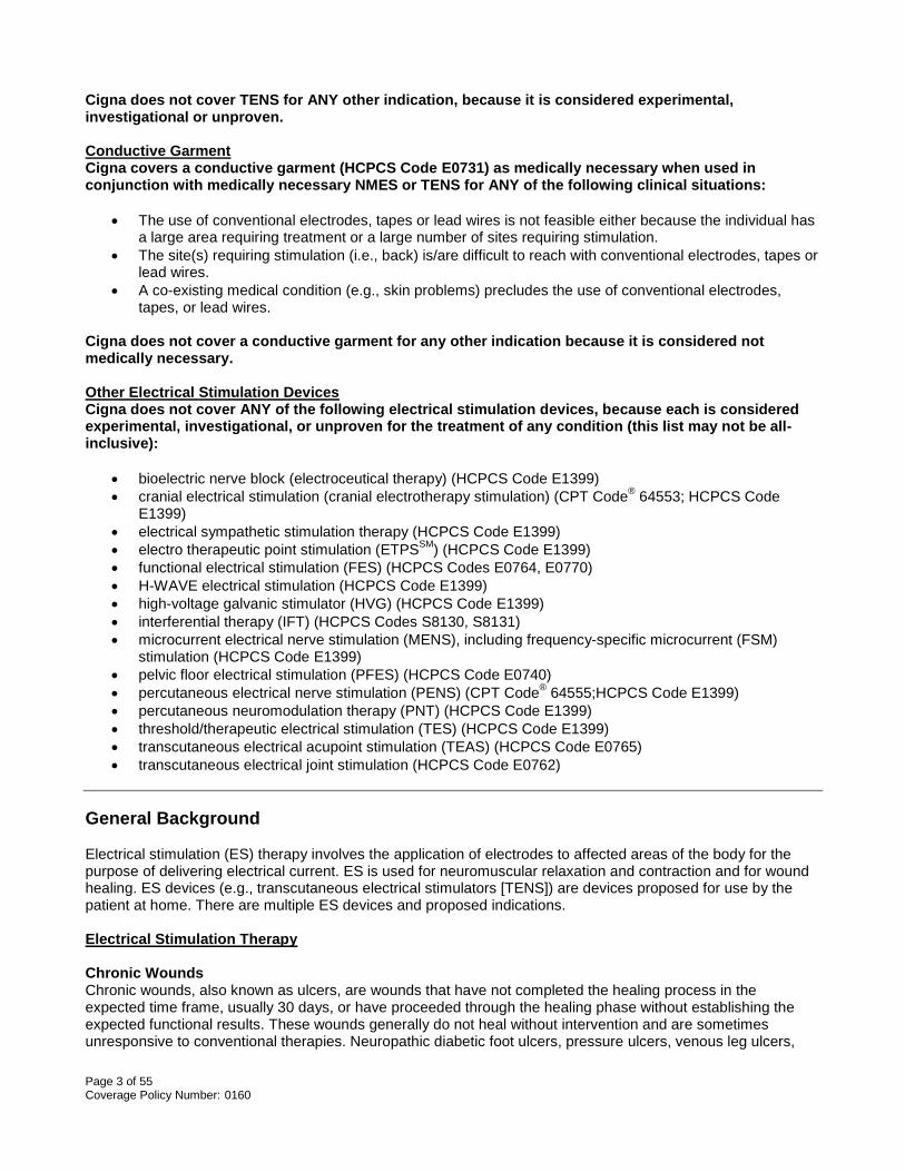
Cigna does not cover TENS for ANY other indication, because it is considered experimental, investigational or unproven. Conductive Garment Cigna covers a conductive garment (HCPCS Code E0731) as medically necessary when used in conjunction with medically necessary NMES or TENS for ANY of the following clinical situations:
• The use of conventional electrodes, tapes or lead wires is not feasible either because the individual has a large area requiring treatment or a large number of sites requiring stimulation.
• The site(s) requiring stimulation (i.e., back) is/are difficult to reach with conventional electrodes, tapes or lead wires.
• A co-existing medical condition (e.g., skin problems) precludes the use of conventional electrodes, tapes, or lead wires.
Cigna does not cover a conductive garment for any other indication because it is considered not medically necessary. Other Electrical Stimulation Devices Cigna does not cover ANY of the following electrical stimulation devices, because each is considered experimental, investigational, or unproven for the treatment of any condition (this list may not be all-inclusive):
• bioelectric nerve block (electroceutical therapy) (HCPCS Code E1399) • cranial electrical stimulation (cranial electrotherapy stimulation) (CPT Code® 64553; HCPCS Code
E1399) • electrical sympathetic stimulation therapy (HCPCS Code E1399) • electro therapeutic point stimulation (ETPSSM) (HCPCS Code E1399) • functional electrical stimulation (FES) (HCPCS Codes E0764, E0770) • H-WAVE electrical stimulation (HCPCS Code E1399) • high-voltage galvanic stimulator (HVG) (HCPCS Code E1399) • interferential therapy (IFT) (HCPCS Codes S8130, S8131) • microcurrent electrical nerve stimulation (MENS), including frequency-specific microcurrent (FSM)
stimulation (HCPCS Code E1399) • pelvic floor electrical stimulation (PFES) (HCPCS Code E0740) • percutaneous electrical nerve stimulation (PENS) (CPT Code® 64555;HCPCS Code E1399) • percutaneous neuromodulation therapy (PNT) (HCPCS Code E1399) • threshold/therapeutic electrical stimulation (TES) (HCPCS Code E1399) • transcutaneous electrical acupoint stimulation (TEAS) (HCPCS Code E0765) • transcutaneous electrical joint stimulation (HCPCS Code E0762)
General Background Electrical stimulation (ES) therapy involves the application of electrodes to affected areas of the body for the purpose of delivering electrical current. ES is used for neuromuscular relaxation and contraction and for wound healing. ES devices (e.g., transcutaneous electrical stimulators [TENS]) are devices proposed for use by the patient at home. There are multiple ES devices and proposed indications. Electrical Stimulation Therapy Chronic Wounds Chronic wounds, also known as ulcers, are wounds that have not completed the healing process in the expected time frame, usually 30 days, or have proceeded through the healing phase without establishing the expected functional results. These wounds generally do not heal without intervention and are sometimes unresponsive to conventional therapies. Neuropathic diabetic foot ulcers, pressure ulcers, venous leg ulcers,

Page 4 of 55 Coverage Policy Number: 0160
and arterial ulcers are examples of chronic wounds. Electrical stimulation (ES) has been proposed as an adjuvant therapy in the treatment of stage III and stage IV pressure ulcers, arterial ulcers, neuropathic (diabetic) ulcers and venous stasis ulcers that are nonresponsive to conventional therapies. Studies have not adequately evaluated the safety and effectiveness of unsupervised home use of electrical stimulation devices by a patient. Risks are uncommon but may occur with unsupervised treatments, including rashes at the site of electrode placement or, in rare cases, burns on the skin. Evaluation of the wound is an integral part of wound therapy. It is recommended that when ES is used as an adjunctive treatment for chronic wound healing, treatment should be conducted under the direct supervision of a medical professional with expertise in wound evaluation and management (Centers for Medicare and Medicaid [CMS], 2002). A pressure ulcer, also known as a decubitus ulcer or bedsore, is the result of pathologic changes in blood supply to the dermal and underlying tissues, usually because of compression of the tissue over a bony prominence. Pressure ulcers are most common over bony prominences, such as the sacrum, heels, hips and elbows (Thomas, 2011, CMS, 2002). When evaluating pressure ulcers, a staging system is typically used that measures tissue destruction by classifying wounds according to the tissue layers involved. In 2007, the National Pressure Ulcer Advisory Panel (NPUAP) redefined the definition of a pressure ulcer and the stages of pressure ulcers, including the original four stages and adding two stages on deep tissue injury and unstageable pressure ulcers. The stages that are supported by the literature for use of electrical stimulation when conventional therapies fail are stages III and IV which are described as follows:
• Stage III: Full thickness tissue loss. Subcutaneous fat may be visible but bone, tendon or muscle are not exposed. Slough may be present but does not obscure the depth of tissue loss. May include undermining and tunneling. The depth of a stage III pressure ulcer varies by anatomical location. The bridge of the nose, ear, occiput and malleolus do not have subcutaneous tissue and stage III ulcers can be shallow. In contrast, areas of significant adiposity can develop extremely deep stage III pressure ulcers. Bone/tendon is not visible or directly palpable.
• Stage IV: Full thickness tissue loss with exposed bone, tendon or muscle. Slough or eschar may be
present on some parts of the wound bed. Often include undermining and tunneling. The depth of a stage IV pressure ulcer varies by anatomical location. The bridge of the nose, ear, occiput and malleolus do not have subcutaneous tissue and these ulcers can be shallow. Stage IV ulcers can extend into muscle and/or supporting structures (e.g., fascia, tendon or joint capsule) making osteomyelitis possible. Exposed bone/tendon is visible or directly palpable.
Arterial (ischemic) ulcers of the lower limb are caused by inadequate arterial blood supply resulting in tissue ischemia and necrosis. Arterial ulcers may be associated with conditions such as arteriosclerosis obliterans, thromboangiitis obliterans (Buerger’s disease), necrotizing vasculitides (e.g., polyarteritis nodosa, rheumatoid arthritis, systemic lupus), sickle cell anemia and diabetes mellitus. Reestablishment of an adequate vascular supply is a key factor to support proper healing. Medical management includes control of diabetes, control of hypertension, smoking cessation, and moderate exercise (CMS, 2002; Bello, 2000). Venous stasis ulcers result from venous hypertension, which is usually caused by valvular incompetence or can develop as a result of thrombosis, obstruction, dilation (varicosities) or hemorrhage. The underlying pathophysiology is venous insufficiency. Treatment regimens focus on increasing venous return and decreasing edema. Generally treatment consists of compression stockings or wraps, combined with frequent elevation of the extremity and avoidance of prolonged standing (Burns, et al., 2007). The major contributors to the formation of diabetic ulcers include neuropathy, foot deformity and ischemia. The neuropathy, both sensory and motor, is secondary to persistently elevated blood glucose levels. Therefore, maintaining optimal blood sugar levels is important. Treatment options include antibiotics if osteomyelitis is present, relief of pressure at the wound site, surgical debridement, control of infection, and arterial reconstruction. Other therapeutic options include Becaplermin (Regranex®), bioengineered skin substitutes and a variety of synthetic dressings (Barbul, 2005).

Page 5 of 55 Coverage Policy Number: 0160
U.S. Food and Drug Administration (FDA): According to the Centers for Medicare & Medicaid Services (CMS) decision memorandum (2003), the FDA granted premarket application (PMA) approvals for electrical stimulators as Class III devices for the indications of bone stimulation and deep brain stimulation. FDA has also cleared electrical stimulators as Class II devices when indicated for muscle stimulation. However, the FDA has not cleared or approved the use of ES for the treatment of wounds. The FDA concluded that the use of these devices for the treatment of wounds is significantly different than the use of these devices for the indications currently covered under a 510(k) clearance. They are considered Class III devices and, as such, require approval via the PMA process. Manufacturers cannot market electrical stimulators for wound healing. However, lack of approval does not preclude physicians and other healthcare providers from providing this therapy as an off-label use. Literature Review: ES is an established treatment option for chronic stage III and stage IV pressure ulcers, venous stasis ulcers, arterial ulcers, and neuropathic diabetic foot ulcers. Although there is a limited number of studies investigating ES for the treatment of chronic wounds, meta-analysis (n=12 studies), systematic reviews, randomized controlled trials (n=34–63) and a nonrandomized comparative study (n=80) reported significant improvement in healing and decrease in wound size or complete healing compared to placebo or no stimulation. Follow-ups occurred for up to three months. There is high variability as to which type of electrical current and application protocol is the most effective for the ulcer type (Agency for Healthcare Research and Quality [AHRQ], 2013; Smith, et al., 2013; Houghton, et al., 2010; Regan, et al., 2010; Jünger, et al., 2008; Janković, et al. 2008; Adunsky, et al., 2005; Houghton, et al., 2003; Akai, et al., 2002; Peters, et al.; 2001). Professional Societies/Organizations: The Association for the Advancement of Wound Care (AAWC) (2010) recommends electrical stimulation as an adjuvant treatment option for venous ulcers and pressure ulcers if healing does not occur within 30 days in response to conventional therapy. In guidelines developed by the National Pressure Ulcer Advisory Panel (NPUAP) and the European Pressure Ulcer Advisory Panel (EPUAP) (2009), electrical stimulation is recommended for the management of recalcitrant stage III and stage IV pressure ulcers to facilitate wound healing. This recommendation is supported by direct scientific evidence from properly designed and implement trials on pressure ulcers, providing statistical results that consistently support the guideline statement. Electrical stimulation is not recommended for the prevention of pressure ulcers. The American College of Foot and Ankle Surgeons (ACFA) (2006) Clinical Consensus Statement for diabetic foot disorders stated that the rationale for using electrical stimulation in wound healing stems from the fact that the body has an endogenous bioelectric system that enhances healing of bone fractures and soft tissue wounds. According to ACFA “laboratory and clinical studies provide an abundance of support for the use of electrical stimulation in wound care”. Auricular Electroacupuncture Auricular electroacupuncture, auricular electrostimulation or electrical auriculotherapy, is electrical stimulation of auricular acupuncture points. It is proposed to treat a specific malfunctioning organ or systemic illness by applying a TENS unit to the correlating part of the external ear. Electrical auriculotherapy has been proposed for smoking cessation, substance abuse, obesity, adrenal disorders, acute and chronic pain control, headaches, arthritis, vertigo, high blood pressure, inflammation, musculoskeletal disorders, relaxation, sciatica, stress, depression and swelling (Electrotherapy Association, 2014). U.S. Food and Drug Administration (FDA): Devices used for electro acupuncture are 510(k) approved by the FDA as a Class II device. Examples of these devices are the ACULIFE/Model IDOC-Ol (Inno-Health Technology, Co., Ltd. Taiwan, Republic of China) and the E-pulse model UH 900 (AMM Marketing LLC, Coral Springs FLA) approved as predicate device for the P-Stim™ (NeuroScience Therapy Corp). The devices are approved “for use in the practice of acupuncture by qualified practitioners of acupuncture as determined by the states” (FDA, Jun 2009; FDA, Dec, 2009). Literature Review: There is insufficient evidence in the published peer-reviewed scientific literature to support the effectiveness of auricular electroacupuncture. A limited number of randomized controlled trials have included small patient populations (n=14–44) with a limited number of sessions (e.g., one) and short-term follow-ups (e.g., three months). Outcomes are conflicting and no significant differences for some outcome measures (e.g.,

Page 6 of 55 Coverage Policy Number: 0160
postoperative laparoscopic pain) have been reported. Studies were conducted to evaluate various conditions and indications including: to decrease the need for anesthesia; treatment for cervical pain, postsurgical gynecological pain and rheumatoid arthritis; to measure vagal activity in men; and for the treatment of depression (Hein, et al., 2013; Holzer, et al., 2011; Tsang, et al., 2011; La Marca, et al., 2010; Sator-Katzenschlager, et al., 2003; Greif, et al., 2002). Sator-Katzenschlager et al. (2004) conducted a randomized controlled trial to compare the results of auricular electroacupuncture (EA) (n=31) to conventional auricular acupuncture (CA) (n=30) for the treatment of chronic low back pain. Common low back pain of muscular origin was noted in 36 subjects and 25 additional patients had skeletal changes. Treatment was administered once a week for six weeks and needles were withdrawn 48 hours after insertion. Follow-up occurred at three months. During the study period and at three months follow-up, patients completed the McGill questionnaire. The Visual Analog Scale was used to assess psychological well being, activity level, quality of sleep, and pain intensity. Analgesic drug use was also documented. Compared to the CA group, the EA group reported a significant improvement in pain relief (p<0.001), psychological well-being, activity, sleep and analgesic consumption (p<0.001). More patients in the CA group returned to work (p=0.0032). There were no reported adverse side effects. An author-noted limitation of the study included the lack of a placebo-controlled group. Additional limitations include the small patient population and short-term follow-up. Transcutaneous Electrical Modulation Pain Reprocessing (TEMPR) Transcutaneous electrical modulation pain reprocessing (TEMPR), also called Scrambler therapy or Calmare® pain therapy, delivers electrical stimulation via the nerve fibers to convey a message of normality to the central nervous system (CNS) by a procedure defined as “scrambling” or “tricking” of information. The device is proposed to send a very low current of electrical stimulation through the nerve fibers, which carries a "no pain" signal to the brain that overrides the previous pain signal. Unlike conventional TENS, the procedure is administered in an outpatient setting and is not intended for home use. The device is proposed to simultaneously stimulate multiple pain areas in a patient. TEMPR has been proposed for the treatment of chemotherapy-induced peripheral neuropathy, intractable cancer pain, failed back surgery syndrome, phantom limb pain, sciatica, post-surgical pain, neuropathic pain, brachial plexus pain, low back pain, neck pain, reflex sympathetic dystrophy and post-herpetic neuralgia (PHN). Recommended treatment regimen for neuropathic pain is 10–12 daily sessions (30–45 minute) and 10–12 treatments for oncologic patients based on the patient’s pain control needs (Competitive Technologies, 2014; Marineo, et al., 2012). U.S. Food and Drug Administration (FDA): The Scrambler Therapy MC-5A TENS device (Competitive Technologies, Inc., Fairfield, CT) was approved by the FDA 510(k) process in 2009 and classified as a multi-channel TENS that allows simultaneous treatment of a number of pain sites. It is indicated for “symptomatic relief of chronic, intractable pain, post-surgical and post-traumatic acute pain”. Literature Review: There is insufficient evidence in the published peer reviewed scientific literature to support the efficacy of TEMPR. Studies comparing TEMPR to conventional treatment options and to sham therapy are lacking. Available studies are primarily in the form of case series with small, heterogeneous patient populations and short-term follow-ups investigating TEMPR for the treatment of various types of pain. In some cases, pain relief was not maintained following therapy (Coyne, et al., 2013; Ricci, et al., 2011; Smith, et al., 2010; Sabato, et al., 2005; Marineo, et al., 2003). Marineo et al. (2012) conducted a randomized controlled trial to compare the effects of Scrambler therapy (n=26) to guideline-based drug management (n=26) (control group) for the treatment of pain (i.e., postsurgical neuropathic pain, postherpetic neuralgia or spinal canal stenosis). Scrambler therapy included one 45-minute session a day for ten days at the maximally tolerated stimulus. The primary outcome was change in visual analogue scale (VAS) pain scores at one month. Secondary outcomes included VAS pain scores at two and three months, pain medication usage and allodynia. At the one-month, two-month and three-month follow-up visits, there was a significant reduction in the mean VAS score for the treatment group compared to the control group (p<0.0001, each). More relapses occurred in patients with polyradicular pain than monoradicular pain. Relapses in the test group were significant (p<0.001) but not in the control group (p>0.05). No adverse effects were observed. Compared to the control group, allodynia significantly reduced in the Scrambler group at one, two and three months (p=0.0017, p=0.0094, p=0.0644, respectively). Scrambler therapy was also associated with significant pain medication reduction and dosage variation was statistically significant (p<0.0001). Author-
Page 7 of 55 Coverage Policy Number: 0160
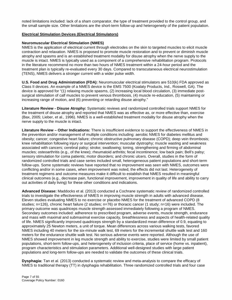
noted limitations included: lack of a sham comparator, the type of treatment provided to the control group, and the small sample size. Other limitations are the short-term follow-up and heterogeneity of the patient population. Electrical Stimulation Devices (Electrical Stimulators) Neuromuscular Electrical Stimulation (NMES) NMES is the application of electrical current through electrodes on the skin to targeted muscles to elicit muscle contraction and relaxation. NMES is proposed to promote muscle restoration and to prevent or diminish muscle atrophy and spasms and is an established treatment modality for disuse atrophy when the nerve supply to the muscle is intact. NMES is typically used as a component of a comprehensive rehabilitation program. Protocols in the literature recommend no more than two hours of NMES treatment within a 24-hour period and the treatment plan is typically re-evaluated every 30 days. Compared to transcutaneous electrical neurostimulation (TENS), NMES delivers a stronger current with a wider pulse width. U.S. Food and Drug Administration (FDA): Neuromuscular electrical stimulators are 510(k) FDA approved as Class II devices. An example of a NMES device is the EMS 7500 (Koalaty Products, Ind., Roswell, GA). The device is approved for “(1) relaxing muscle spasms, (2) increasing local blood circulation, (3) immediate post-surgical stimulation of calf muscles to prevent venous thrombosis, (4) muscle re-education, (5) maintaining or increasing range of motion, and (6) preventing or retarding disuse atrophy.” Literature Review - Disuse Atrophy: Systematic reviews and randomized controlled trials support NMES for the treatment of disuse atrophy and reported that NMES was as effective as, or more effective than, exercise (Bax, 2005; Lieber, et al., 1996). NMES is a well-established treatment modality for disuse atrophy when the nerve supply to the muscle is intact. Literature Review – Other Indications: There is insufficient evidence to support the effectiveness of NMES in the prevention and/or management of multiple conditions including: aerobic NMES for diabetes mellitus and obesity; cancer; congestive heart failure; chronic obstructive pulmonary disease (COPD); deep vein thrombosis; knee rehabilitation following injury or surgical intervention; muscular dystrophy; muscle wasting and weakness associated with cancers; cerebral palsy; stroke; swallowing; toning, strengthening and firming of abdominal muscles; osteoarthritis (e.g., of the knee); rheumatoid arthritis; fecal incontinence; low back pain; Bell’s palsy; sensory stimulation for coma patients; motor disorders; and chronic ulcers. Overall, studies in the form of randomized controlled trials and case series included small, heterogeneous patient populations and short-term follow-ups. Some systematic reviews have reported that no improvement was seen with NMES, outcomes were conflicting and/or in some cases, when improvement was noted, the effects did not last. Heterogeneity of treatment regimens and outcome measures make it difficult to establish that NMES resulted in meaningful clinical outcomes (e.g., decrease pain, functional improvement, improvement in quality of life and ability to carry out activities of daily living) for these other conditions and indications. Advanced Disease: Maddocks et al. (2013) conducted a Cochrane systematic review of randomized controlled trials to investigate the effectiveness of NMES in improving muscle strength in adults with advanced disease. Eleven studies evaluating NMES to no exercise or placebo NMES for the treatment of advanced COPD (8 studies; n=126), chronic heart failure (2 studies; n=76) or thoracic cancer (1 study; n=16) were included. The primary outcome was quadriceps muscle strength assessed immediately following a program of NMES. Secondary outcomes included: adherence to prescribed program, adverse events, muscle strength, endurance and mass with maximal and submaximal exercise capacity, breathlessness and aspects of health-related quality of life. NMES significantly improved quadriceps strength by a standardized mean difference of 0.9, equating to approximately 25 Newton meters, a unit of torque. Mean differences across various walking tests, favored NMES including 40 meters for the six-minute walk test, 69 meters for the incremental shuttle walk test and 160 meters for the endurance shuttle walk test. No serious adverse events were reported. Although the use of NMES showed improvement in leg muscle strength and ability to exercise, studies were limited by small patient populations, short-term follow-ups, and heterogeneity of inclusion criteria, place of service (home vs. inpatient), program characteristics and stimulation parameters. Additional well-designed studies with large patient populations and long-term follow-ups are needed to validate the outcomes of these clinical trials. Dysphagia: Tan et al. (2013) conducted a systematic review and meta-analysis to compare the efficacy of NMES to traditional therapy (TT) in dysphagia rehabilitation. Three randomized controlled trials and four case

Page 8 of 55 Coverage Policy Number: 0160
series (n=291) met inclusion criteria. Outcomes were measured using the Functional Oral Intake Scale (FOIS), Swallow, Functional Scoring System (SFSS), American Speech-Language-Hearing Association National Outcome Measurement System (ASHA NOMS) Swallowing Level Scale, and M.D. Anderson Dysphagia Inventory (MDADI). Four studies compared NMES only to TT and three compared NMES with TT to TT alone. The Swallowing Function Scale of patients treated with NMES were significantly higher compared with patients treated with TT (p=0.02) but subgroup analysis according to etiology (I.e., stoke, cancer and Parkinson’s disease) showed no significant differences between NMES and TT in post-stroke dysphagia. Limitations of the studies included the inclusion of four nonrandomized controlled trials, poor study designs, and heterogeneity of patient population and outcome measures. Due to the limitations, these outcomes need to be validated in well-designed randomized controlled trials with large patient populations and long-term follow-ups. Heart Failure: Arena et al. (2010) conducted a systematic review of the literature to evaluate the evidence supporting NMES and inspiratory muscle training (IMT) for the treatment of systolic heart failure. Thirteen NMES studies met inclusion criteria, ten were randomized controlled trials. Although the studies reported improvement in aerobic capacity, peak oxygen uptake and strength and endurance of muscle groups, the studies were limited by patient population (i.e., mostly males), diverse NMES training protocols, variation in the type of muscle contraction elicited (i.e., titanic vs. twitch), the use of different muscle groups and different comparators. The percent improvement in peak oxygen uptake was consistently greater with conventional therapy (i.e., bicycle/treadmill). Sillen et al. (2009) conducted a systematic review of randomized controlled trials to analyze the role of NMES in strength, exercise capacity, and disease-specific health status in patients with congestive heart failure (n=9 studies) and chronic obstructive pulmonary disease (n=5 studies) with disabling dyspnea, fatigue, and exercise intolerance. The limited number of studies, heterogeneous patient populations and variability in NMES methodology prohibited the use of meta-analysis. Although some of the studies reported significant improvements with NMES compared to no exercise or usual care, outcomes, including adverse events, were conflicting. Additional studies are indicated to provide sufficient evidence to establish the clinical utility of NMES in this patient population. Knee Indications: De Oliveira Melo et al. (2013) conducted a systematic review to identify the evidence for NMES for strengthening quadriceps muscles in elderly patients with knee osteoarthritis (OA). Inclusion criteria were randomized controlled trials comparing pre and post-intervention, elderly patients with clinical diagnosis of knee OA and outcome measurements of quadriceps muscle strength measured preferentially with an isokinetic dynamometer. Six randomized controlled trials (n=35–200) met inclusion criteria. Four studies included ≤ 50 patients. Study designs and outcome measures were heterogeneous and comparators varied. NMES parameters were poorly reported. The trials scored extremely low on the allocation concealment and blinding items. In most of the trials, the randomization methods were not described. Due to the poor methodology of the studies and poor description of the strength measurement methods, no or insufficient evidence was found to support NMES alone or combined with other modalities for the treatment of elderly patients with OA. Due to the study limitations, no meta-analysis was performed. Giggins et al. (2012) conducted a systematic review and meta-analysis to assess the effectiveness of NMES for the treatment of knee osteoarthritis. Nine randomized controlled trials (n=395) and one controlled trial (n=14) were included. Outcome measures included self-reported disease-specific questionnaires and pain scales, strength measurements, knee range of motion, knee and thigh circumference and functional assessments. Two studies were considered of strong quality, four moderate and four weak quality. Overall, there was inconsistent low level evidence that NMES significantly reduced pain and increased strength and function. Pooled analyses of six studies showed that NMES improved levels of self-reported pain and function, but not objective measures of function. The authors noted that the results should be interpreted with caution due to the heterogeneity of studies. Due to the conflicting data, definitive conclusions regarding the effectiveness of NMES for the treatment of knee osteoarthritis could not be made. Kim et al. (2010) conducted a systematic review of randomized controlled trials (n=8) to assess the effectiveness of NMES on “quadriceps strength, functional performance, and self-reported function after anterior cruciate ligament reconstruction.” Control interventions included: therapeutic exercises, EMG biofeedback, TENS plus exercises, and weight-bearing exercises. Quadriceps strength outcomes varied with some studies favoring NMES while others reported equivocal results or favored control interventions. One study each reported

Page 9 of 55 Coverage Policy Number: 0160
functional testing (n=20) and patient self-reported outcomes (n=43). Although some studies reported improvement following NMES, this analysis was limited by the use of various NMES regimens (e.g., treatment duration ranged from three to 11 weeks, number of sessions ranged from 12–105) and overall, only one follow-up visit occurred immediately following completion of treatment sessions. There is insufficient evidence to support clinical meaningful benefit of NMES on functional performance. In a systematic review of randomized controlled trials, Monaghan et al. (2010) assessed the effectiveness of NMES in strengthening quadriceps before and after total knee replacement. Two studies met inclusion criteria. NMES plus exercise resulted in better quadriceps muscle activation compared to exercise alone (n=39), but was not maintained at the 12-week follow-up. No significant differences were reported in either study for maximum voluntary isometric torque or endurance between the NMES group and the control group. In a 2008 systematic review of anterior cruciate ligament reconstruction (ACL) rehabilitation, Wright et al. reported that 14 randomized controlled trials had evaluated postoperative NMES following ACL reconstruction. Because of the variety of parameters in the studies; poor study quality; heterogeneous patient populations; and the lack of randomization, blinding and independent observers, the authors noted that it was difficult to make generalized conclusions regarding NMES, and it did not appear to be a requirement for successful ACL reconstruction rehabilitation. Stroke: In a randomized controlled trial (n=60), Hsu et al. (2010) compared high-NMES and low-NMES to a control group (standard rehabilitation) for the treatment of upper-extremity function in acute stroke patients. The low NMES group received 30 minutes of stimulation per day and the high-NMES group received 60 minutes per day, five times per week, for four weeks. All patients received standard rehabilitation. Compared to the control group, the NMES groups showed significant improvement in the Fugl-Meyer Motor Assessment (p=0.003) and Action Research Arm Test scales (p=0.016) at week four and week 12. There were no significant differences between low- and high-NMES stimulation. No significant differences between the groups were reported on the motor activity log. Limitations of the study include the small patient population, short-term follow-up, and 12 patients lost to follow-up. Transcutaneous Electrical Nerve Stimulation (TENS) A TENS device consists of an electronic stimulus generator that transmits pulses of various configurations through electrodes attached to the skin to stimulate the peripheral nerves for the purpose of pain management. Conventional TENS or high frequency TENS delivers 40–150 hertz (Hz) compared to acupuncture-like TENS that delivers a low frequency at 1–10 Hz. Pulsed TENS uses low-intensity firing in high-frequency bursts at 100 HZ. TENS has been used for a number of applications, including postoperative pain; acute and chronic pain, obstetrical pain, and pain associated with medical procedures. U.S. Food and Drug Administration (FDA): TENS are approved by the FDA 510(k) process as a Class II device for the relief and management of chronic intractable pain. Examples of these devices include the Empi Active Transcutaneous Nerve Stimulator (Empi, Inc., Clear Lake, SD), the StimPad™ TENS System (AEMED, Inc. West Palm Beach, FLA) and the ReBuilder® (Micromed, Inc., Essex Junction, VT). In 2014, FDA announced that it approved the Cefaly Supraorbital Transcutaneous Neurostimulator (Cefaly-Technology, Herstal, Belgium) through the 510(k) de novo premarket review pathway, a regulatory pathway for generally low- to moderate-risk medical devices that are not substantially equivalent to an already legally marketed device. FDA classified the Cefaly as a Class II device indicated for the prophylactic treatment of episodic migraine in patients 18 years of age or older. FDA noted that this is the first TENS device approved for use prior to the onset of pain. Literature Review - Acute Postoperative Pain The evidence in the peer-reviewed literature supports TENS for the treatment of pain in the acute post-operative period (i.e., within 30 days of surgery). Systematic reviews, meta-analysis and randomized controlled trials reported a reduction in pain and analgesic use in the treatment of acute post-operative pain and in some cases, shorter recovery times (Sbruzzi, et al., 2012; Freynet and Falcoz, 2010; Bjordal, et al., 2003). Literature Review - Other Indications: The evidence in the published peer-reviewed scientific literature has not established the effectiveness of TENS for the treatment of any other indications including, but not limited to:

Page 10 of 55 Coverage Policy Number: 0160
chronic low back pain; cervical pain; acute pain; acute and chronic headaches; abdominal pain, asthma, chemotherapy-induced pain, chronic leg ulcers, colonoscopy, drug withdrawal (e.g., opiate addiction), dysmenorrhea, fibromyalgia, fracture healing, hypertension, knee osteoarthritis, mandibular disorders (e.g., neuromuscular orthodontics; temporomandibular joint [TMJ]), motion sickness, nausea and vomiting of pregnancy, postoperative nausea and vomiting; low back pain of pregnancy, pain associated with childbirth (i.e., labor), pelvic pain, post-traumatic acute pain, rotator cuff tendinitis, stroke rehabilitation, suspected placental insufficiency, tinnitus, fecal incontinence, urinary incontinence, vestibulodynia, and unstable angina. Overall, systematic reviews, randomized controlled trials and case series have reported that there was no improvement with TENS for these indications or that conclusions could not be made due to the poor methodology of the studies. Study limitations included small heterogeneous patient populations with short-term follow-ups, insufficient data or conflicting data, and heterogeneity of the application of TENS (e.g., physician applied vs. patient applied, location of electrodes). Evidence supporting TENS for these indications is lacking nor is TENS an established treatment modality. The clinical utility of TENS has not been established for all other indications. Acute Pain: Walsh et al. (2009) assessed the analgesic effectiveness of TENS in acute pain for adults (n=919) in a systematic review of 12 randomized controlled trials. The types of acute pain included procedural pain (e.g. cervical laser treatment, venipuncture, screening flexible sigmoidoscopy) and nonprocedural pain (e.g. postpartum uterine contractions, rib fractures). The authors were unable to make any definitive conclusions due to the insufficient extractable data. Low Back Pain: The Centers for Medicare and Medicaid (2012) conducted a systematic review of the literature to evaluate TENS for the treatment of chronic low back pain. Inclusion criteria included adults with chronic, persistent low back pain (with or without leg pain) for three months or more and used TENS for at least four weeks. Included clinical trials had a patient population of ten or more; well-defined comparators; and used all models, frequencies, and wave patterns of TENS. Studies that examined chronic low back pain in patients with pain related to malignancy, neurodegenerative diseases (e.g. multiple sclerosis) and well-defined rheumatic disorders (except for osteoarthritis) were excluded. Seven systematic reviews and five randomized controlled trials met the inclusion criteria. Relevant clinical practice guidelines were also considered. Following a review of the data, Medicare concluded that TENS did not produce a clinically meaningful reduction in pain, a clinically meaningful improvement in function or a clinically meaningful improvement in any other health outcomes. When compared to TENS, sham units provided equivalent analgesia. The authors also noted that the potential for significant bias in the studies included in this analysis limited their “confidence in the reported results of this body of literature”. Buchmuller et al. (2012) conducted a 21-center, randomized controlled trial to evaluate the efficacy of TENS (n=117) compared to sham (n=119) in improving functional disability in patients with chronic low back pain (LBP), with or without radicular pain. Patients received treatment in four, one-hour daily sessions for three months. The primary outcome measure was improvement of functional status at six weeks based on the Roland–Morris Disability Questionnaire. Secondary outcome measures included functional status at three months, pain relief by weekly visual analogue scale (VAS) assessments, quality of life, use of analgesic and anti-inflammatory medication, satisfaction with the overall treatment strategy and compliance. Treatment was self-administered and recorded stimulation frequency and duration were checked at each study visit to verify compliance. Follow-ups occurred at 15 days, six weeks and three months. An improvement of at least 50% in lumbar pain between the first and last assessments was significantly greater in the TENS group (p=0.0003). The effect on pain intensity was particularly marked in the subgroup of patients with radicular pain. There were no significant differences between the groups in functional status at six weeks (p=0.351) or three months (p=0.816) or in any of the other outcome measures. Skin irritation was reported in 11 TENS patients and three sham patients. The authors noted that “the overall results of this study do not support the use of TENS in the treatment of patients with chronic LBP”. Limitations of the study include the short-term follow-up and heterogeneity of the patients. Khadilkar et al. (2008) conducted a systematic review to determine if TENS was more effective than placebo for the management of chronic low back pain. Four “high-quality” randomized controlled trials (n=585) met inclusion criteria. Due to conflicting evidence, the authors were unable to determine if TENS was beneficial in reducing back pain intensity. Two trials involving 410 patients reported that TENS did not improve back-specific functional status, the level of disability from the pain, the use of medical services or work status. There were no significant differences in outcomes when conventional TENS was compared to acupuncture-like TENS.

Page 11 of 55 Coverage Policy Number: 0160
Cancer Pain: Hurlow et al. (2012) conducted an update review of the 2009 review by Robb et al. One new study met inclusion criteria (n=24). There were significant differences in participants, treatments, procedures and symptom measurement tools used in the studies. The clinical utility of TENS for the treatment of cancer pain has not been established. Robb et al. (2009) conducted a systematic review of the literature to evaluate TENS for the treatment of cancer-related pain. Two randomized controlled trials (n=64) met inclusion criteria. Meta-analysis was not conducted due to the disparities between patient population, mode of TENS, treatment duration, and outcome measures prevented meta-analysis. There is insufficient evidence to support TENS for the treatment of cancer-related pain. Chronic Pain: Nnoaham et al. (2008) conducted a Cochrane systematic review to assess the effectiveness of TENS for the treatment of chronic pain, present for three or more months. A total of 25 randomized controlled trials (n=1281) met inclusion criteria. Included studies compared active TENS to sham TENS controls; active TENS to ’no treatment’ controls; or active TENS to active TENS controls (e.g. High Frequency TENS versus Low Frequency TENS). Due to the poor methodology of the studies, meta-analysis was not possible. Thirteen of 22 inactive control studies, reported a positive analgesic outcome in favor of active TENS treatments. For multiple dose treatment comparison studies, eight of 15 studies reported favorable outcomes for active TENS treatments and seven of nine active controlled studies found no difference in analgesic efficacy between high frequency and low frequency TENS. The authors concluded that “published literature on the subject lacks the methodological rigor or robust reporting needed to make confident assessments of the role of TENS in chronic pain management. Colonoscopy: Amer-Cuenca et al. (2011) conducted a randomized controlled trial (n=90) to evaluate the effectiveness of TENS in controlling pain in unsedated patients undergoing screening colonoscopy. Patients were randomized to one of three groups: control group (n=30), active TENS (n=30), or placebo TENS (n=30). The control group received hospital standard protocol for unsedated colonoscopies without any kind of sedation or analgesia. Pain was assessed five minutes into the procedure and at the end of the procedure using a visual analogue scale (VAS) and a five-point Likert scale. The TENS group reported a ≥ 50% reduction in the VAS scores compared to the placebo and control group (p<0.001). There was also a significant reduction on the Likert scale scores in the TENS group compared to the placebo and control groups (p=0.009). There were no significant differences between the groups in bloating sensation during the procedure and the duration of the procedure. Greater than 50% pain relief was achieved by 17 TENS patients, three placebo patients and six control patients (p<0.001). Author-noted limitations of the study included: the active TENS group’s experience of pain might have been affected by the potential distraction of continuously adapting stimulus intensity and the use of VAS as a measurement of pain. Another limitation is the small patient population. Dementia: Cameron et al. (2003; updated 2005) conducted a systematic review on TENS for the treatment of dementia. Nine randomized controlled trials met inclusion criteria, and three were included in meta-analysis. A statistically significant improvement was reported immediately following therapy in: delayed recall of 8 words and motivation in one trial, each and face recognition in two trials and motivation in one trial. However, the authors concluded that there was insufficient data for definitive conclusions to be drawn. Diabetic Neuropathy: Jin et al. (2010) conducted a systematic review to evaluate the effectiveness of TENS on diabetic peripheral neuropathy. Three randomized controlled trials (n=78) met inclusion criteria. TENS was reported more effective than placebo in the reduction of mean pain score at four and six weeks follow-up but not at 12 weeks. Pieber et al. (2010) conducted a systematic review of the literature to evaluate electrotherapy, including TENS, for the treatment of peripheral neuropathy in patients with diabetes. Three randomized controlled trials (n=76) and one retrospective review (n=54) evaluating TENS met inclusion criteria. The studies included short-term follow-ups and conflicting results. One study reported significant improvement in pain and another study reporting recurrence of pain after cessation of TENS. Due to the small patient populations, short-term treatment duration, short-term follow-up and poor study methodology, large multi-center randomized controlled trials are needed to further evaluate the long-term effect of TENS on diabetic neuropathy. Dysmenorrhea: In a systematic review of seven randomized controlled trials (n=164), Proctor et al. (2009) evaluated the effectiveness of low-frequency TENS (acupuncture-like TENS, 1–4 hertz [Hz]) and high-frequency TENS (conventional TENS, 50–120 Hz) (n=5) for the treatment of primary dysmenorrhea. Studies compared TENS to placebo, no treatment or medical treatment. Overall, high-frequency TENS was reported more effective

Page 12 of 55 Coverage Policy Number: 0160
than placebo TENS for relief of pain. There was no difference in pain relief with low-frequency TENS compared to placebo. There were conflicting results regarding whether high-frequency TENS was more effective than low-frequency TENS. Due to the small patient populations, various methods of the application of TENS, and the lack of precision in the comparisons, clear recommendations for clinical applications could not be made. Labor: Bedwell et al. (2011) conducted a systematic review of randomized controlled trials comparing TENS to routine care or placebo devices for labor pain. Fourteen studies (n=1256) met inclusion criteria. TENS were applied to the back (n=11 studies), acupuncture points (n=2 studies) and in one study to the cranium. Primary outcome measures were pain intensity and patient satisfaction with pain relief. Secondary outcome measures included: duration of labor, cervical dilation on admission to hospital, augmentation of labor, other pain relief, assisted birth or caesarean section, side effects, and sense of control in labor. Outcomes for neonates included Apgar score (<7 at five minutes), cord pH (<7.1) and adverse events. Patients receiving TENS to acupuncture points were less likely to report severe pain. There were no significant differences in use of epidural analgesia or other types of analgesia between the groups, pain ratings and patient satisfaction. None of the studies reported information on Apgar scores or cord pH or women’s sense of control in labor. There was no information that TENS affected any other outcomes on the mother or the baby. No adverse events were reported. The authors concluded that there was limited evidence that TENS reduced pain during labor but the “evidence is neither strong nor consistent”. The use of TENS at home in early labor has not been evaluated. Author-noted limitations of the studies included: small patient populations, unbalanced study groups, heterogeneity of outcome measures, various type of TENS devices were used, TENS was offered alone or as an adjuvant therapy making it difficult to assess the true effect of TENS in some studies, and pain was measured in so many different ways it was not possible to pool results. Mello et al. (2011) conducted a systematic review and meta-analysis to assess the effectiveness of TENS (n=529) compared to placebo or no TENS (n=547) for pain relief during labor including possible maternal and fetal complications. Nine randomized or quasi-randomized clinical trials (n=1076) with more than ten subjects met inclusion criteria. A meta-analysis of six studies demonstrated no evidence that TENS reduced the need for analgesia. There were no statistically significant differences between the groups in pain relief during labor. There was no evidence that TENS interfered in any of the outcomes except the mothers’ desire to use TENS in future deliveries. The use of TENS had no impact on mother or child and no influence on labor. According to the results of this review, there was no evidence that TENS reduced the use of additional analgesia. The authors noted that no study carried out intention-to-treat analyses which may lead to overestimation of the treatment’s clinical effect. Other noted limitations of the studies included a lack of uniformity in frequency or intensity of TENS, heterogeneity of the type of analgesia used, and the difficulty in measuring pain levels. Dowswell et al. (2009) conducted a systematic review on the use of TENS during labor. A total of 19 randomized controlled trials (n=1671) comparing TENS to pharmacotherapy or placebo met inclusion criteria. TENS was applied to the back (n=15), acupuncture points (n=2), and cranium (n=2). Overall, there were no significant differences between pain ratings in the TENS group and the control groups. In cases where TENS was used as an adjunct to epidural analgesia, there was no evidence that it reduced pain. There was no consistent evidence that TENS had any impact on interventions and outcomes of labor. Migraine Headaches: There is insufficient evidence in the peer-reviewed literature to support TENS for the treatment of migraines, including use of Cefaly. Schoenen et al. (2013) conducted a five-center randomized controlled trial to assess the safety and efficacy of Cefaly in the PREvention of Migraine (PREMICE) study using Cefaly. Patients, age 18–65 years old, with migraines, with or without aura, experiencing at least two attacks per month were included in the study. After a one month run-in period, subjects were randomized to Cefaly or sham therapy for 90 days. Primary outcome measures included change in monthly migraine days between the run-in month and the third month of treatment and the percentage of “responders,” (i.e., at least 50% reduction of monthly migraine days). Subjects kept a diary of headache events and had a follow-up visit at day 45 and 90. In both groups, migraine days decreased by an average of 20% during month one. In months two and three the sham group did not maintain decreased migraines while the Cefaly group did. Between run-in and third month of treatment, the mean number of migraine days decreased significantly in the Cefaly group (p=0.023), but not in the sham group (p=0.608). The 50% responder rate was significantly greater in the study group (p=0.023). The number of monthly migraine attacks (p=0.044), monthly headache days (p=0.041) and monthly acute antimigraine drug intake (p=0.007) were significantly reduced in the study group but not in the sham group. There were no reported adverse events. Limitations of the study include self-reported outcomes, heterogeneity

Page 13 of 55 Coverage Policy Number: 0160
in patient demographics between the two groups (e.g., age, duration of migraines) and recruited patients were not the most disabled migraineurs. Published data from randomized controlled trials with large patient populations and long-term outcomes comparing TENS to conventional therapy are needed to establish the effectiveness of TENS/Cefaly for the treatment of migraines. Neck Pain: Escortell-Mayor et al. (2011) conducted a 12-center randomized controlled trial to compare the effectiveness of TENS (n=43) to manual therapy (n=47) for the treatment of subacute or chronic mechanical neck disorders without neurological damage and followed for six months. Over half of the patients reported short-term effects following cessation of either therapy but at six months follow-up, success decreased in one-third of the patients. No significant differences were found between the groups in reduction of pain, decrease of disability or quality of life. No significant adverse events were reported. Following a systematic review of randomized controlled trials regarding electrotherapy, including TENS, for neck pain, Kroeling et al. (2009) concluded that no definitive statements could be made regarding the efficacy and clinical usefulness of these modalities. Eleven TENS trials (n=7-30) met inclusion criteria including: TENS compared to placebo or another modality (i.e., ultrasound, manual therapy, electrical muscle stimulation); TENS plus another therapy (i.e., hot packs, infrared, exercises, neck collar and/or analgesic) compared to the other therapy alone; or different TENS regimens. The authors concluded that “very low quality” evidence showed that TENS might relieve pain better than placebo or electrical muscle stimulation but not as well as exercise and infrared and possibly as well as manual therapy and ultrasound. Osteoarthritis of the Knee: Palmer et al. (2014) conducted a randomized controlled trial (n=224) to evaluate the effectiveness of TENS for the treatment of osteoarthritis (OA) of the knee. Exclusion criteria included comorbidities preventing participation in the knee group, contraindications to TENS or previous use of TENS. Patients, ≥ age 18 years, with OA or suspected OA were randomized to one of three groups: TENS and knee group (n=73), sham TENS and knee group (n=74), or knee group alone (n=77). The knee group participated in a six-week group education and exercise program. The primary outcome was the Western Ontario and McMaster Universities Osteoarthritis Index (WOMAC) function subscale. Secondary outcomes included WOMAC pain, stiffness, and total scores; extensor muscle torque; global assessment of change; exercise adherence; and exercise self-efficacy. All groups improved overtime and the improvements were maintained at the 24-weeks follow-up. There were no significant difference between the outcomes in all three groups (p>0.05). The addition of TENS did not improve outcomes. Rutjes et al. (2009) conducted a systematic review of the literature to evaluate transcutaneous electrical nerve stimulation for the treatment of osteoarthritis of the knee. Thirteen randomized and quasi-randomized trials (n=465) using TENS met inclusion criteria. Due to the heterogeneity of the studies and poor methodology, the authors could not confirm the effectiveness of TENS for this condition. The 2013 American Academy of Orthopedic Surgeons (AAOS) guidelines for treatment of osteoarthritis of the knee stated that evidence from a single low quality study or conflicting findings does not enable AAOS to make recommendations for or against the use of physical agents, including electrotherapeutic modalities (e.g., TENS). The evidence was mixed regarding efficacy and the outcomes in the limited number of studies were conflicting. The American College of Rheumatology’s (ACR) 2012 recommendation on the treatment of osteoarthritis of the hand, hip, and knee, “conditionally” recommended that patients with OA of the knee be instructed in the use of TENS. ACR stated that this modality was only recommended when the patient has chronic moderate to severe pain; is a candidate for total knee arthroplasty and is unwilling to undergo the procedure; or has comorbid medical conditions; or is taking concomitant medications that lead to a relative or absolute contraindication to surgery; or the surgeon does not to recommend the procedure. This recommendation was based on the “consensus judgment of clinical experts”, “informed by available evidence” and “incorporating their preferences and values” (Hochberg, et al., 2012). Phantom Pain and Stump Pain: Mulvey et al. (2010) conducted a systematic review of randomized controlled trials to assess the effectiveness of TENS for the treatment of phantom pain and stump pain following amputation in adults. No studies were identified.

Page 14 of 55 Coverage Policy Number: 0160
Rheumatoid Arthritis: In a systematic review of the literature, Brosseau et al. (2003) evaluated the effectiveness of TENS for the treatment of rheumatoid arthritis of the hand. Three randomized controlled trials (n=78) met inclusion criteria. Conventional TENS (c-TENS) and acupuncture-TENS (acu-TENS) were compared to either placebo or each other. Pain outcomes on the effect of TENS were conflicting. Acu-TENS was beneficial for reducing pain intensity and improving muscle power scores compared to placebo. No clinical benefit on pain was reported with C-TENS compared to placebo. C-TENS resulted in a clinical benefit on the patients’ assessment of change compared to acu-TENS. The authors concluded that more well designed studies with a standardized protocol and adequate numbers of subjects were needed to fully identify the effect of TENS for the treatment of RA of the hand. Stroke: NG and Hui-Chan (2009) conducted a randomized controlled trial (n=109) to determine if TENS would improve functional walking performance (i.e., gait velocity, walking endurance and functional mobility) in hemiparetic stroke patients with spastic plantar flexors. In addition to a control group (n=29), patients were assigned to one of three intervention groups: TENS only (n=28), TENS plus exercise (n=27) or placebo stimulation plus exercise (n=25). Each patient self-administered 20 sessions, five days per week for four weeks. Each group received 60 minutes of TENS and the exercise groups received an additional 60 minutes of exercise following TENS or placebo stimulation. Final follow-up occurred four weeks after the treatment ended. At the final follow-up compared to all other groups, significant improvements were seen in the TENS plus exercise group in gait velocity (p<0.001) and reduction in timed up and go scores (P<0.01). The TENS plus exercise group covered significantly more distance in the 6-minute walk test (6MWT) (p<0.01) compared to the control group and the TENS only group. Additional studies with larger patient populations and long-term follow-up are indicated to validate the results of this study. The generalizability of this study is limited to stroke patients with moderate to severe spasticity in the ankle plantar flexors. The frequency, duration, and intensity of combined rehabilitation programs have not been established. Yan et al. (2009) conducted a randomized controlled trial (n=62) to investigate whether TENS, when applied to acupuncture points in patients after acute stroke, decreased spasticity and/or increased muscle strength and was more effective than placebo stimulation and standard rehabilitation. Patients were randomized to TENS, placebo-TENS, or standard rehabilitation. Stimulation was applied to four acupuncture points in the affected lower leg for 60 minutes, five days a week for three weeks. Compared to placebo or rehabilitation, TENS significantly increased the number of patients with normal tone and ankle dorsiflexor strength and decreased the co-contraction ratio (p<0.05). Overall, the TENS patient walked two to four days earlier than the other patients, but the difference was not significant between the three groups. Limitations of the study include the small patient population and short-term follow-up. Urinary Incontinence and Infections: Monga et al. (2012) conducted a systematic review to evaluate electrical stimulation therapies (i.e., TENS, sacral nerve stimulation, percutaneous posterior tibial nerve stimulation) for the treatment of lower urinary track infections (LUTI). A total of 73 studies including randomized controlled trials (RCTs), case series and retrospective reviews met inclusion criteria. Thirteen studies (n=377), including three RCTs, three comparative studies and seven case series investigated outcomes using TENS. The studies included treatment of pediatric populations, detrusor instability, overactive bladder syndrome, various LUTIs, and irritative voiding dysfunction. Comparators included placebo stimulation, medical therapy, percutaneous neuromodulation, biofeedback or no treatment. The authors concluded that it was not possible to make any meaningful generalizations related to outcomes for the TENS studies due to the significant heterogeneity of the mode of therapy delivery, definition of patient subgroups, and outcome measures. Vestibulodynia: Murina et al. (2008) assessed the efficacy of TENS in the treatment of 40 women with vestibulodynia. The women were randomized to either TENS or sham and received treatment twice a week for 20 sessions. At the three month follow-up, visual analogue scale scores and short-form McGill-Melzack Pain Questionnaire scores improved significantly (p=0.004, p=0.001, respectively) in the TENS group compared to the sham group. Three of 15 women in the TENS group relapsed three months following the end of the study. No adverse events were reported. Limitations of the study include the small patient population and short-term follow-up. Professional Societies/Organizations: Following a systematic review of none-pharmacological treatment modalities for dementia, the Department of Veterans Affairs Health Services Research and Development Services (VA/DOD) (2011) stated that three randomized controlled trials found no significant effects on sleep

Page 15 of 55 Coverage Policy Number: 0160
disturbance or behavioral symptoms following treatment and six-weeks thereafter. Possible benefits of TENS for the treatment of dementia could not be made. The VA/DOD (2010) practice guideline on the management of stroke rehabilitations stated that there was insufficient evidence to support the use of TENS and its mechanism of action for stroke rehabilitation is unknown. However, the guideline stated that TENS could be considered as an adjunctive treatment for enhancing recovery of gait function in this patient population. In practice guidelines for chronic pain management, the American Society of Anesthesiologists Task Force on Chronic Pain Management and the American Society of Regional Anesthesia and Pain Medicine (2010) recommended TENS as part of a multimodal approach to pain management for the treatment of patients with chronic pain (e.g., back pain, neck pain, phantom limb pain). A meta-analysis of randomized trials comparing TENS to sham for back pain reported greater relief for assessment periods of one hour to one month. Observational studies reported that TENS improved pain scores for a variety of conditions for 3–6 months. In a 2010 technology assessment on the efficacy of TENS in the treatment of pain in neurologic disorders, the American Academy of Neurology (AAN) stated that based on the available evidence, TENS is not recommended for the treatment of low-back pain. There are conflicting reports of TENS compared to sham-TENS but the stronger evidence established TENS as ineffective for back pain. Based on two studies comparing TENS to TENS-sham (n=19 and 31) and one study comparing high-frequency muscle stimulation to TENS (n=41), AAN stated that TENS is “probably effective” in reducing diabetic peripheral neuropathy pain. In their chronic pain medical treatment guidelines, the Work Loss Data Institute (2009) recommended TENS as a treatment option for acute post-operative pain in the first 30 days following surgery. They also recommended TENS as a secondary treatment modality on a one-month trial bases for chronic intractable pain of at least three months’ duration for conditions such as neuropathic pain (e.g., diabetic neuropathy, post-herpetic neuralgia), multiple sclerosis, phantom limb pain, complex regional pain syndrome or spasticity). They noted that there is a lack of evidence supporting the efficacy of TENS for chronic pain and stated that TENS should be used as an adjunct to an “evidence-based restorative program.” Following a systematic review of randomized controlled trials of 17 nonpharmacologic therapies for low back pain, the American Pain Society and the American College of Physicians (2007) stated that TENS had not been shown to be effective for acute, subacute or chronic low back pain (Chou and Huffman, 2007). Conductive Garments Conductive garments are fabric electrodes placed between an electrical stimulator and a patient’s skin for the delivery of electrical stimulation. They are an established alternative to standard electrodes and aid in the treatment of patients with chronic pain who have large areas or a large numbers of sites to be stimulated or the frequency is such that it is not feasible to use conventional electrodes, tapes or lead wires. The electrodes may also be indicated when sites requiring stimulation are not accessible by the patient with conventional electrodes, tapes or lead wires (i.e., back) and/or when medical conditions (e.g., skin problems) preclude the use of conventional electrodes, tapes or lead wires. U.S. Food and Drug Administration (FDA): AG Garments (San Diego, CA) conductive electrodes are Class II, 510(k) approved by the FDA “as reusable (by a single patient), cutaneous, flexible, conductive garment/fabric electrodes for interface between electrical stimulators and a patient’s skin for the delivery of electrical stimulation” (FDA, 2002). Other Electrical Stimulation Devices Bioelectric Nerve Block (Electroceutical Therapy) Bioelectric therapy, also known as electromedicine, noninvasive neuron-blockade devices, electroceutical neuron-blockade devices and bioelectric treatment systems, is proposed as a treatment for acute and chronic pain (e.g., back pain, diabetic pain, joint pain, fibromyalgia, headache, and reflex sympathetic dystrophy). Electroceutical treatments use much higher electrical frequencies than TENS units (ranging from one to 20,000 Hz compared to 0.5 to 100 Hz used in TENS).

Page 16 of 55 Coverage Policy Number: 0160
U.S. Food and Drug Administration (FDA): An example of a device used for bioelectric therapy is the Matrix PRO ElecDT (Matrix Electromedical, Inc., Las Vegas, NV) which was 510(k) approved by the FDA as an interferential current therapy device. Literature Review: There is insufficient evidence in the published peer-reviewed scientific studies to support the safety and effectiveness of bioelectric therapy. Well-designed, randomized controlled clinical studies are needed to determine the clinical utility of electroceutical therapy in the treatment of patients with acute or chronic pain. Cranial Electrical Stimulation Cranial electrical stimulation (CES), also called electrotherapy, electrotherapeutic sleep, electrosleep, electric cerebral stimulation, cranial transcutaneous electrical nerve stimulation, cerebral electrotherapy, transcranial electrotherapy, transcranial electrical stimulation, transcranial direct electrical stimulation (tDCS), transcerebral electrotherapy, neuroelectric therapy, and craniofacial electrostimulation, delivers low level electrical stimulation (i.e., microcurrent) to the brain through electrodes that are attached to the ear lobes or behind the ears. It has been proposed that CES’s direct effect on the brain’s limbic system, hypothalamus, reticular activation system, and/or the autonomic nervous system can control the symptoms of various conditions. This therapy is not to be confused with transcranial magnetic stimulation or vagus nerve stimulation. CES has been proposed for the treatment of anxiety, depression, insomnia, substance abuse, fibromyalgia, Alzheimer’s, attention-deficit/hyperactivity disorder (ADHD), asthma, spastic colitis, tension headaches, cluster headaches, migraines, hypertension, tinnitus, preoperative relaxation, aphasia and functional ability following stroke, chemotherapy symptoms in cancer patients, burn patients, and other pain-related disorders. U.S. Food and Drug Administration (FDA): CES devices are approved under the FDA 510(k) class III process for the treatment of insomnia, depression, or anxiety. Examples of these devices include the Cranial Electrical Nerve Stimulator (Johari Digital Healtcare Ltd., Fall CITY, WA), Alpha-Stim® (Electromedical Products, Inc., Hawthorne, CA), and LISS Cranial Stimulator and Fisher-Wallace Cranial Stimulator by Medical Consultants Intl. Ltd. (Glen Rock, NJ). Some devices are approved for use by the patient at home. Literature Review: The evidence in the published peer-reviewed literature does not support the effectiveness of CES for any indication. Studies consist of randomized trials with small patient populations, short-term follow-ups, and conflicting outcomes. Alzheimers: Rose et al. (2009) conducted a randomized controlled trial to compare the short-term effects of CES (Alpha-Stim) (n=19) to sham stimulation (n=19) on sleep disturbance, depressive symptoms, and subjective appraisal in individuals who were the primary caregivers for spouses with Alzheimer’s disease. Subjects used CES 60 minutes per day for four weeks and completed a daily log. At the end of four weeks, there were no significant differences in overall sleep disturbances, sleep quality, or sleep onset latency scores. The CES group did report a nine-minute decrease in sleep onset latency compared to a one minute increase in the sham group. There were no significant differences between the groups in depressive symptoms or in burden, mastery, impact or satisfaction of the care giving situation. Fibromyalgia: Taylor et al. (2013) conducted a randomized controlled trial (n=46) to investigate the effectiveness of microcurrent CES on the pain and symptoms of fibromyalgia. Three groups included active CES (n=17), sham device (n=14) and usual care alone (n=15). All subjects remained on their usual care regimen, including medications. Follow up occurred for eight-weeks. Subjects using CES reported a significantly greater decrease in average pain (p=0.23), fatigue (p=0.71, sleep disturbance (p=0.001) and functional status compared to the other two groups. Limitations of the study include the small patient population, short-term follow-up, self-monitoring and reporting of outcomes, loss to follow-up (n=5), and the study included mainly females (n=43) and cannot be generalized to males. Spinal Cord Injury: Tan et al. (2011) conducted a multi-site randomized controlled trial (n=111) to evaluate the effectiveness of the Alpha-STIM CES in patients with spinal cord injury (SCI) and chronic neuropathic pain at or below the level of injury. Of the 111 enrolled patients, 46 patients were randomized to CES at a sub-threshold level of 100 μA (microamperes) and 56 to sham CES. Patients were trained on the use of the device either in person or via mail and telephone, instructed to apply therapy at home for 21 days and to monitor and record the intensity of pain before and after each treatment. Coordinators contacted the patients via phone on a weekly

Page 17 of 55 Coverage Policy Number: 0160
basis. At the end of 21 days, patients completed a packet of post-intervention questionnaires. Some questionnaires were completed via telephone. At the end of the 21-day study period, patients in the sham group were allowed to use active CES for three weeks at a current setting of the patient’s choice (100–500 μA). Following the 21 days of active CES, patients were also given the option of using CES for up to six months on an as needed basis and were paid to complete the questionnaires. During the three week blinded phase, the active and sham groups did not differ significantly on average daily pain ratings before (p>0.60) or after treatment (p>0.90). However, the active group had a significantly greater average decrease in pain from before to after the daily treatments compared to the sham group (p<0.05). More pain relief was reported by the participants during the open-label phase. In the 40 sham patients who crossed over to CES, a significant reduction in pain was reported (p<0.001). Although an improvement was shown in pain after a session, the improvement did not last until the next day. Less than 14% of patients in either group achieved a 30% or more reduction in pain. The most commonly reported side effects were pulsing, tingling, stinging, itching, and/or a small electric feeling produced by the ear clips. Author noted limitations included: the baseline differences between active and sham groups on several of the outcome measures made group differences in change scores difficult to interpret; loss to follow-up at three (<55%) and six months (>70%); and all of the outcome measures were obtained by self-report. Stroke: Essner et al. (2013b) conducted a Cochrane review of randomized controlled trials to determine the effects of tDCS on generic activities of daily living (ADLs) and motor function in people with stroke. Fifteen studies (n=455) met inclusion criteria. The evidence showed effect in favor of transcranial direct current stimulation (tDCS) at the end of the intervention phase. Although effect was seen at follow-up, the effect was not sustained. Upper limb function showed an effect in favor of tDCS at the end of the intervention phones but not at the three month follow-up. Elsener et al. (2013b) also reviewed randomized controlled trials on tDCS for its effect on improving aphasia in stroke patients. Data from five trials (n=54) showed no evidence that tDCS enhanced current speech and language therapy in these individuals. Electrical Sympathetic Stimulation Therapy Electrical sympathetic stimulation therapy is a form of electrical stimulation of the peripheral nerves by applying eight electrodes bilaterally to the lower legs, feet, arms and hands. The therapy targets the autonomic nervous system and treats systematically as opposed to locally and is proposed for the treatment of chronic, intractable pain. Multiple beat frequencies are generated between 0-1000 Hz. Treatments are typically one hour in duration and may be administered in a physician’s office or at home. U.S. Food and Drug Administration (FDA): Sympathetic therapy devices are approved by the FDA 510(k) process. Two such devices are the Dynatron STS and the Dynatron STS RX, a home device (Dynatronics Corp., Salt Lake city, UT). The devices are indicated for “symptomatic relief of chronic intractable pain and/or management of post-traumatic or post-surgical pain” (FDA, 2001). Literature Review: The evidence in the published peer-reviewed scientific literature does not support the safety and effectiveness of sympathetic therapy. Studies are primarily in the form of case series and retrospective reviews with small patient populations and short-term follow-ups (Guido, 2002). Electro Therapeutic Point Stimulation (ETPSSM) ETPS neuromechanical therapy or neuropathic acupuncture involves the detection and treatment of chronic intractable neuromyofascial pain using the TENS US Unit (Acumed Medical Supplies, LTD, Stanford, CT). The transcutaneous device detects treatment points on the skin and applies brief, concentrated electrical microstimulation in short bursts. Traditional TENS units apply alternating current compared to the direct current applied by ETPS. Depending on how the device is programmed, the therapy is also proposed to decrease circulation and assist in resolution of swelling and pain or to increase circulation to enhance immune response and neural regeneration. The treatments can be self-administered by the patient at home (MedDex Solutions, 2014; Hocking, 2002). U.S. Food and Drug Administration (FDA): The TENS US Unit is approved by the FDA 510(k) Class II device as the TENS Pro 900 (Acumed Medical Supplies, LTD, Stanford, CT) for the treatment of chronic intractable pain.

Page 18 of 55 Coverage Policy Number: 0160
Literature Review: There is insufficient evidence in the published peer-reviewed scientific literature to support the safety and effectiveness of ETPS. The available studies are primarily in the form of case reports and case series with small patient population and short-term follow-ups. Functional Electrical Stimulation (FES) FES or functional neuromuscular stimulation (FNS) attempts to replace stimuli from destroyed nerve pathways to assist neurologically impaired patients (e.g., spinal cord injury, stroke) with functional movement and to suppress spasticity. FES is a high-intensity (25–100 milliamps), short duration therapy that may be delivered for 20 minutes to one hour, several times a week, for months. For the device to be effective, the peripheral nerve must be intact. FES is proposed for multiple indications, including:
• to assist ambulation in paraplegics (e.g., Parastep® I System, Sigmedics, Inc., Fairborn, OH). Parastep is a microcomputer controlled walker proposed to aid standing, walking, balance and stability in individuals with a spinal cord injury for whom gait training and standing are indicated. Using surface electrodes, the device delivers electrical current to peripheral nerves in the lower extremities. Parastep is a proposed alternative to traditional orthotics and bracing;
• as a means of stationary exercise to prevent or reduce muscle atrophy in upper and lower extremities (e.g., ERGYS 2; Therapeutic Alliance, Inc., Fairborn, OH). The ERGYS 2 provides cycling activity proposed to improve muscle strength and circulation in the lower extremities;
• to improve ambulation in patients with gait disorders such as drop foot, hemiplegia due to stroke, cerebral injury, or incomplete spinal cord injury (e.g., Walkaide™ stimulator; Neuromotion, Edmonton, Alberta, Canada; NESS L300 Foot Drop System, Bioness Inc., Valencia, CA). WalkAide is a device that attaches to the leg just below the knee and is proposed to counteract foot drop and improve mobility during walking by stimulating the peroneal nerve. The Ness L300 is a similar device that also attaches below the knee, provides nerve stimulation and is proposed to assist the individual with foot drop to walk with increased balance and speed. The Ness L300 Plus builds on the L300 Foot Drop System by adding a thigh cuff. The thigh cuff is proposed to add control over bending and straightening the knee.
• to provide range of motion and function in patients with upper limb paralysis or hemiplegia (NESS H200 hand rehabilitation system [previously known as the Handmaster], Bioness Inc., Valencia, CA). NESS H200 is for use by an individual with hand paralysis. The device attaches to the lower forearm and is proposed to activate various muscle groups, enhancing grip and allowing opening and closing of the hand.
U.S. Food and Drug Administration (FDA): FES devices, such as the Parastep, that have been proposed for restoring ambulation to paraplegics are regulated by the FDA’s premarket approval (PMA) process. Functional electrical stimulators that are used to provide stationary exercise for paraplegics, to correct gait disorders, or to provide range of motion and function are approved by the FDA 510(k) process as Class II devices. The RT300 FES cycle ergometer (Restorative Therapies, Inc., Baltimore, MD) is approved as a powered muscle stimulator for “general rehabilitation for relaxation of muscle spasms, prevention or retardation of disuse atrophy, increasing local blood circulation and maintaining or increasing range of motion” (FDA, 2009). Other Restorative FES devices include the RT300 Leg, RT300 Leg and Arm, RT300 Arm, RT300 for children. Literature review Assist Ambulation in Paraplegics: Studies investigating FES (i.e., Parastep) were published in 2000 or before and are primarily case series with small patient populations and short-term follow-ups. Brissot et al. (2000) investigated the motor performances of Parastep in 15 thoracic-spine injured patients (T3-T11). Patients had to have a stable neurologic and orthopedic status and be at least six months status-post injury and/or restorative surgery. Two patients did not complete the required training. Follow-up occurred at 40 ± 11 months. After a mean 20 sessions, the patients achieved independent ambulation with a mean walking distance of 52.8 ± 69 meters (m), and a mean speed of 0.15 6 ± 0.14 m/second. At the final follow-up five patients were using the Parastep regularly and all patients used it for physical fitness and not for functional ambulation. According to the authors the high ratio of energy cost of the use of the device may have explained its limited use in daily activity. The authors also noted that “the Parastep approach has very limited applications for mobility in daily life,

Page 19 of 55 Coverage Policy Number: 0160
because of its modest performance associated with high metabolic cost and cardiovascular strain. However, it can be proposed as a resource to keep physical and psychological fitness in patients with spinal cord injury.” Stationary Exercise to Prevent or Reduce Muscle Atrophy in Upper and Lower Extremities: Randomized controlled trials using various FES devices have evaluated FES cycling (MOTOmed®, RECK GmbH, Betzenweiler, Germany) compared to passive cycling (n=35) (Ambrosini, et al., 2011); FES (H200 device) combined with self-directed exercise vs. exercise alone (n=23) (Weber, et al., 2010); FES cycling (device not given) with standard rehabilitation vs. rehabilitation alone (n=20) (Ferrante, et al., 2008); and with arm and hand rehabilitation comparing FES (Compex Motion, Compex SA, Switzerland) to conventional therapy (n=23) (Mangold, et al., 2009). Some studies reported no significant differences with FES. Due to the small patient populations, short-term follow-ups (e.g., 4–12 weeks) and conflicting results, the effectiveness of FES for the treatment of stroke patients has not been established. Improve Ambulation in Patients with Gait Disorders: FES has been proposed for improving ambulation in patients with gait disorders such as drop foot, hemiplegia due to stroke, cerebral injury, or incomplete spinal cord injury. Randomized controlled trials and case series have primarily included small patient populations (n=14-64) with short-term follow-ups and heterogeneous treatment regimens and outcome measures (Esnour, et al., 2010; Nooijen, et al., 2009; Everaert, et al., 2010; Stein, et al., 2010; Barrett, et al., 2009; Postans, et al., 2004). In a randomized controlled trial (n=74), Field-Fote and Roach (2011) evaluated whether there was a difference in walking speed and distance using four locomotor training regimens for patients with chronic spinal cord injuries. The regimens included treadmill-based training with manual assistance (TM) (n=19), treadmill-based training with bilateral electrical stimulation (TS) (Digitimer DS7AH, Digitimer Ltd, Welwyn Garden City, Herts, UK) (n=22), overground training with electrical stimulation (OG) (n=18) (WalkAide™), and treadmill-based training with locomotor robot (LR) (Lokomat Robotic Gait Orthosis, Zurich, Switzerland) (n=15). Training was administered five days per week for 12 weeks. There was a statistically significant improvement in walking speed (p<0.001) in the TM, TS and OG groups and overall time effect on training (p<0.0001). There was a significant improvement in walking distance in the TS and OG groups. Distance gain was greater for OG. Post hoc testing indicated the increase in “time X group” interaction in the OG group was significantly greater than the other groups (p≤0.01). Effect sizes for speed (d=0.43) and distance (d=0.28) were largest with OG. Effect sizes for speed were the same for TM and TS (d=0.28). There was no effect from LR. The Lower Extremity Motor Scores increased 8%–13%, with no significant between group differences. Ten patients were available for an average 20.3 month follow-up (4 OG and 6 in the other groups). These subjects had declined an average of 0.06 meter per second (m/s) in walking speed since completion of training, but were still an average 0.08 m/s faster than before training. Author-noted limitations of the study included: they did not know if the training dosage was optimal for improving walking speed and distance; the focus was on walking rather than other aspects of walking (e.g., producing optimal kinematics); and most of the subjects used a wheelchair as their primary means of mobility so the amount of change qualified as meaningful change may be different from subjects who use other means of mobility; “the training parameters used in the robotic gait orthosis approach were configured to impose a kinematically appropriate gait pattern and stepping proceeded regardless of whether participants contributed effort;” and only ten people returned for longer follow-ups. Other limitations of the study include the small patient population, short-term follow-up, patients lost to follow-up, and the OG group had the largest number of patients who were less impaired. Heart Failure: Smart et al. (2013) conducted a systematic review and meta-analysis of randomized controlled trials to evaluate FES (devices not given) in the treatment of heart failure. Ten studies met inclusion criteria (n=301) which included 158 FES patients, 85 aerobic cycle exercise training and 58 sedentary controls or sham FES. Five studies compared FES to cycle exercise training, two studies compared FES to a sedentary control group and three studies compared active FES to sham FES. Training sessions varied from three to seven sessions per week, FES frequencies varied from 10–50 Hz, off and on intervals ranged from 2–50 seconds, and studies ranged from 5–10 weeks duration. Most studies used FES of the quadriceps and gastrocnemius muscles or hamstrings in the home and exercise training intensity ranged from 50%–80%. FES produced inferior improvements in peak oxygen consumption (VO2) compared to cycling (p=0.04) but superior improvements compared to sedentary or sham FES (p<0.00001). There was no significant difference in change in six minute walk distance (6MWD) between cycling and FES, but following FES 6MWD was significantly greater than sedentary care or sham FES (p=0.0002). There was no significant difference in change in quality of

Page 20 of 55 Coverage Policy Number: 0160
life between cycling and FES, but FES elicited significantly larger improvements in the standardized quality of life score than sedentary or FES sham treatment p<0.00001). The data suggested that in patients with heart failure, FES was inferior to exercise training, but resulted in larger benefits in peak VO2, 6MWD and quality of life compared to placebo. Increasing the number of FES hours improved peak VO2. Author-noted limitations of this review included: studies were small, of “mediocre methodological quality” and of short duration; and analyses of hard end points (e.g., mortality and episodes of hospitalization) were not possible due to insufficient numbers of events. According to the authors, although FES may be a possible modality for heart failure patients who are unable to exercise, the benefits may be smaller than those obtained from conventional exercise training. Sbruzzi et al. (2010) conducted a systematic review and meta-analysis of randomized controlled trials to evaluate FES (devices not given) for the treatment of patients with chronic heart failure (CHF). The aim of the study “was to systematically review the effect of treatment with FES compared with conventional aerobic exercise training (CA) or control group in patients with CHF.” FES has been proposed as an alternative for patients unable to engage in conventional exercise therapy to improve functional capacity and prognosis of this population. Seven studies (n=224) met inclusion criteria. FES was applied to muscles in both legs for 30–60 minutes per day for 5–10 weeks. FES was compared to conventional aerobic exercise (CA) (n= 5 studies) or to a control group, no FES (n=2 studies). FES resulted in a small gain in peak oxygen consumption (VO2) and an increase in peak VO2 of 2.78 milliliters of oxygen per kilogram (ml/kg) per minute, distance of the 6-minute walk test and muscle strength. However, the differences in muscle strength and distance of the 6-minute walk test were not significant. There was insufficient data to conduct a meta-analysis. Limitations of the review included the poor methodology of the studies, small patient populations and short-term follow-up. Stroke Rehabilitation: Pereira et al. (2012) conducted a systematic review of randomized controlled trials to evaluate the effectiveness of FES in improving lower limb function in chronic stroke patients (mean time since stroke ≥ 6 mos). Seven studies (n=231; 12-53 subjects per study) met inclusion criteria. Sufficient data for pooled analysis was only available for the 6-minute walk test (6MWT) and a significant treatment effect was shown for FES (p=0.013). There was no significant effect on 6MWT distance (p=0.10). A subanalysis determined that there was no significant treatment effect of FES on the performance of the 6MWT. Most studies reported significant gains from baseline within their group. Limitations of the studies included variation in FES delivery (i.e., surface vs. intramuscular stimulation) and heterogeneity of the muscles that were stimulated, intensity and type of stimulation, outcome measures and comparators. Outcomes varied and were conflicting. Additional studies are needed to assess the effectiveness of FES in this patient population. Koyuncu et al. (2010) conducted a randomized controlled trial to evaluate FES for the treatment of 50 hemiplegic patients with shoulder subluxation and pain secondary to stroke. All patients received conventional rehabilitation and the study group also received FES stimulation (specific device not mentioned) to the supraspinatus and posterior deltoid muscles on the hemiplegic side, five times a day, one hour each for four weeks. There was a statistically significant decrease in pain during resting and passive range of motion (PROM) in the control group (p<0.05) but not in the study group. Following therapy, radiographic analysis showed a significant improvement in shoulder subluxation and subluxation levels (p<0.001, p<0.05 respectively) in the study group but not in the control group. There were no significant differences in the pre- and post-rehabilitation resting and PROM VAS or active ROM between the groups. Limitations of the study include the small patient population and short-term follow-up. Professional Societies/Organizations: In a guidance document for stroke rehabilitation, the National Institute for Health and Clinical Excellence (NICE) (United Kingdom) (2013) stated that electrical stimulation (ES) for patients with stroke should not be routinely offered for hand and arm rehabilitation. They did however, state that a trial of ES could be considered for patients who had evidence of muscle contraction but could not move their arm against resistance. The trial should be guided by a qualified rehabilitation specialist in the context of a comprehensive program and should only be continued if progress toward “clear functional goals” is demonstrated. The American Heart Association’s (AHA)/American Stroke Association (ASA) scientific statement on rehabilitation of the stroke patient (2010) states that there is evidence to support FES as an adjuvant therapy within the first six months following a stroke. AHA/ASA also stated that the effects of electrical stimulation on the maintenance of functional gains are variable and evidence for wrist and finger rehabilitation over usual care did

Page 21 of 55 Coverage Policy Number: 0160
not show enhanced improvement with FES. AHA/ASA made no recommendations regarding the use of FES in rehabilitation of stroke patients. H-Wave Electrical Stimulation The H-WAVE electrical stimulation device generates a biphasic, exponentially decaying waveform with pulse-wide widths. Its waveform distinguishes it from TENS and other forms of electrical stimulators. H-WAVE is classified as a powered muscle stimulator. The large pulse width theoretically enables contraction in the muscle for extended periods of time at a low fatigue rate and increases circulation, muscle relaxation, pain relief and wound healing. H-wave stimulation has been used in the treatment of pain related to a variety of etiologies, such as diabetic neuropathy, muscle sprains, temporomandibular joint dysfunctions, or reflex sympathetic dystrophy. H-wave electrical stimulation must be distinguished from the H-waves that are a component of electromyography. H-wave devices are available for self-administered home therapy. U.S. Food and Drug Administration (FDA): The H-WAVE® Muscle Stimulator (Electronic Waveform Laboratory, Inc., Huntington Beach, CA) is FDA 510(k) approved is a class II device. Literature Review: There is insufficient evidence in the published peer reviewed scientific literature to support the safety and effectiveness of the H-Wave electrical stimulators. Blum et al. (2008) conducted a systematic review and meta-analysis of randomized and nonrandomized controlled trials to evaluate the safety and efficacy of H-wave therapy. Five studies (n=6535) met inclusion criteria. H-wave was shown to decrease pain across various chronic soft tissue inflammation and neuropathic pain conditions, decrease pain medication intake (n=2 studies) and increase functionality (n=2 studies). However, author-noted limitations of the studies included the heterogeneity of the studies, inconsistency of the effects (e.g., reduction in pain medication, functionality), data were obtained from cross-sectional studies, data were subjective in nature (i.e., there were no formal examination findings, test results and/or laboratory values), various outcome measures, potential selection bias of publications for this review, and due to a lack of reported data it was not possible to statistically evaluate the safety of the therapy. Additional studies and “rigorous, controlled research” are needed to validate the proposed efficacy of H-Wave therapy. In a technology assessment evaluating the use of H-wave for pain management, ECRI (2009) concluded that due to a lack of comparative effectiveness data it was not possible to determine how the efficacy of H-wave compared to other therapies. A search of 22 databases provided two randomized controlled trials (n=54) that met inclusion criteria, and although a statistically significant advantage overall was indicated, the difference was not large enough to be clinically significant. Professional Societies/Organizations: The Work Loss Data Institute (2009) stated that H-wave therapy may be considered on a one-month trial basis as an adjunct to an “evidence-based functional restoration” program for the treatment of diabetic neuropathic pain or chronic soft tissue inflammation. Its use should only be considered following failure of conservative care (e.g., pharmacotherapy, physical therapy, TENS). High Voltage Galvanic Stimulation (HVG) Galvanic stimulation is characterized by high voltage pulsed stimulation and is proposed primarily for local edema reduction through muscle pumping and polarity effect. Edema is comprised of negatively charged plasma proteins, which leak into the interstitial space. The theory of galvanic stimulation is that the high voltage stimulus applies an electrical potential which disperses the negatively charged proteins away from the edematous site, thereby helping to reduce edema. The high voltage and direct current used in HVG differentiates it from the low voltage and alternating current used in TENS or NMES. Besides reducing edema, HVG is also proposed for wound healing and numerous other conditions (Medi-Stem, 2014). U.S. Food and Drug Administration (FDA): HVG stimulators are FDA approved as a 510(k) Class II device. An example of these devices is the CS3102 High Voltage Galvanic Stimulator (Control Solutions, Inc., Northbrook, IL). Literature Review: The few studies that were identified in the literature that addressed HVG were primarily randomized clinical trials and case comparisons published prior to 1997 with small patient populations and

Page 22 of 55 Coverage Policy Number: 0160
short-term follow-up. Patient selection criteria were lacking. There is insufficient evidence in the published peer reviewed scientific literature to support the safety and efficacy of HVG stimulation. Interferential Therapy (IFT) IFT, also known as interferential stimulation (IFS), is a treatment modality that is proposed to relieve musculoskeletal pain and increase healing in soft tissue injuries and bone fractures. Two medium-frequency, pulsed currents are delivered via electrodes placed on the skin over the targeted area producing a low-frequency current. IFT delivers a crisscross current at 4000–4150 pulses per second resulting in deeper muscle penetration. These features are proposed to provide more effective pain control compared to TENS. It is theorized that IFT prompts the body to secrete endorphins and other natural painkillers and stimulates parasympathetic nerve fibers to increase blood flow and reduce edema. U.S. Food and Drug Administration (FDA): Interferential stimulator instruments are approved as 510(k) Class II devices. Examples of FDA-approved devices include the RSJ, RS JC, RS-4i Plus Sequential Stimulator (RS Medical, Vancouver, WA), IF 8000 (Biomotion, Madison, AL), Flex-IT™ (EMSI, Alexander, VA). Literature Review: The evidence in the published peer reviewed scientific literature does not support the safety and effectiveness of IFT for the treatment of multiple conditions including: constipation, urinary incontinence, pain associated with musculoskeletal disorders or injuries, osteoarthritis, dyspepsia, swallowing disorders, stimulation of soft tissue healing, and stimulation of bone fracture healing. Studies are primarily in the form of case reports, case series and some randomized controlled trials with small patient populations, short-term treatment sessions and short-term follow-ups. Randomized controlled trials with large patient populations and long-term follow-ups comparing IFT to established treatment options are lacking. Chronic Low Back Pain: Facci et al. (2011) conducted a randomized controlled trial (n=150) to compare the analgesic effectiveness of TENS and IFC for the treatment of nonspecific chronic low back pain. Patients were randomized to TENS (group 1; n=50), IFC (group 2; n=50) and controls (group 3; n=50). The active therapy groups were treated for a total of ten, 30-minute sessions while the control group received no therapy. Patients were followed for up to two weeks. Outcome measures included visual analog scale (VAS), Brazilian version of the McGill Pain Questionnaire classified according to the number of words chosen (NWC), Pain Rating Index (PRI), Pain Intensity Index (PPI) and Roland-Morris Disability Questionnaire (RMDQ). There was a significant difference in pain reduction in group 1 vs. group 3 (p<0.01) and group 2 vs. group 3 (p<0.01). Recurrence of pain occurred in 4% of groups 1 and 2 and 38% of group 3. Following treatment, the mean PPI, PRI and NWC were significantly improved (p<0.01) in groups 1 and 3, but the differences were the same for groups 1 and 2. There was no significant difference in duration of analgesia between TENS and IFC (p<0.77). There was a significant improvement in RMDQ score in groups 1 and 2 compared to group 3 (p<0.01), but was significantly improved in all three groups (p<0.01). A total of 84% of the patients in group 1, 75% in group 2 and 34% in group 3 stopped using non-steroidal anti-inflammatory drugs (NSAIDs) and analgesic drugs after the treatment. Limitations of the study include the small patient population, patients lost of follow-up (n=13), short-term follow-up and lack of use of therapeutic exercises. The authors noted that studies needed to be conducted to determine what type of equipment is most appropriate for long-term pain relief. Musculoskeletal Pain: Fuentes et al. (2010) conducted a systematic review and meta-analysis of randomized controlled trials (n=20) to evaluate the pain-reducing effectiveness of IFC in the management of musculoskeletal pain. Twenty studies met inclusion criteria. Seven studies assessed IFC for joint pain (e.g., osteoarthritis), nine for muscle pain (e.g., low back pain, neck pain), three for soft tissue shoulder pain (e.g., tendinitis) and one for postoperative pain. Three studies were considered to be of poor methodological quality, 14 of moderate quality and three of high quality. Methodological issues included: small sample sizes; heterogeneity of patient population; inappropriate handling of withdrawals and dropouts; and lack of appropriate randomization, concealment of allocation and blinding of patients and assessors. Fourteen studies (n=1114) were used for meta-analysis. Only three studies reported adverse events (e.g., blisters, burns, bruising, swelling). The authors concluded: whether the analgesic effect of IFC is superior to that of the concomitant interventions was unknown; IFC alone was not significantly better than placebo or other therapy at discharge or follow-up; the heterogeneity across studies and methodological limitations prevented conclusive statements regarding analgesic efficacy; and the results should be viewed with caution due to the limited number of studies that used IFC as a monotherapy.

Page 23 of 55 Coverage Policy Number: 0160
The California Technology Assessment Forum (2005) evaluated the literature on IFT for the treatment of musculoskeletal pain and concluded that this treatment modality has not been shown to be as beneficial as alternative treatments such as nonsteroidal anti-inflammatory drugs and exercise therapy. Although IFT was found to be a generally safe technique, it did not meet the CTAF technology assessment criteria for the treatment of musculoskeletal pain. Osteoarthritis: Gundog et al. (2012) conducted a randomized controlled trial (n=60) to compare the effectiveness of IFC to sham IFC (n=15) for the treatment of osteoarthritis. Active IFC was delivered at 40 Hz (n=15), 100 Hz (n=15) or 180 Hz (n=15), taking into account patient’s age and sex. Treatments were given for twenty minutes each, five times a week, for three weeks. Patients were allowed to use paracetamol during the study. The primary outcome was pain intensity measured by the Western Ontario and McMaster University Osteoarthritis Index (WOMAC). Secondary outcomes included range of motion (ROM) of both knees, time to walk a distance of 15-meters, and the amount of soft-tissue swelling and synovial effusion. Pain at rest, pain on movement, and disability were measured by the Visual Analog Scale. There was a significant improvement in all patients in all outcomes compared to baseline (p<0.05, each) except for ranges of motions. The mean percentage decreases in all outcomes were significantly greater in the active IFC group compared to sham (p<0.05, each). Improvement in WOMAC stiffness subscale was only reported in the IFC group (p<0.05). Intake of paracetamol was significantly higher in the sham group (p<0.05). The effectiveness of the different amplitude-modulated frequency (AMF) of active IFC was not significantly different between the groups. Author-noted limitations of the study included: the small patient population; difficulty finding patients to include in the study who had not experienced any electrotherapy before the study and who were approved to participate in a singular treatment regimen for three weeks; and short-term follow-up. The authors concluded that “these results have to be confirmed by further controlled studies to establish the definitive effectiveness of IFC”. Rutjes et al. (2009) conducted a systematic review of randomized or quasi-randomized controlled trials of electrical stimulation, including IFT (n=4 studies), for the treatment of osteoarthritis of the knee. Due to the poor methodological and reporting quality of the studies, the effectiveness of IFT could not be confirmed. Urinary Incontinence: In a randomized controlled trial, Demirturk et al. (2008) compared IFT (n=20) to Kegel exercises using a biofeedback device (n=20) for the treatment of urinary stress incontinence in women. Treatments lasted 15 minutes per session, three times a week, for 15 sessions. Outcome criteria included pelvic floor muscle strength, one-hour pad test and quality of life questionnaire. Following treatment, all parameters improved significantly (p<0.5 each) in each group There were no significant differences in outcomes between the two groups. No adverse events were reported. Limitations of the study include the small patient population and short-term follow-up. Professional Societies/Organizations: Regarding IFT, the Work Loss Data Institute (2009) stated that “there is no quality evidence of effectiveness except in conjunction with recommended treatments, including return to work, exercise and medications” and the evidence of improvement was limited. The reported results from trials were negative or non-interpretable and study design and methodology were poor. There was a lack of standardized protocol for IFT, and the therapies varied in electrode-placement technique, frequency of stimulation, pulse duration, and treatment time. Microcurrent Electrical Nerve Stimulation (MENS) MENS involves the use of a device that delivers small amounts of electrical current (millionths of an amp) to help relieve pain and heal soft tissues of the body. The application of microcurrent stimulation to an injured area is proposed to realign the body’s electrical current and increase the production of adenosine triphosphate, resulting in increased healing and recovery and blocking of perceived pain. The electrical current is subsensory and usually not felt. MENS differs from TENS in that it uses a significantly reduced electrical stimulation (i.e., 1000 times less current than TENS). The goal of TENS is to block pain, while MENS acts on naturally-occurring electrical impulses to decrease pain by stimulating the healing process (Frequency Specific Microcurrent, 2014). Frequency specific microcurrent (FSM) is a type of microcurrent therapy. The microcurrent device has two separate channels that allow both the frequency and current to be set independently for each channel. FSM is proposed as a treatment option for nerve and muscle pain, shingles, and herpes (Frequency Specific Microcurrent, 2011).

Page 24 of 55 Coverage Policy Number: 0160
U.S. Food and Drug Administration (FDA): The FDA categorizes microcurrent devices as TENS devices intended for pain relief. The device is used to apply an electrical current to electrodes on a patient's skin to treat pain. Precision Microcurrent (Precision Microcurrent, Inc, Newberg, OR) is 510(k) FDA approved as a class II device equivalent to predicate TENS devices. Literature Review: There is insufficient evidence in the published peer-reviewed scientific literature to support the safety and effectiveness of MENS including FSM. Studies include small patient populations and short-term follow-ups with conflicting outcomes and in some cases reported outcomes were no better than placebo (Rajpurohit, et al., 2010; Zuim,et al., 2006). Pelvic Floor Electrical Stimulation (PFES): Although the exact mechanism is not fully understood, it is postulated that electrical stimulation of the bladder floor activates the pudendal nerve, causing contraction of smooth, striated urethral muscles and striated pelvic floor muscles. The electrical stimulation is transmitted via vaginal or anal electrodes intending to improve urethral closure and strengthen the pelvic floor muscles. U.S. Food and Drug Administration (FDA): All devices with surface electrodes used for bladder stimulation are Class II devices. Examples of FDA 510(k) approved, nonimplantable electrical stimulators include the Detrusan® 500 (Innovamed USA, Inc., Lehigh Acres, FL) and the Pathway™ CTS 2000 (Prometheus Group, Duxbury, MA). Literature Review: There is insufficient evidence in the published peer-reviewed scientific literature to support electrical bladder stimulation for the treatment of urinary incontinence. Jerez-Roig et al. (2013) conducted a systematic review of randomized (n=24) and non-randomized controlled trials (n=3) to evaluate the effectives of ES in the treatment of women with urinary incontinence (UI) and overactive bladder syndrome (OAB). The review focused on maximal ES in outpatient and home-based settings as well as, local application of non-implanted transcutaneous electrodes in the pelvic area. Inclusion criteria were women over age 18 years with stress urinary incontinence (SUI), urge urinary incontinence (UUI), mixed urinary incontinence (MUI) and/or overactive bladder (OAB) treated with ES. Outcomes were conflicting with some studies reporting that ES was effective while others reported ES was no more effective than controls. Evidence reported that pelvic floor muscle training was more effective, less effective or not superior to ES for SUI. Four studies reported that vaginal cones were equally effective to ES. Some studies reported ES was well tolerated but others reported adverse events including pain, discomfort, hypersensitivity, irritation, tingling in the thigh, hemorrhage, fecal incontinence, diarrhea, bladder spasms, and vaginal or urinary infection. There was no evidence of which approach (outpatient or home) was more effective. No studies compared different ES treatment regimens therefore; it is unknown as to which parameters are most effective. Due to the heterogeneity of the ES treatment parameters, patient populations and outcome measures, it is difficult to clarify the effectiveness of ES for these indications. Berghmans et al. (2013) conducted a Cochrane review of randomized and quasi-randomized controlled trials to evaluate the effectiveness of electrical stimulation (ES) with non-implanted devices for men with stress, urgency or mixed urinary incontinence. Comparators included no treatment, placebo treatment, or any other solo therapy. The authors also compared ES in combination with other intervention compared to the other intervention alone and the effectiveness of one method of ES compared to another method. Six randomized controlled trial met inclusion criteria. Of the 544 men included in the trial, 305 received ES compared to control or other treatment (n=239). There was some evidence that electrical stimulation (ES) had a short-term effect in reducing incontinence compared with sham treatment but the effects were not maintained at the six-month follow-up. When pelvic floor muscle training (PFMT) with ES was compared to PFMT alone or with biofeedback, there was no statistically significant difference in urinary incontinence and there were more adverse events with combined therapy. It was not possible to determine in one method of ES was better than another. Zhu et al. (2012) conducted a systematic review and meta-analysis of randomized controlled trials to evaluate the role of PFES for the treatment of urinary incontinence (UI) following radical prostatectomy. Four studies (n=210) met inclusion criteria. Two trials compared pelvic floor muscle training (PFMT) with and without PFES, one compared PFMT to extracorporeal magnetic innervation (exMI) plus PFES and the last study compared PFMT to biofeedback plus PFES. Study durations generally ranged from 6–12 months. Pooled analysis did not show that PFES improved UI better than PFMT (p=0.12) nor was there a relative benefit in men treated with PFMT plus PFES in achieving continence (p=0.73). In conclusion, the pooled data suggested no benefit from

Page 25 of 55 Coverage Policy Number: 0160
PFES in the recovery of UI after radical prostatectomy, in the early or late phase of recovery. Author-noted limitations included: most of the studies were of uncertain quality lacking description of randomization concealment and blinding techniques, variability among treatment regimens and outcome measures; treatment regimens and training were not standardized; heterogeneous patient populations; and the “drawbacks of funnel plots to assess for publication bias”. Goode et al. (2011) conducted a three-center randomized controlled trial (n=208) to determine if the addition of PFES to behavioral therapy (behavioral plus) enhanced the effectiveness of behavioral therapy in reducing persistent (1–17 years) post-prostatectomy incontinence. Patients were stratified by site, incontinence type (i.e., stress, urgency or mixed) and severity (i.e. < 5, 5–10, > 10 episodes per week), and randomized to eight weeks of behavioral therapy (i.e., pelvic floor muscle training and bladder control strategies); behavioral therapy plus in-office, dual channel electromyograph biofeedback and daily home pelvic floor electrical stimulation at 20–100 Hz (behavior plus); or delayed treatment (control group). The primary outcome measure was percent reduction in number of incontinence episodes at eight weeks as measured by a seven-day bladder diary. Follow-up occurred for one year after active treatment. Mean incontinence episodes decreased significantly from 28 to 13 per week following behavioral therapy and from 26 to 12 per week following behavior plus therapy (p=001, each). Both reductions were significantly greater than the reduction from 25 to 21 in the control group. However, there was no significant difference in incontinence reduction between the two treatment groups (p=0.69). Improvements were maintained at 12 months in both treatment groups but the difference between the groups was not significant (p=0.32). The addition of biofeedback and PFES did not result in greater effectiveness of incontinence reduction. A limitation of the study noted by the authors was that the study was unblinded. Another limitation is the short-term follow-up. In a Cochrane review of conservative management for post-prostatectomy urinary incontinence, Hunter et al. (2007) reported that “analysis of other conservative interventions such as transcutaneous electrical nerve stimulation and anal electrical stimulation, or combinations of these interventions were inconclusive. Seventeen randomized and quasi-randomized controlled trials met the inclusion criteria, fifteen trials included men after radical prostatectomy (RP), one trial after transurethral resection of the prostate (TURP) and one trial after either operation There were too few data to determine treatment effects on incontinence after TURP. The findings should continue to be treated with caution, as most studies were of poor to moderate quality. The California Technology Assessment Forum (2004) reviewed the evidence on PFES as a treatment for urinary incontinence in women. The report evaluated nine trials that compared electrical stimulation to placebo. Of these studies, three found statistically significant results in favor of PFES. However, because of the variations within the studies, conclusions could not be drawn as to the efficacy of this intervention. The authors found insufficient evidence to conclude that PFES is as beneficial as behavioral or pharmacological therapies. Professional Societies/Organizations: In a guidance document on the management of urinary incontinence in women, the National Institute for Health and Clinical Excellence (NICE, 2006) (United Kingdom) recommended that electrical stimulation not be routinely used in the treatment of women with overactive bladder or be used in combination with pelvic floor muscle training. “While there is no evidence of effectiveness for electrical stimulation” the consensus of the Guideline Development Group was that electrical stimulation may be considered to aid motivation and adherence to therapy in women who cannot actively contract pelvic floor muscles. Percutaneous Electrical Nerve Stimulation (PENS) and Percutaneous Neuromodulation Therapy (PNT): PENS involves the delivery of an electrical current through the insertion of a needle below the skin. PENS is similar to TENS except that the needles are inserted one to four centimeters around or adjacent to the applicable nerve. PENS is generally reserved for patients who fail to obtain pain relief from TENS. PNT is a variation of PENS which was developed as a treatment for neck and back pain. This treatment involves insertion of five pairs of needle-like electrodes into the skin of the neck or back to stimulate nerve fibers in the deep tissues. The treatment regimen typically consists of two to three, 30-minute sessions per week, for two to six weeks. U.S. Food and Drug Administration (FDA): The Vertis PNT System (Vertis Neuroscience Inc., Seattle, WA) was granted marketing approval by the FDA via the 510(k) process. PNT “is indicated for the symptomatic relief

Page 26 of 55 Coverage Policy Number: 0160
and management of chronic or intractable pain and/or as an adjunctive treatment in the management of post-surgical pain and post-trauma pain” (FDA, 2002). The Vertis PNT Control Unit with a cervical electrode and cable also received 510(k) approval. Literature Review: There is insufficient evidence in the published peer-reviewed literature to support the safety and effectiveness of PENS or PNT as a treatment option for chronic pain. Overall, studies have included small patient populations and short term follow-ups. Weiner et al. (2008) conducted a randomized controlled trial (n=200) to evaluate the efficacy of PENS in adults with chronic low back pain. Patients were randomized to either 1) PENS, 2) brief electrical stimulation to control for treatment expectance (control-PENS), 3) PENS plus general conditioning and aerobic exercise (GCAE) or to 4) control-PENS plus GCAE. Treatment was delivered twice a week for six weeks to the 50 participants in each group. All groups reported significantly reduced pain (McGill Pain Questionnaire short form) and disability and improved gait velocity, which was sustained at six months. Significantly fewer fear avoidance beliefs were reported in the CGAE group compared to the non-CGAE group. Comparable reduced pain and function were reported by the PENS and control-PENS group, whether delivered for five minutes or 30 minutes. Thus, the exact dose of electrical stimulation needed for analgesia could not be determined. PENS and GCAE were more effective than PENS alone in reducing fear avoidance beliefs, but not in reducing pain or in improving physical function. There was a statistically significant improvement in chair rise time in the control-PENS plus CGAE compared to control-PENS alone. The overall drop-out rate was 8%. Kang et al. (2007) conducted a single-blinded, randomized study of 63 patients with knee pain secondary to osteoarthritis. Twenty-eight patients were randomly assigned to the sham group and 35 to the live treatment group. The study investigated the efficacy of PNT in reducing knee pain and medication consumption during the first week following treatment. Pain levels were rated on a 100-mm visual analog pain scale. The live group had greater efficacy than the sham group in all time periods; however, only in the immediate post-treatment period did it reach statistical significance (p=0.0361). The overall median pain intensity difference over all periods was 14.5 for the live group and 6.5 for the sham group and reached statistical significance (p=0.0071). At one week follow-up, the live group reported significantly less medication use (p<0.0001) than the sham group. Professional Societies/Organizations: The Work Loss Data Institute (2009) stated that there is a “lack of high quality evidence to prove long-term efficacy” of the use of PENS. They stated that PENS may be considered as an adjunct to an “evidence-based functional restoration” program after all other non-surgical treatments have been tried, failed, or are unsuitable or contraindicate (e.g., exercises, TENS). Threshold/Therapeutic Electrical Stimulation (TES) TES is the application of a low level current (2–10 milliamps) to the muscles in the body. It is typically applied at home while the patient is sleeping, for 8–12 hours per night, for up to six nights a week, for years. Researchers have proposed the use of TES for decreasing neuromuscular spasms that result from involuntary muscle contractions in patients with motor disorders (e.g., cerebral palsy, spina bifida). Proposed outcomes of TES include: improved muscle strength, decreased spasticity, increased joint mobility, and improved bowel and bladder dysfunction. It is also proposed as a treatment option for scoliosis and urinary incontinence (Nakagawa, et al., 2010). U.S. Food and Drug Administration (FDA): TES devices are approved as 510(k) FDA Class II devices. The NT200-TES (Bio-Medical Research LTD, Laurel, MD) is an example of an approved device. Literature Review: The exact mechanism by which threshold electrical stimulation (TES) might improve motor function in children with cerebral palsy or other motor disorders is unclear. Study results are conflicting regarding the potential benefit of TES. There is insufficient published peer-reviewed scientific literature to support TES in the treatment of cerebral palsy or other motor disorders. Negm et al. (2013) conducted a systematic review of randomized controlled trials to determine if low frequency (≤100 Hz) TES by pulsed electrical stimulation (PES) or by pulsed electromagnet field (PEMF) compared to PEMF/PES sham is an effective treatment for osteoarthritis. Seven studies (n=459) met inclusion criteria. Follow-ups ranged from 2–26 weeks and the frequency of PEMF/PES varied from 5–100 Hz. Overall, the evidence suggested that PEMF/PES seemed to improve function but did not significantly decrease pain.

Page 27 of 55 Coverage Policy Number: 0160
However, the studies were of low quality, had a high risk of bias and included small patient populations. Due to heterogeneity of outcome measures, pulsed subsensory threshold electrical stimulation types and treatment regimens, well-designed randomized controlled trials with large patient populations and long-term follow-ups are needed to determine the effectiveness of PEMF/PES for this osteoarthritis. Kerr et al. (2006) conducted a randomized, placebo-controlled trial to assess the efficacy of NMES and TES in strengthening quadriceps muscles of both legs in 60 children with cerebral palsy (CP) with diplegia. The children were randomized into one of three groups: NMES (n=18), TES (n=20), or placebo (n=22). Outcome measures included peak torque of the left and right quadriceps muscles, gross motor function, and impact of disability. They were assessed at baseline, at a six week follow-up visit, and at the end of treatment (16 weeks). No statistically significant difference was noted for NMES or TES versus placebo for strength or function. Statistically significant differences were noted between NMES and TES versus placebo for impact of disability at the end of treatment, but only between TES and placebo at the six week follow-up. The authors noted that further evidence is required to establish the role of NMES and TES as an adjunct therapy, to define patient populations that would benefit from NMES and TES and to determine the appropriate dosing parameters. Dali et al. (2002) conducted a randomized controlled trial to determine whether a group of stable children with CP (i.e., 36 males, 21 females; mean age 10; age range 5–18) would improve their motor skills after 12 months of TES. Two-thirds received active and one-third received inactive stimulators. Tests were videotaped and assessed blindly to record qualitative changes that might not be reflected in performance measurements. Range of motion, degree of spasticity, and muscle growth measured by computed tomography (CT) were evaluated. Fifty-seven of 82 outpatients who were able to walk at least with a walker completed all 12 months of treatment (hemiplegia [n=25]); diplegia [n=32]). There was no significant difference between active and placebo treatment in any of the study groups. Visual and subjective assessments favored TES, whereas objective indices showed the opposite trend. The authors concluded that TES in these CP patients did not have any significant clinical effect during the test period and that additional studies are needed to establish whether or not TES causes improvement in children with other movement disorders than the children with hemiplegia and diplegia in this study. Transcutaneous Electrical Acupoint Stimulation Transcutaneous electrical acupoint stimulation (TEAS), also called electrical acustimulation, and transdermal neuromodulation, involves placing cutaneous electrodes on the skin to deliver an electrical pulse to the median nerve. The median nerve is an acupuncture site (Neiguan point P6) proposed to be associated with nausea and vomiting. Some devices have a watch-type appearance and are worn on the wrist. These devices have been proposed for the relief of nausea and vomiting associated with pregnancy, surgery, chemotherapy and motion sickness. Neurowave Medical Technologies™ (Chicago, IL) offers several of these devices. Nometex™ is proposed for the relief of chemotherapy induced nausea and vomiting, PrimaBella™ for nausea and vomiting associated with pregnancy, Reletex™ for post-operative nausea and vomiting (PONV), and GNV for general nausea and vomiting from motion sickness (ECRI, 2014). U.S. Food and Drug Administration (FDA): The original FDA approval for these devices was for various models of the ReliefBand NST (Woodside Biomedical, Inc., Lake Forest, CA). Approved indications included the treatment of nausea and vomiting due to motion sickness, chemotherapy, pregnancy and therapy related to acquired immune deficiency syndrome (AIDS) (FDA, 1998). A year later, ReliefBand was approved as an adjunct for postoperative nausea and vomiting. In 2007, the product rights for ReliefBand were purchased by Neurowave Medical Technologies (ECRI, 2011; FDA, 2002). Literature Review There is insufficient evidence in the published peer-reviewed scientific literature to support the safety and efficacy of transcutaneous electrical acupoint stimulation (TEAS) for any indication. Studies primarily include small patient populations, short-term follow-ups or no follow-up and conflicting outcomes. Some studies reported that there was no benefit gained from the use of these devices or no lasting benefit when compared to placebo or standard of care. Patient selection criteria and treatment regimens have not been established. Overall, significant reductions in the use of antiemetics and occurrence of vomiting/retching have not been reported with electrical acustimulation.

Page 28 of 55 Coverage Policy Number: 0160
Cancer: Chao et al. (2009) conducted a systematic review to evaluate acupoint stimulation for the management of adverse events in breast cancer. Twenty-six articles addressing acupoint stimulation for various conditions related to anticancer therapies including vasomotor syndrome, chemotherapy-induced nausea and vomiting, lymphedema, post-operation pain, aromatase inhibitors-related joint pain and leucopenia met inclusion criteria. Two randomized controlled trials (RCT) (n=64–67) and one case series (n=27) evaluated electrical acustimulation for the treatment of vomiting. When compared to standard care, one study reported a significant improvement in emesis with acustimulation (p<0.001) at five days but not at day nine. The other RCT reported no significant difference with acustimulation compared to placebo. In a 2007 systematic review, Tipton et al. reviewed strategies for the treatment of chemotherapy-induced nausea and vomiting and concluded that the effectiveness of acustimulation using a wristband device had not been established. One systematic review reported that no benefit was found with the use of the band. Two randomized controlled trials reported positive but inconclusive results, and two reported that there were no significant differences in the outcomes. Ezzo et al. (2006) conducted a systematic review of randomized controlled trials to evaluate all types of acupuncture-point stimulation, including ReliefBand, for the treatment of chemo-therapy induced nausea or vomiting. Eleven studies (n=1247) met inclusion criteria. Based on four studies, acustimulation using ReliefBand showed no benefit. To evaluate the effectiveness of stimulation of P6 for the treatment of chemo-therapy induced nausea and vomiting, Roscoe et al. (2003) randomized 739 patients to either an acupressure band, an acustimulation band (ReliefBand), or no band (control). Patients were chemotherapy naïve and about to begin a cancer treatment regimen. Appropriate pharmacotherapy for symptoms were given as indicated. Compared to no band, patients in the acupressure group had significantly less nausea on the day of treatment (p<0.05), but this reduction was not maintained days 2–5. The acupressure group took fewer antiemetic pills (p<0.05) than the no band group. Men in the acustimulation group reported less vomiting (p<0.05) and less severe nausea (p≤0.05). No differences were reported in the amount of antiemetic medication taken or in delayed nausea in the acustimulation group. In women (n=645), there were no significant differences in all outcomes among the three groups and no significant differences between each treatment group and the control group. Women in the acupressure group experienced less severe nausea overall and in the delayed phase compared to the women in the acustimulation group (p<0.05). Women in the acustimulation group reported more nausea on day three. Expected efficacy of the bands resulted in higher scores in the acupressure group but not in the acustimulation group. The authors noted that the expected benefits appeared at least in part to be a placebo/expectance effect. The results of this study do not support the efficacy of acustimulation and the differences in the outcomes in men and women were unexplained. Postoperative: Larson et al. (2010) conducted a randomized controlled trial to evaluate the effectiveness of pharmacotherapy plus ReliefBand (n=61) compared to pharmacotherapy plus sham (n=61) for the treatment of PONV in patients who underwent outpatient plastic surgery procedures. Acustimulation was only used while the patient was anesthetized. Postoperative questionnaires evaluating PONV were administered at 30, 60 and 120 minutes after surgery. Phone surveys were conducted on postoperative day one. Patients in the ReliefBand group reported significantly lower nausea scores at 30 minutes and 120 minutes postoperatively (p<0.5, each). There were no significant differences in emetic episodes, in rescue medication, or pain in either group. Author-noted limitations of the study included: under representation of men, the study was not double-blinded, and postoperative data collected by questionnaire were subjective. Other limitations include the small patient population and use of phone surveys. Frey et al. (May 2009) conducted a randomized controlled trial to investigate the effectiveness of ReliefBand (n=101) vs. sham (n=99) in relation to known risk factors (i.e., female gender, non-smoker, history of postoperative nausea and vomiting [PONV]/motion sickness, and postoperative morphine usage) of PONV. The subjects, who underwent a vaginal hysterectomy, were randomly subdivided into four subgroups: (1) acustimulation before induction of anesthesia (n=48), (2) acustimulation directly after induction of anesthesia (n=53), (3) sham acustimulation before induction of anesthesia (n=49), and (4) sham acustimulation directly after induction of anesthesia (n=50). Nausea and vomiting/retching were recorded for 24 hours following surgery and stratified by risk factors. The difference in the incidence of PONV and rescue antiemetics were significantly lower in the ReliefBand group (p<0.001; p=0.001 respectively). No significant differences in PONV reducing

Page 29 of 55 Coverage Policy Number: 0160
effects were seen between pre and post-induction of anesthesia. Acustimulation was effective on nausea in patients with three or four risk factors and effective on retching/vomiting only when four risk factors were present. Limitations of the study include the small patient population, potential of patient bias due to the tingling sensation of the active ReliefBand, short-term usage of the device, unknown effects of ReliefBand with other types of surgery or other anesthetic regimens. Frey et al. (Nov 2009) conducted a randomized controlled trial to evaluate acustimulation (ReliefBand) for PONV (n=101) vs. sham (n=99) in patients undergoing laparoscopic cholecystectomy. The ReliefBand group was subdivided into pre-induction (n=57) and post-induction (n=44) and the sham group was also divided into pre-induction (n=55) and post-induction (n=44). ReliefBand was worn for 24 hours following surgery. PONV was recorded at two, six and 24 hours. There was significantly less occurrence of nausea in the first two hours following surgery with ReliefBand (p=0.0.43) compared to the sham group. No significant differences were noted in nausea at the sixth hour follow-up, in retching/vomiting at any stage of follow-up or in the use of antiemetics. Patients with three or four risk factors (female gender, non-smoker, history of PONV/motion sickness, and post-operative morphine usage) had a significant reduction in nausea (p=0.021) and retching/vomiting (p=0.048). There were no significant differences in outcomes based on the use of ReliefBand during pre-induction compared to post-induction. Limitations of the study include the small patient population, short-term use of the band, possible patient bias based on tingling sensation caused by ReliefBand, and the results of this study cannot be generalized to all surgical procedures. Allen and Habib (2008) conducted a systematic review that included six randomized controlled trials (n=649) that compared P6 stimulation for the prevention of intraoperative nausea and vomiting (IONV) and postoperative nausea and vomiting (PONV) in women having cesarean delivery under neuraxial anesthesia. A total of 326 patients received active treatment and 323 received sham/placebo. Five studies reported on IONV. Although two studies reported a significant reduction in nausea and one study reported a significant reduction in rescue antiemetics needed, there were no significant differences in vomiting. Of the four studies that reported PONV outcomes, one reported a significant reduction in nausea, one reported a significant reduction in rescue antiemetics and two reported a significant reduction in vomiting. Only one study included electrical acustimulation. The findings were inconsistent and the heterogeneity of the studies prevented any definitive conclusions from being drawn. Dune and Shiao (2006) conducted a systematic review and meta-analysis of 12 randomized controlled trials (n=1037) in which acustimulation was used for PONV in children who underwent pediatric surgery (i.e., strabismus surgery, tonsillectomy, general surgery, hernia repair, circumcision). Acustimulation included acupressure, acupuncture, laser acupuncture, and electrical acustimulation. Pooled date from two studies that evaluated electrical acustimulation did not show any significant effects in reducing vomiting (p=0.118). Earlier randomized controlled trials investigated ReliefBand for prevention of nausea and vomiting, intraoperatively and/or postoperatively. Conflicting outcomes were reported with some studies reporting no improvement with relief band and other stating nausea was reduced but there was no significant difference in vomiting/retching (Habib, et al., 2006; White, et al., 2005; Coloma, et al., 2002). Pregnancy: Matthews et al. (2014) conducted a Cochrane systematic review of randomized controlled trials to assess the safety and efficacy of interventions for the treatment of nausea, vomiting and retching during the first 20 weeks of gestation. Interventions included acupressure, acustimulation, acupuncture, ginger, vitamin B6 and several antiemetic drugs. Thirty-seven trials (n=5049) met inclusion criteria. Only one study (n=230) evaluated acustimulation and usable data was not reported. Helmreich et al. (2006) conducted a meta-analysis to evaluate the effectiveness of acustimulation on the prevention of nausea and vomiting in pregnant women. Eight randomized controlled trials and six cross over controlled trials met inclusion criteria (n=1655). Only two studies used electrical acustimulation, Rosen et al. 2003 discussed below and a crossover trial with 25 patients. There was insufficient evidence to support electrical acustimulation for the treatment of nausea and vomiting in pregnancy. Professional Societies: In a 2004 (reaffirmed 2013) practice bulletin, the American College of Obstetricians and Gynecologists (ACOG) stated that alternative therapies including P6 acupressure with wristbands could be

Page 30 of 55 Coverage Policy Number: 0160
considered for the treatment of nausea and vomiting in pregnancy. ACOG referenced the Rosen et al. 2003 study discussed above. Transcutaneous Electrical Joint Stimulation Transcutaneous electrical joint stimulation has been proposed as an alternative treatment for osteoarthritis and rheumatoid arthritis. The devices are noninvasive and deliver low-amplitude pulsed electrical stimulation (PES). Proponents theorize that PES can facilitate bone formation and cartilage repair and alter inflammatory cell function to reduce the pain and symptoms associated with osteoarthritis (OA) of the knee and rheumatoid arthritis (RA) of the hand. These devices differ from traditional transcutaneous electrical nerve stimulation (TENS) units in that the TENS units deliver electrical pulses that theoretically block pain or reduce the perception of pain, not repair the underlying cause of the pain. There is insufficient evidence to demonstrate that transcutaneous electrical joint stimulation is effective and facilitates bone formation and cartilage repair. U.S. Food and Drug Administration (FDA) FDA initially approved the BioniCare Bio-1000 System as a substantially equivalent 510(k) Class II predicate device to the transcutaneous electrical nerve stimulator (TENS). The BioniCare System is approved for use as an adjunctive therapy in reducing the level of pain and symptoms associated with osteoarthritis of the knee and for overall improvement of the knee as assessed by the physician’s global evaluation. In 2005, the device was approved as an adjunctive therapy to reduce the level of pain and stiffness associated with rheumatoid arthritis of the hand. The J-Stim1000 (Pain Management Technologies, Akron, OH), was also approved as an adjunctive therapy in reducing the level of pain and symptoms associated with OA of the knee and RA of the hand. Literature Review The evidence in the published peer reviewed scientific literature does not support the efficacy of transcutaneous electrical joint stimulation devices for any indication. The limited numbers of studies are primarily in the form of randomized controlled trials and case series with small patient populations and short-term follow-ups. There was no significant difference in the outcomes compared to placebo (Hungerford, et al., 2013; Fary et al., 2011; Farr, et al., 2006; Mont, et al., 2006). Garland et al. (2007) conducted a randomized, double-blind, controlled study to evaluate the clinical effectiveness of the BioniCare Bio-1000 in patients (n=58) with knee OA. Primary outcome measures included: (1) the percent change from baseline on a 0–100 visual analog scale (VAS) measuring patient global evaluation of arthritis symptoms in the treated knee, (2) the percent change from baseline of pain and other symptoms, and (3) the percent change from baseline on the Western Ontario and McMaster Universities (WOMAC) pain, stiffness, and function subscales. Patients were randomly assigned an active (n=39) or placebo (n=19) device in a 2:1 ratio. All differences in each of the five categories favored the active group over the placebo group. Devices were used for 4–6 hours per day at home and follow-up occurred at three months. According to the study, based on the percentage of patients in each treatment group who experienced 50% or greater improvement in each primary outcome, three of five primary outcome measures showed a statistically significant difference. The exceptions were WOMAC stiffness (p=0.08) and function (p=0.14). Limitations of the study include the small sample size, short-term follow-up and self-reported outcomes. Use Outside of the US Electrical stimulation therapy and devices are available outside of the United States by multiple distributors. For example, the Cefaly device has received the CE (Conformité Européenne) mark for distribution in Europe, a Canadian License Listing, and registration with the Australian Register of Therapeutic Goods. The Bioness L300 Foot Drop System is CE-marked for the European Union, and used in rehabilitation centers worldwide. In a 2009 guidance document on FES for foot drop of central neurological origin, the National Institute for Health and Clinical Excellence (NICE) (United Kingdom) stated that the evidence on safety and efficacy “appears adequate to support” the use of FES for foot drop in terms of improving gait, but the efficacy as it relates to quality of life and activities of daily living needs to be further investigated. Summary Electrical Stimulation Therapy Evidence in the published peer-reviewed scientific literature and professional societies support electrical stimulation therapy for the treatment of stage III or stage IV pressure ulcers, arterial ulcers, neuropathic
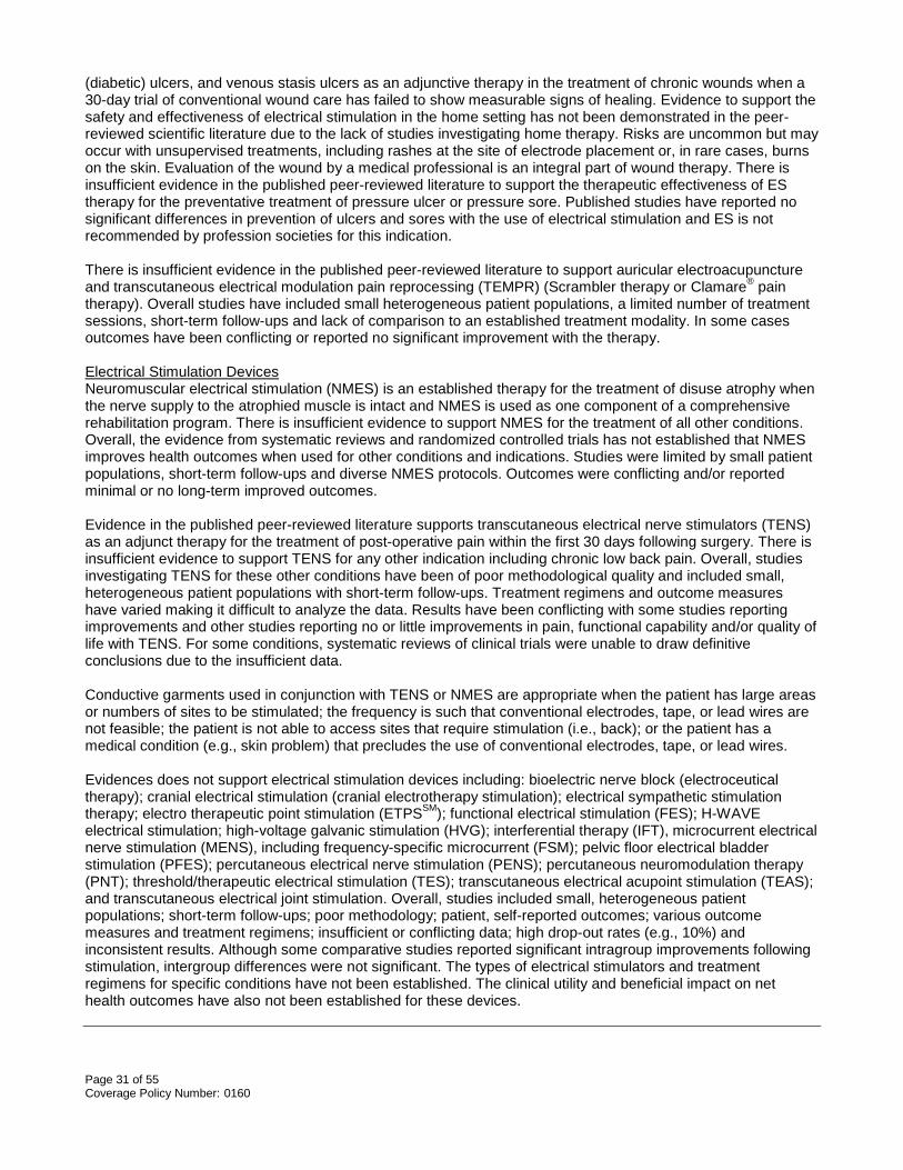
Page 31 of 55 Coverage Policy Number: 0160
(diabetic) ulcers, and venous stasis ulcers as an adjunctive therapy in the treatment of chronic wounds when a 30-day trial of conventional wound care has failed to show measurable signs of healing. Evidence to support the safety and effectiveness of electrical stimulation in the home setting has not been demonstrated in the peer-reviewed scientific literature due to the lack of studies investigating home therapy. Risks are uncommon but may occur with unsupervised treatments, including rashes at the site of electrode placement or, in rare cases, burns on the skin. Evaluation of the wound by a medical professional is an integral part of wound therapy. There is insufficient evidence in the published peer-reviewed literature to support the therapeutic effectiveness of ES therapy for the preventative treatment of pressure ulcer or pressure sore. Published studies have reported no significant differences in prevention of ulcers and sores with the use of electrical stimulation and ES is not recommended by profession societies for this indication. There is insufficient evidence in the published peer-reviewed literature to support auricular electroacupuncture and transcutaneous electrical modulation pain reprocessing (TEMPR) (Scrambler therapy or Clamare® pain therapy). Overall studies have included small heterogeneous patient populations, a limited number of treatment sessions, short-term follow-ups and lack of comparison to an established treatment modality. In some cases outcomes have been conflicting or reported no significant improvement with the therapy. Electrical Stimulation Devices Neuromuscular electrical stimulation (NMES) is an established therapy for the treatment of disuse atrophy when the nerve supply to the atrophied muscle is intact and NMES is used as one component of a comprehensive rehabilitation program. There is insufficient evidence to support NMES for the treatment of all other conditions. Overall, the evidence from systematic reviews and randomized controlled trials has not established that NMES improves health outcomes when used for other conditions and indications. Studies were limited by small patient populations, short-term follow-ups and diverse NMES protocols. Outcomes were conflicting and/or reported minimal or no long-term improved outcomes. Evidence in the published peer-reviewed literature supports transcutaneous electrical nerve stimulators (TENS) as an adjunct therapy for the treatment of post-operative pain within the first 30 days following surgery. There is insufficient evidence to support TENS for any other indication including chronic low back pain. Overall, studies investigating TENS for these other conditions have been of poor methodological quality and included small, heterogeneous patient populations with short-term follow-ups. Treatment regimens and outcome measures have varied making it difficult to analyze the data. Results have been conflicting with some studies reporting improvements and other studies reporting no or little improvements in pain, functional capability and/or quality of life with TENS. For some conditions, systematic reviews of clinical trials were unable to draw definitive conclusions due to the insufficient data. Conductive garments used in conjunction with TENS or NMES are appropriate when the patient has large areas or numbers of sites to be stimulated; the frequency is such that conventional electrodes, tape, or lead wires are not feasible; the patient is not able to access sites that require stimulation (i.e., back); or the patient has a medical condition (e.g., skin problem) that precludes the use of conventional electrodes, tape, or lead wires. Evidences does not support electrical stimulation devices including: bioelectric nerve block (electroceutical therapy); cranial electrical stimulation (cranial electrotherapy stimulation); electrical sympathetic stimulation therapy; electro therapeutic point stimulation (ETPSSM); functional electrical stimulation (FES); H-WAVE electrical stimulation; high-voltage galvanic stimulation (HVG); interferential therapy (IFT), microcurrent electrical nerve stimulation (MENS), including frequency-specific microcurrent (FSM); pelvic floor electrical bladder stimulation (PFES); percutaneous electrical nerve stimulation (PENS); percutaneous neuromodulation therapy (PNT); threshold/therapeutic electrical stimulation (TES); transcutaneous electrical acupoint stimulation (TEAS); and transcutaneous electrical joint stimulation. Overall, studies included small, heterogeneous patient populations; short-term follow-ups; poor methodology; patient, self-reported outcomes; various outcome measures and treatment regimens; insufficient or conflicting data; high drop-out rates (e.g., 10%) and inconsistent results. Although some comparative studies reported significant intragroup improvements following stimulation, intergroup differences were not significant. The types of electrical stimulators and treatment regimens for specific conditions have not been established. The clinical utility and beneficial impact on net health outcomes have also not been established for these devices.

Page 32 of 55 Coverage Policy Number: 0160
Coding/Billing Information Note: 1) This list of codes may not be all-inclusive. 2) Deleted codes and codes which are not effective at the time the service is rendered may not be eligible for reimbursement 3) ICD-10-CM Diagnosis Codes are for informational purposes only and are not effective until 10/01/2015. Electrical Stimulation Therapy Chronic Wound Healing Covered when medically necessary: HCPCS Codes
Description
G0281 Electrical stimulation, (unattended), to one or more areas, for chronic Stage III and Stage IV pressure ulcers, arterial ulcers, diabetic ulcers, and venous stasis ulcers not demonstrating measurable signs of healing after 30 days of conventional care, as part of a therapy plan of care
ICD-9-CM Diagnosis Codes
Description
249.60-249.61
Secondary diabetes mellitus with neurological manifestations
249.70-249.71
Secondary diabetes mellitus with peripheral circulatory disorders
250.60-250.63
Diabetes with neurological manifestations
250.70-250.73
Diabetes with peripheral circulatory disorders
250.80-250.83
Diabetes with other specified manifestations
447.8 Other specified disorders of arteries and arterioles 454.0 Varicose veins of lower extremities with ulcer 454.2 Varicose veins of lower extremities with ulcer and inflammation 459.11 Postphlebitic syndrome with ulcer 459.13 Postphlebitic syndrome with ulcer and inflammation 707.00-707.09
Pressure ulcer
707.10-707.19
Ulcer of lower limbs, except pressure ulcer
707.23 Pressure ulcer stage III 707.24 Pressure ulcer stage IV 707.8 Chronic ulcer of other specified sites 707.9 Chronic ulcer of unspecified site
ICD-10-CM Diagnosis Codes (Effective 10/01/2015)
Description
E08.40-E08.49
Diabetes mellitus due to underlying condition with neurological complications
E08.51-E08.59
Diabetes mellitus due to underlying condition with circulatory complications
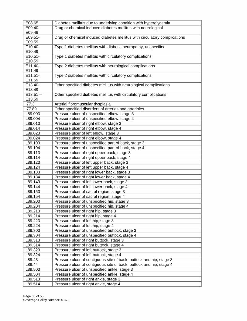
Page 33 of 55 Coverage Policy Number: 0160
E08.65 Diabetes mellitus due to underlying condition with hyperglycemia E09.40-E09.49
Drug or chemical induced diabetes mellitus with neurological
E09.51-E09.59
Drug or chemical induced diabetes mellitus with circulatory complications
E10.40-E10.49
Type 1 diabetes mellitus with diabetic neuropathy, unspecified
E10.51-E10.59
Type 1 diabetes mellitus with circulatory complications
E11.40-E11.49
Type 2 diabetes mellitus with neurological complications
E11.51- E11.59
Type 2 diabetes mellitus with circulatory complications
E13.40-E13.49
Other specified diabetes mellitus with neurological complications
E13.51 – E13.59
Other specified diabetes mellitus with circulatory complications
I77.3 Arterial fibromuscular dysplasia I77.89 Other specified disorders of arteries and arterioles L89.003 Pressure ulcer of unspecified elbow, stage 3 L89.004 Pressure ulcer of unspecified elbow, stage 4 L89.013 Pressure ulcer of right elbow, stage 3 L89.014 Pressure ulcer of right elbow, stage 4 L89.023 Pressure ulcer of left elbow, stage 3 L89.024 Pressure ulcer of right elbow, stage 4 L89.103 Pressure ulcer of unspecified part of back, stage 3 L89.104 Pressure ulcer of unspecified part of back, stage 4 L89.113 Pressure ulcer of right upper back, stage 3 L89.114 Pressure ulcer of right upper back, stage 4 L89.123 Pressure ulcer of left upper back, stage 3 L89.124 Pressure ulcer of left upper back, stage 4 L89.133 Pressure ulcer of right lower back, stage 3 L89.134 Pressure ulcer of right lower back, stage 4 L89.143 Pressure ulcer of left lower back, stage 3 L89.144 Pressure ulcer of left lower back, stage 4 L89.153 Pressure ulcer of sacral region, stage 3 L89,154 Pressure ulcer of sacral region, stage 4 L89.203 Pressure ulcer of unspecified hip, stage 3 L89.204 Pressure ulcer of unspecified hip, stage 4 L89.213 Pressure ulcer of right hip, stage 3 L89.214 Pressure ulcer of right hip, stage 4 L89.223 Pressure ulcer of left hip, stage 3 L89.224 Pressure ulcer of left hip, stage 4 L89.303 Pressure ulcer of unspecified buttock, stage 3 L89.304 Pressure ulcer of unspecified buttock, stage 4 L89.313 Pressure ulcer of right buttock, stage 3 L89.314 Pressure ulcer of right buttock, stage 4 L89.323 Pressure ulcer of left buttock, stage 3 L89.324 Pressure ulcer of left buttock, stage 4 L89.43 Pressure ulcer of contiguous site of back, buttock and hip, stage 3 L89.44 Pressure ulcer of contiguous site of back, buttock and hip, stage 4 L89.503 Pressure ulcer of unspecified ankle, stage 3 L89.504 Pressure ulcer of unspecified ankle, stage 4 L89.513 Pressure ulcer of right ankle, stage 3 L89.514 Pressure ulcer of right ankle, stage 4

Page 34 of 55 Coverage Policy Number: 0160
L89.523 Pressure ulcer of left ankle, stage 3 L89.524 Pressure ulcer of left ankle, stage 4 L89.603 Pressure ulcer of unspecified heel, stage 3 L89.604 Pressure ulcer of unspecified heel, stage 4 L89.613 Pressure ulcer of right heel, stage 3 L89.614 Pressure ulcer of right heel, stage 4 L89.623 Pressure ulcer of left heel, stage 3 L89.624 Pressure ulcer of left heel, stage 4 L89.813 Pressure ulcer of head, stage 3 L89.814 Pressure ulcer of head, stage 4 L89.893 Pressure ulcer of other site, stage 3 L89.894 Pressure ulcer of other site, stage 4 L89.93 Pressure ulcer of unspecified site, stage 3 L89.94 Pressure ulcer of unspecified site, stage 4 L97.101-L97.929
Non-pressure chronic ulcer of lower limb, not elsewhere classified
Experimental/Investigational/Unproven/Not covered indications: ICD-9-CM Diagnosis Codes
Description
All other codes ICD-10-CM Diagnosis Codes (Effective 10/01/2015)
Description
All other codes Experimental/Investigational/Unproven/Not Covered: HCPCS Codes
Description
E0769 Electrical stimulation or electromagnetic wound treatment device, not otherwise classified
G0282 Electrical stimulation, (unattended), to one or more areas, for wound care other than described in G0281
ICD-9-CM Diagnosis Codes
Description
All codes ICD-10-CM Diagnosis Codes (Effective 10/01/2015)
Description
All codes Auricular Electroacupuncture Experimental/Investigational/Unproven/Not Covered: HCPCS Description

Page 35 of 55 Coverage Policy Number: 0160
Codes S8930 Electrical stimulation of auricular acupuncture points; each 15 minutes of
personal one-on-one contact with patient ICD-9-CM Diagnosis Codes
Description
All codes ICD-10-CM Diagnosis Codes (Effective 10/01/2015)
Description
All codes Transcutaneous Electrical Modulation Pain Reprocessing (TEMPR) Experimental/Investigational/Unproven/Not Covered: CPT* Codes Description 0278T Transcutaneous electrical modulation pain reprocessing (e.g., scrambler
therapy) ICD-9-CM Diagnosis Codes
Description
All codes ICD-10-CM Diagnosis Codes (Effective 10/01/2015)
Description
All codes Electrical Stimulation Devices (Electrical Stimulators) Neuromuscular Electrical Stimulation (NMES) Covered when medically necessary: CPT* Codes Description 64565 Percutaneous implantation of neurostimulator electrode array; neuromuscular
HCPCS Codes
Description
E0745 Neuromuscular stimulator, electronic shock unit ICD-9-CM Diagnosis Codes
Description
728.2 Muscular wasting and disuse atrophy, not elsewhere classified ICD-10-CM Diagnosis
Description

Page 36 of 55 Coverage Policy Number: 0160
Codes (Effective 10/01/2015) M62.50-M62.59
Muscular wasting and disuse atrophy, not elsewhere classified
Experimental/Investigational/Unproven/Not Covered: ICD-9-CM Diagnosis Codes
Description
All other codes ICD-10-CM Diagnosis Codes (Effective 10/01/2015)
Description
All other codes Neuromuscular Electrical Stimulation For Scoliosis Experimental/Investigational/Unproven/Not Covered: HCPCS Codes
Description
E0744 Neuromuscular stimulator for scoliosis ICD-9-CM Diagnosis Codes
Description
All codes ICD-10-CM Diagnosis Codes (Effective 10/01/2015)
Description
All codes Transcutaneous Electrical Nerve Stimulator (TENS) Covered when medically necessary: CPT* Codes Description 64550 Application of surface (transcutaneous) neurostimulator
HCPCS Codes
Description
E0720 Tens, two lead, localized stimulation E0730 Transcutaneous electrical nerve stimulation device, four or more leads, for
multiple nerve stimulation ICD-9-CM Diagnosis Codes
Description

Page 37 of 55 Coverage Policy Number: 0160
338.12 Acute post-thoracotomy pain 338.18 Other acute postoperative pain
ICD-10-CM Diagnosis Codes (Effective 10/01/2015)
Description
G89.12 Acute post-thoracotomy pain G89.18 Other acute postprocedural pain
Experimental/Investigational/Not Covered: ICD-9-CM Diagnosis Codes
Description
All other codes ICD-10-CM Diagnosis Codes (Effective 10/01/2015)
Description
All other codes Conductive Garment Covered when medically necessary: HCPCS Codes
Description
E0731 Form fitting conductive garment for delivery of TENS or NMES (with conductive fibers separated from the patient’s skin by layers of fabric)
ICD-9-CM Diagnosis Codes
Description
728.2 Muscular wasting and disuse atrophy, not elsewhere classified 338.12 Acute post-thoracotomy pain 338.18 Other acute postoperative pain
ICD-10-CM Diagnosis Codes (Effective 10/01/2015)
Description
M62.50-M62.59
Muscular wasting and disuse atrophy, not elsewhere classified
G89.12 Acute post-thoracotomy pain G89.18 Other acute postprocedural pain
Experimental/Investigational/Unproven/Not Covered: ICD-9-CM Diagnosis Codes
Description
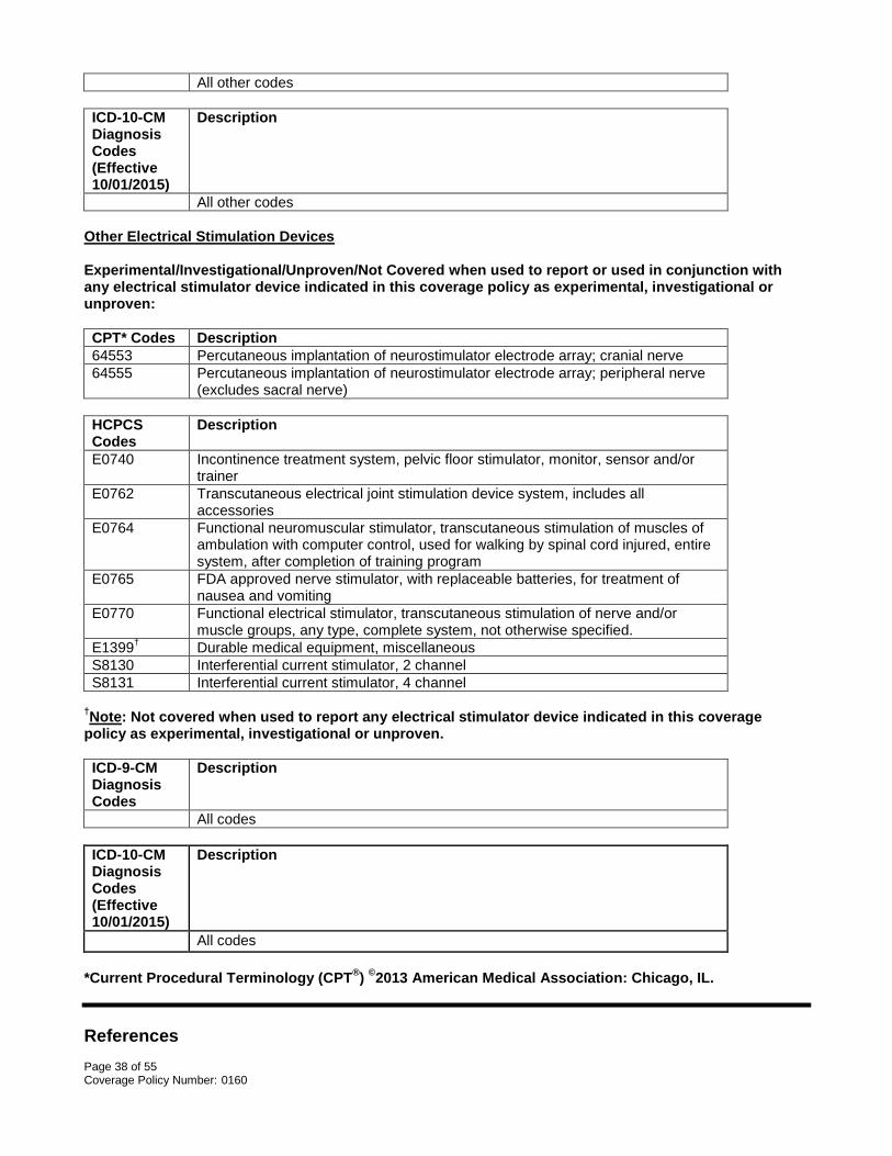
Page 38 of 55 Coverage Policy Number: 0160
All other codes ICD-10-CM Diagnosis Codes (Effective 10/01/2015)
Description
All other codes Other Electrical Stimulation Devices Experimental/Investigational/Unproven/Not Covered when used to report or used in conjunction with any electrical stimulator device indicated in this coverage policy as experimental, investigational or unproven: CPT* Codes Description 64553 Percutaneous implantation of neurostimulator electrode array; cranial nerve 64555 Percutaneous implantation of neurostimulator electrode array; peripheral nerve
(excludes sacral nerve) HCPCS Codes
Description
E0740 Incontinence treatment system, pelvic floor stimulator, monitor, sensor and/or trainer
E0762 Transcutaneous electrical joint stimulation device system, includes all accessories
E0764 Functional neuromuscular stimulator, transcutaneous stimulation of muscles of ambulation with computer control, used for walking by spinal cord injured, entire system, after completion of training program
E0765 FDA approved nerve stimulator, with replaceable batteries, for treatment of nausea and vomiting
E0770 Functional electrical stimulator, transcutaneous stimulation of nerve and/or muscle groups, any type, complete system, not otherwise specified.
E1399† Durable medical equipment, miscellaneous S8130 Interferential current stimulator, 2 channel S8131 Interferential current stimulator, 4 channel
†Note: Not covered when used to report any electrical stimulator device indicated in this coverage policy as experimental, investigational or unproven. ICD-9-CM Diagnosis Codes
Description
All codes ICD-10-CM Diagnosis Codes (Effective 10/01/2015)
Description
All codes *Current Procedural Terminology (CPT®) ©2013 American Medical Association: Chicago, IL. References

Page 39 of 55 Coverage Policy Number: 0160
1. Adunsky A, Ohry A; DDCT Group. Decubitus direct current treatment (DDCT) of pressure ulcers: results
of a randomized double-blinded placebo controlled study. Arch Gerontol Geriatr. 2005. Nov-Dec;41(3):261-9.
2. Agency for Healthcare Research and Quality (AHRQ). Effective health care program. Comparative
effectiveness review: number 90. Pressure Ulcer treatment strategies. May 8, 2013. Accessed Aug 14, 2014. Available at URL address: http://effectivehealthcare.ahrq.gov/search-for-guides-reviews-and-reports/?pageaction=displayproduct&productid=1492
3. Agency for Healthcare Research and Quality (AHRQ). Evidence Report/Technology Assessment
number 187. Treatment of overactive bladder in women. Aug 2009. Accessed Aug 14, 2014. Available at URL address: http://www.ahrq.gov/research/findings/evidence-based-reports/er187-abstract.html
4. Akai M, Hayashi K. Effect of electrical stimulation on musculoskeletal systems: a meta-analysis of
controlled clinical trials. Bioelectromagnetics. 2002;23(2):132-43.
5. Alaven Pharmaceutical. PrimaBella™. 2009. Accessed Aug 14, 2014. Available at URL address: http://www.primabellarx.com/
6. Allen, TK, Habib, AS. P6 stimulation for the prevention of nausea and vomiting associated with
cesarean delivery under neuraxial anesthesia: a systematic review of randomized controlled trials. Anesth Analg. 2008;107(4):1308-1312.
7. Alves-Guerreiro J, Noble J, Lowe A, Walsh D. The effect of three electrotherapeutic modalities upon
peripheral nerve conduction and mechanical pain threshold. Clin Physiol. 2001;21(6):704-11.
8. Ambrosini E, Ferrante S, Pedrocchi A, Ferrigno G, Molteni F. Cycling induced by electrical stimulation improves motor recovery in postacute hemiparetic patients: a randomized controlled trial. Stroke. 2011 Apr;42(4):1068-73.
9. Amer-Cuenca JJ, Goicoechea C, Girona-López A, Andreu-Plaza JL, Palao-Román R, Martínez-Santa
G, Lisón JF. Pain relief by applying transcutaneous electrical nerve stimulation (TENS) during unsedated colonoscopy: a randomized double-blind placebo-controlled trial. Eur J Pain. 2011 Jan;15(1):29-35.
10. American Academy of Neurology. Assessment: efficacy of transcutaneous electric nerve stimulation in
the treatment of pain in neurologic disorders (an evidence-based review). 2010. Accessed Aug 12, 2014. Available at URL address: http://www.aan.com/Guidelines/Home/ByTopic?topicId=22
11. American Academy of Orthopedic Surgeons (AAOS). Clinical practice guideline: Treatment of osteoarthritis of the knee. 2013. Accessed Aug 14, 2014. Available at URL address: http://www.aaos.org/research/guidelines/guide.asp
12. American Association for Chronic Pain. Frequently asked questions. 2014. Accessed Aug 14, 2014.
Available at URL address: http://www.theacpa.org/faqlisting.aspx
13. American College of Foot and Ankle Surgeons (ACFAS). Diabetic foot disorders: a clinical practice guideline. Pathway 3. 2006. Accessed Aug 12, 2014. Available at URL address: http://www.acfas.org/HealthcareCommunity/Content.aspx?id=330
14. American College of Obstetrics and Gynecology. ACOG (American College of Obstetrics and
Gynecology) Practice Bulletin: nausea and vomiting of pregnancy. Obstet Gynecol. 2004 Apr;103(4):803-14. Reaffirmed 2013.
15. American Heart Association’s (AHA)/American Stroke Association (ASA). Comprehensive overview of nursing and interdisciplinary rehabilitation care of the stroke patient. 2010. Accessed Aug 12, 2014.

Page 40 of 55 Coverage Policy Number: 0160
Available at URL address: http://my.americanheart.org/professional/StatementsGuidelines/ByTopic/TopicsQ-Z/Stroke-Statements-Guidelines_UCM_320600_Article.jsp
16. American Society of Anesthesiologists Task Force on Chronic Pain Management; American Society of
Regional Anesthesia and Pain Medicine. Practice guidelines for chronic pain management: an updated report by the American Society of Anesthesiologists Task Force on Chronic Pain Management and the American Society of Regional Anesthesia and Pain Medicine. 2010. Accessed Aug 12, 2014. Available at URL address: http://www.asahq.org/For-Members/Practice-Management/Practice-Parameters.aspx
17. American Society of Plastic Surgeons (ASPS). Chronic wounds of the lower extremity. 2007. Accessed
Aug 14, 2014. Available at URL address: http://www.plasticsurgery.org/For-Medical-Professionals/Legislation-and-Advocacy/Health-Policy-Resources/Evidence-based-GuidelinesPractice-Parameters.html
18. Arena R, Pinkstaff S, Wheeler E, Peberdy MA, Guazzi M, Myers J. Neuromuscular electrical stimulation
and inspiratory muscle training as potential adjunctive rehabilitation options for patients with heart failure. J Cardiopulm Rehabil Prev. 2010 Jul-Aug;30(4):209-23.
19. Association for the Advancement of Wound Care (AAWC). Wound care guidelines. 2010. Accessed Aug
12, 2014. Available at URL address: http://aawconline.org/professional-resources/resources/
20. Atamaz FC, Durmaz B, Baydar M, Demircioglu OY, Iyiyapici A, Kuran B, Oncel S, Sendur OF. Comparison of the efficacy of transcutaneous electrical nerve stimulation, interferential currents, and shortwave diathermy in knee osteoarthritis: a double-blind, randomized, controlled multicenter study. Arch Phys Med Rehabil. 2012 May;93(5):748-56.
21. Barrett CL, Mann GE, Taylor PN, Strike P. A randomized trial to investigate the effects of functional
electrical stimulation and therapeutic exercise on walking performance for people with multiple sclerosis. Mult Scler. 2009 Apr;15(4):493-504.
22. Bax L, Staes F, Verhagen A. Does neuromuscular electrical stimulation strengthen the quadriceps
femoris? A systematic review of randomised controlled trials. Sports Med. 2005;35(3):191-212. 23. Bedwell C, Dowswell T, Neilson JP, Lavender T. The use of transcutaneous electrical nerve stimulation
(TENS) for pain relief in labour: a review of the evidence. Midwifery. 2011 Oct;27(5):e141-8.
24. Bello Y, Phillips T. Recent advances in wound healing. JAMA. 2000 Feb;283:716-8. 25. Berghmans B, Hendriks E, Bernards A, de Bie R, Omar MI. Electrical stimulation with non-implanted
electrodes for urinary incontinence in men. Cochrane Database of Systematic Reviews 2013, Issue 6. Art. No.: CD001202. DOI:10.1002/14651858.CD001202.pub5.
26. Bioness, Inc. NESS L300 Foot Drop System. 2013. Accessed Aug 14, 2014. Available at URL address:
http://www.bioness.com/Healthcare_Professionals/How_L300_Works.php 27. Bjordal JM, Johnson MI, Lopes-Martins RAB, Bogen B, Chow R, Ljunggren AE. Short-term efficacy of
physical interventions in osteoarthritic knee pain. A systematic review and meta- analysis of randomised placebo-controlled trials. BMC Musculoskelet Disord. 2007 Jun 22;8(1):51.
28. Bjordal JM, Johnson MI, Ljunggreen AE. Transcutaneous electrical nerve stimulation (TENS) can
reduce postoperative analgesic consumption. A meta-analysis with assessment of optimal treatment parameters for postoperative pain. Eur J Pain. 2003;7(2):181-8.
29. Blum K, Chen AL, Chen TJ, Waite RL, Downs BW, Braverman ER, Kerner MM, Savarimuthu SM,
DiNubile N. Repetitive H-wave device stimulation and program induces significant increases in the

Page 41 of 55 Coverage Policy Number: 0160
range of motion of post operative rotator cuff reconstruction in a double-blinded randomized placebo controlled human study. BMC Musculoskelet Disord. 2009 Oct 29;10:132.
30. Blum K, Chen AL, Chen TJ, Prihoda TJ, Schoolfield J, DiNubile N, Waite RL, Arcuri V, Kerner M,
Braverman ER, Rhoades P, Tung H. The H-Wave device is an effective and safe non-pharmacological analgesic for chronic pain: a meta-analysis. Adv Ther. 2008 Jul;25(7):644-57.
31. Blum K, Chen TJH, Martinez-Pons M, DiNubile NA, Waite RL, Schoolfield J, et al. The H-Wave small
muscle fiber stimulator, a nonpharmacologic alternative for the treatment of chronic soft- tissue injury and neuropathic pain: an extended population observational study. Adv Ther. 2006 Sep-Oct;23(5):739-49.
32. Brissot R, Gallien P, Le Bot MP, Beaubras A, Laisné D, Beillot J, Dassonville J. Clinical experience with functional electrical stimulation-assisted gait with Parastep in spinal cord-injured patients. Spine (Phila Pa 1976). 2000 Feb 15;25(4):501-8.
33. Brosseau L, Yonge K, Welch V, Marchand S, Judd M, Wells GA, Tugwell P. Transcutaneous electrical
nerve stimulation (TENS) for the treatment of rheumatoid arthritis in the hand. Cochrane Database of Systematic Reviews 2003, Issue 3. Art. No.: CD004377. DOI: 10.1002/14651858.CD004377
34. Brosseau L, Wells GA, Finestone HM, Egan M, Dubouloz CJ, Graham I, Casimiro L, Robinson VA,
Bilodeau M, McGowan J. Clinical practice guidelines for electrical stimulation. Top Stroke Rehabil 2006 Spring;13(2):54-60.
35. Bruce-Brand RA, Walls RJ, Ong JC, Emerson BS, O'Byrne JM, Moyna NM. Effects of home-based
resistance training and neuromuscular electrical stimulation in knee osteoarthritis: a randomized controlled trial. BMC Musculoskelet Disord. 2012 Jul 3;13(1):118.
36. Buchmuller A, Navez M, Milletre-Bernardin M, Pouplin S, Presles E, Lantéri-Minet M, Tardy B, Laurent
B, Camdessanché JP; Lombotens Trial Group. Value of TENS for relief of chronic low back pain with or without radicular pain. Eur J Pain. 2012 May;16(5):656-65.
37. Burns JL, Blackwell SJ. Plastic surgery lower extremity. In: Townsend: Sabiston Textbook of
Surgery.18th ed. Philadelphia, PA: Saunders Elsevier, 2007. Ch. 73. 38. California Technology Assessment Forum (CTAF). Interferential Stimulation for the Treatment of
Musculoskeletal Pain. October 19, 2005. Accessed Aug 13, 2014. Available at URL address: http://ctaf.org/reports?items_per_page=All&=Apply
39. California Technology Assessment Forum (CTAF). Electrical stimulation for the treatment of urinary
incontinence in women. Oct 20, 2004. Aug 13, 2014. Available at URL address: http://ctaf.org/reports 40. Cameron M, Lonergan E, Lee H. Transcutaneous Electrical Nerve Stimulation (TENS) for dementia.
Cochrane Database of Systematic Reviews 2003, Issue 3.
41. Capel ID, Dorrell HM, Spencer EP, Davis MW. The amelioration of the suffering associated with spinal cord injury with subperception transcranial electrical stimulation. Spinal Cord. 2003 Feb;41(2):109-17.
42. Centers for Medicare and Medicaid Services (CMS). Decision memo for electrostimulation for wounds
(CAG-00068R). 2003. Accessed Aug 14, 2014. Available at URL address: http://www.cms.gov/medicare-coverage-database/details/nca-decision-memo.aspx?NCAId=27&NcaName=Electrostimulation+for+Wounds&SearchType=Advanced&CoverageSelection=National&NCSelection=NCA%7cCAL%7cNCD%7cMEDCAC%7cTA%7cMCD&KeyWord=electrical+stimulation&KeyWordLookUp=Doc&KeyWordSearchType=Exact&kq=true&bc=IAAAABAAIAAA&
43. Centers for Medicare & Medicaid Services (CMS). National Coverage Analysis (NCA) for
transcutaneous electrical nerve stimulation for chronic low back pain (CAG-00429N). Jun 8, 2012. Accessed Aug 12, 2014. Available at URL address: http://www.cms.gov/medicare-coverage-
Page 42 of 55 Coverage Policy Number: 0160
database/details/nca-decision-memo.aspx?NCAId=256&NcaName=Transcutaneous+Electrical+Nerve+Stimulation+for+Chronic+Low+Back+Pain&SearchType=Advanced&CoverageSelection=Both&NCSelection=NCA%7cCAL%7cNCD%7cMEDCAC%7cTA%7cMCD&ArticleType=Ed%7cKey%7cSAD%7cFAQ&PolicyType=Final&s=All&KeyWord=NMES&KeyWordLookUp=Doc&KeyWordSearchType=Exact&kq=true&bc=IAAAABAAIAAA&
44. Centers for Medicare and Medicaid Services (CMS). National Coverage Analysis (NCA) for
electrostimulation for wounds (CAG-00068N). Jul 23, 2002. Accessed Aug 12, 2014. Available at URL address: http://www.cms.gov/medicare-coverage-database/details/nca-details.aspx?NCAId=27&NcaName=Electrostimulation+for+Wounds&SearchType=Advanced&CoverageSelection=National&NCSelection=NCA%7cCAL%7cNCD%7cMEDCAC%7cTA%7cMCD&KeyWord=electrical+stimulation&KeyWordLookUp=Doc&KeyWordSearchType=Exact&kq=true&bc=IAAAABAAIAAA&
45. Chao LF, Zhang AL, Liu HE, Cheng MH, Lam HB, Lo SK. The efficacy of acupoint stimulation for the
management of therapy-related adverse events in patients with breast cancer: a systematic review. Breast Cancer Res Treat. 2009 Nov;118(2):255-67.
46. Chou R, Loeser JD, Owens DK, Rosenquist RW, Atlas SJ, Baisden J, Carragee EJ, Grabois M, Murphy
DR, Resnick DK, Stanos SP, Shaffer WO, Wall EM; American Pain Society Low Back Pain Guideline Panel. Interventional therapies, surgery, and interdisciplinary rehabilitation for low back pain: an evidence-based clinical practice guideline from the American Pain Society. Spine (Phila Pa 1976). 2009 May 1;34(10):1066-77.
47. Cipriano G, de Camargo Carvalho AC, Bernardelli GF, Peres PA. Short-term transcutaneous electrical
nerve stimulation after cardiac surgery: effect on pain, pulmonary function and electrical muscle activity. Interact Cardiovasc Thorac Surg. 2008 Apr 16.
48. Coloma M, White PF, Ogunnaike BO, Markowitz SD, Brown PM, Lee AQ, Berrisford SB, Wakefield CA,
Issioui T, Jones SB, Jones DB. Comparison of acustimulation and ondansetron for the treatment of established postoperative nausea and vomiting. Anesthesiology. 2002 Dec;97(6):1387-92.
49. Competitive Technologies. Calmare. 2014. Accessed Aug 12, 2014. Available at URL address:
http://www.calmarett.com/
50. Cooper BC; International College of Cranio-Mandibular Orthopedics (ICCMO). Temporomandibular disorders: A position paper of the International College of Cranio-Mandibular Orthopedics (ICCMO). Cranio. 2011 Jul;29(3):237-44.
51. Coyne PJ, Wan W, Dodson P, Swainey C, Smith TJ. A trial of Scrambler therapy in the treatment of
cancer pain syndromes and chronic chemotherapy-induced peripheral neuropathy. J Pain Palliat Care Pharmacother. 2013 Dec;27(4):359-64.
52. Crowe L, Caulfield B. Aerobic neuromuscular electrical stimulation--an emerging technology to improve haemoglobin A1c in type 2 diabetes mellitus: results of a pilot study. BMJ Open. 2012 Jun 14;2(3). pii: e000219. doi: 10.1136/bmjopen-2011-000219. Print 2012.
53. Dali C, Hansen F, Pedersen S, Skov L, Hilden J, Bjornskov I, et al. Threshold electrical stimulation
(TES) in ambulant children with CP: a randomized double-blind placebo-controlled clinical trial. Dev Med Child Neurol. 2002 Jun;44(6):364-9.
54. De Gennaro M, Capitanucci ML, Mosiello G, Zaccara A. Current state of nerve stimulation technique for
lower urinary tract dysfunction in children. J Urol. 2011 May;185(5):1571-7.
55. Demirtürk F, Akbayrak T, Karakaya IC, Yüksel I, Kirdi N, Demirtürk F, Kaya S, Ergen A, Beksac S. Interferential current versus biofeedback results in urinary stress incontinence. Swiss Med Wkly. 2008 May 31;138(21-22):317-21.

Page 43 of 55 Coverage Policy Number: 0160
56. de Oliveira Melo M, Aragão FA, Vaz MA. Neuromuscular electrical stimulation for muscle strengthening
in elderly with knee osteoarthritis - a systematic review. Complement Ther Clin Pract. 2013 Feb;19(1):27-31.
57. Department of Veterans Affairs Health Services Research and Development Services. A systematic
evidence review of non-pharmacological interventions for behavioral symptoms of dementia. March 2011. Accessed Aug 12, 2014. Available at URL address: www.hsrd.research.va.gov/publications/esp/Dementia-Nonpharm.pdf
58. Department of Veterans Affairs Health Services Research and Development Services. Management of
stroke rehabilitation. 2010. Accessed Aug 12, 2014.. Available at URL address: http://www.healthquality.va.gov/Management_of_Stroke_Rehabilitation.asp
59. Dowswell T, Bedwell C, Lavender T, Neilson JP. Transcutaneous electrical nerve stimulation (TENS) for
pain relief in labour. Cochrane Database of Systematic Reviews 2009, Issue 2. Art. No.: CD007214. DOI: 10.1002/14651858.CD007214.pub2.
60. Dune LS, Shiao SY. Metaanalysis of acustimulation effects on postoperative nausea and vomiting in
children. Explore (NY). 2006 Jul-Aug;2(4):314-20.
61. ECRI Institute. Hotline Response [database online]. Plymouth Meeting (PA): ECRI Institute; Acustimulation wristbands for nausea and vomiting. Jun 17, 2014. Accessed Aug 14, 2014. Available at URL address: http://www.ecri.org.
62. ECRI Institute. Hotline Response [database online]. Plymouth Meeting (PA): ECRI Institute; Cefaly Supraorbital Transcutaneous Neurostimulator (Cefaly-Technology) for Preventing and Treating Migraine Headache. Apr 4, 2014. Accessed Aug 14, 2013. Available at URL address: http://www.ecri.org.
63. ECRI Institute. Technology assessment [database online]. Plymouth Meeting (PA): ECRI Institute; H-
wave device stimulation therapy for pain management. June 2, 2009. Accessed Aug 13, 2014. Available at URL address: http://www.ecri.org.
64. Electrotherapy Association. Auriculotherapy. 2014. Accessed Aug 12, 2014. Available at URL address:
http://www.electrotherapy.com/
65. Elsner B, Kugler J, Pohl M, Mehrholz J. Transcranial direct current stimulation (tDCS) for improving aphasia in patients after stroke. Cochrane Database of Systematic Reviews 2013b, Issue 6. Art.No.: CD009760. DOI: 10.1002/14651858.CD009760.pub2.
66. Elsner B, Kugler J, Pohl M, Mehrholz J. Transcranial direct current stimulation (tDCS) for improving
function and activities of daily living in patients after stroke. Cochrane Database of Systematic Reviews 2013b, Issue 11. Art. No.: CD009645. DOI:10.1002/14651858.CD009645.pub2.
67. Embrey DG, Holtz SL, Alon G, Brandsma BA, McCoy SW. Functional electrical stimulation to
dorsiflexors and plantar flexors during gait to improve walking in adults with chronic hemiplegia. Arch Phys Med Rehabil. 2010 May;91(5):687-96.
68. Emmiler M, Solak O, Kocogullari C, Dundar U, Ayva E, Ela Y, Cekirdekci A, Kavuncu V. Control of acute
postoperative pain by transcutaneous electrical nerve stimulation after open cardiac operations: a randomized placebo-controlled prospective study. Heart Surg Forum. 2008;11(5):E300-3.
69. Escortell-Mayor E, Riesgo-Fuertes R, Garrido-Elustondo S, Asúnsolo-Del Barco A, Díaz-Pulido B,
Blanco-Díaz M, Bejerano-Álvarez E; TEMA-TENS Group. Primary care randomized clinical trial: manual therapy effectiveness in comparison with TENS in patients with neck pain. Man Ther. 2011 Feb;16(1):66-73.

Page 44 of 55 Coverage Policy Number: 0160
70. Esnouf JE, Taylor PN, Mann GE, Barrett CL. Impact on activities of daily living using a functional electrical stimulation device to improve dropped foot in people with multiple sclerosis, measured by the Canadian Occupational Performance Measure. Mult Scler. 2010 Sep;16(9):1141-7.
71. Everaert DG, Thompson AK, Chong SL, Stein RB. Does functional electrical stimulation for foot drop
strengthen corticospinal connections? Neurorehabil Neural Repair. 2010 Feb;24(2):168-77.
72. Eyigor C, Eyigor S, Kivilcim Korkmaz O. Are intra-articular corticosteroid injections better than conventional TENS in treatment of rotator cuff tendinitis in the short run? A randomized study. Eur J Phys Rehabil Med. 2010 Sep;46(3):315-24.
73. Ezzo, JM, Richardson, MA, Vickers, A, Allen, C, Dibble, SL, Issell, BF, Lao, L, Pearl, M, Ramirez, G,
Roscoe, J, Shen, J, Shivnan, JC, Streitberger, K, Treish, I, and Zhang, G. Acupuncture-point stimulation for chemotherapy-induced nausea or vomiting. Cochrane Database Syst Rev. 2006;(2):CD002285.
74. Facci LM, Nowotny JP, Tormem F, Trevisani VF. Effects of transcutaneous electrical nerve stimulation
(TENS) and interferential currents (IFC) in patients with nonspecific chronic low back pain: randomized clinical trial. Sao Paulo Med J. 2011;129(4):206-16.
75. Farr J, Mont MA, Garland D, Caldwell JR, Zizic TM. Pulsed electrical stimulation in patients with
osteoarthritis of the knee: follow up in 288 patients who had failed non-operative therapy. Surg Technol Int. 2006;15:227-33.
76. Fary RE, Carroll GJ, Briffa TG, Briffa NK. The effectiveness of pulsed electrical stimulation in the
management of osteoarthritis of the knee: results of a double-blind, randomized, placebo-controlled, repeated-measures trial. Arthritis Rheum. 2011 May;63(5):1333-42. doi: 10.1002/art.30258.
77. Ferrante S, Pedrocchi A, Ferrigno G, Molteni F. Cycling induced by functional electrical stimulation
improves the muscular strength and the motor control of individuals with post-acute stroke. Europa Medicophysica-SIMFER 2007 Award Winner. Eur J Phys Rehabil Med. 2008 Jun;44(2):159-67.
78. Field-Fote EC, Roach KE. Influence of a locomotor training approach on walking speed and distance in
people with chronic spinal cord injury: a randomized clinical trial. Phys Ther. 2011 Jan;91(1):48-60.
79. Field-Fote EC, Lindley SD, Sherman AL. Locomotor training approaches for individuals with spinal cord injury: a preliminary report of walking-related outcomes. J Neurol Phys Ther. 2005 Sep;29(3):127-37.
80. Frequency specific microcurrent. 2014. Accessed Aug 13, 2014. Available at URL address:
http://www.frequencyspecific.com/
81. Frey UH, Funk M, Löhlein C, Peters J. Effect of P6 acustimulation on post-operative nausea and vomiting in patients undergoing a laparoscopic cholecystectomy. Acta Anaesthesiol Scand. 2009 Nov;53(10):1341-7.
82. Frey UH, Scharmann P, Löhlein C, Peters J. P6 acustimulation effectively decreases postoperative
nausea and vomiting in high-risk patients. Br J Anaesth. 2009 May;102(5):620-5.
83. Freynet A, Falcoz PE. Is transcutaneous electrical nerve stimulation effective in relieving postoperative pain after thoracotomy? Interact Cardiovasc Thorac Surg. 2010 Feb;10(2):283-8.
84. Fuentes JP, Armijo Olivo S, Magee DJ, Gross DP. Effectiveness of interferential current therapy in the
management of musculoskeletal pain: a systematic review and meta-analysis. Phys Ther. 2010 Sep;90(9):1219-38.
85. Gallien P, Brissot R, Eyssette M, Tell L, Barat M, Wiart L, Petit H. Restoration of gait by functional
electrical stimulation for spinal cord injured patients. Paraplegia. 1995 Nov;33(11):660-4.

Page 45 of 55 Coverage Policy Number: 0160
86. Gan TJ, Jiao KR, Zenn M, Georgiade G. A randomized controlled comparison of electro-acupoint stimulation or ondansetron versus placebo for the prevention of postoperative nausea and vomiting. Anesth Analg. 2004 Oct;99(4):1070-5, table of contents.
87. Garland D, Holt P, Harrington JT, Caldwell J, Zizic T, Cholewczynski J. A 3-month, randomized, double-
blind, placebo-controlled study to evaluate the safety and efficacy of a highly optimized, capacitively coupled, pulsed electrical stimulator in patients with osteoarthritis of the knee. Osteoarthritis Cartilage. 2007 Jun;15(6):630-7.
88. Giangregorio L, Craven C, Richards K, Kapadia N, Hitzig SL, Masani K, Popovic MR. A randomized trial
of functional electrical stimulation for walking in incomplete spinal cord injury: effects on body composition. J Spinal Cord Med. 2012 Sep;35(5):351-60.
89. Giggins OM, Fullen BM, Coughlan GF. Neuromuscular electrical stimulation in the treatment of knee
osteoarthritis: a systematic review and meta-analysis. Clin Rehabil. 2012 Feb 9.
90. Goode PS, Burgio KL, Johnson TM 2nd, Clay OJ, Roth DL, Markland AD, Burkhardt JH, Issa MM, Lloyd LK. Behavioral therapy with or without biofeedback and pelvic floor electrical stimulation for persistent postprostatectomy incontinence: a randomized controlled trial. JAMA. 2011 Jan 12;305(2):151-9.
91. Goode P, Burgio K, Lovcher J, Roth D, Umlauf M, Richter H. et al., Varner R, Lloyd L. Effect of
behavioral training with or without pelvic floor electrical stimulation on stress incontinence in women: a randomized controlled trial. JAMA. 2003 Jul;290(3):395-7.
92. Graupe D, Kohn KH. Functional neuromuscular stimulator for short-distance ambulation by certain
thoracic-level spinal-cord-injured paraplegics. Surg Neurol. 1998 Sep;50(3):202-7.
93. Greif R, Laciny S, Mokhtarani M, Doufas AG, Bakhshandeh M, Dorfer L, Sessler DI. Transcutaneous electrical stimulation of an auricular acupuncture point decreases anesthetic requirement. Anesthesiology. 2002 Feb;96(2):306-12.
94. Guido EH. Effects of sympathetic therapy on chronic pain in peripheral neuropathy subjects. Am J Pain
Mgmt. 2002;12:31-34.
95. Gundog M, Atamaz F, Kanyilmaz S, Kirazli Y, Celepoglu G. Interferential current therapy in patients with knee osteoarthritis: comparison of the effectiveness of different amplitude-modulated frequencies. Am J Phys Med Rehabil. 2012 Feb;91(2):107-13.
96. Habib AS, Itchon-Ramos N, Phillips-Bute BG, Gan TJ; Duke Women's Anesthesia (DWA) Research
Group. Transcutaneous acupoint electrical stimulation with the ReliefBand for the prevention of nausea and vomiting during and after cesarean delivery under spinal anesthesia. Anesth Analg. 2006 Feb;102(2):581-4.
97. Hagstroem S, Mahler B, Madsen B, Djurhuus JC, Rittig S. Transcutaneous electrical nerve stimulation
for refractory daytime urinary urge incontinence. J Urol. 2009 Oct;182(4 Suppl):2072-8.
98. Hamza MA, White PF, Craig WF, Ghoname ES, Ahmed HE, Proctor TJ, Noe CE, Vakharia AS, Gajraj N. Percutaneous electrical nerve stimulation: a novel analgesic therapy for diabetic neuropathic pain. Diabetes Care. 2000 Mar;23(3):365-70.
99. Hausdorff JM, Ring H. Effects of a new radio frequency-controlled neuroprosthesis on gait symmetry
and rhythmicity in patients with chronic hemiparesis. Am J Phys Med Rehabil. 2008 Jan;87(1):4-13.
100. Hein E1, Nowak M, Kiess O, Biermann T, Bayerlein K, Kornhuber J, Kraus T. Auricular transcutaneous electrical nerve stimulation in depressed patients: a randomized controlled pilot study. J Neural Transm. 2013 May;120(5):821-7.
Page 46 of 55 Coverage Policy Number: 0160
101. Helmreich, RJ, Shiao, SY, and Dune, LS. Meta-analysis of acustimulation effects on nausea and vomiting in pregnant women. Explore (NY). 2006;2(5):412-421.
102. Hochberg MC, Altman RD, April KT, Benkhalti M, Guyatt G, McGowan J, Towheed T, Welch V, Wells G,
Tugwell P; American College of Rheumatology. American College of Rheumatology 2012 recommendations for the use of nonpharmacologic and pharmacologic therapies in osteoarthritis of the hand, hip, and knee. Arthritis Care Res (Hoboken). 2012 Apr;64(4):465-74.
103. Hocking B. ETPS neuropathic acupuncture. Ch 54. In: Weiner RS (editor). Pain management: a
practical guide for clinicians, sixth edition. CRC Press: Washington D.C. 2002:Pgs 667-690.
104. Holzer A, Leitgeb U, Spacek A, Wenzl R, Herkner H, Kettner S. Auricular acupuncture for postoperative pain after gynecological surgery: a randomized controlled trail. Minerva Anestesiol. 2011 Mar;77(3):298-304.
105. Hosker G, Cody JD, Norton CC. Electrical stimulation for faecal incontinence in adults. Cochrane
Database of Systematic Reviews 2007, Issue 3.
106. Hou C, Tsai L, Cheng K. Immediate effects of various physical therapeutic modalities on cervical myofascial pain and trigger-point sensitivity. Arch Phys Med Rehabil. 2002;83(10):1406-14.
107. Houghton P, Kincaid C, Lovell M, Campbell K, Keast D, Woodbury M, et al. Effect of electrical
stimulation on chronic leg ulcer size and appearance. Phys Ther. 2003;83(1):17-28.
108. Houghton PE, Campbell KE, Fraser CH, Harris C, Keast DH, Potter PJ, Hayes KC, Woodbury MG. Electrical stimulation therapy increases rate of healing of pressure ulcers in community-dwelling people with spinal cord injury. Arch Phys Med Rehabil. 2010 May;91(5):669-78.
109. Hsu SS, Hu MH, Wang YH, Yip PK, Chiu JW, Hsieh CL. Dose-response relation between
neuromuscular electrical stimulation and upper-extremity function in patients with stroke. Stroke. 2010 Apr;41(4):821-4.
110. Hui-Chan CW, Ng SS, Mak MK. Effectiveness of a home-based rehabilitation programme on lower limb
functions after stroke. Hong Kong Med J. 2009 Jun;15(3 Suppl 4):42-6.
111. Hungerford DS1, Maclaughlin EJ, Mines CM, Deveshwar S, Elliott C, Tuber JS, Principe JR, Ford TL, Schechtman J, Zizic TM. Synergistic effect of using a transcutaneous electrical joint stimulator and an unloading brace in treating osteoarthritis of the knee. Am J Orthop (Belle Mead NJ). 2013 Oct;42(10):456-63.
112. Hunter KF,Moore KN, Glazener CMA. Conservative management for postprostatectomy urinary
incontinence. Cochrane Database of Systematic Reviews 2007, Issue 2.
113. Hurley D, Minder P, McDonough S, Walsh D, Moore A, Baxter D. Interferential therapy electrode placement technique in acute low back pain: a preliminary investigation. Arch Phys Med Rehabil. 2001;82(4):485-93.
114. Hurlow A, Bennett MI, Robb KA, Johnson MI, Simpson KH, Oxberry SG. Transcutaneous electric nerve
stimulation (TENS) for cancer pain in adults. Cochrane Database of Systematic Reviews 2012, Issue 3. Art. No.: CD006276. DOI: 10.1002/14651858.CD006276.pub3
115. Imamura M, Abrams P, Bain C, Buckley B, Cardozo L, Cody J, Cook J, Eustice S, Glazener C, Grant A,
Hay-Smith J, Hislop J, Jenkinson D, Kilonzo M, Nabi G, N'Dow J, Pickard R, Ternent L, Wallace S, Wardle J, Zhu S, Vale L. Systematic review and economic modelling of the effectiveness and cost-effectiveness of non-surgical treatments for women with stress urinary incontinence. Health Technol Assess. 2010 Aug;14(40):1-188, iii-iv.

Page 47 of 55 Coverage Policy Number: 0160
116. Janković A, Binić I. Frequency rhythmic electrical modulation system in the treatment of chronic painful leg ulcers. Arch Dermatol Res. 2008 Aug;300(7):377-83. Epub 2008 Jul 16.
117. Jarit G, Mohr K, Waller R. The effects of home interferential therapy on post-operative pain, edema and
range of motion of the knee. Clin J Sport Med. 2003;13(1):16-20.
118. Jerez-Roig J1, Souza DL, Espelt A, Costa-Marín M, Belda-Molina AM. Pelvic floor electrostimulation in women with urinary incontinence and/or overactive bladder syndrome: a systematic review. Actas Urol Esp. 2013 Jul-Aug;37(7):429-44.
119. Jin DM, Xu Y, Geng DF, Yan TB. Effect of transcutaneous electrical nerve stimulation on symptomatic
diabetic peripheral neuropathy: a meta-analysis of randomized controlled trials. Diabetes Res Clin Pract. 2010 Jul;89(1):10-5.
120. Johnson MI, Bjordal JM. Transcutaneous electrical nerve stimulation for the management of painful
conditions: focus on neuropathic pain. Expert Rev Neurother. 2011 May;11(5):735-53.
121. Johnson M, Martinson M. Efficacy of electrical nerve stimulation for chronic musculoskeletal pain: a meta-analysis of randomized controlled trials. Pain. 2007 Jul;130(1-2):157-65.
122. Jünger M, Arnold A, Zuder D, Stahl HW, Heising S. Local therapy and treatment costs of chronic,
venous leg ulcers with electrical stimulation (Dermapulse): a prospective, placebo controlled, double blind trial. Wound Repair Regen. 2008 Jul-Aug;16(4):480-7.
123. Kajbafzadeh AM, Sharifi-Rad L, Baradaran N, Nejat F. Effect of pelvic floor interferential
electrostimulation on urodynamic parameters and incontinency of children with myelomeningocele and detrusor overactivity. Urology. 2009 Aug;74(2):324-9.
124. Kang RW, Lewis PB, Kramer A, Hayden JK, Cole BJ. Prospective randomized single-blinded controlled
clinical trial of percutaneous neuromodulation pain therapy device versus sham for the osteoarthritic knee: a pilot study. Orthopedics. 2007 Jun;30(6):439-45.
125. Karavidas A, Parissis J, Arapi S, Farmakis D, Korres D, Nikolaou M, Fotiadis J, Potamitis N, Driva X,
Paraskevaidis I, Matsakas E, Filippatos G, Kremastinos DT. Effects of functional electrical stimulation on quality of life and emotional stress in patients with chronic heart failure secondary to ischaemic or idiopathic dilated cardiomyopathy: a randomised, placebo-controlled trial. Eur J Heart Fail. 2008 Jul;10(7):709-13.
126. Kerr C, McDowell B, Cosgrove A, Walsh D, Bradbury I, McDonough. Electrical stimulation in cerebral
palsy: a randomized controlled trial. Dev Med Child Neurol. 2006 Nov;48(11):870-6.
127. Khadilkar A, Milne S, Brosseau L, Robinson V, Saginur M, Shea B, Tugwell P, Wells G. Transcutaneous electrical nerve stimulation (TENS) for chronic low-back pain. Cochrane Database of Systematic Reviews 2005, Issue 3.
128. Khadilkar A, Milne S, Brosseau L, Wells G, Tugwell P, Robinson V, et al. Transcutaneous electrical
nerve stimulation for the treatment of chronic low back pain: a systematic review. Spine. 2005 Dec 1;30(23):2657-66.
129. Khadilkar A, Odebiyi DO, Brosseau L, Wells GA. Transcutaneous electrical nerve stimulation (TENS)
versus placebo for chronic low-back pain. Cochrane Database of Systematic Reviews 2008, Issue 4. Art. No.: CD003008. DOI: 10.1002/14651858.CD003008.pub3.
130. Kim KM, Croy T, Hertel J, Saliba S. Effects of neuromuscular electrical stimulation after anterior cruciate
ligament reconstruction on quadriceps strength, function, and patient-oriented outcomes: a systematic review. J Orthop Sports Phys Ther. 2010 Jul;40(7):383-91.

Page 48 of 55 Coverage Policy Number: 0160
131. Kim CM, Eng JJ, Whittaker MW. Effects of a simple functional electric system and/or a hinged ankle-foot orthosis on walking in persons with incomplete spinal cord injury. Arch Phys Med Rehabil. 2004 Oct;85(10):1718-23.
132. Kiratli BJ (author). Therapeutic Interventions. In: Lin VW, Cardenas DD, Cutter NC, Frost FS, Hammond
MC, Lindblom LB, et al. (editors). Spinal Cord Medicine: Principles and Practices. Demos Medical Publishing, Inc. New York; 2003.
133. Kovacs FM, Urrútia G, Alarcón JD. Surgery versus conservative treatment for symptomatic lumbar
spinal stenosis: a systematic review of randomized controlled trials. Spine (Phila Pa 1976). 2011 Sep 15;36(20):E1335-51.
134. Koyuncu E, Nakipoğlu-Yüzer GF, Doğan A, Ozgirgin N. The effectiveness of functional electrical
stimulation for the treatment of shoulder subluxation and shoulder pain in hemiplegic patients: A randomized controlled trial. Disabil Rehabil. 2010;32(7):560-6.
135. Kroeling P, Gross A, Goldsmith CH, Burnie SJ, Haines T, Graham N, Brant A. Electrotherapy for neck
pain. Cochrane Database of Systematic Reviews 2009, Issue 4. Art. No.: CD004251. DOI: 10.1002/14651858.CD004251.pub4.
136. La Marca R, Nedeljkovic M, Yuan L, Maercker A, Ehlert U. Effects of auricular electrical stimulation on
vagal activity in healthy men: Evidence from a three-armed randomized trial. Clin Sci (Lond). 2009 Nov 9.
137. Lan F, Ma YH, Xue JX, Wang TL, Ma DQ. Transcutaneous electrical nerve stimulation on acupoints
reduces fentanyl requirement for postoperative pain relief after total hip arthroplasty in elderly patients. Minerva Anestesiol. 2012 Apr 24.
138. Larson JD, Gutowski KA, Marcus BC, Rao VK, Avery PG, Stacey DH, Yang RZ. Larson JD, Gutowski
KA, Marcus BC, Rao VK, Avery PG, Stacey DH, Yang RZ. The effect of electroacustimulation on postoperative nausea, vomiting, and pain in outpatient plastic surgery patients: a prospective, randomized, blinded, clinical trial. Plast Reconstr Surg. 2010 Mar;125(3):989-94.
139. Laufer Y, Ring H, Sprecher E, Hausdorff JM. Gait in individuals with chronic hemiparesis: one-year
follow-up of the effects of a neuroprosthesis that ameliorates foot drop. J Neurol Phys Ther. 2009 Jun;33(2):104-10.
140. Lee A, Fan LTY. Stimulation of the wrist acupuncture point P6 for preventing postoperative nausea and
vomiting. Cochrane Database of Systematic Reviews 2009, Issue 2. Art. No.: CD003281. DOI: 10.1002/14651858.CD003281.pub3
141. Lichtbroun AS, Raicer MM, Smith RB. The treatment of fibromyalgia with cranial electrotherapy
stimulation. J Clin Rheumatol. 2001 Apr;7(2):72-8; discussion 78.
142. Lieber RL, Silva PD, Daniel DM. Equal effectiveness of electrical and volitional strength training for quadriceps femoris muscles after anterior cruciate ligament surgery. J Orthop Res. 1996 Jan;14(1):131-8.
143. Lyon DE, Schubert C, Taylor AG. Pilot study of cranial stimulation for symptom management in breast
cancer. Oncol Nurs Forum. 2010 Jul;37(4):476-83.
144. Machado LA, Kamper SJ, Herbert RD, Maher CG, McAuley JH. Analgesic effects of treatments for non-specific low back pain: a meta-analysis of placebo-controlled randomized trials. Rheumatology (Oxford) 2009; 48: 520-527.

Page 49 of 55 Coverage Policy Number: 0160
145. Maddocks M, Gao W, Higginson IJ, Wilcock A. Neuromuscular electrical stimulation for muscle weakness in adults with advanced disease. Cochrane Database of Systematic Reviews 2013, Issue 1. Art. No.: CD009419. DOI: 10.1002/14651858.CD009419.pub2.
146. Magis D1, Sava S, d'Elia TS, Baschi R, Schoenen J. Safety and patients' satisfaction of transcutaneous
supraorbital neurostimulation (tSNS) with the Cefaly® device in headache treatment: a survey of 2,313 headache sufferers in the general population. J Headache Pain. 2013 Dec 1;14:95.
147. Mangold S, Schuster C, Keller T, Zimmermann-Schlatter A, Ettlin T. Motor training of upper extremity
with functional electrical stimulation in early stroke rehabilitation. Neurorehabil Neural Repair. 2009 Feb;23(2):184-90.
148. Marineo G. Untreatable pain resulting from abdominal cancer: new hope from biophysics? JOP. 2003
Jan;4(1):1-10.
149. Marineo G, Iorno V, Gandini C, Moschini V, Smith TJ. Scrambler therapy may relieve chronic neuropathic pain more effectively than guideline-based drug management: results of a pilot, randomized, controlled trial. J Pain Symptom Manage. 2012 Jan;43(1):87-95. doi: 10.1016/j.jpainsymman.2011.03.015.
150. Matthews A, Haas DM, O’Mathúna DP, Dowswell T, Doyle M. Interventions for nausea and vomiting in
early pregnancy. Cochrane Database of Systematic Reviews 2014, Issue 3. Art. No.: CD007575. DOI: 10.1002/14651858.CD007575.pub3.
151. MedDex Solutions. ETPS. 2014. Accessed Aug 14, 2014. Available at URL address: http://meddexsolutions.com/etps.html
152. Medi-Stem, Inc. Frequently used modalities. 2014. Accessed Aug 13, 2014. Available at URL address:
http://www.medi-stim.com/overview.html
153. Mehrholz J, Werner C, Kugler J, Pohl M. Electromechanical-assisted training for walking after stroke. Cochrane Database of Systematic Reviews 2007, Issue 4. Art. No.: CD006185. DOI: 10.1002/14651858.CD006185.pub2. Assessed as up-to-date 2009.
154. Meijer JW, Voerman GE, Santegoets KM, Geurts AC. Short-term effects and long-term use of a hybrid
orthosis for neuromuscular electrical stimulation of the upper extremity in patients after chronic stroke. J Rehabil Med. 2009 Feb;41(3):157-61.
155. Mello LF, Nóbrega LF, Lemos A. Transcutaneous electrical stimulation for pain relief during labor: a
systematic review and meta-analysis. Rev Bras Fisioter. 2011 May-Jun;15(3):175-84.
156. Minder P, Noble J, Alves-Guerreiro J. Interferential therapy: lack of effect upon experimentally induced delayed onset muscle soreness. Clin Physiol Funct Imaging. 2002;22(5):339-47.
157. Monaghan B, Caulfield B, O’Mathúna DP. Surface neuromuscular electrical stimulation for quadriceps
strengthening pre and post total knee replacement. Cochrane Database of Systematic Reviews 2010, Issue 1. Art. No.: CD007177. DOI:10.1002/14651858.CD007177.pub2.
158. Monga AK, Tracey MR, Subbaroyan J. A systematic review of clinical studies of electrical stimulation for
treatment of lower urinary tract dysfunction. Int Urogynecol J. 2012 Aug;23(8):993-1005.
159. Mont MA, Hungerford DS, Caldwell JR, Hoffman KC, He YD, Jones LC, Zizic TM. Pulsed electrical stimulation to defer TKA in patients with Knee Osteoarthritis. Orthopedics. 2006;29:887.
160. Mulvey MR, Bagnall AM, Johnson MI, Marchant PR. Transcutaneous electrical nerve stimulation
(TENS) for phantom pain and stump pain following amputation in adults. Cochrane Database of Systematic Reviews 2010, Issue 5. Art. No.: CD007264. DOI: 10.1002/14651858.CD007264.pub2.
Page 50 of 55 Coverage Policy Number: 0160
161. Murina F, Bianco V, Radici G, Felice R, Di Martino M, Nicolini U. Transcutaneous electrical nerve
stimulation to treat vestibulodynia: a randomised controlled trial. BJOG. 2008 Aug;115(9):1165-70.
162. Nakagawa H, Kaiho Y, Namiki S, Ishidoya S, Saito S, Arai Y. Impact of sacral surface therapeutic electrical stimulation on early recovery of urinary continence after radical retropubic prostatectomy: a pilot study. Adv Urol. 2010:102751.
163. National Institute for Health and Clinical Excellence (NICE). Functional electrical stimulation for drop foot
of central neurological origin. Jan 2009. Accessed Aug 13, 2014. Available at URL address: http://guidance.nice.org.uk/IPG278
164. National Institute for Health and Clinical Excellence (NICE). Stroke rehabilitation CG 162. Jun 2013.
Accessed Aug 13, 2014. Available at URL address: http://guidance.nice.org.uk/CG162/NICEGuidance/pdf/English
165. National Institute for Health and Clinical Excellence (NICE). CG 40. Urinary incontinence: the
management of urinary incontinence in women. Oct 2006. Accessed Aug 13, 2014. Available at URL address: http://www.nice.org.uk/CG40
166. National Institute of Arthritis and Musculoskeletal and Skin Disease. Questions and answers about
scoliosis in children and adolescents. 2012. Accessed Aug 14, 2014. Available at URL address: http://www.niams.nih.gov/Health_Info/Scoliosis/default.asp
167. National Pressure Ulcer Advisory Panel (NPUAP). Pressure Ulcer Staging. Feb 2007. Accessed Aug
12, 2014. Available at URL address: http://www.npuap.org/pr2.htm
168. National Pressure Ulcer Advisory Panel (NPUAP) and the European Pressure Ulcer Advisory Panel (EPUAP). International pressure ulcer guidelines. 2009. Accessed Aug 12, 2014. Available at URL address: http://www.npuap.org/resources/educational-and-clinical-resources/international-pressure-ulcer-guidelines/
169. Negm A1, Lorbergs A, Macintyre NJ. Efficacy of low frequency pulsed subsensory threshold electrical
stimulation vs placebo on pain and physical function in people with knee osteoarthritis: systematic review with meta-analysis. Osteoarthritis Cartilage. 2013 Sep;21(9):1281-9.
170. Ng SS, Hui-Chan CW. Does the use of TENS increase the effectiveness of exercise for improving
walking after stroke? A randomized controlled clinical trial. Clin Rehabil. 2009 Dec;23(12):1093-103.
171. Ng MF, Tong RK, Li LS. A pilot study of randomized clinical controlled trial of gait training in subacute stroke patients with partial body-weight support electromechanical gait trainer and functional electrical stimulation: six-month follow-up. Stroke. 2008 Jan;39(1):154-60.
172. Ngai SP, Jones AY, Hui-Chan CW, Ko FW, Hui DS. Effect of Acu-TENS on post-exercise expiratory
lung volume in subjects with asthma-A randomized controlled trial. Respir Physiol Neurobiol. 2009 Jul 31;167(3):348-53.
173. Nightingale EJ, Raymond J, Middleton JW, Crosbie J, Davis GM. Benefits of FES gait in a spinal cord
injured population. Spinal Cord. 2007 Oct;45(10):646-57.
174. Nnoaham KE, Kumbang J. Transcutaneous electrical nerve stimulation (TENS) for chronic pain. Cochrane Database of Systematic Reviews 2008, Issue 3. Art. No.: CD003222. DOI: 10.1002/14651858.CD003222.pub2
175. Nooijen CF, Ter Hoeve N, Field-Fote EC. Gait quality is improved by locomotor training in individuals
with SCI regardless of training approach. J Neuroeng Rehabil. 2009 Oct 2;6:36.

Page 51 of 55 Coverage Policy Number: 0160
176. Palmer S1, Domaille M, Cramp F, Walsh N, Pollock J, Kirwan J, Johnson MI. Transcutaneous electrical nerve stimulation as an adjunct to education and exercise for knee osteoarthritis: a randomized controlled trial. Arthritis Care Res (Hoboken). 2014 Mar;66(3):387-94.
177. Paul L, Rafferty D, Young S, Miller L, Mattison P, McFadyen A. The effect of functional electrical
stimulation on the physiological cost of gait in people with multiple sclerosis. Mult Scler. 2008 Aug;14(7):954-61.
178. Peng T, Li XT, Zhou SF, Xiong Y, Kang Y, Cheng HD. Transcutaneous electrical nerve stimulation on
acupoints relieves labor pain: a non-randomized controlled study. Chin J Integr Med. 2010 Jun;16(3):234-8.
179. Pereira S, Mehta S, McIntyre A, Lobo L, Teasell RW. Functional electrical stimulation for improving gait
in persons with chronic stroke. Top Stroke Rehabil. 2012 Nov-Dec;19(6):491-8.
180. Peters E, Lavery L, Armstrong D, Fleischli J. Electric stimulation as an adjunct to heal diabetic foot ulcers: a randomized clinical trial. Arch Phys Med Rehabil. 2001;82:721-5.
181. Pieber K, Herceg M, Paternostro-Sluga T. Electrotherapy for the treatment of painful diabetic peripheral
neuropathy: a review. J Rehabil Med. 2010 Apr;42(4):289-95.
182. Piquet M, Balestra C, Sava SL, Schoenen JE. Supraorbital transcutaneous neurostimulation has sedative effects in healthy subjects. BMC Neurol. 2011 Oct 28;11:135.
183. Postans N, Hasler J, Granat M, Maxwell D. Functional electric stimulation to augment partial weight-
bearing supported treadmill training for patients with acute incomplete spinal cord injury: a pilot study. Arch Phys Med Rehabil. 2004;85:604-10.
184. Proctor M, Farquhar C, Stones W, He L, Zhu X, Brown J. Transcutaneous electrical nerve stimulation
for primary dysmenorrhoea. Cochrane Database of Systematic Reviews 2002, Issue 1. Art. No.: CD002123. DOI: 10.1002/14651858.CD002123. Updated May 3, 2009.
185. Puhar I, Kapudija A, Kasaj A, Willershausen B, Zafiropoulos GG, Bosnjak A, Plancak D. Efficacy of
electrical neuromuscular stimulation in the treatment of chronic periodontitis. J Periodontal Implant Sci. 2011 Jun;41(3):117-22.
186. Rajpurohit B, Khatri SM, Metgud D, Bagewadi A. Effectiveness of transcutaneous electrical nerve
stimulation and microcurrent electrical nerve stimulation in bruxism associated with masticatory muscle pain - A comparative study. Indian J Dent Res 2010;21:104-6.
187. Regan MA, Teasell RW, Wolfe DL, Keast D, Mortenson WB, Aubut JA; Spinal Cord Injury Rehabilitation
Evidence Research Team. A systematic review of therapeutic interventions for pressure ulcers after spinal cord injury. Arch Phys Med Rehabil. 2009 Feb;90(2):213-31.
188. Ricci M, Pirotti S, Scarpi E, Burgio M, Maltoni M, Sansoni E, Amadori D. Managing chronic pain: results
from an open-label study using MC5-A Calmare® device. Support Care Cancer. 2012 Feb;20(2):405-12. Epub 2011 Mar 11.
189. Robb K, Oxberry SG, Bennett MI, Johnson MI, Simpson KH, Searle RD. A cochrane systematic review
of transcutaneous electrical nerve stimulation for cancer pain. J Pain Symptom Manage. 2009 Apr;37(4):746-53.
190. Roscoe JA, Matteson SE, Morrow GR, Hickok JT, Bushunow P, Griggs J, Qazi R, Smith B, Kramer Z,
Smith J. Acustimulation wrist bands are not effective for the control of chemotherapy-induced nausea in women with breast cancer. J Pain Symptom Manage. 2005 Apr;29(4):376-84.
Page 52 of 55 Coverage Policy Number: 0160
191. Roscoe JA, Morrow GR, Hickok JT, Bushunow P, Pierce HI, Flynn PJ, Kirshner JJ, Moore DF, Atkins JN. The efficacy of acupressure and acustimulation wrist bands for the relief of chemotherapy-induced nausea and vomiting. A University of Rochester Cancer Center Community Clinical Oncology Program multicenter study. J Pain Symptom Manage. 2003 Aug;26(2):731-42.
192. Rose KM, Taylor AG, Bourguignon C. Effects of cranial electrical stimulation on sleep disturbances,
depressive symptoms, and caregiving appraisal in spousal caregivers of persons with Alzheimer's disease. Appl Nurs Res. 2009 May;22(2):119-25.
193. Rosen T, de Veciana M, Miller HS, Stewart L, Rebarber A, Slotnick RN. A randomized controlled trial of
nerve stimulation for relief of nausea and vomiting in pregnancy. Obstet Gynecol. 2003 Jul;102(1):129-35.
194. Rowe DE, Bernstein SM, Riddick MF, Adler F, Emans JB, Gardner-Bonneau D. A meta-analysis of the
efficacy of non-operative treatments for idiopathic scoliosis. J Bone Joint Surg Am. 1997 May;79(5):664-74.
195. Rusy LM, Hoffman GM, Weisman SJ. Electroacupuncture prophylaxis of postoperative nausea and
vomiting following pediatric tonsillectomy with or without adenoidectomy. Anesthesiology. 2002 Feb;96(2):300-5.
196. Rutjes AWS, Nüesch E, Sterchi R, Kalichman L, Hendriks E, Osiri M, Brosseau L, Reichenbach S, Jüni
P. Transcutaneous electrostimulation for osteoarthritis of the knee. Cochrane Database of Systematic Reviews 2009, Issue 4. Art. No.: CD002823. DOI: 10.1002/14651858.CD002823.pub2.
197. Sabato AF, Marineo G, Gatti A. Scrambler therapy. Minerva Anestesiol. 2005 Jul-Aug;71(7-8):479-82.
198. Sator-Katzenschlager SM, Scharbert G, Kozek-Langenecker SA, Szeles JC, Finster G, Schiesser AW,
Heinze G, Kress HG. The short- and long-term benefit in chronic low back pain through adjuvant electrical versus manual auricular acupuncture. Anesth Analg. 2004 May;98(5):1359-64, table of contents.
199. Sator-Katzenschlager SM, Szeles JC, Scharbert G, Michalek-Sauberer A, Kober A, Heinze G, Kozek-
Langenecker SA. Electrical stimulation of auricular acupuncture points is more effective than conventional manual auricular acupuncture in chronic cervical pain: a pilot study. Anesth Analg. 2003 Nov;97(5):1469-73.
200. Sbruzzi G, Ribeiro RA, Schaan BD, Signori LU, Silva AM, Irigoyen MC, Plentz RD. Functional electrical
stimulation in the treatment of patients with chronic heart failure: a meta-analysis of randomized controlled trials. Eur J Cardiovasc Prev Rehabil. 2010 Jun;17(3):254-60.
201. Sbruzzi G, Silveira SA, Silva DV, Coronel CC, Plentz RD. Transcutaneous electrical nerve stimulation
after thoracic surgery: systematic review and meta-analysis of 11 randomized trials. Rev Bras Cir Cardiovasc. 2012 Mar;27(1):75-87.
202. Schoenen J1, Vandersmissen B, Jeangette S, Herroelen L, Vandenheede M, Gérard P, Magis D.
Migraine prevention with a supraorbital transcutaneous stimulator: a randomized controlled trial. Neurology. 2013 Feb 19;80(8):697-704.
203. Seifart A, Unger M, Burger M. The effect of lower limb functional electrical stimulation on gait of children
with cerebral palsy. Pediatr Phys Ther. 2009 Spring;21(1):23-30.
204. Seo HG, Bang MS, Chung SG, Jung SH, Lee SU. Effect of electrical stimulation on botulinum toxin a therapy in patients with chronic myofascial pain syndrome: a 16-week randomized double-blinded study. Arch Phys Med Rehabil. 2013 Mar;94(3):412-8.
Page 53 of 55 Coverage Policy Number: 0160
205. Sillen MJ, Speksnijder CM, Eterman RM, Janssen PP, Wagers SS, Wouters EF, Uszko-Lencer NH, Spruit MA. Effects of neuromuscular electrical stimulation of muscles of ambulation in patients with chronic heart failure or COPD: a systematic review of the English-language literature. Chest. 2009 Jul;136(1):44-61.
206. Singh MK, Pael, J, Gallagher RM.Campagnolo MS, Jun 9, 2011. Chronic Pain syndrome. Accessed Jul
10, 2012. Available at URL address: http://emedicine.medscape.com/article/310834-overview
207. Smart NA, Dieberg G, Giallauria F. Functional electrical stimulation for chronic heart failure: A meta-analysis. Int J Cardiol. Jan 10. 2013 Jul 15;167(1):80-6.
208. Smith ME, Totten A, Hickam DH, Fu R, Wasson N, Rahman B, Motu'apuaka M, Saha S. Pressure ulcer
treatment strategies: a systematic comparative effectiveness review. Ann Intern Med. 2013 Jul 2;159(1):39-50.
209. Smith TJ, Coyne PJ, Parker GL, Dodson P, Ramakrishnan V. Pilot trial of a patient-specific cutaneous
electrostimulation device (MC5-A Calmare®) for chemotherapy-induced peripheral neuropathy. J Pain Symptom Manage. 2010 Dec;40(6):883-91.
210. Solak O, Turna A, Pekcolaklar A, Metin M, Sayar A, Solak O, Gurses A. Transcutaneous electric nerve
stimulation for the treatment of postthoracotomy pain: a randomized prospective study. Thorac Cardiovasc Surg. 2007 Apr;55(3):182-5.
211. Stein RB, Everaert DG, Thompson AK, Chong SL, Whittaker M, Robertson J, Kuether G. Long-term
therapeutic and orthotic effects of a foot drop stimulator on walking performance in progressive and nonprogressive neurological disorders. Neurorehabil Neural Repair. 2010 Feb;24(2):152-67.
212. Tan C1, Liu Y, Li W, Liu J, Chen L. Transcutaneous neuromuscular electrical stimulation can improve
swallowing function in patients with dysphagia caused by non-stroke diseases: a meta-analysis. J Oral Rehabil. 2013 Jun;40(6):472-80.
213. Tan G, Rintala DH, Jensen MP, Richards JS, Holmes SA, Parachuri R, Lashgari-Saegh S, Price LR.
Efficacy of cranial electrotherapy stimulation for neuropathic pain following spinal cord injury: a multi-site randomized controlled trial with a secondary 6-month open-label phase. J Spinal Cord Med. 2011;34(3):285-96.
214. Tan G, Rintala DH, Thornby JI, Yang J, Wade W, Vasilev C. Using cranial electrotherapy stimulation to
treat pain associated with spinal cord injury. J Rehabil Res Dev. 2006 Jul-Aug;43(4):461-74.
215. Tanovic E. Effects of functional electrical stimulation in rehabilitation with hemiparesis patients. Bosn J Basic Med Sci. 2009 Feb;9(1):49-53.
216. Taylor AG1, Anderson JG, Riedel SL, Lewis JE, Kinser PA, Bourguignon C. Cranial electrical
stimulation improves symptoms and functional status in individuals with fibromyalgia. Pain Manag Nurs. 2013 Dec;14(4):327-35.
217. Thomas DR. Pressure ulcers. In: Rakel RE, Bope ET, editors. Conn’s Current Therapy. 1st ed.
Philadelphia, PA: W.B. Saunders; 2011. Section 13.
218. Thompson AK. Doran B. Stein RB. Short-term effects of functional electrical stimulation on spinal excitatory and inhibitory reflexes in ankle extensor and flexor muscles. Experimental Brain Research. 2006;170(2):216-26.
219. Tipton JM, McDaniel RW, Barbour L, Johnston MP, Kayne M, LeRoy P, Ripple ML. Putting evidence
into practice: evidence-based interventions to prevent, manage, and treat chemotherapy-induced nausea and vomiting. Clin J Oncol Nurs. 2007 Feb;11(1):69-78.
Page 54 of 55 Coverage Policy Number: 0160
220. Thrasher TA, Flett HM, Popovic MR. Gait training regimen for incomplete spinal cord injury using functional electrical stimulation. Spinal Cord. 2006;44:357-61.
221. Tsang HC, Lam CS, Chu PW, Yap J, Fung TY, Cheing GL. A randomized controlled trial of auricular
transcutaneous electrical nerve stimulation for managing posthysterectomy pain. Evid Based Complement Alternat Med. 2011;2011:276769.
222. U.S. Food and Drug Administration (FDA). 510(k) database. Accessed Aug 12, 2014. Available at URL
address: http://www.accessdata.fda.gov/scripts/cdrh/cfdocs/cfPMN/pmn.cfm
223. U.S. Food and Drug Administration (FDA). Premarket approval (PMA) database. Accessed Aug 12, 2014. Available at URL address: http://www.accessdata.fda.gov/scripts/cdrh/cfdocs/cfPMA/pma.cfm
224. van der Linden ML, Hazlewood ME, Hillman SJ, Robb JE. Functional electrical stimulation to the
dorsiflexors and quadriceps in children with cerebral palsy. Pediatr Phys Ther. 2008 Spring;20(1):23-9.
225. van der Salm A, Nene A, Maxwell D, Veltink P, Hermens H, IJzerman M. Gait impairments in a group of patients with incomplete spinal cord injury and their relevance regarding therapeutic approaches using functional electrical stimulation. Artif Organs. 2005 Jan;1:8-14.
226. van Middelkoop M, Rubinstein SM, Kuijpers T, Verhagen AP, Ostelo R, Koes BW, van Tulder MW. A
systematic review on the effectiveness of physical and rehabilitation interventions for chronic non-specific low back pain. Eur Spine J. 2011 Jan;20(1):19-39. Epub 2010 Jul 18.
227. van Swigchem R, Vloothuis J, den Boer J, Weerdesteyn V, Geurts AC. Is transcutaneous peroneal
stimulation beneficial to patients with chronic stroke using an ankle-foot orthosis? A within-subjects study of patients' satisfaction, walking speed and physical activity level. J Rehabil Med. 2010 Feb;42(2):117-21.
228. Walls RJ, McHugh G, O'Gorman DJ, Moyna NM, O'Byrne JM. Effects of preoperative neuromuscular
electrical stimulation on quadriceps strength and functional recovery in total knee arthroplasty. A pilot study. BMC Musculoskelet Disord. 2010 Jun 14;11:119.
229. Walsh DM, Howe TE, Johnson MI, Sluka KA. Transcutaneous electrical nerve stimulation for acute pain.
Cochrane Database of Systematic Reviews 2009, Issue 2. Art. No.: CD006142. DOI: 10.1002/14651858.CD006142.pub2.
230. Weber DJ, Skidmore ER, Niyonkuru C, Chang CL, Huber LM, Munin MC. Cyclic functional electrical
stimulation does not enhance gains in hand grasp function when used as an adjunct to onabotulinumtoxinA and task practice therapy: a single-blind, randomized controlled pilot study. Arch Phys Med Rehabil. 2010 May;91(5):679-86.
231. Weiner DK, Perera S, Rudy TE, Glick RM, Shenoy S, Delitto A. Efficacy of percutaneous electrical
nerve stimulation and therapeutic exercise for older adults with chronic low back pain: a randomized controlled trial. Pain. 2008 Nov 30;140(2):344-57.
232. White PF, Ghoname EA, Ahmed HE, Hamza MA, Craig WF, et al. The effect of montage on the
analgesic response to percutaneous neuromodulation therapy. Anesth Analg. 2001 Feb;92(2):483-7.
233. White PF, Hamza MA, Recart A, Coleman JE, Macaluso AR, Cox L, Jaffer O, Song D, Rohrich R. Optimal timing of acustimulation for antiemetic prophylaxis as an adjunct to ondansetron in patients undergoing plastic surgery. Anesth Analg. 2005 Feb;100(2):367-72.
234. Work Loss Data Institute (WLDI), California. Medical treatment utilization schedule regulations. Chronic
pain medical treatment guidelines. May 2009. Accessed Aug 12, 2014. Available at URL address: http://www.dir.ca.gov/dwc/DWCPropRegs/MTUS_Regulations/MTUS_ChronicPainMedicalTreatmentGuidelines.pdf

Page 55 of 55 Coverage Policy Number: 0160
235. Wright RW, Preston E, Fleming BC, Amendola A, Andrish JT, Bergfeld JA, Dunn WR, Kaeding C, Kuhn
JE, Marx RG, McCarty EC, Parker RC, Spindler KP, Wolcott M, Wolf BR, Williams GN. A systematic review of anterior cruciate ligament reconstruction rehabilitation: part II: open versus closed kinetic chain exercises, neuromuscular electrical stimulation, accelerated rehabilitation, and miscellaneous topics. J Knee Surg. 2008 Jul;21(3):225-34.
236. Xu M, Zhou SJ, Jiang CC, Wu Y, Shi WL, Gu HH, Cai HD, Wang XQ. The Effects of P6 Electrical
Acustimulation on Postoperative Nausea and Vomiting in Patients After Infratentorial Craniotomy. J Neurosurg Anesthesiol. 2012 Jun 22.
237. Yamanishi T, Mizuno T, Watanabe M, Honda M, Yoshida K. Randomized, placebo controlled study of
electrical stimulation with pelvic floor muscle training for severe urinary incontinence after radical prostatectomy. J Urol. 2010 Nov;184(5):2007-12.
238. Yan T, Hui-Chan CW. Transcutaneous electrical stimulation on acupuncture points improves muscle
function in subjects after acute stroke: a randomized controlled trial. J Rehabil Med. 2009 Apr;41(5):312-6.
239. Zhu YP, Yao XD, Zhang SL, Dai B, Ye DW. Pelvic floor electrical stimulation for postprostatectomy
urinary incontinence: a meta-analysis. Urology. 2012 Mar;79(3):552-5.
240. Zuim PR, Garcia AR, Turcio KH, Hamata MM. Evaluation of microcurrent electrical nerve stimulation (MENS) effectiveness on muscle pain in temporomandibular disorders patients. J Appl Oral Sci. 2006 Jan;14(1):61-6.
The registered marks "Cigna" and the "Tree of Life" logo are owned by Cigna Intellectual Property, Inc., licensed for use by Cigna Corporation and its operating subsidiaries. All products and services are provided by or through such operating subsidiaries and not by Cigna Corporation. Such operating subsidiaries include Connecticut General Life Insurance Company, Cigna Health and Life Insurance Company, Cigna Behavioral Health, Inc., Cigna Health Management, Inc., and HMO or service company subsidiaries of Cigna Health Corporation.