chronic obstructive pulmonary disease (copd) · • summarize the components of a long-term...
TRANSCRIPT

Continuing Education (CEU) course for healthcare professionals. View the course online at wildirismedicaleducation.com for accreditation/approval information, course availability and other details, and to take the test for CE credit. The information provided in this course is to be used for educational purposes only. It is not intended as a substitute for professional healthcare.
Contact Hours: 5
Chronic Obstructive Pulmonary Disease (COPD)
COPYRIGHT © 2016, WILD IRIS MEDICAL EDUCATION, INC. ALL RIGHTS RESERVED. BY Leslie R. Crane, EdD, MSN, RN; Michael Jay Katz, MD, PhD COURSE OBJECTIVE: The purpose of this course is to enable healthcare professionals to understand the causes of and the current treatments for chronic obstructive pulmonary disease. LEARNING OBJECTIVES Upon completion of this course, you will be able to:
• Differentiate the anatomy and function of normal lungs with lungs damaged by COPD.
• Distinguish between the two major forms of COPD.
• Identify the causes of and preventive measures for COPD.
• Review the cardiovascular complications of COPD.
• Describe characteristic findings in the history, physical exam, and lab values of a patient with COPD.
• Describe the differential diagnoses related to COPD.
• Summarize the components of a long-term treatment plan, including pulmonary rehabilitation.
• Explain interventions to help patients with COPD stop smoking.
• Discuss acute exacerbations of COPD and their treatment.
WHAT IS COPD? Chronic obstructive pulmonary disease is a condition that makes it difficult to move air into and out of a person’s lungs. Difficulty moving air in the lungs is called airflow obstruction or airflow resistance. COPD is characterized by a progressively increasing airflow obstruction that cannot be fully reversed, although it can sometimes be temporarily improved by medications. In almost

wildirismedicaleducation.com COPD 2
! © 2016 WILD IRIS MEDICAL EDUCATION, INC.
all cases, COPD has been caused by the long-term inhalation of pollutants, especially cigarette smoke (Tashkin, 2015a). COPD affects 12 to 16 million people in the United States and is the third leading cause of death and disease burden in the world (Kim & Criner, 2013). The specific form that COPD takes falls along a spectrum. At one end of the spectrum, people get emphysema, and at the other end of the spectrum, people get chronic bronchitis. Many people with COPD have a mix of both emphysema and chronic bronchitis. (These two forms of COPD are discussed in detail below.) Regardless of its form, COPD causes dyspnea (difficulty breathing). Dyspnea feels like shortness of breath. Initially, shortness of breath occurs only during vigorous exercise. Subsequently, the dyspnea begins to happen with mild exercise. Eventually, normal activities of daily living cause dyspnea. Finally, a person with COPD is short of breath even when at rest. This relentless increase of dyspnea gradually limits a person’s activities, and at some point it becomes hard for a person with COPD to do anything but sit or lie down (Almagro et al., 2015). Patients with COPD have little to no reserve capacity or volume in their lungs, placing them at greater risk of developing hypoxemia. Hypoxemia occurs when air peripheral oxyhemoglobin saturation (SpO2) (normal range 94% to 100%) and arterial oxygen tension (PaO2) (normal range 80% to 100%) are less than normal (Sahu et al., 2015). This causes a reduction of oxygen in the blood. Respiratory infections, increases in inhaled pollutants, and the occurrence of other medical problems will further reduce the lung’s ability to absorb oxygen and to expel carbon dioxide. These problems can send patients with COPD into hypoxemia. Such stresses are unavoidable, so patients with COPD suffer repeated episodes of significantly worsened symptoms called acute exacerbations. Acute exacerbations resolve slowly over weeks or months even with medical treatment, and sometimes acute exacerbations must be managed in a hospital. After COPD has become symptomatic, the disease is treated with bronchodilators, which can ease the patient’s dyspnea so that a wider range of activities remains tolerable. Using the combination of a corticosteroid inhaler and a muscarinic antagonist can reduce the severity and duration of exacerbations and the occurrence of hospital admissions. However, COPD follows a relentless downward course. Supplemental oxygen therapy can prolong some patients’ lives, and a few select patients can benefit temporarily from lung surgery. Acute exacerbations continue for all patients, and most patients eventually succumb to an acute exacerbation that cannot be reversed (Tashkin, 2015b).
Airflow Obstruction: The Essence of COPD In the past, patients with COPD with emphysema were said to have type A COPD and were sometimes called “pink puffers.” Patients with COPD with chronic bronchitis were said to have type B COPD and were sometimes called “blue bloaters.” Although these names are still used, the division of COPD into two alternative types is too simple because many patients have a mix of emphysema and chronic bronchitis. Currently, the

wildirismedicaleducation.com COPD 3
! © 2016 WILD IRIS MEDICAL EDUCATION, INC.
emphasis is on the common feature of all patients with COPD: airflow obstruction. Whether it appears as emphysema, as chronic bronchitis, or as a mixture of the two, COPD is characterized by chronic, worsening, and irreversible airflow obstruction (WHO, 2015).
Prevention COPD can be almost entirely prevented by avoiding long-term inhalation of pollutants, mainly cigarette smoke. As they age, all people suffer a decline in lung function. Smokers who quit before developing symptoms of COPD can often reduce the decline in lung function to nearly normal levels within a few years of remaining smoke free, although established damage will not improve (WHO, 2015).
COPD INCIDENCE COPD is the most common serious lung disease in the United States. Over the last few decades, there has been an increase in the percentage of Americans with COPD. Currently, 12 to 16 million adults in the United States have a diagnosis of COPD, and an equal number of Americans with COPD may still be undiagnosed. Three million people a year worldwide die from COPD. Among people with COPD, significantly more have the chronic bronchitis form than the emphysematous form (Kim & Criner, 2013; WHO, 2015).
Age of Onset Eighty percent of deaths of patients with COPD occur as a result of smoking (CDC, 2014). The characteristics of the population of people with COPD are the same as the characteristics of the population of people who have been long-time smokers. COPD is most common in older people because symptomatic COPD usually takes more than 20 pack-years of smoking to develop. A lower percentage of adults aged 40 to 59 years have had any lung obstruction (13.6%) compared with adults aged 60 to 79 years (17.0%). Today, 21% of adult Americans are smokers, and 1 of 5 high school students has tried smoking in the last month (CDC, 2015a). The current generation of older adults has done a record-breaking amount of cigarette smoking. Although many elderly Americans have stopped smoking, even those who quit can develop symptoms of COPD and suffer a greater-than-normal decline in their breathing ability late in life (CDC, 2015a).
PACK-‐YEARS
A person’s smoking intensity is measured in pack-years. The typical patient with COPD has a smoking history of more than 40 pack-years. One “pack-year” means that a person has smoked approximately one pack (20 cigarettes) per day for one year. Smoking one half pack a day for

wildirismedicaleducation.com COPD 4
! © 2016 WILD IRIS MEDICAL EDUCATION, INC.
one year is equivalent to one half pack-years, and smoking two packs a day for 10 years is equivalent to 20 pack-years.
Gender The increased level of smoking by women over the past 30 years is causing the women’s death rate from COPD to rise. The occurrence of COPD is somewhat higher in men than in women in the 40- to 79-year-old age range. In that age range, 16.1% of men and 13.4% of women have some degree of lung obstruction. Women were diagnosed with COPD with lower pack-years, fewer comorbidities, and less bronchial obstruction, but worse diffusion capacity impairment (a pulmonary function test to evaluate the degree of lung disease) (CDC, 2015a).
Race The prevalence of COPD follows the history of the level of smoking in a population. In the United States, higher rates of COPD are found among those who have had the highest levels of smoking: white people, blue-collar workers, and people with less formal education. White adults had a higher percentage of any lung obstruction (16.3%) than black adults (13.5%), who in turn had a higher percentage than Hispanic adults (7.7%) (CDC, 2015a).
Mortality Rates COPD is the third leading cause of death in the United States. More women than men die of COPD for these reasons: more women are smoking than previously, smaller lungs and estrogen contribute to the severity of lung disease in women, and women are often misdiagnosed. Approximately one half of patients with COPD die within 10 years of their initial diagnosis (ALA, 2015; Janssen et al., 2015).
PATHOPHYSIOLOGY OF COPD In COPD, the body’s reaction to inhaled pollutants (mainly smoke) results in chronic inflammation of the bronchial tree. Inflammation is a natural protective reaction, but it is useless against air pollutants. Instead of helping, the persistent inflammatory reactions damage the lungs.
Normal Lungs Before exploring the details of COPD’s inflammatory damage, here is a review of the structure and function of normal lungs.

wildirismedicaleducation.com COPD 5
! © 2016 WILD IRIS MEDICAL EDUCATION, INC.
LUNG STRUCTURE The two lungs comprise millions of microscopic alveoli clustered at the ends of tiny air tubes. The lung tubes begin at the trachea and branch into successively narrower, shorter, and more numerous tubules. The central tubes are the bronchi and bronchioles. The most peripheral tubes are the respiratory bronchioles, which are lined with alveoli. It is through the walls of the alveoli that gases are exchanged between the inspired air and the blood in the surrounding capillaries.
Figure A: Locations of the respiratory structures in the body. Figure B: Enlarged image of airways, alveoli, and their capillaries. Figure C: Location of gas exchange between the capillaries and alveoli. (Source: National Institutes of Health.) The medium and large bronchi are wrapped with smooth muscle, which tightens to narrow the airways and relaxes to widen the airways. The walls of all the airways are lined by ciliated epithelial cells with interspersed secretory cells, which coat the inner walls of the airways with mucus. All the cilia of the pseudostratified epithelial cells beat in the direction of the trachea and throat, so mucus and trapped particles are continuously moved up and out of the lungs. Healthy lungs are lightweight, soft, spongy, and elastic. The anatomy of the lungs promotes gas exchange between the circulatory system and the source of oxygen in air. Normally, the chest walls stretch the lungs and keep them expanded to three times their relaxed size. When the chest is surgically opened, however, the lungs recoil, as the innate elasticity of the lungs pulls them back to their resting size (Taylor, 2015). When an adult takes a full breath, the volume of air in the lungs is about six liters. During life, the lung is never completely airless: even after a complete exhalation, there are about 2.5 liters of air left.

wildirismedicaleducation.com COPD 6
! © 2016 WILD IRIS MEDICAL EDUCATION, INC.
LUNG FUNCTION Lungs are the organs through which oxygen is absorbed into and carbon dioxide is expelled from the bloodstream. These gas exchanges occur through the walls of the alveoli and the terminal respiratory airways, which make up the distal-most air spaces inside the lungs. Maintaining healthy levels of blood gases are the lungs’ primary function, and the lungs contain an extensive capillary system to provide more than the necessary surface for gas exchange. The lung tissue itself is very thin and delicate, and most of the volume inside a normal lung is taken up by air. Since lung tissue is thin and air is light, most of the weight of a lung can be attributed to the blood circulating in it (Martin, 2014; Taylor, 2015). People with healthy lungs rarely use all the gas-exchange potential of their lungs. During the most strenuous activity, a healthy person will use only 60% to 70% of their maximal ventilatory capacity. Strenuous exercise does cause temporary dyspnea (shortness of breath), but the 30% to 40% ventilatory reserve quickly relieves the dyspnea of a healthy person after a short rest. Even the dyspnea caused by strenuous exercise in a healthy person is not as debilitating as the dyspnea in a person with severe COPD. Healthy lungs function less efficiently as they age. As people get older their chest walls stiffen, their bones become weaker, and their respiratory muscles weaken. These changes make breathing almost twice as much work for a 70-year-old as for a 20-year-old (Martin, 2014). The forced vital capacity (VC or FVC) and the amount of air that can be exhaled in one second (1-second forced expiratory volume, or FEV1) gradually and progressively decline during a person’s lifetime. In a healthy person, none of these natural lung changes approaches the dramatic declines caused by COPD. The natural decline in lung function worsens the already compromised breathing of those elderly people who have COPD (Almagro et al., 2014; Tashkin, 2015b).
Lungs with COPD COPD slowly destroys the lungs and makes it increasingly difficult for a patient to breathe. The most serious effect of COPD is a progressive obstruction of airflow. In COPD the airways leading into the alveoli become narrowed and less flexible, and they are often clogged with mucus. Eventually, many alveoli coalesce into larger, useless airspaces because the walls separating the alveoli become damaged or destroyed.

wildirismedicaleducation.com COPD 7
! © 2016 WILD IRIS MEDICAL EDUCATION, INC.
Upper right: Healthy alveoli. Lower right: Alveoli with COPD. (Source: National Institutes of Health.) The specific form that COPD takes varies from person to person. The two predominant forms of COPD are emphysema (destruction of alveoli) and chronic bronchitis (inflammation of the conducting air tubules). EMPHYSEMA For some people, COPD causes significant destruction of the terminal airways and air sacs (alveoli). This form of COPD is called emphysema. In emphysema, the overall architecture of the lung is altered dramatically and the lung becomes honeycombed with useless spaces. These air spaces are created when the walls of the small respiratory airways and their alveoli are torn, allowing neighboring airways and alveoli to merge. In the process, the surrounding capillaries become damaged, resulting in reduced pulmonary perfusion. Another characteristic of emphysema is decreased elasticity of lung tissue. Besides reducing the lung area available for gas exchange, emphysema leads to hyperinflated lungs and obstructed airflow (Heuper et al., 2015). CHRONIC BRONCHITIS The other main type of COPD involves inflamed airways that become clogged with mucus produced by the goblet cells in the lungs. Patients with this variant of COPD develop a chronic cough that brings up sputum. This manifestation of COPD is a form of chronic bronchitis, which is defined as a persistent mucus-filled cough that has occurred frequently for at least three months per year in two consecutive years and that is not caused by another disease such as an infection, cancer, or congestive heart failure. It is characterized by an increase in the number and the size of mucous glands in the airways of the lung.

wildirismedicaleducation.com COPD 8
! © 2016 WILD IRIS MEDICAL EDUCATION, INC.
Chronic bronchitis can occur without COPD. More than one third of smokers have chronic bronchitis, but the disorder is only considered a form of COPD when there is also significant obstruction to airflow within the lungs (Kim & Criner, 2013).
Contributors to COPD In the industrialized world, cigarette smoking is the main cause of COPD. In underdeveloped countries, smoke from plant products that are burned for indoor cooking or heating is as much a cause of COPD as is cigarette smoking. Other causes of or contributors to COPD include air pollution, second-hand smoke, and occupational exposure to dust and chemicals (Lo Tam Loi et al., 2013). In the United States, chronic lung diseases, including COPD, account for 73% of smoking-related conditions. Even among smokers who have quit, chronic lung disease accounts for 50% of smoking-related conditions. Fifteen to twenty percent of all smokers develop COPD (Lo Tam Loi et al., 2013). Other smoking-related diseases or conditions include throat cancer, stroke, heart attack, and asthma (CDC, 2015d). The longer and more intensely people smoke, the more likely they are to develop COPD. Many long-term smokers eventually develop COPD, but the severity of the disease varies from person to person, even among heavy smokers. People living in the same environment and smoking the same amount can differ in their propensity for developing COPD. Two factors have been suggested as the basis for this difference: individual physical characteristics and genetic factors (Lo Tam Loi et al., 2013). INFLAMMATORY RESPONSE Cigarette smoking causes COPD by inciting a chronic inflammatory response to the pollutants in the smoke. Eventually, this persistent inflammation is caused by the release of proteases in the lungs that lead to destruction of lung tissue, accumulation of mucus, and thickening of small airways. Smoke also flattens the cilia in the airways and prevents them from removing mucus and fluid. Prolonged pulmonary inflammation is eventually accompanied by systemic inflammation. Other factors such as diet, sedentary lifestyle, and infections may also contribute to systemic inflammation in someone with COPD. The severity of inflammation may necessitate the use of corticosteroids. Patients with COPD are more resistant to the effects of corticosteroids, requiring higher doses and more prolonged use than more healthy smokers or nonsmokers (Lo Tam Loi et al., 2013).
Destruction of Lung Tissue Lungs with COPD produce less enzymes that promote the formation of myofibroblast cells that aid in the healing of wounds and tissue. In the absence of this enzyme, diseased

wildirismedicaleducation.com COPD 9
! © 2016 WILD IRIS MEDICAL EDUCATION, INC.
lung tissue in COPD is repaired more slowly. The progressive destruction of lung tissue leads to the emphysematous form of COPD, which is characterized by:
• Destruction of alveoli • Loss of lung elasticity • Loss of lung supporting tissue • The collapse of small airways
(Karvonen et al., 2013) Thickening of Small Airways The hallmark of COPD is the increased resistance it causes for airflow in the lungs. In the chronic bronchitis form of COPD, much of the airflow obstruction comes from a progressive thickening and stiffening of the small airways. The pathologic process underlying the narrowing of airways is fibrosis. With fibrosis, excess collagen accumulates in and around the airways, making them fatter and more rigid. Extra collagen is secreted as a natural repair response to tissue damage. The chronic bronchitis form of COPD includes changes in the small airways. These changes reduce airway volume. Specifically:
• Mucous cells proliferate and become larger; this generates excess mucus. • The smooth muscle in the airway walls thickens. • The airway walls bulge with invading inflammatory cells.
AIRWAY SENSITIVITY People differ in their airway sensitivities, that is, in how readily their airways constrict when exposed to a variety of irritants such as pollen, dust, and chemicals. Asthma is the most common disease of people who have abnormally sensitive airways. People with COPD also tend to have sensitive and reactive airways. Although asthma and COPD are different diseases, smokers with asthma or with the tendency to develop asthma are more likely to develop COPD and are more likely to have COPD that worsens quickly (Lo Tam Loi et al., 2013). ALPHA-‐1 ANTITRYPSIN (AAT) DEFICIENCY Besides airway sensitivity, certain families carry other genetic factors that make them especially susceptible to developing COPD. One of these genetic propensities is alpha-1 antitrypsin deficiency. AAT deficiency allows the chronic inflammation caused by inhaled smoke to do considerable damage to the lungs; specifically, AAT deficiency fosters the destruction that causes emphysema. The gene for AAT is recessive. Therefore, someone with one normal and one faulty allele for the deficiency would be a carrier but not more susceptible to COPD. The deficiency is diagnosed by

wildirismedicaleducation.com COPD 10
! © 2016 WILD IRIS MEDICAL EDUCATION, INC.
a blood level of the protein or ATT phenotype, or genetic testing. The serum concentration of A-1 antitrypsin below 15% to 20% of the normal value suggests the presence of an AAT deficiency (Vestbo et al., 2013). Long-time smokers typically develop COPD when they are 50 to 60 years old. Smokers who are born with AAT deficiency, however, develop symptomatic COPD 10 to 20 years earlier, at an average age of 40 years. Elastase is so destructive that emphysema can even develop in nonsmokers if they have a severe AAT deficiency (Baraldo et al., 2015).
Functional Effects of COPD REDUCED FEV1 When inhaling, a person stretches his or her chest and lung tissues. During exhalation, the elastic recoil of the chest and lungs is a major contributor to the force that pushes air out of the lungs. In COPD, fibrosis reduces lung elasticity. Therefore, a patient with COPD needs to replace the lost elastic force with extra muscular effort, and the extra effort must be sustained for a longer time. The narrowed airways in lungs with COPD carry smaller volumes of air, and people with COPD take longer to empty their lungs. The extent of airway obstruction can be quantified for patients with COPD. One standard assessment measures the patient’s one-second forced expiratory volume (FEV1), the volume of air that can be pushed out of the lungs during the first second after a full inhalation. (See “Lung Function Tests” below.) A persistent, irreversible low FEV1 is the most characteristic objective finding in COPD (Terzano et al., 2014; Vestbo, 2013). HYPERINFLATION OF THE LUNGS In COPD, the difficulty of breathing is worsened by excessively expanded (hyperinflated) lungs. Most people with COPD have some degree of emphysema, and part of each breath flows into nonfunctioning spaces, where it is unusable. To get sufficient oxygen into their system, people with COPD need to take larger breaths. People with COPD also take longer exhaling, and after taking a large breath, there is not enough time to fully exhale the air. Excess air remains in their lungs during each breathing cycle. Wasted air space and excess residual air lead to hyperinflated lungs. Hyperinflated lungs change the shape of the chest and diaphragm, making the mechanics of breathing more difficult. With hyperinflated lungs, breathing can be exhausting.
wildirismedicaleducation.com COPD 11
! © 2016 WILD IRIS MEDICAL EDUCATION, INC.
HYPOXEMIA AND HYPERCAPNIA Together, the obstructed airflow and the hyperinflated lungs of COPD make breathing hard work. When COPD is severe, just the breathing required for slow walking could use one third of the body’s total oxygen intake. In COPD, patients may not have enough energy to pull in all the oxygen they need or to expel all the carbon dioxide they produce. Compounding the problem of maintaining adequate gas exchange, COPD destroys alveoli and the small capillaries that surround them, making each breath even less effective. As a result, people with severe COPD become chronically hypoxemic (too little circulating oxygen in the blood) and hypercapnic (too much circulating carbon dioxide in the blood). People with moderate COPD become hypoxemic during modest exercise, and as the disease worsens, they can become unable to exercise at all. A 2014 study of patients with COPD showed subjects’ hypoxemia with PaO2 ranges measuring 64.2 +/- 4.6 mmHg in patients with moderate COPD and 56.5 +/- 2.96 mmHg in patients with severe COPD (Terzano, 2014). PaO2 normal range is 80% to 100% (Sahu et al., 2015). The same study subjects exhibited hypercapnia with PaCO2 ranges measuring 55.2 +/- 3.5 mmHg in patients with moderate COPD and 72.6 +/- 5.3 mmHg in patients with severe COPD. Normal range for PaCO2 is 35 to 45 mmHg (Normalbreathing.com, 2015). DYSPNEA AND ITS SPIRALING EFFECTS Over the years, patients with COPD become less and less able to do even modest exercise without developing dyspnea. Dyspnea, the feeling of breathlessness, is the most frequently reported symptom in patients with moderate and severe COPD. The symptom burden for patients with COPD can be compared to the symptom burden of lung cancer patients. It comes from a mix of three sensations:
• The urge to breathe. This sensation is triggered by exercise or by the metabolic results of exercise: hypoxemia, hypercapnia, and metabolic acidosis.
• Difficulty breathing. This sensation is produced by excess chest movement and by unusual effort required by the muscles of respiration during breathing.
• Anxiety. This sensation can be caused by a fear of suffocating or by a memory of past discomfort with breathlessness. (The anxiety of dyspnea can also come from entirely different sources of stress that are happening at the time.) (Bailey et al., 2013; Janssen et al., 2015)
Breathlessness is upsetting. It stops people from exercising, and it is the main reason that people with COPD limit their activities. Dyspnea with exercise gets worse as COPD progresses. The degree of perceived breathlessness is proportional to respiratory effort. The degree of dyspnea is self-reported by the patient, as much as pain levels. Exertional dyspnea may be caused by hyperinflation of the lungs from trapped air, resulting in reduced inspiratory volume (Bailey et al., 2013). Patients begin to spend all their time either sitting in a chair or lying in bed, and after

wildirismedicaleducation.com COPD 12
! © 2016 WILD IRIS MEDICAL EDUCATION, INC.
months of inactivity, patients with COPD become deconditioned as their muscles and circulatory system settle into sedentary states. It is a spiraling problem: dyspnea causes lack of exercise, lack of exercise causes deconditioning, and deconditioning makes it harder to exercise. When they have become deconditioned, patients with COPD get severe leg tiredness and leg discomfort when they try to exercise. Leg problems become yet another limiting factor when deconditioned people with COPD attempt to exercise. To break this cycle, people with COPD must exercise. Pulmonary rehabilitation, which includes gradually increasing, supervised training regimens, can reverse muscle weakness, reduce leg pain, and increase exercise tolerance (see “Pulmonary Rehabilitation” below).
Damage Beyond the Lungs Patients with COPD have problems with organ systems other than their lungs. COPD leads to chronic hypoxemia, it drains energy reserves, and it is a source of chronic inflammation. These problems cause total body muscle weakness and weight loss. Chronic hypoxemia strains the heart and reduces the ability of the heart’s ventricles to respond to the demands of exercise. This may lead to ischemic tissue. Caution must be taken in the pulmonary rehabilitation phase of treatment to prevent symptomatic ischemia. Chronic inflammation initiates a generalized prothrombotic condition in the circulation. This makes blood clots more likely to form, and patients with COPD are at increased risk for developing myocardial infarctions, strokes, deep-vein thromboses (DVTs), and pulmonary emboli. CASE A 72-year-old woman presents to the emergency department with shortness of breath; tachypnea; and pain, heat, and redness in her right calf. She has a 40 pack-year history of smoking and quit 10 years ago when she was diagnosed with COPD. Contrast venography is performed to the right leg, and the radiologist diagnoses deep vein thrombosis (DVT). During the history taking, the patient states she has recently returned from vacation in Europe and that the leg pain started soon after an 11-hour flight. The emergency department nurse explains to the patient and her family that patients with COPD are at higher risk for DVT due to the chronic inflammation in the blood vessels caused by cigarette smoking. Once the COPD process starts, quitting smoking does not improve the problem. The patient will be admitted to the hospital and started on anticoagulant therapy to prevent more clots from forming and an exercise regime initiated by physical therapy.
wildirismedicaleducation.com COPD 13
! © 2016 WILD IRIS MEDICAL EDUCATION, INC.
PULMONARY HYPERTENSION COPD:
• Destroys lung capillaries • Thickens the walls of small pulmonary blood vessels
• Constricts lung arteries due to chronic hypoxia and acidemia (a blood pH of <7.35 caused by greater-than-normal concentration of hydrogen ions)
• Constricts lung arteries due to the physical pressure of hyperinflated lungs These changes increase the arterial resistance inside the lungs. More force is needed to push blood through the lungs, and the person develops pulmonary hypertension. In a normal adult lung, the mean pulmonary artery pressure is <16 mmHg. Pulmonary arterial hypertension (PAH) is chronic, progressive, and results in an increased pulmonary arterial pressure. In a lung with pulmonary hypertension, the mean pulmonary artery pressure is >20 mmHg. COPD also affects the blood vessels in the lung. Pulmonary hypertension is especially hard on the right ventricle of the heart, which hypertrophies in response. Pulmonary hypertension can exist comorbidly with other diseases such as heart failure and COPD. As the strain on the right ventricle persists, the heart can fail. Heart failure secondary to lung problems is called cor pulmonale, and COPD is the leading cause of cor pulmonale. The incidence of pulmonary hypertension in COPD is around 20% but more than 50% in severe chronic bronchitis (Terzano et al., 2015; Vestbo, 2013). DEPRESSION In addition, people with COPD have a high incidence of clinical depression. The depression is not only a psychological reaction to their increasingly restricted lifestyles. There is a demonstrated increase in patients with COPD with depression when there is evidence of clinical determinants such as poorer health, younger age, female gender, severity of airflow limitation, and current smoking status. The metabolic and inflammatory changes of COPD make depression more likely biochemically (Janssen et al., 2014).
CLINICAL APPEARANCE OF STABLE COPD
The Typical Patient with COPD The “typical” American patient with moderate to severe COPD is a middle-aged, non-Hispanic white male with less than a high school education (CDC, 2015a) and a history of smoking at least one pack of cigarettes a day for more than 40 years. He complains of general tiredness and becomes short of breath when exercising. His legs bother him while walking, so he spends most of his time sitting. If you ask him to exhale quickly, it takes him an unnaturally long time.

wildirismedicaleducation.com COPD 14
! © 2016 WILD IRIS MEDICAL EDUCATION, INC.
Other aspects of the “typical” picture range along a spectrum:
• If this person is on the emphysematous end of the spectrum, he will tend to be thin and have a wide, barrel-shaped chest. He will always feel a great deal of dyspnea. When he coughs, he will not produce much sputum. On chest examination, this person’s breath sounds will be distant and relatively clear.
• If this person is on the chronic bronchitis end of the spectrum, he will tend to be of
normal weight or overweight. He will cough frequently and will bring up sputum. On chest examination, his breath sounds will include rales (dry crackles), rhonchi (harsh, wet sounds), and wheezes. A COPD patient with chronic bronchitis has exacerbations usually related to bacterial respiratory infections (Izquierdo-Alonso et al., 2013).
CASE Roy Evans presents to the urgent care clinic with a fever of 102.5 °F, tympanic diaphoresis, severe dyspnea with a respiratory rate of 28, a heart rate of 122, blood pressure of 158/92, and an oxygen saturation of 89% on room air. He is moderately obese. Upon assessment, the nurse auscultates his lungs and finds diminished bases and expiratory wheezes throughout all fields. He is sitting on the examination table bent forward, audibly wheezing, and using accessory chest muscles to breathe. He displays equilateral expansion of his chest. He states he is coughing up more secretions than usual and that they are yellower and thicker. Mr. Evans is known to have chronic bronchitis-type COPD. He takes acetylcysteine (Mucomyst) to thin secretions to make them easier to bring up and the antibiotic azithromycin (Zithromax) every day to prevent infections, in addition to his daily inhalers. He is diagnosed with community-acquired pneumonia on top of his chronic COPD and given a nebulizer treatment and is given a prescription for an additional antibiotic to treat his pneumonia. The nurse demonstrates the nebulizer machine to Mr. Evans and his wife, as they will have one delivered to their home for self-administered treatments until his condition improves. The nurse discusses the current medication regime and the new additions and questions the patient for an understanding on taking his meds correctly.
Chief Complaints Patients with COPD usually present with the complaints of dyspnea and coughing. DYSPNEA Dyspnea during mild exercise is the most common reason that people with COPD first seek out a doctor. This dyspnea will have appeared gradually over a period of years. The dyspnea of COPD reflects at least two sensations:
• The urge to breathe. Patients with COPD have airway obstruction, and they cannot fully

wildirismedicaleducation.com COPD 15
! © 2016 WILD IRIS MEDICAL EDUCATION, INC.
empty their lungs before they need to take another breath. The residual air, which keeps the lungs hyperinflated, dilutes the oxygen content of the newly inhaled air. Thus, these people feel hypoxemic.
• Difficulty breathing. Patients with COPD have hyperinflated lungs. Their chests remain overly expanded in the resting state (i.e., after exhaling). It is difficult for the respiratory muscles to expand their chest farther when attempting to take a new breath. Thus, these people put an unusual effort into breathing.
Sometimes a patient with COPD will come to the healthcare provider reporting that a recent illness has triggered dyspnea. Illnesses, especially respiratory illnesses, worsen dyspnea. If the patient actually has COPD, a careful review of the history of the patient’s exercise tolerance usually turns up evidence of increasing dyspnea before the illness (Bailey et al., 2013; Janssen et al., 2015). COUGH While dyspnea is the symptom that most often brings patients with COPD to visit a healthcare provider, coughing is the most common symptom found in patients with early COPD. The cough of COPD is usually worse in the mornings. Early in the disease, the cough produces only a small amount of colorless sputum (i.e., mucus and lung secretions that are expelled into the throat by coughing). Coughing typically begins earlier in the development of COPD than dyspnea, but unlike dyspnea, coughing may or may not limit the patient’s daily activities; it depends on what the patient needs to do in a day. (For example, if they teach or preach, coughing may interfere with their work.) Coughing is stimulated by irritation of the bronchial tree. The sudden onset of new coughing is usually caused by irritation from a respiratory infection and is accompanied by fever, tachycardia, and tachypnea. This type of cough typically lasts less than three weeks, although in some people coughs can hang on as long as two months after a respiratory illness. The coughing of COPD, however, occurs intermittently for years. CASE Shelley Bradley made an appointment with her family nurse practitioner (FNP) because of increased dyspnea after a viral respiratory infection she came down with in spite of getting her annual flu shot. She told the FNP that she has had a persistent cough for three weeks after the first flu-like symptoms appeared. Shelley was diagnosed with COPD four years ago. She quit smoking at that time and has a 32-pack-year history of smoking. She has no signs of infection and undergoes a chest X-ray, which shows no infection and no change in her airway. She is given a prescription for an ipratropium (Atrovent) inhaler to use in addition to her longer-acting salmeterol (Serevent) inhaler. She is instructed to exhale deeply before administering the medication and to hold her breath after each inhalation of the medication. The FNP has her return the demonstration to show she understands proper technique.
wildirismedicaleducation.com COPD 16
! © 2016 WILD IRIS MEDICAL EDUCATION, INC.
Shelley’s FNP discusses the importance of protecting herself from contracting a respiratory infection in the future. She discusses the availability of flu and pneumonia vaccines and the value of frequent handwashing and avoiding proximity to people with signs of respiratory infections. The FNP discusses Shelley’s medication regimes.
Medical History HISTORY OF THE CHIEF COMPLAINT Almost as a rule, the health system first sees patients with COPD when they are in their late 40s to mid-50s and with chief complaints of dyspnea and excessive coughing. In retrospect, their symptoms have been going on for at least a decade, with coughing having shown up first. At one time the dyspnea had only been noticed during heavy exertion, but eventually it began to interfere with even mild activities. A thorough medical history of an asthma patient may include risk factors, previous medical history, pertinent family history, history of symptom progression, prior exacerbations and hospitalizations, comorbidities, and support available to the patient (Vestbo, 2013). During the medical history, most patients with COPD state that typical symptoms are exacerbated upon arising, usually in the morning. These symptoms may include, in descending order of occurrence, dyspnea, sputum, cough, wheezing, and chest tightness (Roche et al., 2013). Many patients with COPD will report that typical respiratory infections are now occurring more frequently, lasting longer, and seeming more severe. Colds bring on shortness of breath, wheezing, and coughing as the most common symptoms (Althani et al., 2013). SMOKING The key element in taking the history of a patient with COPD is inquiring about smoking. The first symptoms of COPD appear after about 20 pack-years of smoking, and the disease usually becomes clinically significant after 40 pack-years of smoking. OTHER IMPORTANT INFORMATION Besides asking about chronic diseases and heart conditions, a few other specific problems should be explicitly investigated when taking the history of a patient with COPD:
• Allergy history. Asthma and other allergic syndromes that affect the respiratory system can worsen (or mimic) COPD.
• Symptoms of clinical depression. Depression is more common in people with chronic illnesses such as COPD. Symptoms of anxiety and depression—such as poor appetite, persistent sadness, inability to focus, restlessness, lethargy, poor self-image, somnolence, suicidal ideation, thoughts of harming self, exhaustion, self-loathing, unexplained weight

wildirismedicaleducation.com COPD 17
! © 2016 WILD IRIS MEDICAL EDUCATION, INC.
loss, and insomnia—may be found in the medical history and have been treated effectively by pulmonary rehabilitation (CESD-R, 2015; Tselebis et al., 2013).
Physical Exam A patient with mild COPD may have no signs of the disease when sitting quietly, and their physical exam may be normal. In contrast, the physical exam of a person with severe COPD can be diagnostic. The physical exam may include measurements of height and body mass as well as spirometry to measure lung function, including forced vital capacity (FVC) and forced expiratory volume (FEV) (Pływaczewski et al., 2015). The following may also be included in the physical examination:
GENERAL APPEARANCE
Patients with emphysematous COPD are typically thin but barrel-chested. They tend to breathe through pursed lips, and they sit leaning forward in a “tripod” position, supporting the upper body on the elbows or the extended arms. This posture widens the chest as much as possible by forcing the diaphragm down and forward.
The tripod position. Patient leans forward, resting on elbows or hands, in an effort to expand the chest and ease breathing. (Source: Jason M. Alexander, MFA. © 2007, Wild Iris Medical Education.) Patients with chronic bronchitis COPD are typically of normal weight or overweight. They have a productive cough and may be cyanotic. At rest, their rate of respirations is high, often more than 20 breaths per minute. Patients may present as dull and irritable because their state of consciousness can be clouded by hypoxemia.

wildirismedicaleducation.com COPD 18
! © 2016 WILD IRIS MEDICAL EDUCATION, INC.
WEIGHT The patient’s weight will influence the treatment recommendations. Obesity worsens the symptoms of COPD. On the other hand, many patients with COPD, especially patients with the emphysematous form of COPD, are cachectic and underweight and have wasted muscles. In these cases, nutritional therapy will be important. CHEST A patient with COPD with chronic bronchitis but little emphysema may have a normal-sized chest. Significant emphysema, however, leads to a wide, barrel-shaped chest with a flattened diaphragm. In a patient with emphysema, the chest remains perpetually in the position of inhalation. To take a new breath, emphysematous patients must expand their chests beyond the normal position of inhalation. This requires using accessory respiratory muscles of the shoulder, neck, and back. LUNGS The chest of an emphysematous patient is unusually resonant to percussion and the breath sounds are distant. At the other end of the spectrum, the chest of a chronic bronchitis patient can have dull spots when percussed, and their lungs will be noisy with rales, rhonchi, and wheezing. The common feature of all forms of COPD is airway obstruction that worsens as the disease becomes more severe. A simple, direct measure of airway obstruction is the time it takes a patient to exhale an entire lungful of air. A normal person has a forced expiratory time (FET) of <3 seconds. An FET of >4 seconds suggests obstruction. An FET of >6 seconds indicates considerable airway obstruction, at the level of moderate-to-severe COPD. HEART COPD can injure the heart in two major ways:
• The chronic inflammatory state of COPD predisposes a person to develop coronary artery disease. Therefore, the history and physical examination of a patient with COPD should look for evidence of ischemic heart problems.
• COPD can cause pulmonary hypertension that strains the right ventricle of the heart. Pulmonary hypertension will intensify the pulmonary component of the second heart sound. In addition, pulmonary hypertension can cause tricuspid valve insufficiency, which can be heard as a holosystolic murmur loudest along the left sternal border. When pulmonary hypertension causes right-sided heart failure (cor pulmonale), the patient will have jugular venous distension and edema of the legs and ankles.

wildirismedicaleducation.com COPD 19
! © 2016 WILD IRIS MEDICAL EDUCATION, INC.
CASE Carl Messenger is a 72-year-old admitted to the intensive care unit following a myocardial infarction. He has a history of type 2 diabetes mellitus, hypertension, coronary artery disease, hypercholesterolemia, cor pulmonale, and COPD. He presently lies comfortably in bed without pain or difficulty breathing on two liters per minute of oxygen by nasal cannula. His cardiac monitor shows sinus tachycardia with a heart rate of 110 and occasional premature ventricular contractions. Upon physical exam by the critical care nurse, Mr. Messenger displays clear but diminished breath sounds, a systolic heart murmur, 2+ radial pulses, 1+ pedal pulses, 3+ pitting edema half-way to the knees, jugular vein distension while upright, and clubbing of the fingertips. As his condition is stable, he will be transferred to the step-down unit as soon as a monitored bed is available.
Laboratory Findings The key chemistry values in a person with COPD are the levels of blood gases—oxygen and carbon dioxide—and the pH of the blood. BLOOD OXYGEN LEVELS The severity of a patient’s COPD can be estimated by the degree that the blood gases deviate from normal. In the early stages of the disease, the amount of oxygen in arterial blood is usually within normal limits. Oxygen concentration in arterial blood is measured as its partial pressure (PaO2), and a normal oxygen partial pressure (or oxygen tension) is 80 to 100 mmHg (Sahu et al., 2015). As COPD worsens, the PaO2 can drop below 60 mmHg. This level signals respiratory distress to the brain and it strongly activates the respiratory centers. When the PaO2 is below 60 mmHg, a person hyperventilates in an attempt to reverse the hypoxemia by breathing in more air. Unfortunately, hyperventilation due to hypoxemia expels too much carbon dioxide from the bloodstream and causes respiratory alkalosis, a pH imbalance in the blood. Hypoxemia with alkalosis is found in the middle phase of the course of COPD. In later stages of COPD, the patient does not have the energy to hyperventilate, so carbon dioxide builds up in the blood, with the PaCO2 often reading >50 mmHg. Now the hypoxemia is accompanied by hypercapnia (excess blood carbon dioxide), and the patient develops chronic respiratory acidosis, an ominous sign. Hypoxemia with acidosis is found in the late phase of the course of COPD (Terzano et al., 2014).
Arterial Blood Gases Early in the course of COPD, arterial blood gases (ABGs) do not need to be checked regularly. However, an early set of baseline values should be taken because they can be

wildirismedicaleducation.com COPD 20
! © 2016 WILD IRIS MEDICAL EDUCATION, INC.
used as a comparison to evaluate the degree of change brought on by an acute exacerbation.
NORMAL ABG RANGES
ABG Normal Range pH 7.35–7.45 PaO2 80–100 PaCO2 35–45 HCO3- (bicarb) 22–26 Base excess (BE) -2 to +2 O2 saturation (sat) 94%–100%
Pulse Oximetry Accurately measuring a person’s blood oxygen tension requires drawing arterial blood and testing it in a laboratory. Pulse oximetry is a quicker, noninvasive way to test blood oxygenation. A pulse oximeter has a small probe that can be clipped onto a patient’s finger or earlobe. Using measurements of transmitted light, the oximeter determines the percentage of the patient’s hemoglobin (Hgb) that is saturated with oxygen. Pulse oximeters are not as accurate as direct oxygen tension measurements from arterial blood gases, and the percentage of hemoglobin saturation measured by an oximeter is not the same as a person’s PaO2. Nonetheless, the two values are related. A person with a normal PaO2 (80–100 mmHg as determined from blood gases) will have a hemoglobin saturation of 94% to 100% (as determined by pulse oximetry). A person with hypoxemia of 60 mmHg will have a hemoglobin saturation of approximately 86%. Normal range of oxygen saturation is 94% to 100%, but a person with moderate to severe COPD may run lower-than-normal saturation levels when breathing room air. In COPD, dynamic hyperinflation at the end of expiration leads to lower-than-normal oxygen saturation readings, causing exercise intolerance and exertional dyspnea (Zafar et al., 2013).
HEMATOCRIT Routine blood analyses are not needed to manage most cases of COPD. Some people with severe COPD produce excess red blood cells (polycythemia) in response to their chronic hypoxia. This leads to hematocrit readings of >52% in men (normal is 43%–52%) and >48% in women (normal is 37%–48%). ALPHA-‐1 ANTITRYPSIN (AAT) LEVELS Patients who develop emphysema at an early age (under 40 years old) and nonsmokers of any age who develop emphysema are usually tested for their blood levels of the enzyme AAT.

wildirismedicaleducation.com COPD 21
! © 2016 WILD IRIS MEDICAL EDUCATION, INC.
Deficiency of this enzyme makes a person unusually susceptible to emphysematous COPD. AAT deficiency is not common. When it is found, the patient and family should be educated about the genetics of this disease. It is sometimes possible to treat AAT deficiency with replacement doses of the enzyme (Baraldo et al., 2015; Kohn & Margolis, 2015).
Imaging Studies COPD is a disease that is defined as having structural and functional abnormalities: COPD causes progressively worsened airflow obstruction in the lungs. Therefore, breathing measurements are better diagnostic indicators of the disease than are static pictures of the lung. Nonetheless, imaging studies play a role in evaluating patients with COPD and their pathological processes and physiological consequences. The most commonly used images for evaluating and managing COPD are chest X-rays and computed tomography (CT) scans. Other modalities that are sometimes used include magnetic resonance imaging (MRI), positron emission tomography (PET), single-photon emission computed tomography (SPECT), electrical impedance tomography (EIT), and optical coherence tomography (OCT). CHEST X-‐RAYS Chest X-rays are used to rule out other causes of airway obstruction, such as mechanical obstruction, tumors, infections, effusions, or interstitial lung diseases. In acute exacerbations of COPD, chest X-rays are used to look for pneumothorax, pneumonia, and atelectasis (collapse of part of a lung). In its later phases, COPD produces a number of changes that can be seen in chest X-rays:
• When COPD includes significant emphysema, the chest is widened, the diaphragm is flattened, and the lung fields have fainter and fewer vascular markings. Emphysema can make the heart look long, narrow, and vertical, and the airspace behind the heart can be enlarged.
• When COPD includes significant chronic bronchitis, chest X-rays have a “dirty” look. There are more vascular markings and more nonspecific bronchial markings, and the walls of the bronchi look thicker than normal when viewed end-on. Often, the heart appears enlarged.
CHEST COMPUTED TOMOGRAPHY SCANS CT scans are now the imaging technique of choice for lung evaluations. CT scans, especially high-resolution scans, are better than chest X-rays at resolving the details of the lung abnormalities caused by COPD. Specifically, CT scans can help define which areas of a patient’s lungs are predominately emphysematous and which are predominately bronchiolitic. CT scans are also better than chest X-rays at identifying other diseases, such as tumors or infections, that
wildirismedicaleducation.com COPD 22
! © 2016 WILD IRIS MEDICAL EDUCATION, INC.
may be complicating a patient’s COPD. Late in the disease, CT scans are used to evaluate patients with COPD who are to be treated with lung volume reduction surgery (Milne & King, 2014).
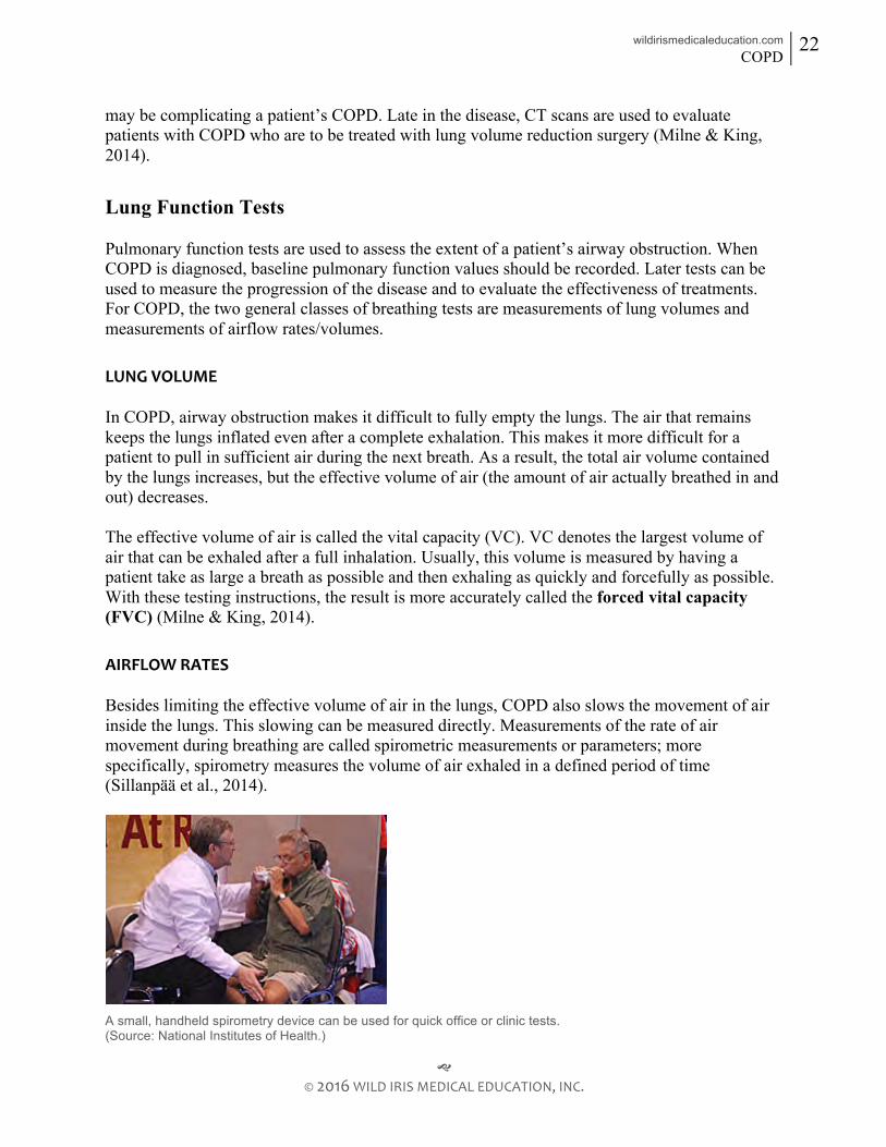
Lung Function Tests Pulmonary function tests are used to assess the extent of a patient’s airway obstruction. When COPD is diagnosed, baseline pulmonary function values should be recorded. Later tests can be used to measure the progression of the disease and to evaluate the effectiveness of treatments. For COPD, the two general classes of breathing tests are measurements of lung volumes and measurements of airflow rates/volumes. LUNG VOLUME In COPD, airway obstruction makes it difficult to fully empty the lungs. The air that remains keeps the lungs inflated even after a complete exhalation. This makes it more difficult for a patient to pull in sufficient air during the next breath. As a result, the total air volume contained by the lungs increases, but the effective volume of air (the amount of air actually breathed in and out) decreases. The effective volume of air is called the vital capacity (VC). VC denotes the largest volume of air that can be exhaled after a full inhalation. Usually, this volume is measured by having a patient take as large a breath as possible and then exhaling as quickly and forcefully as possible. With these testing instructions, the result is more accurately called the forced vital capacity (FVC) (Milne & King, 2014). AIRFLOW RATES Besides limiting the effective volume of air in the lungs, COPD also slows the movement of air inside the lungs. This slowing can be measured directly. Measurements of the rate of air movement during breathing are called spirometric measurements or parameters; more specifically, spirometry measures the volume of air exhaled in a defined period of time (Sillanpää et al., 2014).
A small, handheld spirometry device can be used for quick office or clinic tests. (Source: National Institutes of Health.)
wildirismedicaleducation.com COPD 23
! © 2016 WILD IRIS MEDICAL EDUCATION, INC.
The most common spirometric measurement used for COPD is the one-second forced expiratory volume (FEV1). This is the maximum amount of air that a patient can breathe out in the first second of a forced exhalation after having taken a full breath. Spirometry is helpful in evaluating the severity of airflow obstruction in patients with symptomatic COPD. On the other hand, spirometry does not add much to the evaluation of asymptomatic patients with COPD because treatments (other than smoking cessation) are not typically begun until after a patient becomes symptomatic (Sillanpää et al., 2014).
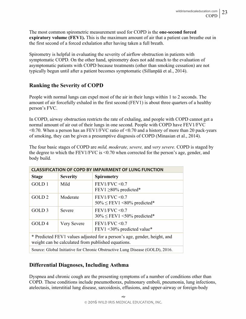
Ranking the Severity of COPD People with normal lungs can expel most of the air in their lungs within 1 to 2 seconds. The amount of air forcefully exhaled in the first second (FEV1) is about three quarters of a healthy person’s FVC. In COPD, airway obstruction restricts the rate of exhaling, and people with COPD cannot get a normal amount of air out of their lungs in one second. People with COPD have FEV1/FVC <0.70. When a person has an FEV1/FVC ratio of <0.70 and a history of more than 20 pack-years of smoking, they can be given a presumptive diagnosis of COPD (Minasian et al., 2014). The four basic stages of COPD are mild, moderate, severe, and very severe. COPD is staged by the degree to which the FEV1/FVC is <0.70 when corrected for the person’s age, gender, and body build. CLASSIFICATION OF COPD BY IMPAIRMENT OF LUNG FUNCTION Stage Severity Spirometry GOLD 1 Mild FEV1/FVC <0.7
FEV1 ≥80% predicted* GOLD 2 Moderate FEV1/FVC <0.7
50% ≤ FEV1 <80% predicted* GOLD 3 Severe FEV1/FVC <0.7
30% ≤ FEV1 <50% predicted* GOLD 4 Very Severe FEV1/FVC <0.7
FEV1 <30% predicted value* * Predicted FEV1 values adjusted for a person’s age, gender, height, and weight can be calculated from published equations. Source: Global Initiative for Chronic Obstructive Lung Disease (GOLD), 2016.
Differential Diagnoses, Including Asthma Dyspnea and chronic cough are the presenting symptoms of a number of conditions other than COPD. These conditions include pneumothorax, pulmonary emboli, pneumonia, lung infections, atelectasis, interstitial lung disease, sarcoidosis, effusions, and upper-airway or foreign-body
wildirismedicaleducation.com COPD 24
! © 2016 WILD IRIS MEDICAL EDUCATION, INC.
obstructions. A patient with COPD may also have other comorbidities such as lung masses, respiratory infections, increased incidence of atrial fibrillation, arterial hypertension, heart failure, and ischemic heart disease (Terzano, 2014). Most of these conditions can be identified using imaging studies, such as chest X-rays, and clinical signs. Anemia or metabolic acidosis can also cause chronic dyspnea, and both of these can be identified by blood studies. Asthma, which is another common obstructive airway disease, is high on the list of differential diagnoses for conditions presenting with both dyspnea and cough. Asthma usually cannot be distinguished from COPD by chest X-rays, clinical signs, or blood studies. Patients with asthma have hypersensitive airways that are always slightly inflamed, edematous, and filled with immune cells, characteristically eosinophils. Certain inhaled allergens and a variety of stresses can trigger these primed immune cells, causing a flare of the disease (an asthmatic attack) that brings on edema, mucus, and narrowed airways. Like COPD, asthmatic attacks will obstruct airways and impede airflow; but unlike COPD, the airway restrictions of an asthmatic attack can be, at least in young people, quickly and almost entirely reversed by bronchodilators. As people with asthma age, however, their airway obstruction sometimes becomes more fixed and less reversible. Clinically, these people’s disease begins to share more features with COPD, and the two diseases may be hard to distinguish. Determining which disease is present can be important for a patient’s treatment. For example, the dyspnea of asthmatic patients tends to improve markedly when the patient is given steroids, but the chronic dyspnea of most patients with COPD does not improve following steroids (Althani et al., 2013; Fu et al., 2013). Some useful distinctions between asthma and COPD include:
• Asthma usually appears in people <30 years of age, while COPD typically appears in people >40 years of age.
• Asthmatic attacks are reversed quickly and completely by medications, while the symptoms of COPD are reversed only modestly and temporarily by medications.
• Asthma often runs in families, while COPD usually does not.
• Only 20% to 30% of asthmatic patients have been smokers, and those who smoke have less than a 20-pack-year history. On the other hand, 90% to 95% of COPD patients have been smokers, and most have greater than a 20-pack-year history of smoking.

wildirismedicaleducation.com COPD 25
! © 2016 WILD IRIS MEDICAL EDUCATION, INC.
COMPARISON OF RESPIRATORY DISORDERS Disorder Symptoms / Relationship to COPD Smoking a Factor? COPD • Dyspnea, cough with sputum production, exercise
intolerance 80% with emphysema
Asthma • Dyspnea, chest tightness, cough with mucus production, wheezing
• May be indistinguishable from COPD
20%–30%
Lung masses • Hoarseness, hemoptysis, dyspnea, cough, chest pain or tightness, fever, weight loss, clubbing
• Shared; genetic dispositions and inflammation may predispose patients with COPD to lung cancer
85%
Effusions • Dyspnea, hypoxia, tachypnea, pain with inspiration
• Inflammation and chronic irritation may cause fluid to accumulate around the lungs
Possibly
Congestive heart failure
• Dyspnea, edema, jugular vein distention, reduced ejection fraction, orthopnea, hypertension, gallop
• Share the same pathophysiological mechanisms: inflammation and skeletal muscle alterations
Possibly
Pneumonia • Dyspnea, chest pain with inspiration, hypoxemia, cough, sputum production, fever
• Inflammation, suppressed immune system, and increased secretions put patients at risk for infections
Possibly
Source: Cavailles et al., 2013.
LONG-TERM TREATMENT OF COPD COPD is a life-long disease. It requires special medical treatment during acute exacerbations, and after the disease reaches the “moderate” level, it requires daily medications and permanent adjustments to a patient’s lifestyle. GOLD guidelines offer a comprehensive framework for the management of COPD (Vestbo et al., 2013). The goals of long-term COPD treatments are:
• Slow the progression of the disease • Ease the symptoms • Increase the patient’s ability to be mobile and carry out activities of daily living • Prevent acute exacerbations

wildirismedicaleducation.com COPD 26
! © 2016 WILD IRIS MEDICAL EDUCATION, INC.
Education is important to improve quality of life and reduce hospital admissions. All patients with COPD should learn about their disease and understand that smoking and air pollution will further damage their lungs. Patients need to make a special effort to avoid respiratory infections and to get yearly influenza vaccinations (Ko et al., 2015). In addition to yearly influenza vaccinations, it is recommended that all adults obtain a pneumonia vaccination after reaching age 65. Those at higher risk for pneumonia, such as patients with COPD, are urgently recommended to get the vaccination, often earlier than age 65 (CDC, 2015c; Swartz, 2015). At each stage of the disease, there are some characteristic medical therapies:
• Mild COPD is usually treated with short-acting bronchodilators, which are used as needed for dyspnea.
• Moderate COPD requires regular treatments with bronchodilators, sometimes with the addition of inhaled corticosteroids. At this stage, patients are often enrolled in a pulmonary rehabilitation program.
• Severe COPD typically requires two or more bronchodilators regularly. Inhaled corticosteroids are added to the regimen to prevent repeated acute exacerbations.
• Very severe COPD usually needs the addition of long-term oxygen therapy. Surgical treatments can be appropriate at this stage.
(Source: National Institutes of Health.)
Therapeutic Lifestyle Changes Medications are the fundamental day-to-day tools for controlling the symptoms of COPD, but there are also five effective nonpharmaceutical techniques for treating COPD: patient education, smoking cessation, keeping airways clear, nutritional therapy, and pulmonary rehabilitation (Engelke, 2012). Following are guidelines a clinician can follow in each of these five areas:

wildirismedicaleducation.com COPD 27
! © 2016 WILD IRIS MEDICAL EDUCATION, INC.
PATIENT EDUCATION / ENERGY CONSERVATION Teach your patients with COPD about their disease. Explain that the disease causes irreversible and progressive problems. Warn patients that they will have episodes in which the symptoms—difficulty breathing, wheezing, productive cough, and tiredness—get worse for days or even weeks. Assure patients that they will be helped by medications that make breathing easier. Tell them there are several things they can do to slow the progression of the disease and to lessen the number of acute exacerbations. The most important step is to stop smoking. Although smoking has already damaged their lungs, continued smoking will increase the damage and will make their COPD worsen more quickly. Let patients with COPD know that they should make every effort to stay active while recognizing the need to monitor and time their efforts throughout the day. In addition, give them practical suggestions that will help them to cope with the inevitable limitations posed by COPD. For example, tell them:
• Don’t push yourself. Slow the speed at which you do things, and stop and rest when you
are tired.
• Pace your activities and plan strenuous activities for times when you have the most energy. For example, you will feel best soon after you take your bronchodilator medicines. On the other hand, wait an hour after meals before you do activities.
• Sit on a chair or stool in the shower, don’t stand. Likewise, sit while you shave, comb your hair, and brush your teeth.
• Don’t use products that are hard on the lungs, such as hair sprays, spray-on deodorants, or strong perfumes.
• Use the exhaust fan in your kitchen to make it less likely that you will breathe smoke and cooking vapors.
• Wear slip-on shoes so you don’t have to bend over to tie laces.
• Make sure your occupation does not require more physical exercise than you can actually do. Consider setting smaller goals at work and allow more time to finish tasks.
• Find out how to get a daily air pollution report, and don’t go outside on days with moderate or severe pollution.
• Ask people not to smoke in your home or work area. (Engelke, 2012)
SMOKING CESSATION The cornerstone of management of COPD is smoking cessation (Cavailles et al., 2013).

wildirismedicaleducation.com COPD 28
! © 2016 WILD IRIS MEDICAL EDUCATION, INC.
Healthcare professionals are vital to the success of a smoking cessation program. Approximately 42.1 million people (18% of adults aged 18 years or older) in the United States smoke (Smalls et al., 2015). Most patients with COPD have a long smoking history and many will still be smoking when they are under medical care. From day one, strongly urge patients to stop smoking. Quitting can be difficult, since the nicotine in tobacco smoke is powerfully addictive. In addition, the rituals of smoking fill basic psychological needs. Therefore, when caregivers merely tell patients to stop smoking, their patients succeed over the long term only 5% of the time. Smoking cessation programs significantly improve the odds. Long-term success rates of greater than 20% to 40% can be achieved by comprehensive programs that include behavioral therapy and medications. Begin by saying to patients, “COPD cannot be cured, but if you continue smoking, the disease will worsen much more quickly. Have you thought about quitting smoking?” Regardless of the answer, follow it with the offer, “When you’re ready to stop smoking, I’ll be happy to work with you to set up as effective a program as possible.” Successful smoking intervention programs begin by asking the patient to set a specific quitting date. The programs then maintain continued contact with the patient to provide medication, counseling, support, advice, and a modicum of social pressure. (See “Resources” at the end of this course).
The “Five As” for Counseling Smokers Clinicians use the Five As when counseling their patients who smoke. Taking even one step is constructive.
1. Ask about the tobacco use and identify and document tobacco use status of every patient at every visit.
2. Advise the patient to quit and provide information on the benefits of quitting.
3. Assess whether the tobacco user is willing to quit at this time. Are there any challenges to remaining abstinent?
4. Assist the patient with finding resources and coming up with a cessation plan. Offer medication and provide or refer for counseling or additional behavioral treatment to help the patient quit. For patients unwilling to quit at this time, provide motivational interventions designed to increase future quit attempts. For the recent quitter and anyone with remaining challenges, provide relapse prevention.
5. Arrange follow-up to help the patient follow through with quitting. (Smalls et al., 2015)

wildirismedicaleducation.com COPD 29
! © 2016 WILD IRIS MEDICAL EDUCATION, INC.
Pharmacologic Therapy for Smoking Cessation The pharmacologic aspect of smoking cessation programs attempts to ease the effects of nicotine withdrawal. Smokers who need their first cigarette within a half-hour of getting up in the morning are likely to be highly addicted to nicotine. When these people stop smoking, they become anxious, irritable, easily angered, easily tired, and depressed. Their sleep is disrupted. They have difficulty concentrating. These withdrawal effects are common during the first two to three weeks after quitting. There are seven FDA-approved medications for the treatment of tobacco use:
• Nicotine gum • Nicotine inhaler • Nicotine lozenge • Nicotine nasal spray • Nicotine patch • Bupropion (Wellbutrin, Zyban) SR • Varenicline (Chantix)
(Smalls et al., 2015) Nicotine Replacement Therapy. To lessen withdrawal symptoms, nicotine can be taken in low doses without smoking to relieve the symptoms of craving. Nicotine replacements are available as gum, lozenges, transdermal patches, inhalers, and nasal sprays. These should be used on a regular schedule and PRN (as needed for cigarette cravings) for about two weeks, and then the doses should be tapered. Nicotine patches are marketed as Habitrol and NicoDerm CQ; nicotine gum includes Nicorette. The gum, lozenges, and inhaler help to satisfy oral cravings, and the inhaler raises nicotine blood levels more rapidly than the other routes of administration. As nicotine is a vasoconstrictor, people with coronary artery disease are advised not to use any nicotine replacement therapy. Antidepressants. The antidepressant bupropion SR (sustained-release) (brand names Zyban or Wellbutrin) is approved by the FDA to help patients for whom nicotine replacement therapy has not worked. Bupropion raises levels of dopamine in the brain, which helps to relieve nicotine cravings. Nicotine agonists. In 2006, varenicline (Chantix), a nicotine agonist, was approved by the FDA for anti-smoking therapy. Varenicline binds to nicotine receptors and prevents nicotine from activating the receptors, while producing a smaller stimulant effect than nicotine. As varenicline contains no nicotine, it does not cause vasoconstriction that can reduce blood flow to the myocardium, making it the drug of choice for patients with a cardiac history. It also stimulates the release of dopamine. Electronic or e-cigarettes contain vaporized liquid nicotine and are believed to aid in smoking cessation, but there is not yet sufficient research to support this. No prescription is needed.

wildirismedicaleducation.com COPD 30
! © 2016 WILD IRIS MEDICAL EDUCATION, INC.
CHANTIX AND ZYBAN HAVE FDA WARNINGS
On July 1, 2009, the U.S. Food and Drug Administration (FDA) announced that it would require manufacturers to put a “Boxed Warning” on the prescribing information for the smoking cessation drugs Chantix (varenicline) and Zyban (bupropion). The warning highlights the risk of serious mental health events, including changes in behavior, depressed mood, hostility, and suicidal thoughts, when taking these drugs. “The risk of serious adverse events while taking these products must be weighed against the significant health benefits of quitting smoking,” said Janet Woodcock, M.D., director of the FDA’s Center for Drug Evaluation and Research. “Smoking is the leading cause of preventable disease, disability, and death in the United States, and we know these products are effective aids in helping people quit.”
Source: FDA, 2013.
CASE Faith Jeffries, RN, has a patient in her unit, Mrs. Hunter, who struggles with quitting smoking in spite of being diagnosed with moderate COPD. She has tried nicotine patches and gum, the nicotine agonist Chantix (varenicline), hypnotherapy, acupuncture, and counseling. Each method has been temporarily successful, and then Mrs. Hunter started smoking again. Mrs. Hunter states that the patches caused skin irritation and scarring, the gum didn’t work, Chantix caused severe nausea, and the hypnotherapist “couldn’t put her under.” At present, the patient’s physician has ordered the antidepressant Zyban (bupropion) to reduce nicotine cravings. Although Mrs. Hunter understands the dangers of smoking and the effects on her health, she returns to smoking in times of stress. Faith sits with Mrs. Hunter to discuss the types of stressors that trigger her addiction and some strategies to avoid them or handle them in other ways.
KEEPING AIRWAYS CLEAR Patients with COPD with significant chronic bronchitis must keep their airways clear. They should be encouraged to cough up sputum, and they should not get in the habit of using cough suppressants or sedatives. Postural drainage can help patients who cannot clear their secretions by coughing. This is a technique patients can be taught to employ at home in which they place themselves in a variety of body positions that encourage gravity-assisted drainage of the lungs. Most people’s lungs secrete extra mucus in response to inhaled irritants. To avoid stimulating excess secretions, patients with COPD need to stay out of smoke-filled rooms, and they should stay indoors during air pollution alerts. Home air conditioners and air filters are effective at keeping indoor air clear of particulates.

wildirismedicaleducation.com COPD 31
! © 2016 WILD IRIS MEDICAL EDUCATION, INC.
NUTRITIONAL THERAPY The symptoms of COPD improve when patients who are overweight lose weight. Some patients with COPD, however, have the opposite problem: they have become thin and malnourished. In part, this cachexia results from the high energy cost of breathing with COPD. In addition, the chronic inflammatory state underlying COPD tends to put the body’s metabolism into a catabolic state, in which larger molecules such as tissue are broken down in to smaller molecules. This constant breakdown of tissue increases the body’s metabolic rate, causing further weight loss. To help maintain a healthy body weight, thin patients with COPD should be given dietary counseling that includes specific recommendations for meals that are nutritionally balanced and that contain sufficient calories to make up for the work of breathing (Engelke, 2012; Nordén et al., 2015). (For more information on nutrition, see also “Resources” at the end of this course.) PULMONARY REHABILITATION AND INTEGRATED CARE Pulmonary rehabilitation (PR) is the term for a group of techniques used to improve patients’ conditioning and to ease their exercising difficulties. It is a comprehensive, evidence-based, multidisciplinary program designed to assist patients with COPD who are having difficulty with breathing and activities of daily living. Most PR programs involve a respiratory therapist, occupational therapist, physical therapist, and dietitian. Physicians, pharmacists, and nurses may also be involved, but not at every meeting with the patient. PR programs are delivered in inpatient, outpatient, clinic, physician office, telehealth, and home settings (Camp et al., 2015; Spruit et al., 2015). PR programs include assessment, exercise therapy, education, and psychological support. Education sessions are important parts of rehabilitation programs; in these sessions, patients and their families learn details about COPD and its treatment. The benefits are maximization of functional status and the reduction of healthcare cost by promoting self-management of symptoms. Some rehabilitation programs continue for an extended time, but most run for a few weeks and then give patients individualized instructions for continuing at home. The primary objective of a PR program is to restore individual patients to as independent a level of function as possible with an improved health-related quality of life. It is evidence-based that dyspnea symptoms improve in patients with COPD who undergo a PR regime. PR is proven to be a cost-effective treatment model and reduces the number of hospital admissions, but it cannot be substantiated that PR extends the life of patients with COPD (Camp et al., 2015).
Education in Pulmonary Rehabilitation Patient and family education is central to all PR programs, although it has been demonstrated that education alone does not improve outcomes. Education should inform the patient and family how to self-manage the disease in collaboration with the various PR disciplines. Education topics may include understanding chronic lung disease,

wildirismedicaleducation.com COPD 32
! © 2016 WILD IRIS MEDICAL EDUCATION, INC.
medications, breathing control, oxygen therapy, heart health, falls prevention, diagnostic tests, and advance care planning (Spruit et al., 2015). Muscle and Endurance Training A goal of PR is to optimize the functional status of a patient with COPD by exercise training and collaborative self-management. Exercise training supervised by occupational and physical therapists does not improve lung functioning, but it can reduce COPD symptoms and increase the amount of exercise that the patients can do without being stopped by dyspnea. It can also reduce the number of hospitalizations for acute exacerbations. Physical inactivity is the greatest source of the muscle weakness that plagues patients with COPD, causing exercise intolerance and the wasting of skeletal and respiratory muscles. Although people with COPD have irreversible breathing difficulties, exercise training—including interval training, strength training, upper and lower limb training, and transcutaneous neuromuscular electrical stimulation—can significantly increase a patient’s strength and endurance and reduce their fatigability. These improvements result from increased muscle size (specifically, cross-sectional area), increased blood flow to muscles, increased oxidative enzyme capacity, and reduction of lactic acid production during exercise. Endurance training in the form of walking and cycling retards the progression of activity intolerance in patients with COPD, as do unsupported upper extremity exercises, such as cross-body weight lifting (Spruit et al., 2013; Spruit et al., 2015). Typical Programs Comprehensive PR programs start with a patient assessment by a physician, advanced practice nurse, or physician assistant. The initial patient assessment is performed to determine the best PR program with regard to duration of the program (usually 4 to 12 weeks) and number of sessions per week, type of exercise modalities (duration of each exercise, repetitions, progression for increasing exercise), need for nutritional counseling, need for psychological support, and need for oxygen supplementation. Pulmonary rehabilitation programs are tailored to the needs of each individual. Typically, the programs include graded aerobic exercises, such as regular sessions of walking or stationary bicycling three times weekly. The walking exercise program, for example, might begin with slow treadmill walking for only a few minutes. Gradually, the length and speed of the walking is increased. The goal would be for the patient to walk in gradually increased increments without needing to stop because of shortness of breath. At that point, the patient would be assigned a maintenance exercise program to be done at home. Other exercises may include stretching, weight training, and a stationary cycle (Lareau & Fahey, 2013).

wildirismedicaleducation.com COPD 33
! © 2016 WILD IRIS MEDICAL EDUCATION, INC.
Rehabilitation sessions also include breathing instruction that teaches patients how to slow their rate of breathing by pursing their lips and how to rest the upper respiratory muscles by using abdominal breathing instead of chest breathing.
PURSED-‐LIP BREATHING
Pursed-lip breathing improves breathing, releases air trapped in the lungs, expands the airways longer, and decreases effort. This breathing control exercise prolongs expiration to reduce the respiratory rate and improves breathing by maximizing inspiration. Pursed-lip breathing relieves shortness of breath and promotes general relaxation. This technique can be used during the difficult part of any activity, such as bending, lifting, or stair climbing. It should be practiced four to five times a day to establish the correct breathing pattern. The patient with COPD should be instructed to:
• Relax the neck and shoulder muscles • Inhale slowly through the nose for two counts, keeping the mouth closed • Take normal breaths while counting “one, two”
• Pucker or “purse” the lips as if whistling or gently flickering the flame of a candle
• Exhale slowly and gently through pursed lips while counting “one, two, three, four”
Source: Cleveland Clinic Foundation, 2014.
Oxygen Supplementation Supplemental oxygen is recommended during exercise for patients who experience severe exercise-induced hypoxemia. Oxygen saturation may need to be monitored in patients whose oxygen dependency may otherwise prevent them from exercising. Increased flow rates may enable oxygen-dependent patients to exercise longer and with less dyspnea. This may improve exercise endurance during a high-intensity exercise program. Some studies using a helium-oxygen mix showed mixed results for improved functional exercise capacity (Spruit et al., 2013). Neuromuscular Electrical Stimulation (NMES) Some patients with COPD have such poor lung function or such weak musculature that they cannot take part in the usual aerobic exercise training programs. Small studies suggest that transcutaneous electrical stimulation of the patients’ lower limbs can improve their muscle strength and exercise tolerance. This is an individualized protocol in which the intensity (amplitude), frequency, duration, and waveform of the stimulus is determined for each patient. Precise patient and family education is needed to ensure the device is properly used.
wildirismedicaleducation.com COPD 34
! © 2016 WILD IRIS MEDICAL EDUCATION, INC.
NMES increases muscle strength, exercise capacity, and reduces dyspnea of outpatients with severe COPD and poor baseline exercise tolerance and can be used during acute COPD exacerbations. This has worked even for bedridden patients. Neuromuscular stimulation routines are safe for most patients, inexpensive, and can be done at home (Spruit et al., 2013). Treatment of Anxiety and Depression Clinical depression as a comorbidity with COPD occurs internationally at a rate of 30% to 50% compared to a 6% to 8% occurrence in the background population. Multiple studies have proven there is a direct correlation between reduction of measurable anxiety and depression levels in patients with COPD when they participate in PR. Therefore, PR is recommended as a viable adjunct to other treatments for patients with COPD at any stage of the severity of the disease. The degree of psychological improvement brought about by the program warrants PR be included in treatment of anxiety and depression in this population (Tselebis et al., 2013; Spruit et al., 2013). Advance Care Planning A PR program can provide the opportunity for patients with COPD, families, and primary care providers to discuss end-of-life issues. Advance directives, living wills, and durable powers of attorney can be explained and decided upon. The subject of invasive and noninvasive ventilation is a particularly necessary subject for this patient population (Spruit et al., 2013).
CASE Stuart Moody is being discharged from the hospital with a new diagnosis of mild to moderate emphysema-type COPD. His physician has ordered that Mr. Moody start a pulmonary rehabilitation program with a follow-up visit to the doctor’s office in six weeks. The nurse explains to Mr. Moody that a PR program consists of education about exercise training, nutritional counseling, medications, how to self-measure peak flows, breathing training, panic control, and airway control. Mr. Moody will see a registered dietitian in consultation before discharge to help him maintain or achieve his correct weight. He will also work with a physical therapist to learn progressive aerobic exercise. Mr. Moody is fortunate to be started on PR at this time. Previously, PR was only ordered for moderate to severe COPD. He is also fortunate that Medicare now covers PR.
Medications The medicines currently available for COPD focus on long-term therapy with prevention of symptoms. Stepped therapy, the process by which additional medications are added as symptoms progress, is the standard in treating COPD. Inhaled and systemic drugs for patients with COPD

wildirismedicaleducation.com COPD 35
! © 2016 WILD IRIS MEDICAL EDUCATION, INC.
include beta-adrenergic agents, cholinergic antagonists, methylxanthines, corticosteroids, nonsteroidal anti-inflammatory drugs (NSAIDs), and mucolytic agents. Drug therapy is used to reduce the extent to which dyspnea restricts a patient’s activities. Most COPD drugs work by keeping airways as wide open as possible. Medications (bronchodilators) used to reduce airflow obstruction are not typically given to asymptomatic patients with COPD. BRONCHODILATORS Bronchodilators are the workhorses of the COPD medications. Although spirometry shows that bronchodilators only modestly reduce airway obstruction in most patients with COPD, regular doses of bronchodilators relieve dyspnea sufficiently for patients with COPD to increase their levels of activity. Bronchodilators work by relaxing the smooth muscles in the walls of the lungs’ airways; this widens the airways and allows air to move through them more easily. Short- and fast-acting bronchodilators are used as “rescue” medicines to relieve sudden bouts of dyspnea and coughing. Long-acting bronchodilators are used in daily, regularly scheduled drug regimens. All symptomatic patients are prescribed a short-acting rescue bronchodilator that they can use to recover from a bout of suddenly worsening dyspnea. Either short-acting parasympatholytic or short-acting sympathomimetic bronchodilators can be used as fast-relief medications (Lilley et al., 2014).
Parasympatholytic Bronchodilators The parasympatholytic bronchodilators are anticholinergic drugs, which relax the smooth muscles in the airway by blocking the effect of acetylcholine, produced by the parasympathetic nervous system. The most commonly prescribed short-acting anticholinergic bronchodilator is ipratropium (Atrovent). Ipratropium is relatively inexpensive and widely available. It is usually administered via a metered-dose inhaler (MDI), although there are other formulations. It can be used as a PRN medication. It takes effect in 15 to 30 minutes, has its peak action in 1 to 2 hours, and lasts 4 to 6 hours. Traditionally, ipratropium has also been used as the main anticholinergic in long-term drug regimens. However, studies have shown that tiotropium (Spiriva) is a more effective drug. Tiotropium is a longer-acting anticholinergic bronchodilator. It is more expensive than ipratropium, but a typical dose lasts an entire day. Tiotropium is helpful when used alone and is even more effective in combination with a long-acting beta agonist (Keating, 2012). Tiotropium is inhaled as a powder via a dry powder inhaler (DPI).
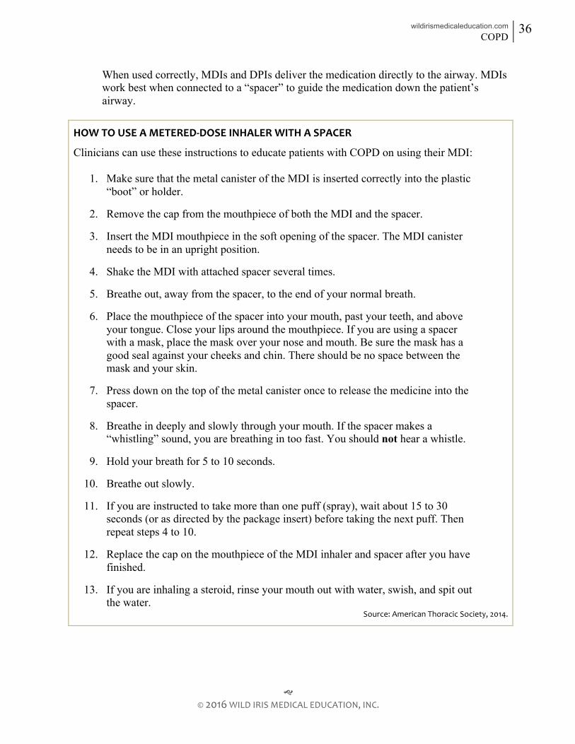
wildirismedicaleducation.com COPD 36
! © 2016 WILD IRIS MEDICAL EDUCATION, INC.
When used correctly, MDIs and DPIs deliver the medication directly to the airway. MDIs work best when connected to a “spacer” to guide the medication down the patient’s airway.
HOW TO USE A METERED-‐DOSE INHALER WITH A SPACER
Clinicians can use these instructions to educate patients with COPD on using their MDI:
1. Make sure that the metal canister of the MDI is inserted correctly into the plastic “boot” or holder.
2. Remove the cap from the mouthpiece of both the MDI and the spacer.
3. Insert the MDI mouthpiece in the soft opening of the spacer. The MDI canister needs to be in an upright position.
4. Shake the MDI with attached spacer several times.
5. Breathe out, away from the spacer, to the end of your normal breath.
6. Place the mouthpiece of the spacer into your mouth, past your teeth, and above your tongue. Close your lips around the mouthpiece. If you are using a spacer with a mask, place the mask over your nose and mouth. Be sure the mask has a good seal against your cheeks and chin. There should be no space between the mask and your skin.
7. Press down on the top of the metal canister once to release the medicine into the spacer.
8. Breathe in deeply and slowly through your mouth. If the spacer makes a “whistling” sound, you are breathing in too fast. You should not hear a whistle.
9. Hold your breath for 5 to 10 seconds.
10. Breathe out slowly.
11. If you are instructed to take more than one puff (spray), wait about 15 to 30 seconds (or as directed by the package insert) before taking the next puff. Then repeat steps 4 to 10.
12. Replace the cap on the mouthpiece of the MDI inhaler and spacer after you have finished.
13. If you are inhaling a steroid, rinse your mouth out with water, swish, and spit out the water.
Source: American Thoracic Society, 2014.

wildirismedicaleducation.com COPD 37
! © 2016 WILD IRIS MEDICAL EDUCATION, INC.
HOW TO USE A DRY POWDER INHALER
Clinicians can use these instructions to educate patients with COPD on using their DPI:
1. Remove all candy, food, or gum from your mouth.
2. Stand up straight.
3. Hold the inhaler level to the floor.
4. Open the inhaler with the mouthpiece facing you.
5. Slide the lever away from you until you hear it click. This means the medicine has been released. Be careful not to tip the inhaler or slide the lever again; if you do, the medicine will fall out and be wasted.
6. Take a deep breath in and then breathe out.
7. Place the inhaler in your mouth, seal your lips tightly around it, and take a quick, deep breath in.
8. Hold your breath for 10 seconds and then breathe out. Source: Respiratory Health Association, 2015.
Sympathomimetic Bronchodilators Beta2 Adrenergic Agonists One class of sympathomimetic bronchodilators, the beta2 agonists, acts by mimicking the effect of norepinephrine on airway muscles. Beta2 agonists cause smooth muscles to relax, widening the airways. Muscle tremors and heart palpitations are the most common side effects of beta2 agonists, but when the medicines are inhaled (as opposed to taken in oral formulations), the side effects are usually mild. The short-acting beta2 agonists—which include albuterol (Accuneb, ProAir, Proventil, and Ventolin) and metaproterenol (Alupent)—are the most commonly prescribed sympathomimetic bronchodilators. These drugs are usually administered via either MDI or DPI. Short-acting beta2 agonists such as albuterol and metaproterenol take effect in 5 to 15 minutes and last for 2 to 4 hours. Short-acting beta2 agonists are used as rescue medicines when a patient needs immediate relief from sudden episodes of increased dyspnea. A short-acting beta2 agonist can also be added to an anticholinergic drug as part of a regular drug regimen. The long-acting beta2 agonist bronchodilators include formoterol (Foradil) and salmeterol (Serevent). These drugs are more expensive than albuterol or metaproterenol, but a typical dose lasts for at least 12 hours. Inhalation is the recommended route for administering the long-acting beta2 agonists.
wildirismedicaleducation.com COPD 38
! © 2016 WILD IRIS MEDICAL EDUCATION, INC.
Phosphodiesterase Inhibitors Another class of sympathomimetic bronchodilators, the phosphodiesterase inhibitors or xanthines, acts by stimulating the release of norepinephrine, which then relaxes smooth muscles in the airways of the lung. For COPD, the phosphodiesterase inhibitor theophylline (Elixophyllin, Theo-Dur) is used to dilate airways, stimulate the respiratory centers of the brain, and improve the function of respiratory muscles. Theophylline is taken orally and side effects are common; among them are sleeplessness and gastrointestinal upset, including nausea and vomiting. Occasionally, theophylline causes serious cardiac arrhythmias or seizures, especially when liver disease has decreased the body’s ability to metabolize the drug. Older people are more likely to get theophylline toxicity. Two newer phosphodiesterase inhibitors, cilomilast (Ariflo) and roflumilast (Daxas or Daliresp), appear to be safer than theophylline.
Bronchodilator Regimens Patients vary in their response to bronchodilators, so the most effective drug regimens are those that have been individually tailored. Finding the right drug or set of drugs is empirical. Patients may try many different inhalers until finding a combination that works best for them. When drug combinations are being tried, it is best to introduce the drugs one at a time to learn the patient’s response to that drug only. For patients with chronic stable COPD, short-acting bronchodilators will eventually be insufficient to control their symptoms. Currently, the long-acting anticholinergic drug tiotropium is usually recommended as the first drug to try in a regular daily medication regimen. It is taken once daily and it does not have the side effects of sympathomimetic drugs, but it is generally not as effective as the beta agonists and is only recommended if the COPD patient cannot tolerate the beta agonist side effects. Concurrently, a short-acting beta2 agonist, such as albuterol, is usually prescribed as a rescue drug. If this initial regimen is insufficient, the short-acting beta2 agonist is added to the regularly scheduled drug regimen rather than being used only when needed. The combination of ipratropium and albuterol is available commercially (DuoNeb) as an inhalant. As COPD progresses, most patients do better with combinations of two or three bronchodilators. In American and Western European medicine, theophylline (or another phosphodiesterase inhibitor) is usually the last bronchodilator to be added. If they are to be followed faithfully, drug regimens must be realistic. Bronchodilator therapy with two or three drugs is expensive. In addition, using inhalers can be physically difficult for some people, especially the elderly, and physicians may need to modify an optimal pharmacologic therapy to make it practical for a particular patient (Lilley et al., 2014).

wildirismedicaleducation.com COPD 39
! © 2016 WILD IRIS MEDICAL EDUCATION, INC.
BRONCHODILATORS
Short-acting beta agonists • Action: relax bronchiolar smooth muscle
• Drugs: albuterol (Proventil), metaproterenol (Alupent), erbutenol (Maxair), terbutaline (Brethine)
• Side effects: tachycardia, palpitations, anxiety, muscle tremors
• Comments: fast-acting rescue drug; take 5 minutes before other inhalers; used for treatment
Long-acting beta agonists
• Action: relax bronchiolar smooth muscle • Drugs: salmeterol (Serevent) • Side effects: tachycardia, palpitations, anxiety, muscle tremors • Comments: do not use for acute onset of symptoms; shake inhaler, since drug
separates; used for prevention Anticholinergic agents
• Action: inhibit parasympathetic nervous system • Drugs: ipratropium (Atrovent) • Side effects: cough, dry mouth • Comments: may exacerbate cardiac symptoms; shake inhaler, since drug separates;
used for prevention; must carry at all times if used as a rescue drug Phosphodiesterase inhibitors (methylxanthines)
• Action: similar to caffeine • Drugs: aminophylline (Theophylline, Theo-Dur)
• Side effects: stomach upset, heartburn, insomnia, headache, nervousness or irritability, tachycardia, tachypnea
• Comments: do not use caffeine; monitor blood levels Sources: Everyday Health, 2012; Lilley et al., 2014.
CORTICOSTEROIDS Corticosteroids, also called glucocorticoids, are two-edged swords. On the one hand, they are effective anti-inflammatory medicines and can be used to tone down the inflammatory response that underlies or exacerbates many diseases. On the other hand, the continued use of corticosteroids causes Cushing’s syndrome, glaucoma, cataracts, myopathy, ulcers, osteoporosis, hyperglycemia, poor wound healing, and the inability to overcome infections.

wildirismedicaleducation.com COPD 40
! © 2016 WILD IRIS MEDICAL EDUCATION, INC.
In stable COPD, the problems that come from the long-term use of oral or systemic corticosteroids usually outweigh the drugs’ benefits. Inhaled steroids—such as fluticasone (Flovent), beclomethasone (Beclovent, Beconase), and budesonide (Pulmicort Turbuhaler)—have fewer adverse effects than oral formulations, and approximately 10% of people with COPD find that regularly inhaled steroids reduce their airway obstruction. For this population of patients, inhaled steroids can be a useful addition to the other regularly scheduled bronchodilators. The regular use of inhaled corticosteroids is usually reserved for patients with severe COPD. In people with severe COPD, steroids will reduce the number of exacerbations and the rate of mortality. For people with severe COPD, inhaled corticosteroids are typically combined with a long-acting beta2 agonist in a regular treatment regimen. Regular use of inhaled corticosteroids for COPD does, however, increase a patient’s risk of developing pneumonia (Lilley et al., 2014). The usefulness of corticosteroid therapy cannot be predicted in advance for any one patient. At the moment, spirometrically testing a patient’s response to the medication is the only way to identify in advance those patients with COPD who will be helped by adding inhaled steroids to their regular regimen of bronchodilators.
ANTI-‐INFLAMMATORIES
Corticosteroids • Action: disrupt inflammatory pathways • Drugs:
o Inhaled: fluticasone (Flovent) o Oral: prednisone (Deltasone)
• Side effects: o Inhaled: coughing, hoarseness, dry mouth, sore throat o Oral: glaucoma, edema, hypertension, mood swings, weight gain,
cataracts, hyperglycemia, infections, osteoporosis and fractures, menstrual irregularities, suppressed adrenal gland hormone production, thin skin, easy bruising and slower wound healing
• Comments: must be taken every day, even if there are no symptoms; do not stop taking suddenly; take with food; reduces local immunity, may increase risk for local infections like candida (yeast)
Nonsteroidal anti-inflammatory drugs (NSAIDs)
• Action: stabilize mast cell membranes to prevent inflammation • Drugs: nedocrimil (Tiladel)
• Side effects: dyspepsia, nausea, hyperacidity; in higher doses, MI, CVA, rash, GI bleeding

wildirismedicaleducation.com COPD 41
! © 2016 WILD IRIS MEDICAL EDUCATION, INC.
• Comments: must be taken by inhaler every day for prophylaxis, even if there are no symptoms
Sources: Everyday Health, 2012; Lilley et al., 2014.
PREMIXED COMBINATION INHALERS
• Action: combine the effects of bronchodilators and corticosteroids • Drugs:
o Short-acting: ipratropium, albuterol (DuoNeb), ipratropium fenoterol (DuoVent) o Long-acting: formoterol, budesonide (Symbicort), salmeterol, fluticasone
(Advair) • Side effects:
o Short-acting: cough, dry mouth; tachycardia, palpitations, anxiety, muscle tremors
o Long-acting: coughing, hoarseness, dry mouth, sore throat; tachycardia, palpitations, anxiety, muscle tremors;
Sources: Everyday Health, 2012; Lilley et al., 2014.
MUCOLYTIC AGENTS Patients with COPD often have thick, tenacious mucus that is very difficult to expectorate, particularly during an acute exacerbation. Mucolytic agents can be given by respiratory nebulizer treatments, sometimes mixed with normal saline to thin secretions. They can also be given orally to produce a systemic effect. Acetylcysteine (Mucomyst) or dornase alfa (Pulmozyme) are commonly given by inhaled nebulizer treatment, and guaifenesin (Robitussin) is given by mouth to promote expectoration.
MUCOLYTIC AGENTS
• Action: thin secretions to promote expectoration • Drugs:
o Inhaled: acetylcysteine (Mucomyst), dornase alfa (Pulmozyme) o Oral: guaifenesin (Robitussin)
• Side effects: foul smell, sticky nebulizer mask, white patches or sores inside mouth or on lips, nausea, fever, nasal drainage, sore throat, drowsiness, rash, or clammy skin
• Comments: must be instructed in home nebulizer use; may interact with some vitamins, minerals, and herbs
Sources: Everyday Health, 2012; Lilley et al., 2014.

wildirismedicaleducation.com COPD 42
! © 2016 WILD IRIS MEDICAL EDUCATION, INC.
VACCINATIONS
People with COPD are at higher risk for serious, even life-threatening complications that are preventable by vaccination. As protection against serious respiratory illnesses, people with COPD should get an influenza vaccination each year (CDC, 2015a). During outbreaks of strains of flu not covered by the annual vaccination, people with COPD should probably receive prophylactic antiviral treatment such as amantadine (Symmetrel), rimantadine (Flumadine), oseltamivir (Tamiflu), or zanamivir (Relenza) (Lilley et al., 2014). Pneumococcal vaccinations are also recommended for people with COPD before age 65. A second and even third dose is recommended for people 65 years and older who got their first dose when they were younger than 65 and it has been five or more years since the first dose (CDC, 2015b).
Source: OAC 4723-‐4-‐01.
Oxygen Therapy Supplemental oxygen improves levels of blood oxygenation and reduces the rate at which patients need to breathe. No other medical treatment has proved as effective in improving survival rates of patients with COPD. For people with COPD, supplemental oxygen also slows the rate at which muscles fatigue. These effects make it easier for patients to breathe more deeply and to exercise for longer periods. Oxygen therapy is expensive and involves special equipment. Therefore, when people with COPD can maintain a blood oxygenation level of PaO2 >55–60 mmHg (an oxygenation saturation of more than ~89%), this is considered adequate oxygenation (unless the patient is symptomatic) and supplemental oxygen therapy is not routinely prescribed (Sharma et al., 2015). CONTINUOUS OXYGEN Eventually, however, supplemental oxygen will be necessary. For some patients with COPD, oxygen is needed to participate in regular exercise programs. For other patients, oxygen is needed to carry out the typical activities of daily living. If they live long enough, all patients with COPD lose sufficient lung function to the point that they will be hypoxemic at rest even on an optimal regimen of regular bronchodilator treatments. For these people, continuous oxygen therapy can prolong their lives and reduce hospitalizations. When a patient’s blood PaO2 is <55–60 mmHg (an oxygen saturation of less than ~85%–89%) at rest, it is recommended that supplemental oxygen be given continuously as a life-saving measure (Meena et al., 2015; Sharma et al., 2015). Low-flow (2–3 liter/min) oxygen inhaled through nasal cannulas is usually sufficient to raise a COPD patient’s blood PaO2 to 65–80 mmHg (an oxygen saturation of 89%–94%). In addition to increasing survival rates by about 50%, this level of supplemental oxygen lowers the person’s hematocrit toward a normal range, makes sleep easier, and improves exercise tolerance.

wildirismedicaleducation.com COPD 43
! © 2016 WILD IRIS MEDICAL EDUCATION, INC.
Home oxygen therapy is also recommended for patients with COPD with heart failure, pulmonary hypertension, or erythrocytosis (i.e., a hematocrit >56%), even when their PaO2 is >55 mmHg. Some patients who maintain a higher level of arterial oxygen during the day drop to a PaO2 <55 mmHg when they sleep. For people whose hemoglobin desaturates at night, nocturnal oxygen therapy is helpful. HOME OXYGEN DELIVERY SYSTEMS Home oxygen can be purchased as an oxygen concentrator, compressed oxygen cylinder, or liquid oxygen. All of these methods can supply an oxygen concentration of 90% or more to the individual and enrich the local environment. The cost of continuous home oxygen therapy can be $500 or more per month. In many cases, Medicare will cover 80% of the cost for supplemental oxygen for patients with desaturation (oxygen saturation <94%) during sleep or physical activity (Meena et al., 2015; Sharma et al., 2015). Patients usually breathe supplemental oxygen via a continuous-flow nasal cannula. Devices that “conserve oxygen”—reservoir cannulas such as moustache-configured oximizers or oximizer pendants, electromechanical demand pulse delivery devices, transtracheal oxygen delivery—are especially efficient because they provide all the supplemental oxygen early in each inhalation. Some patients who have trouble keeping low blood levels of carbon dioxide can be fitted with facemasks from machines that deliver supplemental oxygen at continuous positive pressure; these systems provide noninvasive positive pressure ventilation (Meena et al., 2015). A home system is usually adjusted to deliver two to three liters of oxygen per minute, and in most cases this will maintain a patient’s oxygen saturation at >89%. For patients who continue to have dyspnea at night, the flow rate is raised by 1 liter/min during sleep. One goal of oxygen therapy is to allow patients to remain active. Inside the home, long tubes can connect the nasal cannulas to stationary oxygen delivery units so patients can move around. For more freedom and to go outdoors, patients can carry portable tanks of compressed oxygen or liquid oxygen. The risks of home oxygen therapy are hypercapnia, oxygen toxicity, and burns secondary to the flammable nature of oxygen (Sharma et al., 2015). HAZARDS Medical. There is a small risk that too high a concentration of inspired oxygen will suppress the respiratory drive of patients with COPD. Long-term low-flow oxygen therapy is probably safest when the amount of oxygen delivered gives the patient a PaO2 of 60–65 mmHg, which is toward the low end of the acceptable range of inspired oxygen (Sharma et al., 2015). Physical. Concentrated oxygen is flammable and poses a fire hazard. Patients and their families cannot smoke or use open flames near the oxygen equipment. The long oxygen tubing may also constitute a fall risk.

wildirismedicaleducation.com COPD 44
! © 2016 WILD IRIS MEDICAL EDUCATION, INC.
AIR TRAVEL Commercial planes maintain an internal air pressure equivalent to 5,000–8,000 feet above sea level. For those patients with COPD whose resting arterial blood oxygen concentration is low (PaO2 <69 mmHg) even at sea level, the cabin concentration of oxygen will usually not be high enough to avoid hypoxemia. Airlines can provide supplemental oxygen, and some airlines will allow patients to bring small oxygen delivery systems with them, although patients must make arrangements with the airline in advance.
Surgery for COPD Surgery is risky in people with severe COPD. Postoperatively, many normal patients temporarily have reduced lung volumes, rapid shallow breathing, and an impaired ability to take in oxygen and expel carbon dioxide. These routine postoperative problems add additional stress to the already compromised respiratory systems of patients with COPD. One result is that patients with severe COPD develop postoperative pneumonia 13 times more often than patients with normal lung function. (Preoperative antibiotics can reduce the high rate of postoperative pneumonia.) The lack of alternative treatments for severe COPD has led to the development of three surgical procedures that attempt to improve and prolong the lives of patients with COPD. The techniques are lung transplantation, lung volume reduction surgery, and bullectomy. LUNG TRANSPLANTATION People with severe COPD are the most common recipients of lung transplants. Candidates for lung transplantation are patients with severe COPD who have exhausted medical therapies and have life expectancies of ≤2 years. The BODE Index is usually used to estimate a COPD patient’s life expectancy (see box below). Typically, patients should also be younger than 65 years. Three quarters of patients with COPD who receive lung transplants live for ≥2 years after the operation and many of the survivors have substantially improved abilities to exercise.
BODE INDEX
The updated BODE Index (body-mass index, airway obstruction, dyspnea, and exercise capacity) uses four measurements to assign patients with COPD to 1 of 15 groups, each with a different estimated survival rate. This mortality prediction index is a multistage scoring system that provides prognostic data in patients with COPD. The BODE index is better than the FEV1 at predicting the risk of death among patients with COPD. The measurements are:
1. Body-mass index 2. Degree of airflow obstruction (FEV1 calculated by spirometric measurement)

wildirismedicaleducation.com COPD 45
! © 2016 WILD IRIS MEDICAL EDUCATION, INC.
3. Amount of dyspnea (using the Modified Medical Research Council dyspnea scale) 4. Exercise capacity (distance walked in 6 minutes)
Two years after a BODE assessment is made, estimated mortality rates for long-standing patients with COPD are approximately:
• 30% for a BODE score >7 • 15% for a BODE score 5–6 • 10% for a BODE score <5
Source: Sakar et al., 2015.
LUNG VOLUME REDUCTION SURGERY As noted earlier, the lungs of an emphysematous patient become hyperinflated with air spaces that contribute little to gas exchange. The widened chest caused by hyperinflated lungs is difficult for the patient to expand farther when attempting to inhale. By removing lung tissue that contains dead air space, surgery can sometimes reduce the patient’s work of breathing by improving airflow obstruction. In lung volume reduction surgery, 20% to 30% of the lung volume is removed from both sides of the chest. As a result, survivors can usually exercise more than they could before the surgery. Those patients who have mainly upper-lung emphysema also have an increased lifespan after this surgery. The major postoperative complication of lung volume reduction surgery is continuing air leakage from the lungs into the chest. Other complications that may occur are pneumothorax or the formation of a fistula (Clarenbach et al., 2015). BULLECTOMY In some cases, individual large empty air spaces (bullae) can be surgically removed. Typical bullae in a patient with emphysema are a few centimeters in diameter. Occasionally, however, bullae can be huge, taking up as much as a third of the chest space. These giant bullae squeeze the healthier lung tissue and compress the adjacent blood vessels. By removing giant bullae, the remaining lung tissue can reexpand, and some of the circulation will be restored. As with lung volume reduction surgery, a major postsurgical complication of bullectomy is persistent air leakage. Premedication with antibiotics greatly reduces the postoperative incidence of pneumonia (Ideno et al., 2015). According to the National Institutes of Health (2014), the average hospital stay after most lung surgeries without complications is 5 to 7 days.

wildirismedicaleducation.com COPD 46
! © 2016 WILD IRIS MEDICAL EDUCATION, INC.
ACUTE EXACERBATION OF COPD Patients with COPD have little or no ventilatory reserve, and a further compromise of their respiratory system can send them into hypoxemia. The normal wear and tear of daily life puts respiratory compromises in everyone’s path periodically. People with COPD respond poorly to these respiratory problems and often experience an increase in dyspnea, cough, and sputum production. Such episodes of suddenly worsening symptoms are called acute exacerbations. The significance to healthcare personnel of patients’ exacerbations is their deleterious effect on patients’ health, the healthcare economy, increased hospital readmissions, accelerated rate of disease progression, future exacerbation risk, and mortality (Tashkin, 2015b).
Causes of Acute Exacerbations Acute exacerbations of COPD can be brought on by a variety of factors. Infections, especially respiratory infections from colds to pneumonias, are common triggers. Smoking, passive smoking, reactivity of airways, occupational factors, and air pollution are the risk factors of COPD and can be the cause of an acute exacerbation. Acute exacerbations can also be triggered by other medical conditions, especially when these conditions impinge on the cardiovascular or respiratory systems. Pneumothorax, pulmonary emboli, congestive heart failure, heart arrhythmias, chest trauma, lung atelectasis, and pleural effusions will all worsen a patient’s COPD. At the same time, however, many acute exacerbations cannot be easily explained.
Signs and Symptoms of an Acute Exacerbation During an acute exacerbation, patients become more breathless than usual. They may have chest tightness, begin to wheeze or cough, and find it difficult to talk. In addition, their airways can become clogged with sputum, which may be yellowish or greenish and filled with white cells. A sudden decrease in the ability to breathe efficiently makes patients tachycardic and sweaty, and their percentage of oxygenated hemoglobin (measured by pulse oximetry) decreases. In serious cases, patients become hypercapnic because they cannot get rid of sufficient carbon dioxide, making them acidotic and lethargic.
Treatment of an Acute Exacerbation A patient’s regularly scheduled medications will not reverse an acute exacerbation. Instead, extra rescue medicines (short-acting bronchodilator) and systemic corticosteroids are needed. To prevent ventilatory decompensation from worsening, further medical assistance, including hospitalization and mechanical ventilation, can be needed to treat an acute exacerbation and its cause (Ngando et al., 2015).

wildirismedicaleducation.com COPD 47
! © 2016 WILD IRIS MEDICAL EDUCATION, INC.
Unlike attacks of asthma, which can usually be reversed quickly, acute exacerbations of COPD improve slowly even when the patient gets prompt medical help. On average, it will take a week for a person to recover from an exacerbation of COPD, and recovery from 1 out of 4 acute exacerbations takes more than a month. For patients with severe COPD, an acute exacerbation can be fatal. RESCUE MEDICATIONS As a first step in counteracting the sudden worsening of their lung functions, patients are usually advised to take a predetermined “rescue dose” of a short-acting bronchodilator. Typically, it is a beta2 agonist (albuterol, pirbuterol, or terbutaline), ipratropium, or the combination of albuterol and ipratropium. Patients should be advised to always keep their quick-relief inhaler with them (Lilley et al., 2015). EMERGENCY EVALUATION When a sudden worsening of the ability to breathe is not improved by rescue therapy, the patient needs to be seen quickly by a doctor. Besides their COPD, the patient could be experiencing a medical emergency such as pneumothorax, pulmonary embolism, anaphylaxis, airway obstruction, or myocardial infarction. When the person with COPD does not improve with the usual rescue medications or home oxygen (if available), the patient or family should call the physician or 911 or report to the emergency department, depending on the severity of symptoms. Anyone with the sudden onset of severe dyspnea should be evaluated as a medical emergency. First, it must be ascertained that the patient has a clear airway. Diagnostic tests may include chest X-ray, electrocardiography, arterial blood gasses, complete blood count, electrolytes, cardiac enzymes, peak flow measurement, pulse oximetry, and pulmonary function testing (McKenna et al., 2015). At the same time, an intravenous line should be established and a cardiac monitor connected. If the patient’s pulse oximetry shows an oxygen saturation of <88%, supplemental oxygen should be given. The possibility of a pulmonary embolus should always be considered when there is a sudden increase in dyspnea and hypoxia. The patient should be medically stabilized. Patients with a serious instability or decompensation are admitted to an intensive care unit and the workup continues there. More than 25% of patients with COPD will eventually be admitted to an ICU (Schmidt et al., 2014). MEDICAL MANAGEMENT For patients experiencing an acute exacerbation of COPD, the immediate goals are to maintain an adequate level of blood oxygen and an appropriate blood pH. For some patients with COPD, their exacerbation will be sufficiently mild that bronchodilators, steroids, and oxygen will lead to a rapid improvement. Other pharmacological treatments may

wildirismedicaleducation.com COPD 48
! © 2016 WILD IRIS MEDICAL EDUCATION, INC.
include a short-acting beta agonist, a short-acting inhaled anti-cholinergic, corticosteroids, and antibiotics. If no treatable trigger is found for this episode, the patients can often be sent home and followed outside the hospital (McKenna et al, 2015). Other patients’ lung functioning will have deteriorated sufficiently that these persons need to be supported in a hospital. COPD leads to chronic respiratory failure, and acute exacerbations can lead to the superposition of acute respiratory failure. The result has been called “acute-on-chronic respiratory failure.” In acute-on-chronic respiratory failure, patients have increasing dyspnea and may eventually develop an altered mental state or even respiratory arrest. Acute-on-chronic respiratory failure typically produces an acidosis, with pH <7.35 (normal pH is 7.35 to 7.45). For acute-on-chronic respiratory failure patients, hospital therapy includes bronchodilator treatments, systemic steroids, controlled oxygen, and often, intravenous antibiotics. When necessary, steps must be taken to maintain the patient’s ventilation and circulation. Supplemental oxygen for hypoxemia in patients with COPD is given to keep blood oxygenation levels of 88%–92%. Meanwhile, attempts are made to identify and reverse the precipitating factors; if a specific infection has not been identified, antibiotics are sometimes given prophylactically (McKenna et al, 2015).
ANTIBIOTICS FOR ACUTE EXACERBATIONS OF COPD
Respiratory infections are frequent causes of acute exacerbations of COPD. When an acute exacerbation includes signs of infection (e.g., fever, elevated white blood-cell count, purulent sputum, or a suggestive chest X-ray), the empirical administration of antibiotics is usually recommended. In fact, antibiotics are the most common medications ordered for patients who present to the emergency department or hospital. The most commonly ordered classifications of antibiotics ordered are macrolides, fluoroquinolones, and doxycycline. When patients with COPD are hospitalized for acute exacerbations, the early use of antibiotics will reduce mortality and treatment failures (McKenna et al., 2015). Likely microbes include Streptococcus pneumoniae, Haemophilus influenzae, Moraxella catarrhalis, and Pseudomonas aeruginosa, and appropriate antibiotics include:
• Cefuroxime (Zinacef) • Azithromycin (Zithromax) • Clarithromycin (Biaxin)
In severe cases, noninvasive positive pressure mechanical ventilation (also called noninvasive ventilatory support, or NIVS) with a facemask or nasal cannula will often improve gas exchange without having to intubate the patient. Noninvasive ventilation leads to fewer secondary pneumonias and is easier to wean than endotracheal intubation (Lee et al, 2015).

wildirismedicaleducation.com COPD 49
! © 2016 WILD IRIS MEDICAL EDUCATION, INC.
END-STAGE CARE Palliative care becomes a valuable adjunct of medical care in patients with COPD in the absence of curative treatment, progressive and limiting dyspnea, and the use of opioids for the treatment of severe and burdensome symptoms. This means that maintaining and, when possible, improving a patient’s quality of life should always be a prime motivator of therapy. Patients with COPD experience psychological and emotional suffering and high morbidity and mortality rates. In spite of this, they do not always receive adequate palliative care (Lobato & Alises, 2015). Palliative care is a rapidly growing, multidiscipline specialty that is used to improve quality of life for seriously ill patients and their families. The various disciplines involved include doctors, nurses, social workers, chaplains, and other specialties, when appropriate. Early palliative care also means that patients and their families should be encouraged to consider end-of-life options early in the disease process, before the patient becomes mentally compromised or the family becomes emotionally worn out. Decisions that patients and their families will face include whether to participate in drug trials, what type of ventilation to use and for how long, whether to consider lung transplantation, whether to take advantage of hospice, and what type of end-of-life palliation is desired. Counseling may be provided by specially trained hospice nurses or in-hospital palliative care nurses (Campion et al., 2015).
PROGNOSIS COPD develops quietly. Early in their disease, patients have measurable declines in their lung function before they develop symptoms. The first symptoms are usually an intermittent cough and some shortness of breath during exercise. Patients often dismiss these as temporary lung irritations or as a lack of physical conditioning. After many years, the cough becomes chronic or the spells of breathlessness become more frequent. Typically, this is the stage at which people first seek medical help. As time progresses, even with bronchodilator therapy, the patient’s lung function continues to gradually decline. Occasional episodes of debilitating exacerbations become more frequent. Patient age, sex, smoking history, and severity of disease affect the eventual prognosis of the disease (McKenna et al., 2015). Eventually, dyspnea limits a COPD patient to only minimal activity. Patients are continually fatigued, they lose weight, and at some point they succumb to a respiratory illness, cor pulmonale, heart failure, renal failure, diabetes with neuropathy, acute respiratory failure, or lung cancer. The established factors for poor prognosis in patients with COPD include reduced pulmonary function (FEV1 <30%), arterial blood gas measures, and cor pulmonale with pulmonary hypertension (Duenk et al., 2014).

wildirismedicaleducation.com COPD 50
! © 2016 WILD IRIS MEDICAL EDUCATION, INC.
QUESTIONS PATIENTS MAY ASK
About COPD Q. What is COPD? A. COPD is an abbreviation for “chronic obstructive pulmonary disease.” This disease is caused by inflammation of the lungs from many years of breathing in cigarette smoke or other types of pollution. The airways in the lungs become narrowed, and in some people, the airways become clogged with mucus. These problems make it harder and harder to move air into and out of the lungs. A person with COPD frequently feels short of breath. COPD makes normal breathing tiring, and it can make it so difficult to breathe that exercise becomes too tiring to do. COPD continues to worsen over time, especially if the person is still smoking. Q. What causes COPD? A. Smoking is the most common cause of COPD. Cigarette, cigar, and pipe tobacco can all cause COPD when the smoke is inhaled. Other kinds of air pollution can be just as bad as smoke if the pollution is inhaled for many years. Anyone can get COPD from smoking, although it usually takes many years of smoking for the disease to be noticeable. A small number of people have an inherited genetic defect called alpha-1 antitrypsin deficiency that makes them more likely to get the disease after only a few years of smoking or sometimes without having ever smoked at all. Q. Is COPD contagious? A. No. Q. Do children inherit COPD? A. Most types of COPD are not inherited. COPD is usually caused by cigarette smoking. Teaching children not to smoke will protect them from getting COPD. A small number of people inherit a genetic defect called AAT deficiency, which makes them unusually susceptible to developing COPD. When these people get COPD, it is the emphysema type of COPD, and it usually shows up early, in people younger than 40 years old. If you think you may have this problem, your doctor can test you to find out. Q. Can COPD be cured? A. There is no cure for COPD, and it is a major cause of illness and death.

wildirismedicaleducation.com COPD 51
! © 2016 WILD IRIS MEDICAL EDUCATION, INC.
Q. What is a good way to get trustworthy information about COPD? A. The American Lung Association has a COPD center online that is full of useful information. Another good source is the COPD website of the National Heart, Lung, and Blood Institute. (See “Resources” at the end of this course.)
COPD Diagnosis and Treatment Q. How do I know if I have COPD? A. The signs and symptoms of COPD are different for each person, but common symptoms are cough, coughing up mucus, shortness of breath, wheezing, and chest tightness. COPD usually occurs in people who are at least 40 years old and who have smoked for many years. To make the diagnosis, a physician or nurse practitioner will administer a physical exam and a set of breathing tests. Q. What is spirometry? A. Spirometry measures how much air you breathe and how quickly you can get air into and out of your lungs. Spirometry tests are easy and painless. You breathe forcefully into a tube, and the machine at the other end measures how much air you are moving. Spirometry can detect COPD even before you have many symptoms. Q. I have COPD—so what do I do to fix it? A. COPD cannot be cured but it can be treated to make your life more comfortable. See your primary care provider and get set up with a treatment plan tailored specifically for you. Meanwhile, quitting smoking is the single most important thing you can do to slow the progress of the disease. Q. I have COPD. What should I do if I am having more trouble than usual catching my breath or if I am coughing more than usual? A. If you have a set of rescue medicines that you have been told to take, go ahead and use them. Then call your primary care provider right away. Q. I have COPD. What do I do when I’m getting sick, like with a fever or a cold? A. Call your primary care provider right away.

wildirismedicaleducation.com COPD 52
! © 2016 WILD IRIS MEDICAL EDUCATION, INC.
Q. How often do I have to get flu shots for my COPD? A. Flu can cause serious problems in people with COPD, and flu shots can reduce your chances of getting the flu. You should get a flu shot every year. In addition, you should have a pneumococcal vaccination, usually every five years. Q. I have COPD. How do I know when I need emergency help? A. People with COPD will have episodes called “acute exacerbations.” During these times, you will have a much harder time catching your breath. You may also have chest tightness, more coughing, a change in your sputum, or a fever. It is important to call your primary care provider if you have any of those signs or symptoms. Specifically, you should get emergency help or advice if:
• You have taken your rescue medicines and you still feel as if you can’t breathe • You find that it is suddenly hard to talk or to walk • You are coughing up more mucus and it is yellow, green, or brown • You develop a fever • You get unusual chest pain or chest tightness • Your heart is beating very quickly or irregularly for more than a few minutes • Your lips or fingernails are gray or blue • Your breathing is fast and hard, even after you have used your medicines
• Your mind is getting cloudy or you are getting tired and sleepy at the wrong time
Because it is likely that you will have an acute exacerbation at some time, be prepared. Plan now and have these things easily available:
• Your rescue medicines for sudden spells of difficult breathing
• Phone numbers of your primary care provider and of people who can take you to your primary caregiver’s office or to a nearby emergency department
• Directions to your primary caregiver’s office and to a nearby emergency department
• A list of the medicines that you usually take Q. I have COPD. Can I still use my fireplace at home? A. Unless your fireplace is the only way for you to heat your home, you should not burn wood or kerosene in your home. It is important to keep the air in your house clean. Keep your windows closed and stay indoors when there is a lot of pollution or dust outside. When you cook, keep smoke and cooking vapors out of the air with an exhaust fan or open a window or a door. Don’t let anyone smoke in your house. Avoid using any aerosol (spray) products. Don’t use strong

wildirismedicaleducation.com COPD 53
! © 2016 WILD IRIS MEDICAL EDUCATION, INC.
perfumes. When your house is being painted or is being sprayed for insects, stay away from the house for as long as possible until the fumes dissipate. Q. What can be done for my COPD? A. Treatment for COPD helps prevent complications, prolong life, and improve a person’s quality of life. Quitting smoking, staying away from people who are smoking, and avoiding exposure to other lung irritants are the most important ways to reduce your risk of developing COPD or to slow the progress of the disease if you have it. Treatment for COPD includes medicines such as bronchodilators, steroids, flu shots, and pneumococcal vaccine to avoid or to reduce further complications. As the symptoms of COPD get worse over time, a person may have more difficulty walking and exercising. You should talk to your primary care provide about exercise programs. Ask whether you will benefit from a pulmonary rehab (PR) program—a coordinated program of exercise, physical therapy, disease management training, advice on diet, and counseling. Oxygen treatment and surgery (to remove part of a lung or even to transplant a lung) may be recommended for patients with severe COPD. Q. Exactly what is pulmonary rehab? A. Pulmonary rehabilitation (pulmonary rehab or PR) is a program that includes regular exercise, training in how to manage your disease, and practical advice, all of which help you to stay active and to remain able to carry out your day-to-day activities. After some medical breathing evaluations, you will meet with a pulmonary rehab team and make a plan that is best for your disease and your lifestyle. Usually, there are meetings, exercise classes, suggestions for long-term improvements in your lifestyle, and an advisor whom you can always contact for advice.
About Smoking Q. Why should I quit smoking? A. People who stop smoking live longer. If you quit smoking before you are 35, you will live about six years longer. Even if you quit at age 55, you can still add two years to your life. By quitting smoking, you reduce your chances of getting lung disease, heart disease, and cancer. You will feel better and healthier. Smoking injures your senses of taste and smell, and quitting smoking will even make food taste better. Q. Frankly, I like to smoke, and I know people who have lived a long time even though they were smokers. Why should I go through the agony of stopping something I enjoy? Besides, I may not even be able to quit.

wildirismedicaleducation.com COPD 54
! © 2016 WILD IRIS MEDICAL EDUCATION, INC.
A. Cigarettes are legal addictive drugs, and they are easier to buy and less expensive than illegal drugs—but smoking is gambling, with bad odds. As a smoker, you have a 1-in-3 chance of dying earlier than you would if you quit. When you do die, it will most likely be of heart disease, stroke, cancer, or COPD. Smoking is responsible for about 1 out of every 5 deaths in the United States, and almost a half million Americans die each year from diseases caused by smoking. Your smoking can also hurt the people around you. Breathing in another person’s smoke can cause lung problems in children and cancer and heart disease in adults. Pregnant women and new mothers and fathers can protect their baby’s health by stopping smoking now. Sure, it is tough to quit smoking. Staying healthy and protecting the health of the people around you is difficult. But don’t hide behind the excuse that you can’t stop smoking. Studies suggest that everyone can quit smoking. Q. What is the first thing I need to do once I’ve decided I want to quit? A. You should set a quit date. Then make an appointment to see your primary care provider before the quit date. He or she will help you devise a plan that will make quitting easier. Also, plan to join a support group or a stop-smoking program. The American Lung Association has an online stop-smoking program called “Freedom from Smoking Online.” Another helpful organization is Nicotine Anonymous, which runs 12-step programs with group support. (See “Resources” at the end of this course.) Here are some other general tips:
• Pick a good time to quit, a time when you won’t be under a lot of stress.
• Face the fact that it may not be easy and that you may have uncomfortable symptoms for a few weeks. You may get headaches or be sleepy or dizzy. You may become irritable or nervous. You will probably have cravings for a cigarette.
• Add some extra exercise to your quitting program. Walking, for example, is a great stress reducer.
• Tell your friends and family you are trying to quit smoking. Get their help to distract you, to keep up your spirits, and to be there when you need to complain.
Q. What medicines should I take when I’m trying to stop smoking? A. There are a variety of antismoking medicines, and your primary care provider can suggest the best one for you. Nicotine is an addictive drug. For many people, nicotine replacements help to keep withdrawal symptoms to a minimum. Nicotine replacements come as patches, gums, lozenges, and an inhaler. Your primary care provider can also prescribe a nicotine-free tablet called Chantix, which reduces withdrawal symptoms. Some people get help from an antidepressant called bupropion, which is a prescription medicine.
wildirismedicaleducation.com COPD 55
! © 2016 WILD IRIS MEDICAL EDUCATION, INC.
Q. Will I gain weight if I quit smoking? A. Many smokers gain weight when they quit, but it is usually less than 10 pounds. Eat a healthy diet, stay active, and try not to let weight gain distract you from your main goal—quitting smoking. Some of the medications that help you quit may also help to delay weight gain. Remember, smoking will hurt your health much more than a few extra pounds of weight. Q. Aren’t nicotine replacement products just as bad as smoking? A. No, nicotine replacements do not have all the tars and poisonous gases that are found in cigarettes. Furthermore, these medicines give you less nicotine than a smoker gets from cigarettes. Nicotine replacement products (patches, gums, lozenges, or inhalers) should not be used by pregnant or nursing women. People with other medical conditions should check with their primary care provider before using any nicotine replacement product. It is important that smokers quit smoking completely before starting to use nicotine replacements.
CONCLUSION COPD is one of the leading causes of death, caused primarily by cigarette smoking. It leads to breathing difficulty even with mild exercise and during routine activities of daily living, eventually making it hard for the person with COPD to do anything more than sit or lie down. There is no cure. Healthcare professionals can encourage all patients to stop smoking immediately as a form of prevention or, in those with COPD, to slow down the progression of the disease and improve their quality of life. COPD’s first symptom is typically coughing, although dyspnea is what usually first prompts someone to seek care. COPD is a life-long disease, and treatment focuses both on long-term goals (slowing its progression, easing symptoms, improving quality of life, preventing acute exacerbations) and managing acute exacerbations. Therapeutic lifestyle changes are fundamental, and healthcare professionals can assist patients though education, supporting smoking cessation, and pulmonary rehabilitation programs. Primary care providers introduce drug therapy to reduce restrictions in a patient’s activities. Bronchodilators are used to relieve dyspnea and are typically administered via inhalers. Corticosteroids are also effective as anti-inflammatories, generally used in patients with severe COPD. Eventually, oxygen therapy may be required. Surgery (lung transplantation, lung volume reduction, and bullectomy) may be also recommended in cases of severe COPD that no longer respond to medication therapies. Because COPD is a chronic condition affecting millions of people, healthcare providers can provide effective care by helping their patients understand the disease, quit smoking in order to slow its progression, learn how to manage their long-term treatment, and prepare for and know what actions to take in the event of an acute exacerbation.

wildirismedicaleducation.com COPD 56
! © 2016 WILD IRIS MEDICAL EDUCATION, INC.
" RESOURCES
COPD COPD Foundation http://www.copdfoundation.org/ Disease-‐related resources: COPD (American Thoracic Society) http://www.thoracic.org/professionals/clinical-‐resources/disease-‐related-‐resources/copd.php GOLD (Global Initiative for Chronic Obstructive Lung Disease) http://www.goldcopd.org Lung diseases and conditions (National Heart, Lung, and Blood Institute) http://www.nhlbi.nih.gov/health-‐pro/resources/lung#content Lung health and diseases: COPD (American Lung Association) http://www.lung.org/lung-‐health-‐and-‐diseases/lung-‐disease-‐lookup/copd/ Nutritional guidelines for people with COPD (Cleveland Clinic) http://my.clevelandclinic.org/health/diseases_conditions/hic_Understanding_COPD/hic_Coping_with_COPD/hic_Nutritional_Guidelines_for_People_with_COPD SMOKING Freedom From Smoking Online http://ffsonline.org Helping Smokers Quit: A Guide for Clinicians http://www.ahrq.gov/professionals/clinicians-‐providers/guidelines-‐recommendations/tobacco/clinicians/references/clinhlpsmkqt/index.html Nicotine Anonymous http://www.nicotine-‐anonymous.org SmokeFree http://www.smokefree.gov REFERENCES
Almagro P, Martinez-‐Camblor P, Soriano JB, Marin JM, Alfageme I, Casanova C, et al. (2014). Finding the best thresholds of FEV1 and dyspnea to predict 5-‐year survival in COPD patients: the COCOMICS study. PLoS One, 9(2), e89866. doi: http://dx.doi.org/10.1371/journal.pone.0089866 Althani A, Bushra S, Saath N, & Sataar HA. (2013). Characterisation of winter viral respiratory infections in patients with asthma and COPD in Qaatar. Archives of Virology, 158(5), 1079–83. doi: 10.1007/s00705-‐012-‐1576-‐4 American Lung Association (ALA). (2015). How serious is COPD? Retrieved from http://www.lung.org/lung-‐health-‐and-‐diseases/lung-‐disease-‐lookup/copd/learn-‐about-‐copd/how-‐serious-‐is-‐copd.html
wildirismedicaleducation.com COPD 57
! © 2016 WILD IRIS MEDICAL EDUCATION, INC.
American Thoracic Society (ATS). (2014). Using your metered dose inhaler. American Journal of Respiratory Critical Care Medicine, 190, 5–6. Bailey PH, McMillan Boyles C, Duff G, Cloutier J, Bartlett A, Goodridge D, et al. (2013). Best practice in nursing care of dyspnea: the 6th vital sign in individuals with COPD. Journal of Nursing Education and Practice, 3(1), 108. doi: 10.5430/jnep.v3n1p108 Baraldo S, Turato G, Lunardi F, Bazzan E, Schiavon M, et al. (2015). Immune activation in alpha-‐1 antitrypsin-‐deficiency emphysema: beyond the protease-‐antiprotease paradigm. American Journal of Respiratory and Critical Care Medicine, 191(4), 402–9. Camp PG, Hernandez P, Bourbeau J, Kirkham, A, Debigare R, et al. (2015). Pulmonary rehabilitation in Canada: a report from the Canadian Thoracic Society COPD Clinical Assembly. Canadian Respiratory Journal, 22(3), 147–52. Campion EW, Kelley AS, & Morrison RS. (2015). Palliative care for the seriously ill. New England Journal of Medicine, 373(8), 747–55. Cavailles A, Brinchault-‐Rabin G, Dixmier A, Goupil F, Gut-‐Gobert C, et al. (2013). COPD and comorbidities. European Respiratory Review, 22(130), 454–75. doi: 10.1183/09059180.00008612
Centers for Disease Control and Prevention (CDC). (2015a). Lung obstruction among adults aged 40–79: United States, 2007–2010. Retrieved from http://www.cdc.gov/nchs/data/databriefs/db180.htm Centers for Disease Control and Prevention (CDC). (2015b). COPD, asthma, and adult vaccines. Retrieved from http://www.cdc.gov/vaccines/hcp/adults/downloads/fs-‐asthma-‐vaccines.pdf Centers for Disease Control and Prevention (CDC). (2015c). Adult vaccination: an important step in protecting your health. Retrieved from http://www.cdc.gov/features/vaccineschronicconditions/ Centers for Disease Control and Prevention (CDC). (2015d). Health effects of cigarette smoking. Retrieved from http://www.cdc.gov/tobacco/data_statistics/fact_sheets/health_effects/effects_cig_smoking/index.htm#disparities Centers for Disease Control and Prevention (CDC). (2014). Smoking and respiratory diseases. Retrieved from http://www.cdc.gov/tobacco/data_statistics/sgr/50th-‐anniversary/pdfs/fs_smoking_respiratory_508.pdf CESD-‐R. (2015). The Center for Epidemiologic Studies Depression Scale Revised. Retrieved from http://cesd-‐r.com/ Clarenbach CF, Sievi NA, Brock M, Schneiter D, Weder W, et al. (2015). Lung volume reduction surgery and improvement of endothelial function and blood pressure in patients with chronic obstructive pulmonary disease: a randomized controlled trial. American Journal of Respiratory and Critical Care Medicine, 192(3), 307–14. Cleveland Clinic Foundation. (2014). Pursed-‐lip breathing. Retrieved from https://my.clevelandclinic.org/health/diseases_conditions/hic_Understanding_COPD/hic_Pulmonary_Rehabilitation_Is_it_for_You/hic_Pursed_Lip_Breathing Duenk RG, Heijdra Y, Verhagen SC, Dekhuijzen RPNR, Vissers KCP, & Engels Y. (2014). PROLONG: a cluster controlled trial to examine identification of patients with COPD with poor prognosis and implementation of proactive palliative care. BMC Pulmonary Medicine 2014, (14)54. doi: 10.1186/1471-‐2466-‐14-‐54

wildirismedicaleducation.com COPD 58
! © 2016 WILD IRIS MEDICAL EDUCATION, INC.
Engelke Z. (2012). Patient education: teaching the patient with chronic obstructive pulmonary disease (COPD). Cinahl Information Systems, April, 1–5. Everyday Health. (2012). Medications for COPD. Retrieved from http://www.everydayhealth.com/copd/copd-‐medications.aspx Fu J, Gibson PG, Simpson JL, & McDonald VM. (2013). Longitudinal changes in clinical outcomes in older patients with asthma, COPD and asthma-‐COPD overlap syndrome. Respiration, 87, 63–74. doi: 10.1159/000352053 Global Initiative for Chronic Obstructive Lung Disease (GOLD). (2016). Pocket guide to COPD diagnosis, management and prevention: a guide for health care professionals. Retrieved from http://www.goldcopd.org/uploads/users/files/WatermarkedPocket%20Guide%202016%282%29.pdf Hueper K, Vogel-‐Claussen J, Parikh MA, Austin JHM, Bluemke DA, et al. (2015). Pulmonary microvascular blood flow in chronic obstructive pulmonary disorder and emphysema The MESA COPD study. American Journal of Respiratory and Critical Care Medicine, 192(5), 570–80. doi: 10.1164/rccm.201411-‐2120OC Ideno S, Hatori E, Takeda J, & Morisaki H. (2015). Anesthetic management of a patient with Hermansky-‐Pudlak syndrome undergoing video-‐assisted bullectomy. Journal of Clinical Anesthesia, 27(3), 243–6. Izquierdo-‐Alonso JL, Rodriguez-‐González Moro JM, de Lucas-‐Ramos P, Unzueta I, Ribera X, et al. (2013). Prevalence and characteristics of three clinical phenotypes of chronic obstructive pulmonary disease (COPD). Respiratory Medicine, (107)5, 724–31. Janssen DJ, de Hosson SM, de Vaate EB, Moore KJM, & Baas AAF. (2015). Attitudes toward opioids for refractory dyspnea in COPD among Dutch chest physicians. Chronic Respiratory Disease 12(2), 85–92. doi: 10.1177/1479972315571926 Janssen DJ, Müllerova H, Agusti A., Yates JC, Tal-‐Singer R, Rennard SI, et al. (2014). Persistent systemic inflammation and symptoms of depression among patients with COPD in the ECLIPSE cohort. Respiratory Medicine, 108(11), 1647–54. doi:http://dx.doi.org/10.1016/j.rmed.2014.07.013 Karvonen HM, Lehtonen ST, Harju T, Sormunen RT, Lappi-‐Blanco E, Mäkinen JM, Laitakari K, Johnson S, & Kaarteenaho RL. (2013). Myofibroblast expression in airways and alveoli is affected by smoking and COPD. Respiratory Research, 14(1), 84. Keating GM. (2012). Tiotropium bromide inhalation powder. Drugs, 72(2), 273–300. Kim V & Criner GJ. (2013). Chronic bronchitis and chronic obstructive pulmonary disease. American Journal of Respiratory and Critical Care Medicine, 187(3), 228–37. Ko FWS, Ngai JCN, Ng SSS, Chan K, Cheung R, Leung M, et al. (2014). COPD care programme can reduce readmissions and in-‐patient bed days. Respiratory Medicine, 108(12), 1771–8. doi: http://dx.doi.org/10.1016/j.rmed.2014.09.019 Kohn R & Margolis ML. (2015). Localized pulmonary densities in a patient with alpha-‐1 antitrypsin deficiency. Annals of the American Thoracic Society, 12(6), 940–4. doi: 10.1513/AnnalsATS.201411-‐509CC Lareau SC & Fahey B. (2013). Pulmonary rehabilitation. American Journal Respiratory Critical Care Medicine, 188, 5–6.

wildirismedicaleducation.com COPD 59
! © 2016 WILD IRIS MEDICAL EDUCATION, INC.
Lee AL, Williamson HC, Lorensini S, & Spencer LM. (2015). The effects of oscillating positive expiratory pressure therapy in adults with stable non-‐cystic fibrosis bronchiectasis: a systematic review. Chronic Respiratory Disease, 12(1), 36–46. doi: http://dx.doi.org/10.1177/1479972314562407 Lilley LL, Rainforth-‐Collins S, & Snyder JS. (2014). Pharmacology and the nursing process. St. Louis: Elsevier Mosby. Lobato S & Alises S. (2015). Palliative care in COPD patients: a challenge in modern medicine. Lung India, 32(6). Lo Tam Loi AT, Hoonhorst SJM, Franciosi L, et al. (2013). Acute and chronic inflammatory responses induced by smoking in individuals susceptible and non-‐susceptible to development of COPD: from specific disease phenotyping towards novel therapy. Protocol of a cross-‐sectional study. British Medical Journal, (3)2, 1–10. doi: 10.1136/ bmjopen-‐2012-‐002178 Martin LJ. (2014). Aging changes in the lungs. U. S. National Library of Medicine. Retrieved from https://www.nlm.nih.gov/medlineplus/ency/article/004011.htm McKenna P, MacLeod K, Le C, Tok K, Ursenbach J, Sutherland L, et al. (2015). Management of acute exacerbation of COPD in rural Alberta emergency departments. Canadian Journal of Rural Medicine, 20(1), 7–14. Retrieved from http://search.proquest.com/docview/1658212623?accountid=458 Meena M, Dixit R, Kewlani JP, Kumar S, Harish S, et al. (2015). Home-‐based long-‐term oxygen therapy and oxygen conservation devices: An updated review. National Journal of Physiology, Pharmacy, and Pharmacology, 5(4), 267–74. Milne S & King GG. (2014). Advanced imaging in COPD: insights into pulmonary pathophysiology. Journal of Thoracic Disease, 6(11), 1570–85. doi: 10.3978/j.issn.2072-‐1439.2014.11.30 Minasian AG, van den Elshout, FJ, Dekhuijzen PR, Vos PJ, Willems B, Frank F, et al. (2014). Serial pulmonary function tests to diagnose COPD in chronic heart failure. Translational Respiratory Medicine, 2(1), 1–8. doi: 10.1186/s40247-‐014-‐0012-‐5 National Institutes of Health (NIH). (2014). Lung surgery. Retrieved from https://www.nlm.nih.gov/medlineplus/ency/article/002956.htm Ngando I, Liu G, Garavaglia J, Quedado J, Hodder C, & McKnight R. (2015). Evaluating methylprednisolone doses in ICU patients for acute COPD exacerbation. Critical Care Medicine, 43(12), 189. doi: 10.1097/01.ccm.0000474578.04771.e7 Nordén J, Grönberg AM, Bosaeus I, Forslund HB, Hulthén L, Rothenberg E, et al. (2015). Nutrition impact symptoms and body composition in patients with COPD. European Journal of Clinical Nutrition, 69(2), 256–61. doi: http://dx.doi.org/10.1038/ejcn.2014.76 Normalbreathing.com. (2015). Hypercapnia (hypercarbia): causes and treatment. Retrieved from http://www.normalbreathing.com/hypercapnia-‐hypercarbia.php Pływaczewski R, Maciejewski J , Bednarek M, Zieliński J, Górecka D, & Śliwiński P. (2015). National Institute of Tuberculosis and Lung Diseases, 83(3), 193–202. doi: 10.5063/PiAP.2015 Respiratory Health Association. (2015) Using a dry powder inhaler (DPI or Diskus). Retrieved from http://www.lungchicago.org/site/files/487/54229/419920/661042/WYTNK_Using_a_Dry_Powder_Inhaler.pdf

wildirismedicaleducation.com COPD 60
! © 2016 WILD IRIS MEDICAL EDUCATION, INC.
Roche R, Chavannes NH, & Miravitlies M. (2013). COPD symptoms in the morning: impact, evaluation, and management. Respiratory Research, 14, 112. doi: 10.1186/1465-‐9921-‐14-‐112 Sahu M, Bisoi A, Chander N, Agarwala S, & Chauhan S. (2015). Abernethy syndrome, a rare case of hypoxia: a case report. Annals of Pediatric Cardiology, 8(1), 64–6. Sakar SK, Basuthakur S, Das SK, Das A, Das S, et al. (2015). Evaluation of correlation of BODE index with health-‐related quality of life among patients with stable COPD attending a tertiary care hospital. Lung India, 32(1), 24–8. doi: http://dx.doi.org/10.4103/0970-‐2113.148434 Schmidt MM, Demoule A, Delandes-‐Boutmy E, Chaize M, de Miranda S, Bele N, et al. (2014). Intensive care unit admission in chronic obstructive pulmonary disease: patient information and the physician’s decision-‐making process. Critical Care, 18(3), 115. doi: 10.1186/cc13906 Sharma G, Goodwin JS, Zhang W, Kuo Y, et al. (2015). Burn injury associated with home oxygen use in patients with chronic obstructive pulmonary disease. Mayo Clinic Proceedings, 90(4), 492–9. Sillanpää, E, Stenroth L, Bijlsma AY, Rantanen T, Mcphee JS, Maden-‐Wilkinson T, et al. (2014). Associations between muscle strength, spirometric pulmonary function and mobility in healthy older adults. Age, 36(4), 9667. doi: http://dx.doi.org/10.1007/s11357-‐014-‐9667-‐7 Smalls TD, Broughton AD, Hylick EV, Woodard TJ. (2015). Providing medication therapy management for smoking cessation patients. Journal of Pharmacy Practice 2015, 28(1), 21–5. doi: 10.1177/0897190014562381 Spruit MA, Pitta F, McAuley E, ZuWallack RL, & Nici L. (2015). Pulmonary rehabilitation and physical activity in patients with chronic obstructive pulmonary disease. American Journal of Respiratory and Critical Care Medicine, 192(8), 924–33. doi: 10.1164/rccm.201505-‐0929CI Spruit MA, Singh SJ, Garvey C, ZuWallack R, Nici L, et al. (2013). An official American Thoracic Society/European Respiratory Society statement: key concepts and advances in pulmonary rehabilitation. American Journal Respiratory Critical Care Medicine, 188(8), 13–64. doi: 10.1164/rccm.201309-‐1634ST Swartz AW. (2015). Vaccine against pneumococcal pneumonia in adults. New England Journal of Medicine, 373(1), 91–3. Tashkin DP. (2015a). Does marijuana pose risks for chronic airflow obstruction? Annals of the American Thoracic Society, 12(2), 235–6. doi: http://dx.doi.org/10.1513/AnnalsATS.201412-‐581 Tashkin DP. (2015b). Dual anti-‐inflammatory agents prevent COPD exacerbations. The Lancet, (385)9971, 832–4. Taylor T. (2015). Lungs. Inner Body. Retrieved from https://www.innerbody.com/anatomy/respiratory/lungs Terzano C, Romani S, Gaudio C, Pelliccia F, Serao M, et al. (2014). Right heart functional changes in the acute, hypercapnic exacerbations of COPD. BioMed Research International, 2014, 596051. Tselebis A, Bratis D, Pachi A, Moussas G, Ilias I, et al. (2013). A pulmonary rehabilitation program reduces levels of anxiety and depression in COPD patients. Multidisciplinary Respiratory Medicine, 8, 41. doi: 10.1186/2049-‐6958-‐8-‐41 U.S. Food and Drug Administration (FDA). (2013). Chantix and Zyban to get boxed warning on serious mental health events. Retrieved from http://www.fda.gov/NewsEvents/Newsroom/PressAnnouncements/ucm170100.htm

wildirismedicaleducation.com COPD 61
! © 2016 WILD IRIS MEDICAL EDUCATION, INC.
Vestbo J, Hurd SS, Agustí AG, Jones PW, Vogelmeier C, et al. (2013). Global strategy for the diagnosis, management, and prevention of chronic obstructive pulmonary disease: GOLD executive summary. American Journal of Respiratory and Critical Care Medicine, 187(4), 347–65. World Health Organization (WHO). (2015). Chronic obstructive pulmonary disease (COPD). Retrieved from http://www.who.int/mediacentre/factsheets/fs315/en/ Zafar MA, Tsuang W, Lach L, Eschenbacher W, & Panos RJ. (2013). Dynamic hyperinflation correlates with exertional oxygen desaturation in patients with chronic obstructive pulmonary disease. Lung 191(2), 177–82. doi: 10.1007/s00408-‐012-‐9443-‐3
" DISCLOSURE Wild Iris Medical Education, Inc., provides educational activities that are free from bias. The information provided in this course is to be used for educational purposes only. It is not intended as a substitute for professional healthcare. Neither the planners of this course nor the author have conflicts of interest to disclose. (A conflict of interest exists when the planners and/or authors have financial relationship with providers of goods or services which could influence their objectivity in presenting educational content.) This course is not co-‐provided. Wild Iris Medical Education, Inc., has not received commercial support for this course. There is no “off-‐label” use of medications in this course. All doses and dose ranges are for adults, unless otherwise indicated. Trade names, when used, are intended as an example of a class of medication, not an endorsement of a specific medication or manufacturer by Wild Iris Medical Education, Inc., or ANCC. Product trade names or images, when used, are intended as an example of a class of product, not an endorsement of a specific product or manufacturer by Wild Iris Medical Education, Inc., or ANCC. Accreditation does not imply endorsement by Wild Iris Medical Education, Inc., or ANCC of any commercial products or services mentioned in conjunction with this activity. ABOUT THIS COURSE You must score 70% or better on the test and complete the course evaluation to earn a certificate of completion for this CE activity. ABOUT WILD IRIS MEDICAL EDUCATION Wild Iris Medical Education offers a simple CE process, relevant, evidence-‐based information, superior customer service, personal accounts, and group account services. We’ve been providing online accredited continuing education since 1998. ACCREDITATION INFORMATION FOR WILD IRIS MEDICAL EDUCATION

wildirismedicaleducation.com COPD 62
! © 2016 WILD IRIS MEDICAL EDUCATION, INC.
TEST [ Take the test online at wildirismedicaleducation.com ]
1. Chronic obstructive pulmonary disease is characterized by:
a. Greater absorption of oxygen by the lungs. b. Airflow obstruction in the lungs. c. Acute lung injury. d. Increased lung reserve capacity.
2. The most effective action in preventing the development of COPD is teaching patients to:
a. Wear a mask around industrial irritants. b. Switch from smoking to chewing tobacco. c. Live in an area with little or no air pollution. d. Quit smoking.
3. A 70-year-old female patient states that she quit smoking five years ago after having smoked regularly for many years. Which statement likely best explains why she is experiencing greater-than-normal difficulty breathing for her age?
a. The patient’s advanced age is compromising her breathing ability. b. The presence of a respiratory infection could be causing symptoms of chronic obstructive pulmonary disease. c. The patient is probably still smoking, just not as much as before. d. The course of chronic obstructive pulmonary disease is irreversible, with symptoms sometimes delayed until later in life.
4. One “pack-year” is the equivalent of having smoked one pack of cigarettes:
a. Each month for a year. b. Each week for a year. c. Each day for a year. d. Over the course of one year.
5. In the breath of a patient with normal lungs:
a. No air is left inside the lungs after exhalation. b. Some air is left inside the lungs after exhalation. c. Saliva and postnasal drip move down into the airways. d. The airways of the lungs maintain a dry environment.

wildirismedicaleducation.com COPD 63
! © 2016 WILD IRIS MEDICAL EDUCATION, INC.
6. Emphysema is characterized by: a. Destruction of alveoli, decreased lung tissue elasticity, and hyperinflated lungs. b. Chronic cough, excess sputum production, and increased lung area for gas exchange. c. Development of compensatory pulmonary capillaries and alveoli. d. Destruction of proximal airway structures and deflated lungs.
7. Chronic bronchitis is characterized by:
a. An intermittent, dry cough occurring for at least six months per year. b. Large, useless air spaces in the lung. c. An increase in the number and size of mucous glands in the lung. d. Unobstructed airflow within the lungs.
8. When a patient with asthma also smokes cigarettes, he or she is:
a. Protected somewhat from developing chronic obstructive pulmonary disease. b. Less likely than a nonsmoker to develop chronic obstructive pulmonary disease. c. More likely than a nonsmoker to develop chronic obstructive pulmonary disease. d. At equal risk with a nonsmoker to develop chronic obstructive pulmonary disease.
9. A male patient with chronic emphysema asks the clinician why he becomes so tired “just sitting and taking deep breaths.” The clinician knows the patient has understood the explanation of the physiology leading to his clinical symptoms when the patient responds:
a. “I’m going to have to take it easy; I must be exerting myself too much.” b. “I guess my deflated lungs are making my breathing more difficult.” c. “My lungs are working so hard trying to get the air in and out.” d. “Maybe I need some iron pills.”
10. During the pulmonary rehabilitation phase of treatment, the physical therapist instructs the patient with chronic obstructive pulmonary disease to monitor symptoms related to a prothrombotic state such as:
a. Swelling or pain in the calf that is unrelated to exercising. b. Increased fatigue associated with exercise training. c. Sore throat and a productive cough. d. Shortness of breath after exercising.

wildirismedicaleducation.com COPD 64
! © 2016 WILD IRIS MEDICAL EDUCATION, INC.
11. Pulmonary hypertension is associated with a: a. Larger-than-normal resistance to air flow in the passages of the lungs. b. Larger-than-normal resistance to blood flow in the arteries of the lungs. c. Thinning of the walls of the small blood vessels in the lungs. d. Formation of new capillaries in the lungs.
12. The main reason a patient with chronic obstructive pulmonary disease presents for care is:
a. Weight loss. b. Dyspnea. c. Coughing. d. Weight gain.
13. The health histories of patients with chronic obstructive pulmonary disease most frequently reveal:
a. Bouts of clinical depression. b. Seasonal allergies. c. Many years of cigarette smoking. d. Symptoms of reflux disease.
14. Patients with emphysematous chronic obstructive pulmonary disease tend to sit in a tripod position in order to:
a. Concentrate on what is being said to them. b. Expectorate mucus or phlegm. c. Maintain a neutral sitting position. d. Expand the chest to breathe more easily.
15. Upon assessment of the patient with chronic obstructive pulmonary disease, the clinician suspects cor pulmonale when observing which symptom?
a. Hemoptysis b. Edema of the legs and ankles c. Rales d. Severe cyanosis of the fingers and toes

wildirismedicaleducation.com COPD 65
! © 2016 WILD IRIS MEDICAL EDUCATION, INC.
16. The blood gas analysis for a patient in the later stages of chronic obstructive pulmonary disease will usually show:
a. A normal oxygen level due to the use of oxygen therapy. b. A normal oxygen level and a low carbon dioxide level. c. An acid-base balance. d. A low oxygen level and a high carbon dioxide level.
17. Which description explains how a pulse oximeter works?
a. A probe on a patient’s finger or earlobe measures the percentage of hemoglobin saturated with oxygen. b. An arterial blood sample from a patient is collected and then sent to the laboratory to be evaluated. c. Oxygen saturation is measured by the cardiac electrodes on a cardiac monitor. d. A precise measurement of oxygen from the surface capillaries of a patient is drawn and analyzed.
18. The percentage of forced vital capacity (FVC) that can be exhaled in one second after taking a full breath:
a. Does not change in a patient with chronic obstructive pulmonary disease. b. Increases with normal aging. c. Decreases in a patient with chronic obstructive pulmonary disease. d. Averages about one quarter of a healthy person’s total FVC.
19. Which statement by the patient’s husband indicates that he understands how best to slow the progression of his wife’s chronic obstructive pulmonary disease?
a. “The most important thing is using the inhalers.” b. “The best thing we can do is to both quit smoking.” c. “Now that she’s not smoking anymore, she’ll get better.” d. “She gets tired easily and should get all the rest she can.”
20. As part of a patient education program about chronic obstructive pulmonary disease, a clinician will encourage patients to:
a. Finish tasks quickly so that they can rest. b. Use spray deodorants instead of roll-ons. c. Stay indoors on high-pollution days. d. Avoid showering or bathing in the evening.

wildirismedicaleducation.com COPD 66
! © 2016 WILD IRIS MEDICAL EDUCATION, INC.
21. A patient develops angina while using nicotine replacement to stop smoking. Which alternative pharmacologic therapy is the patient’s physician likely to prescribe next?
a. Sertraline HCL, an antidepressant b. Nicotine nasal spray c. Varenicline, a nicotine agonist d. Electronic cigarettes
22. Nicotine replacement therapy is effective in helping patients to stop smoking by:
a. Creating a placebo effect. b. Easing the withdrawal symptoms of nicotine. c. Providing an alternative product that must be taken long-term. d. Replacing an addiction to cigarettes with an addiction to pure nicotine.
23. The clinician teaches a patient with chronic bronchitis to avoid certain medications such as:
a. Sedatives and cough suppressants. b. Nicotine patches. c. Nonsteroidal anti-inflammatory drugs. d. Mucolytic agents.
24. A female patient with emphysema is having difficulty maintaining an adequate weight. A dietitian recommends that she:
a. Consume more calories to replace the ones burned in the work of breathing. b. Carefully review her calories now that she is less physically active. c. Gain weight and build muscle mass to improve her respiratory symptoms. d. Take supplemental vitamins since she has less of an appetite.
25. Pulmonary rehabilitation is recommended for a patient who is newly diagnosed with chronic obstructive pulmonary disease to provide a comprehensive program of:
a. Replacing nonfunctional parts of the lung. b. Administering oxygen therapy. c. Exercise and education. d. Lung lavage.

wildirismedicaleducation.com COPD 67
! © 2016 WILD IRIS MEDICAL EDUCATION, INC.
26. A patient with chronic obstructive pulmonary disease is recommended to begin treatment with a bronchodilator via a metered-dose inhaler (MDI). Which instruction does the clinician provide when teaching the patient how to use the MDI?
a. “Take two puffs of the medication and hold your breath after each one.” b. “Float the inhaler in water to see how much of the medication is left in it.” c. “Use this inhaler every time you get short of breath.” d. “Attach this spacer to the inhaler and put your lips around the spacer mouthpiece.”
27. A male patient with chronic obstructive pulmonary disease has a partial pressure of arterial oxygen (PaO2) level that intermittently falls below 55 mmHg when he is home, especially during the night and with activity. The respiratory therapist teaches the patient to administer oxygen therapy:
a. When he becomes dyspneic, by nasal cannula at 2–3 L/min. b. Before bedtime, by O2 mask at 5–6 L/min. c. When he starts wheezing, by nasal cannula at 4–5 L/min. d. During the day, by BiPAP mask at 5–7 L/min.
28. Surgical procedures for patients with chronic obstructive pulmonary disease are:
a. Risky for most patients. b. Low-risk, with few postoperative complications. c. Performed to expand the bullae. d. Best done early in the disease process.
29. Acute exacerbations of COPD are commonly caused by:
a. Myocardial infarction. b. Rare medical conditions. c. Nausea and vomiting. d. Respiratory infections.
30. Patients with chronic obstructive pulmonary disease who have an acute exacerbation:
a. May take a week or more to recover. b. Bounce back quickly after rescue treatment with corticosteroids. c. Are unlikely to recover. d. Should always be treated in an intensive-care unit.