chronic adaptation of atrial structure and function in … hypertrophy is demonstrated in hdhs...
TRANSCRIPT
Chronic Adaptation of Atrial Structure and Function in Elite Male Athletes
Mr Gavin McClean1, Prof Keith George1, Ms Rachel Lord1, Dr Victor Utomi1, Dr Nigel Jones2,
Prof John Somauroo1,3, Ms Sarah Fletcher4 and Dr David Oxborough1
1 Research Institute for Sport and Exercise Sciences, Liverpool John Moores University, Tom
Reilly Building, Byrom Street, Liverpool, UK
2 Aintree University Hospitals NHS Trust and British Boxing Board of Control
3 Countess of Chester Hospital, NHS Trust, Chester, UK
4Cardiology Department, Airedale General Hospital, Keighley, UK
Address for Correspondence:
Dr David Oxborough, Reader in Cardiovascular Physiology Research Institute for Sport and Exercise Sciences, Tom Reilly Building, Liverpool John Moores University, Liverpool, L3 3AF Email: [email protected] Tel: 0151 904 6231
ABSTRACT
Aims: To establish the degree of structural and functional adaptations in the left (LA) and right
atria (RA) in elite male athletes engaged in “high dynamic:high static” (HDHS) and “low
dynamic:high static” (LDHS) sporting disciplines compared to sedentary controls.
Methods and Results: 18 male, elite HDHS athletes (13 boxers and 7 triathletes), 18 male,
elite LDHS athletes (8 weightlifters and 10 Akido) and 20 male, age-matched sedentary
controls were assessed using conventional 2D and myocardial speckle tracking (MST)
echocardiography. Absolute LA and RA volumes (end systole (VOLes), Pre A (VOLpreA) and
end diastole (VOLed)) as well as the functional indices of reservoir (RESvol), conduit (CONvol)
and booster volumes (BOOvol) were defined. MST allowed the assessment of atrial strain (ε)
during the reservoir (RESε), conduit (CONε) and booster (BOOε) phases of the cardiac cycle.
Both LA and RA size were significantly larger in HDHS compared to LDHS and controls (P <
0.05) across all structural and functional volume parameters with no significant difference
between LDHS and controls (LAVOLes 35 ± 8 ml/m2, 26 ± 10 ml/m2 and 23 ± 5 ml/m2;
RAVOLes 37 ± 10 ml/m2, 26 ± 9 ml/m2 and 23 ± 5 ml/m2, LARESvol 35 ± 9 ml, 25 ± 11 ml
and 23 ± 7 ml, RARESvol 41 ± 11 ml, 34 ± 11 ml and 28 ± 7 ml for HDHS, LDHS and controls
respectively). RA:LA ratios were greater than 1 in all groups due to a comparatively larger RA
volume (RAVOLes : LAVOLes 1.05 ± 0.26, 1.12 ± 0.55 and 1.04 ± 0.28 for HDHS, LDHS and
controls (P > 0.05)). There was no significant between group differences for any ε parameter.
Conclusion: Bi-atrial hypertrophy is demonstrated in HDHS athletes and not LDHS athletes
suggesting that the dynamic component to training is the primary driver for both LA and RA
adaptation. Although functional data derived from volume shifts suggest augmented function
in HDHS athletes, MST imaging demonstrated no difference in intrinsic atrial ε in any of the
groups.

Key Words: Athletes Heart; Left Atrium; Right Atrium; Strain Imaging; Echocardiography
INTRODUCTION
The athlete’s heart (AH) has been relatively well described with particular attention to the
structure and function of both the left (LV) and right ventricles (RV)1. The predominant
adaptation appears to be one of chamber enlargement (eccentric hypertrophy) affecting
endurance trained athletes to a greater extent than those athletes predominantly involved in
resistance training2,3,4. Whilst data on the ventricles has been forthcoming, there is limited
comprehensive structural and functional data available on the left (LA) and right atrium (RA)
of elite male athletes of varying training types5,6,7. In addition, the RV has previously been
shown in endurance athletes to adapt disproportionately when compared to the LV4, however
the relationship of RA to LA size has not been explored.
Previous studies that have assessed cardiac adaptation in athletes have described training
type as either endurance or resistance1,9, however the definitions of these groups are often ill-
defined. In reality, sports exist within various forms, and as such classification as “endurance”
or “resistance” may be overly simplistic, ineffectively reflecting the haemodynamic volume load
of training and competition in any sport. The task force classification of the American College
of Cardiology (ACC)10, developed a more complex classification system based on the
exposure to acute dynamic (isotonic) and/or static (isometric) muscle component. Dynamic
exercise is a whole body exercise causing a marked increase in oxygen consumption, with a
moderate increase in blood pressure whilst static exercise results in a smaller increase in
oxygen consumption, with significant increases in blood pressure. In this way athletes can be
differentiated by both the dynamic and static components of their training (for example a boxer
is defined as high dynamic/high static [HDHS; group CIII] whilst a weightlifter is defined as low
dynamic:high static [LDHS; Group AIII]) It is therefore believed that athletes from these
contrasting training groups pose the ideal model of comparison for structural and functional
adaptations within the atria.
When compared to the non-athletic population, athletes have been documented to be at a
higher risk of developing atrial fibrillation (AF)11. The specific mechanisms have not been fully
determined, however atrial size and function may be a contributing factor. It is therefore clear
that a greater understanding of atrial physiology in a well-defined training specific athletic
population may provide some insight into those that are at a higher risk of AF development.
In view of this, the study aims to establish LA (RA) structure and function in HDHS and LDHS
athletes and sedentary controls. This broad aim leads to three specific hypotheses.
1) HDHS athletes will have larger atrial volumes during ventricular systole and therefore
greater functional volumes than LDHS athletes and sedentary controls
2) HDHS athletes will have superior atrial function when compared to LDHS athletes and
sedentary controls
3) Relative RA to LA ratio will be greater in HDHS compared to LDHS athletes and sedentary
controls
METHODS
Study design and Population
This study utilised a cross-sectional design consisting of two groups of elite athletes as
classified by the task force classification of the ACC11. Following an apriori sample size power
calculation aimed at discerning a 5% difference in indexed atrial volume and atrial strain (ε),
18 male HDHS athletes (CIII) (> 70% VO2max, > 50% maximum voluntary contraction (MVC))
included 11 boxers and 7 triathletes; (mean age, 28 ± 8 years; range, 16 - 41 years) and 18
male LDHS athletes (AIII) (< 40% VO2max, > 50% MVC included 8 weightlifters and 10 aikido
athletes; (mean age, 26 ± 7 years; 17 - 40 range years) were prospectively recruited. Average
weekly training hours per week were 13 ± 5 hrs/week and 10 ± 3 hrs/week for HDHS and
LDHS athletes respectively. The number of competitive training years were 10 ± 7 years and
11 ±7 years for HDHS and LDHS athletes respectively. In addition, 20 male age matched
sedentary controls (CON), (defined as < 3 hours exercise per week) were recruited (mean age
27 ± 8 years; range 20 - 43 years). All subjects were healthy and free from known
cardiovascular disease and not taking any form of prescribed medication. All subjects provided
written informed consent to participate, and ethics approval was granted by the Liverpool John
Moores University Ethics Committee.
Procedures
After a full explanation of procedures weight (Seca 217, Hannover, Germany) and height
(Seca Supra 719, Hannover, Germany) were recorded. Following 5 minutes of seated rest,
left brachial artery blood pressure (BP) was obtained (GE Dinamap Pro 300 V2 Vital Signs
Monitor, USA). A resting 12-lead electrocardiogram (ECG) was performed (CardioExpress
SL6, Spacelab Healthcare Washington, US) followed by an echocardiographic examination.
All echocardiographic images were acquired by a single experienced sonographer using a
commercially available ultrasound system (Vivid Q; GE Healthcare, Horten, Norway) with a
1.5-MHz to 4-MHz phased-array transducer and heart rate (HR) was acquired from the ECG
inherent to the ultrasound system.. All images were acquired with the subject lying in the left
lateral decubitus position and recorded to DVD in a raw Digital Imaging and Communications
in Medicine format (DICOM). All data were analysed offline by a single experienced operator
using commercially available software (EchoPAC version 6.0, GE Healthcare, Horten,
Norway).
Conventional 2D Echocardiography
Standard 2D echocardiographic parameters were obtained from parasternal and apical
acoustic windows. All settings were optimised to obtain maximum signal-to-noise ratio and
optimal endocardial delineation. LA volumes were obtained using the acoustic windows of
apical 4 and 2-chambers, with the biplane Simpsons method, according to the American
Society of Echocardiography guidelines12 whilst RA volumes were acquired using the 4-
chamber orientation with a monoplane Simpsons method. For both chambers, volumes were
calculated at end ventricular systole (LA(RA)VOLes), pre-atrial contraction (LA(RA)VOLpreA)
and at end ventricular diastole (LA(RA)VOLed). Volumes permitted calculation of atrial
reservoir volume (LA(RA)RESvol) defined as the difference between LA(RA)VOLes and
LA(RA)VOLed, atrial conduit volume (LA(RA)CONvol) defined as the difference between LV
stroke volume (measured using a biplane Simpsons method) and LA(RA)RESvol and atrial
booster pump volume (LA(RA)BOOvol) defined as the difference between LA(RA)VOLpreA
and LA(RA)VOLed as previously described13. LA linear dimension (LAd) was measured from
the parasternal long axis orientation. To obtain accurate values for chamber structural size, all
volumes and dimensions were indexed for body surface area (BSA)14. Relative ratio of RA to
LA (RA:LA) was established from the volumes of LA(RA)es, LA(RA)preA and LA(RA)ed.
Myocardial Speckle Tracking Echocardiography
Myocardial Speckle Tracking (MST) software was used for the assessment of atrial ε data.
For acquisition an apical 4-chamber orientation was used with frame rates maintained
between 40-90 frames per second (FPS). The focal point was positioned at the mid atrial level
and all images were optimised to ensure optimal endocardial delineation. Using dedicated
software (EchoPAC, version 6.0, GE Healthcare, Horten, Norway), a region of interest (ROI)
for both RA and LA was created by tracing around the endocardial surface of the atrial lateral
wall, superior wall, and atrial septum using a manually traced point-and-click technique.
Tracking quality was determined by both the software and the operator and if any segments
were considered unacceptable the participant was excluded from the study. Global LA(RA) ε
was reported as an average of 6 myocardial segments allowing assessment during the
reservoir phase (defined as the peak positive value during ventricle systole) (LA(RA)RESε),
the conduit phase (the difference between peak positive ε and the starting point of diastasis)
(LA(RA)CONε) and the booster phase (the difference between terminal diastolic strain and
end diastole (immediately following the P wave on the ECG) (LA(RA)BOOε) (see Figure 1).
Data Analysis
Following assessment for normal distribution using a Kolmogorov-Smirnov test, demographic
and echocardiographic data from the 3 groups were analysed using a one-way between-
subjects ANOVA with an alpha value set to p=0.05. In order to establish the impact of training
longevity on atrial remodelling a Pearson’s bivariate correlation was undertaken. All data was
analysed using the Statistical Package for Social Sciences software program (SPSS) (version
20). Previous data collected in our laboratory demonstrated excellent intra-observer reliability
for peak atrial ε with a coefficient of variation (CoV) = 6% and intra-class correlation coefficient
(ICC) = 0.96915.
RESULTS
All participant demographics are presented in Table 1. All resting 12-lead ECG’s were
considered normal as defined by the European Society of Cardiology16. There was no
difference between any of the groups for age, systolic and diastolic BP and BSA whilst there
was no difference in training years between the athlete groups. HDHS athletes had a
significantly lower heart rate (HR) than both LDHS athletes and controls (50 ± 9, 72 ± 18 and
63 ± 9 beats.min-1, respectively) whilst training hours per week were higher in HDHS athletes
when compared to LDHS athletes (13 ± 5, and 10 ± 3 hrs/wk, respectively). There was no
significant correlation of training years to any parameter of atrial structure.
Conventional Echocardiography
All atrial structural data are presented in Table 2. HDHS athletes had higher indexed LAd,
LA(RA)VOLes, LA(RA)VOLpreA, LA(RA)VOLed when compared to both LDHS athletes and
controls (P < 0.05). There were no significant differences for any of the parameters of atrial
structure between LDHS and controls. RA:LA ratios were greater than 1 for all parameters of
size but not significantly different between any of the groups.
Functional volume data are presented in Table 2. HDHS athletes had a significantly larger
LA(RA)RESvol than LDHS athletes, as well as significantly larger LA(RA)CONvol LABOOVol
and RARESvol compared to controls. There were no significant differences for any of the
functional volume parameters between LDHS athletes and sedentary controls.
Myocardial Speckle Tracking
Atrial ε values are presented in Table 3. There were no statistically significant differences for
any of the ε indices between the three groups.
DISCUSSION
The main findings of this study were 1) HDHS athletes have larger atrial dimensions and
volumes than both LDHS athletes and controls, which subsequently provided this group with
larger functional volumes, 2) there are no significant differences in atrial ε as determined by
MST between any of the groups and 3) although RA:LA structural ratio data were greater than
1 for all variables none of these were different between athlete groups or sedentary controls.
Atrial Structure
LA enlargement has previously been documented in athletes engaged in high-dynamic
training17 and the current study confirms this, with the additional value of demonstrating that
LA size is consistently larger throughout the cardiac cycle. In addition, novel data from the
current study highlighted that the RA adapts in a similar fashion, supporting the concept that
chronic dynamic training contributes to a “bi-atrial” hypertrophy of the myocardium1. Atrial
enlargement is likely related to the sustained elevation in preload experienced during dynamic
training that causes a repetitive volume challenge18. This enlargement permits an increased
capacity to meet the high-intensity workload through an amplified atrial ejection volume to the
simultaneously dilating ventricle19. Enlargement may be further compounded by increased
expression levels of the B-myosin heavy chain isoform (fundamental to chamber enlargement)
as evidenced from chronic dynamic training within animal studies20.
In contrast, structural remodelling was not observed in LDHS athletes. This could be explained
by the limited elevation in preload during static training, due to its intermittent nature of
repetitions with sets and work-to-rest ratios. Additionally, a Valsalva manoeuvre may be
integrated into a static exercise which would have the impact of increasing intra-thoracic
pressures and thereby concomitantly reducing atrial preload21. Our data suggest that for an
athlete to undergo physiological structural remodelling of the atrium, a chronic sustained
elevation of preload must be present.
In contrast to our original hypothesis an increased RA:LA ratio for all structural variables was
demonstrated across all groups. This suggests that the RA is larger than the LA in both
conditioned and sedentary individuals throughout the cardiac cycle. The consistency of RA:LA
ratios across athletic and sedentary populations is at odds with some data observed in the
ventricles4. Disproportionate RV remodelling in response to high-dynamic training is thought
to be related to the divergent wall stress that the ventricles are exposed to during exercise22.
In view of our findings we can speculate that dilatation of the RV during prolonged exercise23,24
may protect the RA and venous system from any relative elevation in afterload. It is clear that
future studies aimed at assessing RA structure and pressure during exercise are important in
determining the mechanisms involved in this process.
There is limited data pertaining to RA size in healthy individuals however a previous small
study suggests that absolute volumes derived from 2D echocardiography are similar between
both the RA and LA25. Our findings of a larger RA than LA in all groups are of interest and
raise clinical / diagnostic issues which require further studies to establish normal RA volumes
in a large heterogeneous population.
Atrial Function
Data from the current study demonstrates that chronic HD training contributes to increased
functional volumes of the LA and the RA. HDHS athletes exhibited higher passive and active
emptying volumes compared to LDHS athletes and controls, whilst also demonstrating a larger
reservoir for pulmonary venous return during LV contraction and isovolumetric relaxation. This
improved volumetric flow may be a consequence of increased flexibility and compliance of the
ventricular muscle and increased myocardial distensibility at end diastole3. In turn it is likely
that this would improve atrial function through its dependence on myocardial compliance,
preload, and descent of the ventricular base26. That aside the increased volumes are likely
due to a greater initial starting volume and may not fully reflect superior intrinsic functional
capacity. In view of this, ε imaging was undertaken in order to establish a less load dependent
measurement of atrial myocardial function.
We observed no difference in myocardial ε during any of the phases of the cardiac cycle. This
is at odds with previous studies assessing LA ε in highly trained female athletes have
demonstrated reduced values at rest when compared to controls27 whilst others have
demonstrated increased LA diastolic ε in elite soccer players when compared to controls6. The
disparity with these studies is difficult to explain but may be partially related to gender, sample
size and training type and volume. Here we have utilised a male population specifically defined
by the ACC task force criteria as HDHS and LDHS whereas soccer players are defined as
HDLS. It may well be a combination of the HS and HD components that create a balanced
volume challenge on the atria that maintains intrinsic function. It is clear that further work in
this area is required. It is also important to note that ε has previously been reported to be less
dependent on volume load28 and related to a greater extent to true intrinsic myocardial function
and is very likely to explain, in part, normal ε in the presence of larger atrial functional volumes.
Clinical Implications
Structural remodelling of the LA and RA has been identified as the main contributor for
initiation and persistence of AF29 and there is strong evidence of an increased prevalence in
‘masters’ endurance athletes11. In view of bi-atrial enlargement being specific to HDHS
athletes, it is therefore pertinent to speculate that athletes involved in high dynamic training
may be more susceptible to AF and hence additional longitudinal research in the masters
HDHS athlete groups would add value to the current evidence base. Interestingly our data
was unable to highlight any association between training years and the magnitude of atrial
remodelling and thus provides some assurance that training longevity is not the primary driver
for atrial enlargement.
Atrial enlargement is also an indicator of underlying pathology secondary to raised ventricular
filling pressures in conditions such as hypertrophic cardiomyopathy (HCM) and
arrhythmogenic right ventricular cardiomyopathy (ARVC)31. In view of these conditions
accounting for 35% and 8% of sudden cardiac death in athletes19, it is important to ensure that
any atrial enlargement in athletes involved in HDHS activity is physiological in nature. This can
be achieved by ensuring that reservoir and conduit functional volumes are equally enlarged,
RA:LA ratios are only mildly increased above 1 and atrial ε is within normal limits. Equally any
atrial enlargement in athletes engaged in LDLS activity should be interpreted with caution and
corroborative investigations may be warranted.
Limitations
There are some specific limitations to the study. This study is constrained to a relatively small
population of male athletes and therefore in order to fully establish differences in atrial
structure and function a much larger sample size in a more diverse athletic population would
be required. The model used for MST, provides only an approximation of the global
characteristics of the atrial wall, despite the fact that RA and LA structure is complex with a
non-contractile atrial septum. The use of a global ε value was utilised to ensure parity with
other studies in this area, however, it could be argued that the assessment of individual
segments may be beneficial. Another important limitation relates to the use of linear scaling to
BSA as an index of structure, when in reality biologic relationships rarely conform to such
linearity1,32. We chose to undertake linear ratio scaling in order to conform with clinical
guidelines, however an allometric approach may provide added value. The use of BSA as a
scaling variable is also problematic in that the body mass component, if predominantly based
on fat, rarely influences cardiac size31. It would be more accurate to utilise fat-free mass,
however the challenges in obtaining the measurement often deem it impossible in the clinical
setting.
CONCLUSION

To our knowledge this is the first study that has assessed both LA and RA structure and
function, in combination with using novel ε imaging in specific athletes groups described within
the ACC task force classification10. The novel findings from this study include bi-atrial
hypertrophy throughout the cardiac cycle as well as increased functional volumes in a well-
controlled model of high dynamic exercise. The lack of this finding in LDHS athletes suggests
that the dynamic component to training is the primary driver for atrial adaptation. Although
volumetric function was increased throughout the cardiac cycle in HDHS athletes, ε imaging
demonstrated further novel findings with no significant reduction in intrinsic atrial function in
any of the groups. This data may aid pre-participation screening of the athlete in upper normal
limits for physiological atrial adaptation and furthermore highlights the potential for a higher
risk of AF development in athletes engaged in high-dynamic training loads.
ACKNOWLEDGMENTS
We would like to thank Mr William Dedes, Mr Daniel Green, Mr Punit Mistry and Mr Matthew
Smith for their help in recruiting participants.
CONFLICTS OF INTEREST
None declared.
REFERENCES
1. Utomi V, Oxborough D, Whyte G, Somauroo J, Sharma S, Shave R, et al. Systematic
review and meta-analysis of training mode, imaging modality and body size influences on
the morphology and function of the male athlete's heart. Heart 2013; 99: 1727-1733.
2. Utomi V, Oxborough D, Ashley E, Lord R, Fletcher S, Stembridge M, Shave R, et al.
Predominance of normal left ventricular geometry in the male ‘athletes heart’. Heart 2014;
in press
3. D’Andrea A, Caso P, Galderisi M, Di Maggio D, Cicala, S, D’Andrea L, et al. Assessment
of myocardial response to physical exercise in endurance competitive athletes by pulsed
Doppler tissue imaging. Am J Cardiol 2001; 8: 1226–1230.
4. Oxborough, D. Birch, K. Sharma, S. Shave, R. Whyte, G. George, K. Artis, N. et al. The
right ventricle of the endurance athlete: The relationship between morphology and
deformation. J Am Soc Echocardiogr 2012; 25: 263-271.
5. D'Andrea A, Riegler L, Cocchia R, Scarafile R, Salerno G, Gravino, R, et al. Left atrial
volume index in highly trained athletes. Am Heart J 2010; 159: 1155-1161.
6. D'Ascenzi F, Cameli M, Zacà V, Lisi M, Santoro A, Causarano A, et al. Supernormal
diastolic function and the role of left atrial myocardial deformation analysis by 2D Speckle
Tracking Echocardiography in elite soccer players. Echocardiography 2011; 28: 320-326.
7. Pagourelias E, Kouidi E, Efthimiadis G, Deligiannis A, Geleris P, Vassilikos V, Right
ventricular and atrial adaptations to training in male Caucasian athletes: an
echocardiographic study. J Am Soc Echocardiogr 2013; 26: 1344-1352.
8. Saraiva R, Demirkol S, Buakhamsri A, Greenberg N, Popović Z, Thomas J, et al. Left atrial
strain measured by two-dimensional speckle tracking represents a new tool to evaluate
left atrial function. J Am Soc Echocardiogr 2010; 23: 172-180.
9. Pluim B, Zwinderman A, Van Der Laarse A, Van Der Wall E.E. The Athlete's Heart: A
meta-analysis of cardiac structure and function. Circulation 2001; 101: 336-344.
10. Mitchell M, Haskell W, Snell P, van Camp S. Task Force 8: Classification of Sports. J Am
Coll Cardiol 2005; 45: 1364-1367.
11. Andersen K, Farahmand B, Ahlbom A, Held C, Ljunghall S, Michaëlsson K, et al. Risk of
arrhythmias in 52 755 long-distance cross-country skiers: a cohort study. Eur Heart J 2013;
34: 3624-3631.
12. Lang R, Bierig M, Devereux R, Flachskampf F, Foster E, Pellikka P, et al.
Recommendations for chamber quantification: a report from the American Society of
Echocardiography’s Guidelines and Standards Committee and the Chamber
Quantification Writing Group, developed in conjunction with the European Association of
Echocardiography, a branch of the European Society of Cardiology. Eur J Echocardiogr
2006; 7: 79-108.
13. Ogawa K, Hozumi T, Sugioka K, Iwata S, Otsuka R, Takagi Y, et al. Automated
assessment of left atrial function from time-left atrial volume curves using a novel speckle
tracking imaging method. J Am Soc Echocardiogr 2009; 22: 63-9.
14. Dubois D, Dubois EF. A formula to estimate the approximate surface area if height and
weight be known. Arch Intern Med 1916 ;17 :863–71.
15. Oxborough D, George K, Birch K. Intraobserver Reliability of Two‐Dimensional Ultrasound
Derived Strain Imaging in the Assessment of the Left Ventricle, Right Ventricle, and Left
Atrium of Healthy Human Hearts. Echocardiography, 2012; 29: 793-802.
16. Corrado D, Pelliccia A, Heidbuchel H, Sharma S, Link MS, Basso C et al. European
Society of Cardiology (ESC) classification of ECG abnormalities in athletes. Eur Heart J
2010;31: 243-259.
17. Pelliccia A, Maron B, Di Paolo F, Biffi A, Filippo M, Pisicchio C, et al. Prevalence and
clinical significance of left atrial remodeling in competitive athletes. J Am Coll Cardiol 2005;
46: 690-696.
18. Ekblom B, Hermansen I. Cardiac output in athletes. J Ap Physiol 1969; 25: 619–625.
19. Maron B, Pelliccia A. The heart of trained athletes: cardiac remodelling and the risks of
sports, including sudden death. Circulation 2006; 114: 1633-44.
20. Hoit B, Shao Y, Gabel M, Walsh R. Left atrial mechanical and biological adaptation
to pacing induced heart failure. Cardiovascular Research 1995; 29: 469-474
21. Haykowsky M, Eves N, Warburton D, Findlay M. Resistance exercise, the Valsalva
maneuver, and cerebrovascular transmural pressure. Med Sci Sports Exerc 2003; 3: 65-
68.
22. La Gerche A, Heidbuchal, H, Burns A, Mooney D, Taylor A, Pfluger H, et al.
Disproportionate Exercise Load and Remodeling of the Athlete's Right Ventricle. Med Sci
Sports Exerc 2012; 43: 974-981.
23. Oxborough D, Shave R, Warburton D, Williams K, Oxborough A, Charlesworth S, et al.
Dilatation and Dysfunction of the Right Ventricle Immediately After Ultraendurance
Exercise: Exploratory Insights From Conventional Two-Dimensional and Speckle Tracking
Echocardiography. Circ Cardiovac Imaging 2011; 4: 253-263.
24. Oxborough D, George K, Utomi V, Lord R, Morton J, Jones N, et al. Acute Response and
Chronic Stimulus for Cardiac Structural and Functional Adaptation in a Professional Boxer.
Oxford Medical Case Reports 2014; in press
25. Wang Y, Guttman J, Heilbron D, Whar D, Schiller N. Atrial volume in a normal adult
population by two-dimensional echocardiography. Chest 1984; 86: 595-601
26. Oxborough D, Birch K, Whyte G, George K, Wilson M, O'Hanlon R, et al. A depression in
left ventricular diastolic filling following prolonged strenuous exercise is associated with
changes in left atrial mechanics. J Am Soc Echocardiogr 2010; 23: 968-976.
27. D'Ascenzi F, Pelliccia A, Natali BM, Zacà V, Cameli M, Alvino F, et al. Morphological and
Functional Adaptation of Left and Right Atria Induced by Training in Highly Trained Female
Athletes. Circ Cardiovasc Imaging – In press
28. Andersen N, Terkelsen C, Sloth E, Poulsen, S, et al. Influence of preload alterations on
parameters of systolic left ventricular long-axis function: a Doppler tissue study. J Am Soc
Echocardiogr 2004; 17: 941-947.
29. Sitges M, Teijeira V, Scalise A, Vidal B, Tamborero D, Collvinent B, et al. Is there an
anatomical substrate for idiopathic paroxysmal atrial fibrillation? A case–control
echocardiographic study. Europace 2007;9: 294-298.
30. Harris K, Spirito P, Maron M, Zenovich A, Formisano F, Lesser J, et al. Prevalence, clinical
profile, and significance of left ventricular remodelling in the end-stage phase of
hypertrophic cardiomyopathy. Circulation 2006;114: 216-225.
31. Batterham A, George K, Whyte G, Sharma S, McKenna W. Scaling cardiac structural data
by body dimensions: a review of theory, practice and problems. Int J Sports Med 1999; 20:
495-502.

FIGURE AND TABLE LEGENDS
Table 1 - Demographic variables for HDHS athletes, LDHS athletes and sedentary controls.
Table 2 – Left and right atrial structure and functional volumes for HDHS athletes, LDHS
athletes and controls.
Table 3 - Left and right atrial strain (ε) for HDHS athletes, LDHS athletes and sedentary
controls
Figure 1 - Myocardial strain (ε) for a single participant of both the left (A) and right atrium (B)
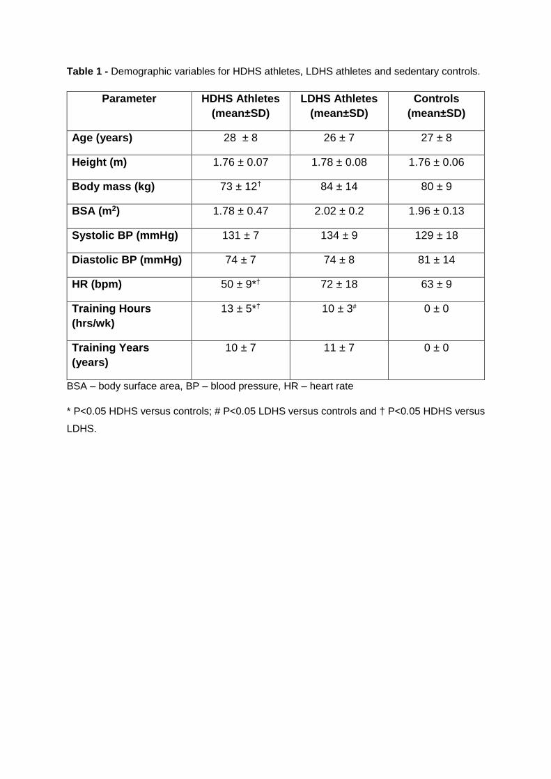
Table 1 - Demographic variables for HDHS athletes, LDHS athletes and sedentary controls.
BSA – body surface area, BP – blood pressure, HR – heart rate
* P<0.05 HDHS versus controls; # P<0.05 LDHS versus controls and † P<0.05 HDHS versus
LDHS.
Parameter HDHS Athletes (mean±SD)
LDHS Athletes (mean±SD)
Controls (mean±SD)
Age (years) 28 ± 8 26 ± 7 27 ± 8
Height (m) 1.76 ± 0.07 1.78 ± 0.08 1.76 ± 0.06
Body mass (kg) 73 ± 12† 84 ± 14 80 ± 9
BSA (m2) 1.78 ± 0.47 2.02 ± 0.2 1.96 ± 0.13
Systolic BP (mmHg) 131 ± 7 134 ± 9 129 ± 18
Diastolic BP (mmHg) 74 ± 7 74 ± 8 81 ± 14
HR (bpm) 50 ± 9*† 72 ± 18 63 ± 9
Training Hours (hrs/wk)
13 ± 5*† 10 ± 3# 0 ± 0
Training Years (years)
10 ± 7 11 ± 7 0 ± 0
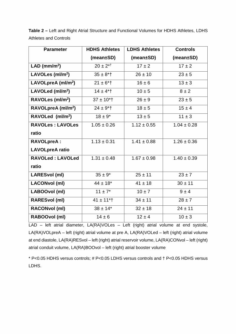
Table 2 – Left and Right Atrial Structure and Functional Volumes for HDHS Athletes, LDHS
Athletes and Controls
Parameter HDHS Athletes (mean±SD)
LDHS Athletes (mean±SD)
Controls (mean±SD)
LAD (mm/m2) 20 ± 2*† 17 ± 2 17 ± 2
LAVOLes (ml/m2) 35 ± 8*† 26 ± 10 23 ± 5
LAVOLpreA (ml/m2) 21 ± 6*† 16 ± 6 13 ± 3
LAVOLed (ml/m2) 14 ± 4*† 10 ± 5 8 ± 2
RAVOLes (ml/m2) 37 ± 10*† 26 ± 9 23 ± 5
RAVOLpreA (ml/m2) 24 ± 9*† 18 ± 5 15 ± 4
RAVOLed (ml/m2) 18 ± 9* 13 ± 5 11 ± 3
RAVOLes : LAVOLes ratio
1.05 ± 0.26 1.12 ± 0.55 1.04 ± 0.28
RAVOLpreA : LAVOLpreA ratio
1.13 ± 0.31 1.41 ± 0.88 1.26 ± 0.36
RAVOLed : LAVOLed ratio
1.31 ± 0.48 1.67 ± 0.98 1.40 ± 0.39
LARESvol (ml) 35 ± 9* 25 ± 11 23 ± 7
LACONvol (ml) 44 ± 18* 41 ± 18 30 ± 11
LABOOvol (ml) 11 ± 7* 10 ± 7 9 ± 4
RARESvol (ml) 41 ± 11*† 34 ± 11 28 ± 7
RACONvol (ml) 38 ± 14* 32 ± 18 24 ± 11
RABOOvol (ml) 14 ± 6 12 ± 4 10 ± 3
LAD – left atrial diameter, LA(RA)VOLes – Left (right) atrial volume at end systole,
LA(RA)VOLpreA – left (right) atrial volume at pre A, LA(RA)VOLed – left (right) atrial volume
at end diastole, LA(RA)RESvol – left (right) atrial reservoir volume, LA(RA)CONvol – left (right)
atrial conduit volume, LA(RA)BOOvol – left (right) atrial booster volume
* P<0.05 HDHS versus controls; # P<0.05 LDHS versus controls and † P<0.05 HDHS versus
LDHS.
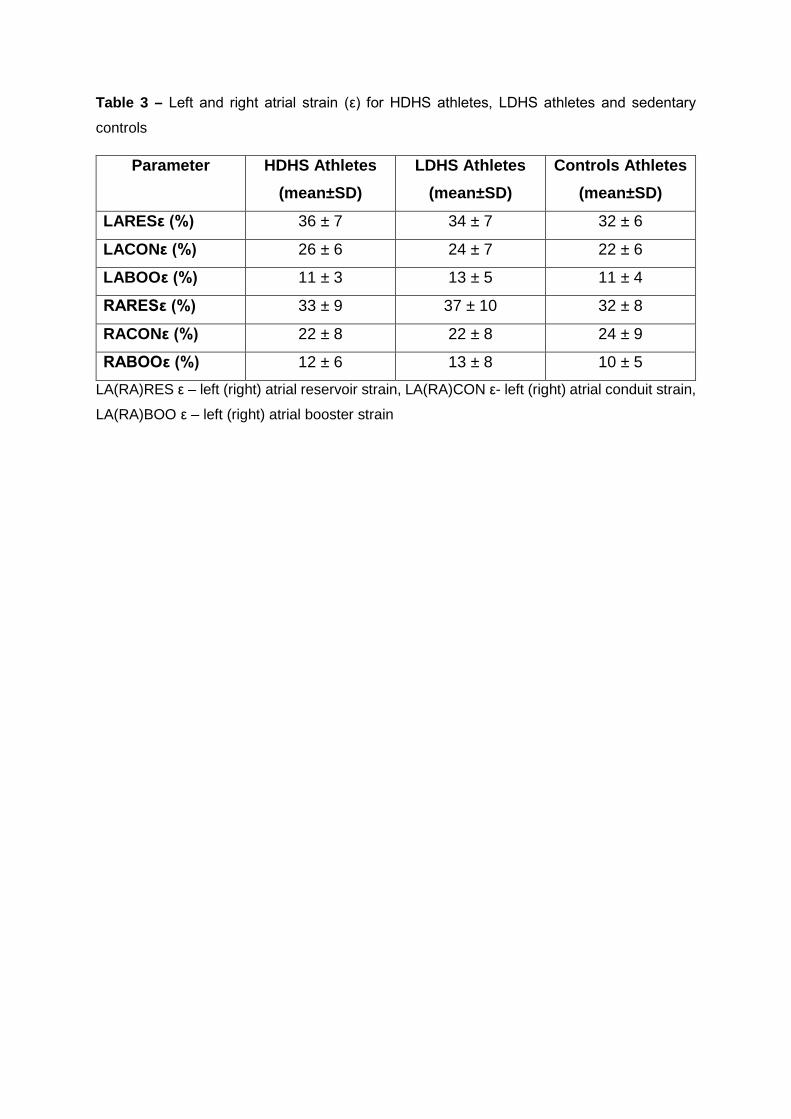
Table 3 – Left and right atrial strain (ε) for HDHS athletes, LDHS athletes and sedentary
controls
Parameter HDHS Athletes (mean±SD)
LDHS Athletes (mean±SD)
Controls Athletes (mean±SD)
LARESε (%) 36 ± 7 34 ± 7 32 ± 6
LACONε (%) 26 ± 6 24 ± 7 22 ± 6
LABOOε (%) 11 ± 3 13 ± 5 11 ± 4
RARESε (%) 33 ± 9 37 ± 10 32 ± 8
RACONε (%) 22 ± 8 22 ± 8 24 ± 9
RABOOε (%) 12 ± 6 13 ± 8 10 ± 5
LA(RA)RES ε – left (right) atrial reservoir strain, LA(RA)CON ε- left (right) atrial conduit strain,
LA(RA)BOO ε – left (right) atrial booster strain

