chromosomal mapping of the mink cell focus-inducing...
TRANSCRIPT

MouseChromosomal Mapping of the Mink Cell Focus-Inducing and Xenotropic env Gene Family in the
Cila Blatt, Karen Mileham, Martin Haas, Muriel N. Nesbitt, Mary E. Harper, and Melvin I. Simon
doi:10.1073/pnas.80.20.6298 1983;80;6298-6302 PNAS
This information is current as of December 2006.
www.pnas.org#otherarticlesThis article has been cited by other articles:
E-mail Alerts. click hereright corner of the article or
Receive free email alerts when new articles cite this article - sign up in the box at the top
Rights & Permissions www.pnas.org/misc/rightperm.shtml
To reproduce this article in part (figures, tables) or in entirety, see:
Reprints www.pnas.org/misc/reprints.shtml
To order reprints, see:
Notes:

Proc. Natl. Acad. Sci. USAVol. 80, pp. 6298-6302, October 1983Genetics
Chromosomal mapping of the mink cell focus-inducing andxenotropic env gene family in the mouse
(retroviruses/recombination/polymorphism/xenotropic cell-surface antigen/leukemia induction)
CILA BLATT*, KAREN MILEHAM*, MARTIN HAASt, MURIEL N. NESBITTf, MARY E. HARPER*, ANDMELVIN I. SIMON**The Agouron Institute, 505 Coast Boulevard South, La Jolla, CA 92037; and tCancer Center and fBiology Department, University of California at San Diego,La Jolla, CA 92093
Communicated by Dan L. Lindsley, July 14, 1983
ABSTRACT Chromosomal locations of members of the xeno-tropic-related env gene family in the mouse genome have beendetermined. Endonuclease restriction site polymorphisms de-tected by molecular hybridization were used to.study the inher-itance of mink cell-focus inducing and xenotropic env gene-relatedsequences in recombinant inbred strains of mice. Some of the en-dogenous env sequences, appear to be eloselylinked to genes de-termining leukemia virus induction and to genes involved in theimmune response, such as the heavy and light chains of the im-munoglobulin molecules or allotypic determinants on B and T lym-phocytes. The use of probes that detect restriction fragment lengthpolymorphisms in a small family of dispersed sequences promisesto yield a large number of markers that can be used together withrecombinant inbred strains for efficient mapping of the mousegenome.
The expression of specific retroviral functions is inherited in astable fashion in inbred strains of mice. Some of these functionshave been attributed to integrated ecotropic proviruses and othersto xenotropic viral sequences. Ecotropic viruses can proliferatein murine cells, whereas xenotropic viruses primarily infectheterologous cells. Genetic loci associated with ecotropic virusinduction and with stably integrated proviruses have beenmapped in the mouse genome by following levels of virusexpression or induction in different strains (1, 2). More recentlyDNA-DNA hybridization has been used to follow the patternof segregation of restriction fragment length polymorphisms tomap integrated viral sequences (3, 4).
All mice strains have sequences related to xenotropic virusesand these sequences are stable-components of the mouse ge-nome (5, 6). They appear to be comprised of a family of dis-persed genes. Three xenotropic-virus-associated functions havebeen mapped by following the induction of virus in inbred mousestrains (7-9), and hybridization with envelope (env)-specificprobes suggests that there are more than 15 sites at which thesesequences reside (5, 6). The resident xenotropic sequences, andparticularly those related to the env gene, appear to be asso-ciated with-a variety of important functions in the mouse. Theyhave. been implicated in the immune response (10) and the ap-pearance of differentiation-specific cell-surface antigens (11),and they play a role in the formation of leukemogenic viruses.The onset of leukemia is associated with the appearance of anew class of viruses, the dualtropic mink cell focus-inducing(MCF) viruses (12). Analysis of the structure of the MCF vi-ruses suggests that they arise by a recombination event thatsubstitutes an endogenous xenotropic-like sequence for themurine leukemia virus (MuLV) env gene (13-15). To further
characterize the xenotropic-like env sequences and their rela-tionship to the immune system, cell-surface antigen formation,and specific gene rearrangement, we sought to identify theirlocations on the mouse chromosomes.We were able to determine chromosomal locations for some
of the endogenous MCF env gene sequences by following en-donuclease restriction site polymorphisms in DNA of recom-binant inbred (RI) strains of mice. This approach has been ap-plied to map a number of genes (3, 4). The B x D RI strainswere derived by crossing the C57BL/6J (B) and DBA/2J (D)strains, followed by systematic inbreeding of the progeny, be-ginning with chosen pairs from the F2 generation (16, 17). Thesestrains are now highly inbred and have been used extensivelyfor gene mapping (3, 18, 19). The probability of recombinationoccurring between any two loci in the course of inbreeding isrelated to the map distance between them (16). In using RI strainsto map DNA-level variants, polymorphic patterns are first de-termined in the parental strains B and D. The DNA restrictionpattern of each one of the B x D RI strains is then screenedfor resemblance to either parental pattern (B or D). The resultof this analysis is summarized as the strain distribution pattern(SDP) for a given locus and is then compared with SDPs de-rived from previously mapped genes. Closely linked genes willhave similar SDPs.
Using this method and probing with a MCF xenotropic-spe-cific env gene clone, we were able to assign map positions to15 env gene sequences in the mouse genome. Some of thesewere closely linked to genes involved in the immune responseand others appeared to be linked to genes involved in virus in-duction.
MATERIALS AND METHODSMice. Mice were purchased from The Jackson Laboratory
(3).Restriction Endonuclease Digestion. Digestion of high mo-
-lecular weight liver DNA was carried out in a 4- to 6-fold en-zyme excess. The enzymes EcoRI, Pst I, and Bgl II were pre-pared in our laboratory. Other enzymes were purchased fromBethesda Research Laboratories.
Southern. Blot and Hybridization Analysis. Digested DNAwas electrophoresed through 0.8% agarose gels (5-10 kug perlane) and transferred to nitrocellulose as described (20). A re-.combinant pBR322 plasmid subclone. that contains a 700-basepair BamHI-EcoRI fragment of the env gene clone of Molo-ney-MCF virus pMo-MCFI-16 (21) was used as probe. The sub-
Abbreviations: MuLV, murine leukemia virus; MCF, mink cell focus-inducing; RI, recombinant inbred; SDP, strain distribution pattern; cM,centimorgan(s); kb, kilobase(s); MLC, mixed lymphocyte culture; LPS,lipopolysaccharide.
6298
The publication costs of this article were defrayed in part by page chargepayment. This article must therefore be hereby marked "advertise-ment" in accordance with 18 U.S.C. §1734 solely to indicate this fact.
Proc. Natl. Acad. Sci. USA 80 (1983) 6299
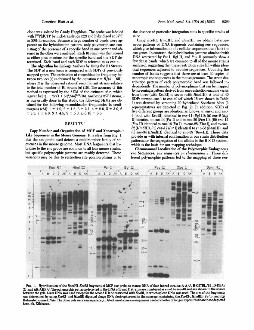
clone was isolated by Candy Haggblom. The probe was labeledwith [32P]dCTP by nick-translation (22) and hybridized at 370Cin 50% formamide. Because a large number of bands were ap-parent on the hybridization pattern, only polymorphisms con-sisting of the presence of a specific band in one parent and ab-sence in the other were analyzed. Each RI strain was then scoredas either plus or minus for the specific band and the SDP de-termined. Each band and each .SDP is referred to as env n.The Algorithm for Linkage Analysis by Using the RI Strains.
The SDP of a new locus is compared with SDPs of previouslymapped genes. The estimation of recombination frequency be-tween two loci (r) is obtained by the equation r = R/(4 - 6R),where R is the observed ratio; of recombinant -strains relativeto the total number of RI strains (n) (16). The accuracy of thismethod is expressed by the SEM of the estimate of r, whichis given by [r(l + 2r)(1 + 6r)2/4n]'/2 (16). Analyzing 25 RI strains,as was usually done in this study, the following SEMs are ob-tained for the following recombination frequencies in centi-morgans (cM): 1 ± 1.0, 2 ± 1.6, 3 + 2.1,-4 ± 2.6, 5 ± 3.0, 6± 3.5, 7 + 4.0, 8 + 4.5, 9 ± 5.0, and 10 ± 5.5.
RESULTS
Copy Number and Organization of MCF and Xenoti-opic-Like Sequences in the Mouse Genome. It is clear from Fig. 1that the env probe used detects a multimember family of se--quences in the mouse genome. Most DNA fragments that hy-bridize to the env probe are common to all four mouse strains,but specific polymorphic patterns are readily detected. Thesevariations may be due to restriction site polymorphisms or to
the absence of particular integration sites in specific strains ofmice.
Using EcoRI, HindIII, and BamHI, we obtain heteroge-neous patterns of DNA fragments containing env sequences,which give information on the cellular sequences that flank theenv genes. In contrast, the hybridization patterns obtained withDNA restricted by Pst I, Bgl II, and Pvu II primarily show afew dense bands, which are common to all of the mouse strainsanalyzed, suggesting that these restriction sites fall within iden-tical sequences adjacent to env-like sequences. Counting thenumber of bands suggests that there are at least 30 copies ofxenotropic env sequences in the mouse genome. The strain dis-tribution pattern of each polymorphic band was followed in-dependently. The number of polymorphisms that can be mappedby'screening a pattern derived from one restriction enzyme variesfrom three (with EcoRI) to seven (with HindIII). A total of 40SDPs termed env-1 to env-40 (of which -16 are shown in Table1) was derived by screening 20 hybridized Southern.blots (2representatives are depicted in Fig. 2). In addition, SDPs offive different groups are identical as follows: (i) env-1 and env-4 (both with EcoRI) identical to env-11 (Bgl II); (ii) env-9 (BglII) identieal to env-14 (Pst I) and to env-20 (Pvu II); (iii) env-12(Pvu II) identical to env-16 (Pst I), to env-28 (Xba I), and to env-32 (HindIII); (iv) env-17 (Pst I) identical to env-38 (BamHI); and(v) ent-34 (HindIII) identical to env-39 (BamHI). These dataprovide us with internal confirmation of our strain distributionpatterns for the segregation of the alleles in the B x D system,which is the basis for our mapping technique.Chromosomal Localization of the Polymorphic Endogenous
env Sequences. env sequences on -chromosome 1. Three dif-ferent polymorphic patterns led to the mapping of three env
Eco RI Hind EEl~ _
kb A B D D AK A B D AK
PstII Bgl IIA 8 D AK A B D AK
Pvu I Xba I
A B D AK A B D AK
9I.9- E Z -29
200_-28
2 ~~~~~~~~39\-37-,
'27 lo
38- _35-
qw, 4w~~~I--7 3
4-
_q
pL*22- -26
23-24-
v
40-
FtG. 1. Hybridization of the BamHI-EcoRI fragment of MCF env probe to mouse DNA of four inbred strains: A-A/J, B-C57BL/6J, D-DBA/2J, and AK-AKR/J. The polymorphic patterns detected in the DNA ofB and D strains are numbered as env-1 to env-40 and are shown in the spaces
between the gels. Liver DNA was used except for the second D lane restricted with EcoRI, in which spleenDNA was used.. The size of the fragmentswas determined by using EcoRI- and HindIl-digested phage DNA electrophoresed in the same gel containing the EcoRI-, HindH-, Pst I-, and Bgl1-digested mouse DNAs. The other gels were run separately. Detection ofsome env sequences needed shorter or longer exposures than those depicted
here. kb, Kilobases.
A
Bam HIAI BA B D AK
22 -B6 -
14 -
8o-B -I
6 -l 3-
2-I-
4 -
3 -
2 -j
4- EU0
Genetics: Blatt et al.
Proc. Natl. Acad. Sci. USA 80 (1983)
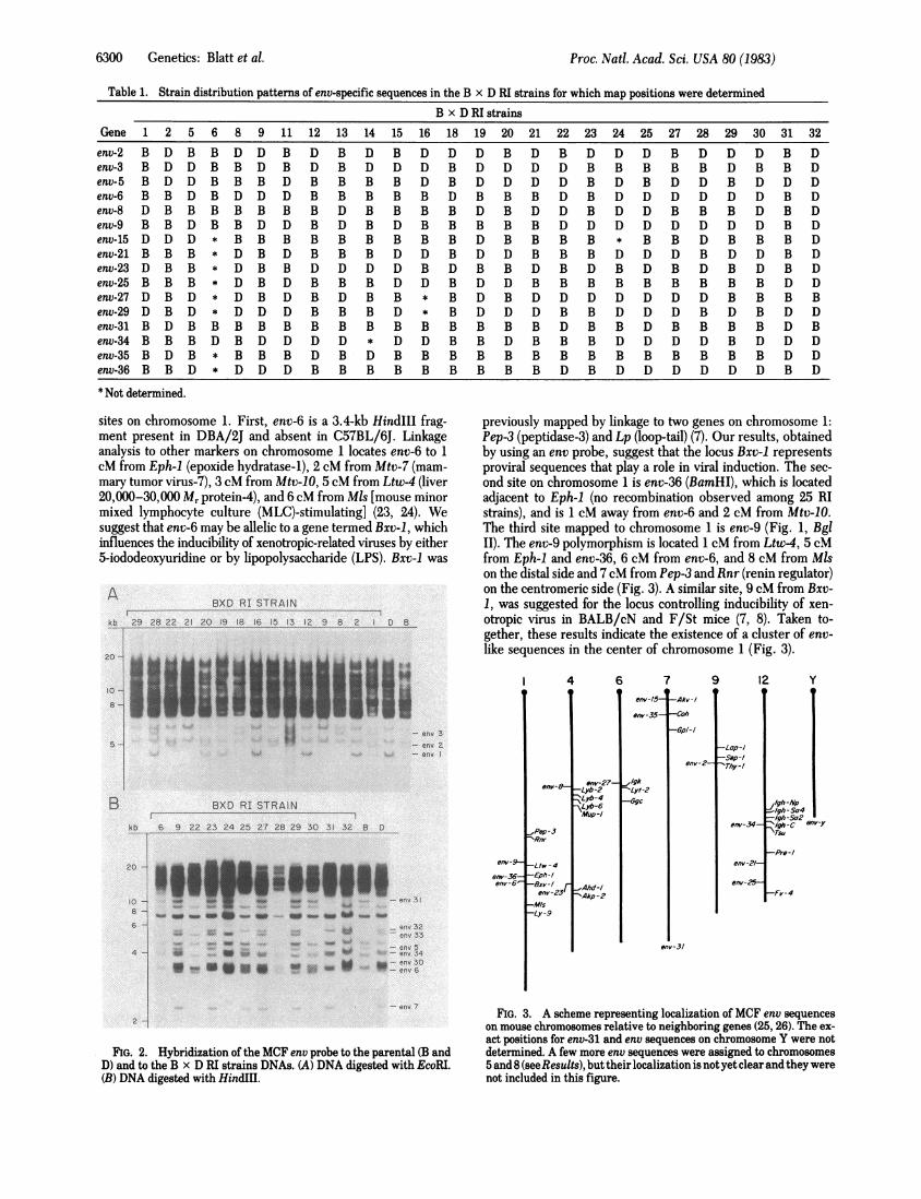
Table 1. Strain distribution patterns of env-specific sequences in the B x D RI strains for which map positions were determined
B x D RI strainsGene 1 2 5 6 8 9 11 12 13 14 15 16 18 19 20 21 22 23 24 25 27 28 29 30 31 32env-2 B D B B D D B D B D B D D D B D B D D D B D D D B Denv-3 B D D B B D B D B D D D B D D D D B B B B B D B B Denv-5 B D D B B B D B B B B D B D D D D B D B D D B D D Denv-6 B B D B D D D B B B B B D B B B D B D D D D D D B Denv-8 D B B B B B B B D B B B B D B D D B D D B B B D B Denv-9 B B D B B D D B D B D B B B B B D D D D D D D D B Denv-15 D D D * B B B B B B B B B D B B B B * B B D B B B Denv-21 B B B * D B D B B B D D B D D B B B D D D B D D B Denv-23 D B B * D B B D D D D B D B B D B D B D B D B D B Denv-25 B B B * D B D B B B D D B D D B B B B B B B B B D Denv-27 D B D * D B D B D B B * B D B D D D D D D D B B B Benv-29 D B D * D D D B B B D * B D D D B B D D D B D B D Denv-31 B D B B B B B B B B B B B B B B D B B D B B B B D Benv-34 B B B D B D D D D * D D B B D B B B D D D D B D D Denv-35 B D B * B B B D B D B B B B B B B B B B B B B B D Denv-36 B B D * D D D B B B B B B B B B D B D D D D D D B D* Not determined.
sites on chromosome 1. First, env-6 is a 3.4-kb HindIII frag-ment present in DBA/2J and absent in C57BL/6J. Linkageanalysis to other markers on chromosome 1 locates env-6 to 1cM from Eph-l (epoxide hydratase-1), 2 cM from Mtv-7 (mam-mary tumor virus-7), 3 cM from Mtv-10, 5 cM from Ltw-4 (liver20,000-30,000 Mr protein-4), and 6 cM from Mls [mouse minormixed lymphocyte culture (MLC)-stimulating] (23, 24). Wesuggest that env-6 may be allelic to a gene termed Bxv-1, whichinfluences the inducibility of xenotropic-related viruses by either5-iododeoxyuridine or by lipopolysaccharide (LPS). Bxv-1 was
ABXD RI STRAIN
kb 29 28 22 21 20 19 18 16 15 13 12 9 8 2 18B
Iill - env 3
i f-il' - env 2-env
BXD RI STRAINB
kb: 6 9 22 23 24 25 27 28 2!9 30 31 32 8 CD
20-
10 -
6
4-
env-
env-36-env-6~
en v 3
w SW NW go_MO___env32
env 33
|"env 5
W"'. ANO env 34
env 3 0
previously mapped by linkage to two genes on chromosome 1:Pep-3 (peptidase-3) and Lp (loop-tail) (7). Our results, obtainedby using an env probe, suggest that the locus Bxv-1 representsproviral sequences that play a role in viral induction. The sec-ond site on chromosome 1 is env-36 (BamHI), which is locatedadjacent to Eph-1 (no recombination observed among 25 RIstrains), and is 1 cM away from env-6 and 2 cM from Mtv-10-.The third site mapped to chromosome 1 is env-9 (Fig. 1, BglII). The env-9 polymorphism is located 1 cM from Ltw-4, 5 cMfrom Eph-1 and env-36, 6 cM from env-6, and 8 cM from Mlson the distal side and 7cM from Pep-3 and Rnr (renin regulator)on the centromeric side (Fig. 3). A similar site, 9 cM from Bxv-1, was suggested for the locus controlling inducibility of xen-otropic virus in BALB/cN and F/St mice (7, 8). Taken to-gether, these results indicate the existence of a cluster of env-like sequences in the center of chromosome 1 (Fig. 3).
env-8-
.Pep-3'Rm-
-Ltw-4-Eph-I-Sxv-/ r
env-23-M/s-Ly -9
69
env-27--Lyb-2Lyb-4\Lb-6Mup-I
_Ahd-/-Akp-2
7env-/5
env-35
,Igk _-LLyt-2-Ggc
-Aktv-/
-Coh
-op/-I
env-2-
9 129
-Lop-I-Sep-I-hy -/
env-34-
env-2/-
env-25-
yS
/lgh-/JpJIgh -So4I-Igh-S52V/gh-C env-.y
-Pre-/
-Fv-4
env-31
env 7
2
IFIG. 2. Hybridization of the MOF env probe to the parental (B and
D) and to the B x D RI strains DNAs. (A) DNA digested with EcoRI.(B) DNA digested with HindIII.
FIG. 3. A scheme representing localization of MCF env sequenceson mouse chromosomes relative to neighboring genes (25, 26). The ex-
act positions for env-31 and env sequences on chromosome Y were notdetermined. A few more env sequences were assigned to chromosomes5 and 8 (seeResults), but their localization is notyet clear andthey werenot included in this figure.
20 -
10
8-
5-
B
6300 Genetics: Blatt et al.

Proc. Natl. Acad. Sci. USA 80 (1983) 6301
env sequences on chromosome 4. Two env polymorphismsare located on chromosome 4. The first, env-8 (Bgl II), showstight linkage to a unique region of genes specifying B-lym-phocyte alloantigens. env-8 maps 1 cM from Lyb-2, 1 cM fromLyb-6, 4 cM from Lyb-4, and 5 cM from Mup-1 (major urinaryprotein-i) (23, 24). The Lyb genes apparently control antigenicdeterminants on cells of the B-lymphocyte lineage. Lyb-2 mayrepresent a cell-surface marker for very early B cells (27). Lyb-4 is an alloantigen that appears to be involved with lymphocyte-activating determinants in MLC reactions (27), and antiserumto Lyb-6 determinants was shown to be mitogenic for B cells(28). Therefore, it is not surprising to find Lps, a gene for LPSresponsiveness, in close association with the above genes (23).Our linkage data suggest that env-8 is located either among thesegenes, between Lyb-2 and Lyb-6, or on the centromeric sideof the cluster, 1 cM from Lyb-2 (Fig. 3).The second site for an env sequence on chromosome 4 is env-
23 (Pvu II), which is located 2 cM from Akp-2 (alkaline phos-phatase-2) and Ahd-1 (aldehyde dehydrogenase-1), 7 cM fromGpd-1 (glucose phosphate dehydrogenase-i), and 11 cM fromFv-1 (Friend virus susceptibility-i) (Fig. 3). This env sequencemay be close to or overlap with a gene controlling the level ofexpression of xenotropic cell-surface antigen (24, 29).
env sequences on chromosome 5. Several env sequences showloose linkage to markers on chromosome 5. For example, env-5 (HindIII) maps 7 cM from Pgm-1 (phosphoglucomutase-1) (23)and 11 cM from Afp (a-fetoprotein) (unpublished data) and istentatively placed between these two genes. The polymor-phism represented by env-3 (EcoRI) could be located 9 cM fromAfp, but it does not show significant linkage to Pgm-1 (14 cM)or R* (Rickettsia resistance) (15 cM).
env sequences on chromosome 6. Restriction with Xba I givesrise to env-27, which maps 2 cM from Lyt-2 (T-lymphocyte al-loantigen-2) (24). The distance of 8 cM from Ggc (y-glutamylcyclotransferase) places env-27 on the centromeric side of Lyt-2 and close to Igk, the gene coding for immunoglobulin lightchain K (23, 24).
env sequences on chromosome 7. The centromeric part ofchromosome 7 includes at least two env sequences. env-35(BamHI) maps to the locus of Coh (coumarin hydroxylase) (norecombination among 23 mice tested) and 4 cM from Gpi-1(glucose phosphate isomerase-i) (23). The second locus, env-15(Pst I), is located 6 cM from Coh. Lack of significant linkage toGpi-1 suggests that env-15 is close to the centromere and verynear to Akv-1, a locus influencing inducibility of AKR leukemiavirus (1, 23). A third locus is env-31 (HindIII), which showslinkage of 8 cM to Coh, 7 cM to Tam-1 (tosyl arginine meth-ylesterase-i), and 14 cM to Gpi-1. Thus, the exact position ofenv-31 is not clear.
env sequences on chromosome 8. The results of digestion withXba I define env-29, which shows linkage to Blv-1 (4 cM) onchromosome 8. The locus Blv-1 designates an integration siteof ecotropic MuLV in the DNA of C57BL/6J (4, 24).
env sequences on chromosome 9. The hybridization patternof the env probe with EcoRI fragments of mouse DNA led tothe mapping of env-2 on chromosome 9, 2 cM from Sep-1 (serumprotein-i) and 8 cM from Lap-1 (leucine arylamino-peptidase-1) (23). These results locate env-2 very near or at the locus de-fined for Thy-1 (6 antigen) (30).
env sequences on chromsome 12. Further analysis of SDPsled to the finding that env-34 (Fig. 2B, HindIII) and env-39(Fig. 1, BamHI) are identical and map to the region coding forthe heavy chain of the immunoglobulin molecule on chromo-some 12. This site is located 3 cM from Igh-Sa2 and 4 cM fromIgh-Sa4 and Igh-c on the centromeric side and 7 cM from Pre-I (prealbumin component-i), which is on the distal side of
chromosome 12 (23) (Fig. 3). The locus suggested for env-34 isalso the site suggested for a cluster of T-cell alloantigens Tsuand Tind (31, 32). A second site on chromosome 12 is derivedfrom the SDP of env-21 (Pvu II). This site is located 4 cM fromPre-i. A third env gene sequence, env-25 (Pvu II), is linked toLyb-7 (B-lymphocyte alloantigen-7) [no recombination was ob-served for the eight RI lines scored for Lyb-7 (23)] and is looselylinked to env-21 (9 cM) and to Pre-i (12 cM). Lack of linkageto the immunoglobulin genes suggests that env-21 and env-25may be located on the distal part of chromosome 12.
env sequences on chromosome Y. Three DNA fragments ofstrains A/J and C57BL/6J restricted with Bgl II hybridize withthe env probe but are missing from DNA of DBA/2J mice (Fig.1). These DNA fragments segregate in concordance with the Ychromosome in DNA of 40 males and females representing RIstrains derived from A/J and C57BL/6J (data not shown).
Fifteen other SDPs were derived from the polymorphic pat-terns observed, but significant linkage with other markers (<10cM) was not detected. However, as more genes are assigned tospecific loci, we will be able to localize these 15 env gene se-quences, whose SDPs contribute to the expansion of the link-age map of the mouse genome.
DISCUSSION
Recombinant inbred strains of mice have been used to map re-striction length polymorphisms, including polymorphisms as-sociated with ecotropic viral insertions in the mouse germ line(4). Using this technique we were able to look at the chro-mosomal distributions of sequences that correspond to a genefamily-i.e., the MCF and xenotropic env gene sequences. Thetechnique requires that the xenotropic sequences exist in a sta-ble configuration in the parental and the RI mice. Hoggan etal. (6), using a probe similar to the one we used, concluded thatthere was minimal polymorphism of env sequences betweeninbred mouse strains. Corroboration for the chromosomal as-signments reported here comes from studies in our laboratorywith another set of RI strains (A x B and B X A), in which someof the same map positions have been found. Furthermore, us-ing six other RI strains, Wejman et al. also found some of thesame map positions with a similar env-specific probe (J. Wej-man, B. A. Taylor, N. Jenkins, and N. Copeland, personal com-munication).The resolution of the RI mapping technique establishes link-
age to within 1 cM of a known marker. On the molecular level,1 cM corresponds to hundreds of thousands of base pairs.Nonetheless, the RI method allows us to develop testable hy-potheses about the relationships between MCF sequences andadjacent functions. For example, the polymorphisms env-6, env-9, and env-36 appear to cluster near the site of the Bxv-1 locus,which has been shown to be involved in the induction of xen-otropic virus (7). A second cluster of polymorphisms associatedwith env map on chromosome 7 adjacent to the Akv-i locus,which is involved in ecotropic MuLV induction. The associationof structural env gene sequences with loci determining virusinducibility suggests that molecular cloning and sequence anal-ysis of these specific genomic DNA fragments may help to es-tablish the relationship between MCF sequences and themechanisms involved in the regulation of leukemia virus in-duction.
Another group of polymorphisms associated with env se-quences map at or very close to loci that express genes involveddirectly in the immune response. The sequence env-34 is tightlylinked to immunoglobulin heavy chain genes on chromosome12, whereas env-27 is linked to the light chain gene on chro-mosome 6 (Fig. 3). Recently, it has been shown that specific
Genetics: Blatt et al.

Proc. Natl. Acad. Sci. USA 80 (1983)
translocations into the immunoglobulin regions on chromosome6 or 12, involving endogenous oncogenes such as c-myc or c-mos, were associated with plasmacytomas in mice (25, 26, 33).Our data suggest close linkage between endogenous env se-quences and the immunoglobulin genes and raise the possi-bility that MCF sequences may play some role in the specifictranslocations that are frequently found in B-cell tumors.
It is interesting to note that the env loci on chromosomes 6and 12 are also closely linked to genes that have been relatedto T-cell surface markers. In particular, env-27 is located 2 cMfrom Lyt-2 and Lyt-3 on chromosome 6, which are specificallyexpressed by cytotoxic and suppressor T cells. The env se-quence on chromosome 12, env-34, is adjacent to the genes thathave been associated with T-cell alloantigens Tsu and Tind, whichare apparently involved in suppression or augmentation of theimmune response (31, 32). In addition, env-2 appears to be ad-jacent to Thy-i, the 6 antigen on chromosome 9, which is alsoa marker expressed on thymocytes. Furthermore, env-8 is linkedto a cluster of Lyb genes on chromosome 4, which are involvedin the expression of B-cell alloantigens. Howe et al. (25) sug-gested that the Lyb-4 locus may reflect a viral integration sitein order to account for a different chromosomal location for Lyb-4 in C3H mice. The close association of a number of env se-quences with loci involved in the immune response may be for-tuitous, or the presence of these genes in a region of the chro-mosome that is activated in the immune response may lead totheir gratuitous coexpression with immune-specific functions.Nevertheless, there is evidence to suggest that the viral en-velope glycoprotein gp 70 is a constituent of the surface of nor-mal cells (34-36). Its expression appears to be restricted mainlyto lymphoid and epithelial cells and it could play a role in dif-ferentiation and development (34). Work by Moroni et al. (10)has documented the synergistic induction or inhibition of xen-otropic env gene products and immune functions. The closechromosomal linkage between MCF env gene sequences andgenes involved in various functions of the immune responsemay reflect a functional interaction between them. The cell-surface localization of xenotropic env gene products suggeststhat they may function as membrane receptors on some cells.Alteration of such receptors (36, 37) resulting from env generecombination could play a role in cellular differentiation andoncogenesis. Alternatively, the genetic linkage may be adven-titious. A closer analysis of the degree of linkage and the detailsof gene expression in specific regions involved in the immuneresponse should resolve this question.We thank Dr. Marguerite Vogt for the MCF env probe and for help-
ful discussions. Dr. B. A. Taylor kindly provided a compilation of knownSDPs in the B X D mice. We thank Jessica Dausman, Lisa Jahn, andSteve Marks for technical assistance and Michele Platten for manuscriptpreparation. This work was supported by National Institutes of HealthGrant GM29797.
1. Rowe, W. P., Hartley, J. W. & Bremner, T. (1972) Science 178,860-862.
2. Ihle, J. N., Joseph, D. R. & Dormotor, J. J. (1979) Science 204,71-73.
3. Traina, V. L., Taylor, B. A. & Cohen, J. C. (1981)J. Virol. 40, 735-744.
4. Jenkins, N. A., Copeland, N. G., Taylor, B. A. & Lee, B. K. (1981)Nature (London) 293, 370-374.
5. Chattopadhyay, S. K., Cloyd, M. W., Linemeyer, D. L., Lander,M. R., Rands, E. & Lowy, D. R. (1982) Nature (London) 295, 25-31.
6. Hoggan, M. D., Buckler, C. E., Sears, J. F., Rowe, W. P. & Mar-tin, M. A. (1983) J. Virol. 45, 473-477.
7. Kozak, C. A. & Rowe, W. P. (1980)J. Exp. Med. 152, 219-228.8. Morse, H. C., Kozak, C. A., Yetter, R. A. & Hartley, J. W. (1982)
J. Virol. 43, 1-7.9. Yetter, R. A., Hartley, J. W. & Morse, H. C. (1983) Proc. Natl.
Acad. Sci. USA 80, 505-509.10. Moroni, C., Matter, A., Stoye, J. P., Monckton, R. P., Dela-
marter, J. F. & Schumann, G. (1980) Cell. Immunol. 54, 107-114.11. Tung, J. S., Vitetta, E. S., Fleissner, E. & Boyse, E. A. (1975)J.
Exp. Med. 141, 198-205.12. Hartley, J. W., Wolford, N. K., Old, L. J. & Rowe, W. P. (1977)
Proc. Natl. Acad. Sci. USA 74, 789-792.13. Elder, J. H., Gautsch, J. W., Jensen, F. C., Lerner, R. A., Hart-
ley, J. W. & Rowe, W P. (1977) Proc. Natl. Acad. Sci. USA 74, 4676-4680.
14. Rommelaere, J., Faller, D. V. & Hopkins, N. (1978) Proc. Nati.Acad. Sci. USA 75, 495-499.
15. Chien, Y. H., Verma, I. M., Shih, T. Y., Scolnick, E. M. &Davidson, N. (1978) J. Virol. 28, 352-360.
16. Taylor, B. A. (1978) in Origin of Inbred Mice, ed. Morse, C. H.(Academic, New York), pp. 423-438.
17. Bailey, D. W. (1971) Transplantation 11, 325-327.18. Festenstein, H., Bishop, C. & Taylor, B. A. (1977) Immunoge-
netics 5, 357-361.19. Taylor, B. A., Bailey, D. W., Cherry, M., Riblet, R. & Weigert,
M. (1975) Nature (London) 256, 644-646.20. Southern, E. M. (1975) J. Mol. Biol. 98, 503-517.21. Bosselman, R. A., Van Straaten, F., Van Beveren, C., Verma, I.
M. & Vogt, M. (1982) J. Virol. 44, 19-31.22. Maniatis, T., Jeffrey, A. & Kleid, D. G. (1975) Proc. Natl. Acad.
Sci. USA 72, 1184-1188.23. Roderick, T. H. & Davisson, M. T. (1982) Mouse News Lett. 67,
7-9.24. Womack, J. E. (1982) Genetic Maps 2, 286-294.25. Calame, K., Kim, S., Lalley, P., Hill, R., Davis, M. & Hood, L.
(1982) Proc. Natl. Acad. Sci. USA 79, 6994-6998.26. Rechavi, G., Givol, D. & Canaani, E. (1982) Nature (London) 300,
607-611.27. Howe, R. C., Ahmed, A., Fladetta, T. J., Byrnes, J. E., Rogan,
K. M., Dorf, M. E., Taylor, B. A. & Humphreys, R. E. (1979)Immunogenetics 9, 221-232.
28. Kessler, S. W., Ahmed, A. & Scher, I. (1978) in B Lymphocytesin the Immune Response, eds. Cooper, M., Mosier, D., Scher, I.& Vitetta, E. (Elsevier/North-Holland, New York), pp. 47-54.
29. Kozak, C. A. & Rowe, W. P. (1980) in Animal Virus Genetics, eds.Fields, B. & Jaenisch, R. (Academic, New York), pp. 171-176.
30. Douglas, T. C. & Dawson, P. E. (1979) J. Hered. 70, 250-254.31. Owen, F. L., Riblet, R. & Taylor, B. A. (1981)J. Exp. Med. 153,
801-810.32. Owen, F. L., Spurll, G. M. & Panageas, E. (1982) J. Exp. Med.
155, 52-60.33. Taub, R., Kirsch, I., Morton, C., Lenoir, G., Swan, G., Tronick,
S., Aaronson, S. & Leder, P. (1982) Proc. Natl. Acad. Sci. USA 79,7837-7841.
34. Elder, J. H., Jensen, F. C., Bryant, M. L. & Lerner, R. A. (1977)Nature (London) 267, 23-28.
35. Hara, I., Izui, S., McConhey, P. J., Elder, J. H., Jensen, F. C.& Dixon, F. J. (1981) Proc. Natl. Acad. Sci. USA 78, 4397-4401.
36. Roman, J. M., Hirsch, J., Readhead, C., Levy, D., DeOgny, L.& Dreyer, W. J. (1981) Transplant. Proc. 13, 1782-1786.
37. Kemp, M. C., Famulari, N. G. & Compans, R. W. (1981)J. Virol.39, 463-470.
6302 Genetics: Blatt et al.