chromatography of dns derivatives on pre-coated high-performance thin-layer chromatographic plates
TRANSCRIPT

Journal ofChromotogrrophy, 131 (1977) 109419 @ JZkevier Scientik Publishing Company, Amsterdam - Printed in The Netherlands
CHROW 9364
CHROMATOGRAPHY OF Dns DERIVATIVES ON PRE-COATED HIGH- PERFORMANCE THIN-LAYER CHROMATOGRAPHK PLATES
N. SElLER and B. KNGDGEN
Mnr-planck-Im&itut f? Htinforschung7 Arbeitsgruppe Neurochemie, FrankfmtjM (G-F-R.)
(Received May 31st, 1976)
SUMMARY
5-Dimethylaminonaphthalene-I-sulphonyl (Dns) derivatives of amino acids and amines have been separated on high-performance thin-layer chromatographic (HPTLC) plates. Due to interaction of the matrix, especially with polar compounds and solvents, relative mobilities on HPTLC plates differ from those on silica gel G layers; as these differences are small for non-polar compounds, HPTLC plates can be used for the determination of aliphatic polyamines and allied compounds when using known solvent systems for separation. This paper describes (i) solvent systems for the two-dimensional separation of Dns-amino acids that are suitable for HPTLC and normal silica gel plates, (ii) a horizontaEdeveiopment technique that allows preparation of several two-dimensional chromatograms on a 10 x 10 cm plate, and (iii) an extraction procedure for the optical evaluation of compounds separated by thin-layer chromatography. The obvious advantages of HPTLC plates are rapid development, high capacity and increase in the signal-to-noise ratio.
INTRODUCTION
5-Dimethylaminonaphthalene-1-sulphonyl chloride (Dns-Cl) is currently among the most important reagents for fluorescent labelling of low-moiecular-weight compounds’. Since the introduction of thin-layer chromatography (TLC) for the separation of Dns-amino acid and -amine derivativeszs3, many separations of Dns derivatives have been carried out on silica gel-coated plates and many solvents have been devised for the separation of Dns-amino acids44 and Dns-amines’s’. An im- provement in the detection of amino acids was the introduction of polyamide sheetsg, on which the small spots that can be produced allow the detection sensitivity to be improved; the separation procedure of Woods and Wang9, or versions of this method, have become a standard technique for end-group work”. However, the low capacity of this layer material, together with the restriction to distribution-chromato- graphy systems, limits the usefulness of polyamide sheets in the analysis of biologica materials. Nevertheless, they have been used for the detection of amino acids and cer- tain amines in tissues”+_
The recently developed high-performance thin-layer chromatography (HPTLC)

110 N. SEILJZR, B. KNCiDG
pre-coated plates combine the high capacity and versatility of normal plates with the smalI spot-size typical of polyamide layers. It was therefore to ascertain to what extent the experiences of and procedures for assayin arnines and amino acids could be applied to these plates.
MATERIALS
The silica gel 60 F2s4 pre-coated plates (HPTLC) (silica gel pore size 60 A, Iayer thickness 200 pm), silica gel G and solvents were obtained from Merck (Darm- stadt, G.F.R.); An automated spreader (Camag, Muttenz, Switzerland) was used to prepare 20 x 20 cm TLC plates (layer thickness 3OOpm). The Dns-CI and Dns derivatives of amines were prepared in our laboratory according to known proce- dures4J’. Dns-amino acids were obtained from Sigma (St_ Louis, MO., U.S.A.), spermine phosphate, spermidine phosphate and putrescine hydrochloride from Fluka (Buchs, Switzerland) and Paminobutyric acid from Serva (Heidelberg, G.F.R.); monoacetyl putrescine was prepared as described by Tabor et aZ.13.
RESULTS f
Some characteris f its of HPTL C plates I The purity of HPTLC plates is inadequate for quantitative work. As shown
in Fig. 1, a considerable amount of absorbing and Suorescing material can be moved I with methanol to the solvent front, even if the plate is developed immediately after it has been taken out of a fresh package. Washing with methanol is, however, sufficient to purify the plate to an extent adequate for the determination of Dns derivatives. In I addition to optical characteristics, mass spectrometry was used as a criterion for ade quate purity of the thin layer. Mass spectra were recorded of 1-nmole amounts o bis-Dns-putrescine, which were eluted with 50~1 of ethyl acetate_ The background of.the spectra was Iow, especially in the region of significance (m/e > 150); it was only negligibly higher in the spectrum from the spot of a methanol-washed HPTLC plate as compared with that of a normal (untreated) silica gel G plate.
The data summarized in Fig. 1 show that the layer thickness is homogenous if the plate edge is disregarded. In situ evaluation of the spots can therefore be used even by transmittance measurements.
The electropherograms of non-derivatized biogenic amines, obtained with use of 0.5 M pyridinium acetate buffer of pH 4.8 (see ref. 14), show considerable interaction of the HPTLC layer with the amines (see Fig. 2). Even the order of rel- ative electrophoretic mobilities is reversed on the HPTLC, as compared with normal silica gei (and celhrlose) Iayers. Interaction of the Dns derivatives with the gel matrix is much weaker, so that the relative mobilities are similar on all types of silica gel plates (see Fig. 3), provided that the solvent mixture contains only components of low, or relatively lo-w, polarity (ahphatic or aromatic hydrocarbons, ethers, esters or halogenzted hydrocarbons). As poIar solvents (such as triethylamine, methanol or water) are preferentially adsorbed by the highly active HPTLC layer, the order of relative mobilities can be changed (see Fig. 4); binders present in commercial plates have much less influence in this respect. Nevertheless, most solvents that have been suggested for the separation of Dns-amines4*8 can still be used for chromatography

I , NPTLC OF Dns-AMINO ACIDS AND -AMINES
I Ill
‘c_
fiOOnm
Activation: * 365 nm
Fig. 1. Transmittance and fluorescence of a commercial HPTLC plate. A fresh pIate was developed with methanol for a distance indicated by the arrow. The transmittance at different wavelengths and the total fluorescence were scanned with the device described by SeilerJl. Absorbing and fluorescing material accumulates at the solvent front. The Iow transmittance near one edge of the plate is due to inhomogeneity of the layer.
Fig. 2. Comparison of elestrophoretic mobilities of some non-derivatized biogenic amines and amino acids on HFMX and silica gel G 1500 plates (Schleicher & Schtill, Dassel, G.F.R.) with 0.5 M pyridmium acetate buffer of pH 4.8. HPTLC plate: 40 V/cm, 15 min; silica gel G 1500 plate: 20 V/cm, 45 min. 1 = Putrescine; 2 = spermidine; 3 = spermine; 4 = monoacetylputrescine; 5 = putreanine; 6 = homocamosine; 7 = 4-aminobutyrate: 8 = mixture of putrescine. spermidine. spermine, monoacetylputrescine and 4-aminobutyrate. Visualization by reaction with ninhydqine.

112 N. SEILER, B. KNODGEN
Origin
Fig. 3. Relative mobilities of Dns-amines on HPTLC and 3OO+m silica gel G layers. Solvent: ethyl acetate-cyclohexane (3:2). Development in horizontal ‘anks (solvent-vapour-saturated atmosphere) for 5 and 10 cm, respectively. 1 = Ammonia; 2 = putrescine; 3 = spermidine; 4 = spermine; 5 = methylamine; 6 = dimethylamine; 7 = ethanolamine; 8 = 2-oxopyrrolidine (reaction product of Caminobutyrate); 9 = histamine; 10 = 5-hydroxytryptamine; 11 = phenethylamine; 12 = tyra- mine. Amounts: about 2 nmoles on the silica gel and 1 nmole on tbe HPTLC plate-
t (LlO cm ,I
1 2 3 4 6 i
_~z._qn _ J i ._ .I ? Fig. 4. Relative mobilities of Dns-amines on HPTLC and 3OOqm silica gel G layers. Solvent: ben- zene-triethylamine (5:l). Other conditions, compounds and amounts as specified in Fig. 3.
i
I

HPTLC OF DIE-AMINO ACIDS AND -AMINES 113
on HPTLC plates. Fig. 5 shows a two-dimensional separation of Dns-amines with a solvent combination useful for the separation of Dns-2-oxopyrrolidine. the reaction product of Caminobutyric acid with Dns-C1r5-“; the slight differences in the distri- bution patterns on normal and HPTLC plates are of no practical consequence. Simi- larly, the polyamines spermidine and spermine are separable from Dns-treated tissue extracts on HPTLC plates (see Fig. 6) by using previously described solvents4~8JB~1g. The high capacity of the HPTLC plates allows the separation of relatively large amounts of Dns derivatives without effect on the regular shape of the spots. It is possible to detect tissue components present at low concentration in the presence of large amounts of other tissue constituents.
________. ._._.; _ _ ;4;Amiridbtityiate _
I
Methylamine 1 Apmonia
t 2nd
Putrescine Spermidine Sper mine Ethanolamine Serotonin
imine L Histo
FOrigin
Fig. 5. Two-dimensional separation of some Dns derivatives by using solvents proposed for sepa- rating the reaction product of Caminobutyrate from other Dns tissue constituent@‘*“. Solvents: first dimension, benzene-cyclohexene-methanol (85:15:2) (two runs); second dimension: diethyl ether-cyclohexane (3 : 1). Ascending development (filter-paper-lined Camag tanks).
Separation of Dns-amino acids Most solvents previously suggested for the separation of Dns-amino acids on
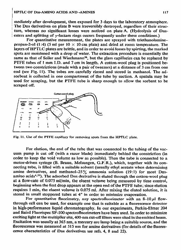
silica gel or related active layers44 are mixtures of polar with non-polar compounds. Because of limited mutual solubility and preferential adsorption of the polar com- ponent by the HPTLC layer, many of these solvents separated into two phases on the plate and +vere therefore only of limited value. By using chloroform-acetic acid- water (lo:9 : 1) in the first dimension, and methyl acetate-propan-2-01-25 oA ammonia (9:7:2) in the second (with activation of the layer for 10 min at 100” between runs), adequate separation of the most important Dns-amino acids was achieved, both on normal and on HPTLC plates (see Fig. 7).
I14 N. SEILER, B. KNt)DGEN
>.
1 I
5u
I +llkii - Putrescine - Scknidine
Ethanolamine
. oiigin i .
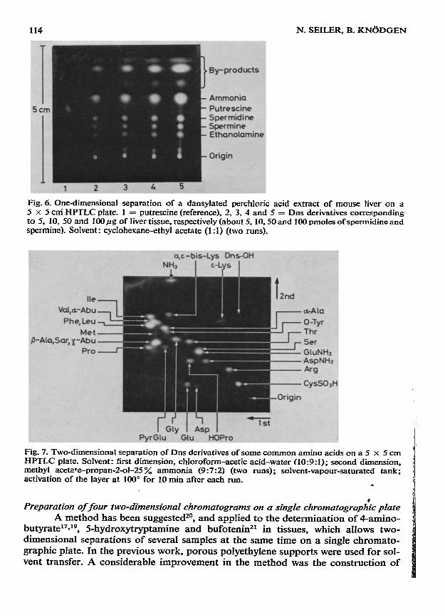
Fig. 6. One-dimensional separation of a dansylated perchloric acid extract of mouse liver on a 5 x 5 cni HPTLC plate. 1 = putrescine (reference), 2, 3, 4 and 5 = Dns derivatives corresponding to 5, 10, 50 and IOOpg of liver tissue, respectively (about 5. 10.50 and 100 pmoles of spermidine and spermine). Solvent: cyciohexane-ethyl acetate (1 :l) (two runs).
c&la
o-Tyr Thr SW GluNHz
*r
CysSOaH
Fig. 7. Two-dimensional separation of Dns derivatives of some common amino acids on a 5 x 5 cm HPTLC plate. Solvent: first dimension, chloroform-acetic acid-water (10:9:1); second dimension, methyl aceWe-propan-2-ol-25 % ammonia (9:7:2) (two runs); solvent-vapour-saturated tank; activation of the layer at 100” for 10 min after each run.
Preparation of four two-dimensional chromatograms on a singIe cht-omatograph:c plate A method has been suggested20, and applied to the determination of Camino-
butyrate”*“, 5-hydroxytryptamine and bufoteninzl in tissues, which allows two- dimensional separations of several samples at the same time on a single chromato- graphic plate. In the previous work, porous polyethylene supports were used for sol- vent transfer. A considerable improvement in the method was the construction of

HPTLC OF Dns-AMINO ACIDS AND -AMINES 115
supports from stainless-steel, the supports being covered by a sheet of filter paper (fibre direction vertical to the direction of solvent movement); details of the applica-
tion of such a support can* be seen in Fig. 8, and Fig. 9 shows separations of dan- sylated tissue sampIes. The channels scored in the layer divide the plate area into, e.g., four km x 5-cm areas, and samples are applied at an appropriate distance (e.g., 1 cm) from the plate corners. The plates are developed by placing them (layer downward) on a support of appropriate size, until the solvent fronts reach the scored channels from two opposite plate edges. After drying (or activation at elevated tem- perature), separation can be performed in the second direction.
Fig. 8. Use of the horizontal tank with stainless-steel support: (a) tank lid; (b) tank containing sol- vent: (c) stainless-steel support, bent to hold a sheet of filter paper (d) in place at c2, and to support the chromatographic plate (e) at each end (q); (e3 thin layer; (e2) narrow channels scored in the layer.
One of the obvious advantages of this technique, besides its rapidity, is that saturation of the layer with sokent vapour is ensured, even with mixtures containing solvents differing greatly in density. Moreover, many samples can be separated uni- dimensionally at the same time, if they are applied at two opposite plate edges. Evi- dently, the entire length of the plate can be utilized if the layer is removed from one plate edge (or if the filter paper sheet is removed from one end of the stainless-steel support). .
Quantitative evaluation The principles of quantitative measurement of Dns derivatives separated by-
TLC have been described in detai14.8~22.u, and advantages of in situ methods for the quantitative evaluation of HPTLC pIat& have been discussed24-Z.
Under certain circumstances, extraction procedures are desirable, or even a

N. SEILER, B. KNODGEN
Is*_-- -~ .-’ -- . =- <r+ *~;__-: _ ._
_ _ ____ ___-_z -__ .- -.--_ -L^--~.-.;~:_-,;-
Fig. 9. Four two-dimensional separations on a singIe IO-cm x 1O-cn1 HPTJX plate prepared with a stainless-steel support in a horizontal tank. A: Reference compounds (Dns derivatives), 1-12 as in Fig. 3; 13 = tryptamine; 14 = 4-aminovaleraldehyde (reaction product of iysine); 15 = side- reaction product (structure unknown) of proline with Dns-Cl. B: Dns derivatives of mouse-brain tis- sue; C: Dns derivatization products of mouse-liver tissue; D: Side-products of the derivatization reaction (blank). Solvents: first dimension, cyclohexane-ethyl acetate (1 :I); second dimension, ben- zene-triethylamine (5 : 1).
pre-requisite, e.g., if a fluorescence-scanning device is not accessible, or if such a meth- od as mass spectrometry27=28 is used for the determination of Dns derivatives.
The stability of Dns derivatives adsorbed on active surfaces is limited. Increase in fluorescence intensity and stabilization can be achieved by spraying with triethanoi- amine-propan-2-01 (1:4)=. Fig. 10 shows two chromatograms with nanomole amounts of various Dns-amines. Plate A was sprayed with the triethanolamine reagent im-
_- .= A - ~-:I_ _-- _g .(
Fig. IO. Effect of spraying with triethanolamine on the stability of Dns derivatives_ A: layer sprayed with 5 ml of a solution of triethanolamine-propan-2-o1(1:4); B: dried at room temperature and ex- posed, together with plate A, for 3 days to laboratory atmosphere. Developing solvent and com- pounds (about 2 nmoles each) as in Fig. 3.
HPTLC OF Dns-AMINO ACIDS AND -AMMES 117
mediately after development, then exposed for 3 days to the laboratory atmosphere. The Dns derivatives on plate B were irreversibly destroyed, regardless of their struc- ture, whereas no significant losses were noticed on plate A. (Hydrolysis of Dns- esters and splitting of y-lactam rings occurs frequently under these conditions_)
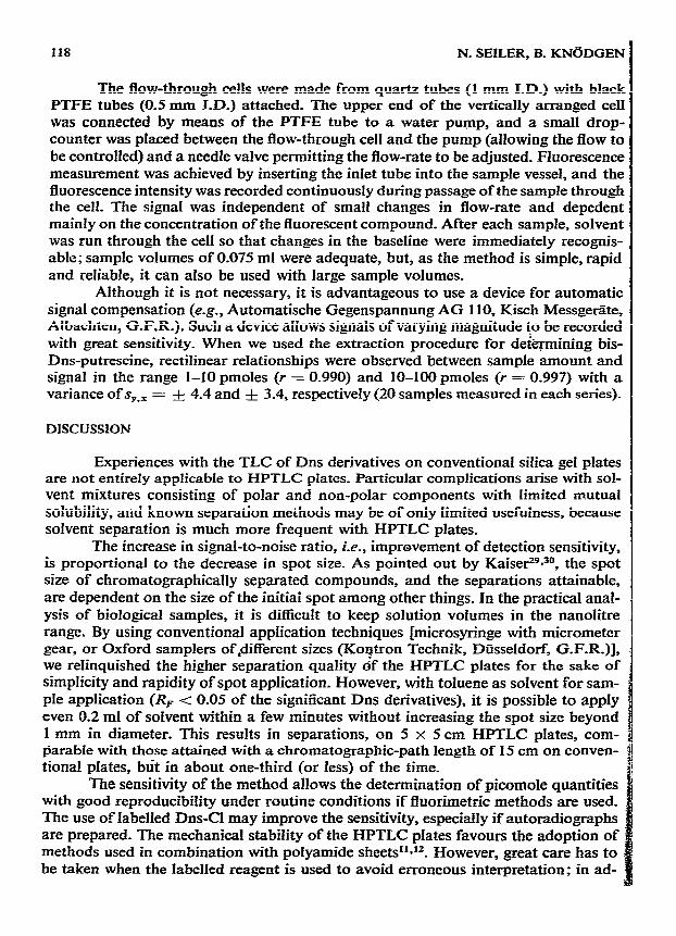
For quantitative measurement, the plates are sprayed with triethanolamine- propan-2-01 (1:4) (5 ml per 10 x 10 cm plate) and dried at room temperature. The layers of HPTLC plates are brittle, and in order to avoid losses by spirting, the marked spots are moistened with a drop of water. The extraction procedure is essentially the same as that of Seiler and Wiechmann r6, but the glass capillaries can be replaced by PTFE tubes of 1 mm I.D. and 7 cm in length. A cotton-wool plug is positioned be- tween two constrictions (made with a pair of tweezers) at a distance of 3 cm from one end (see Fig. 11). The tubes are.carefully rinsed and stored in methanol. The ad- sorbent is collected in one compartment of the tube by suction. A spatula may be used for scraping, but the PTFE tube is sharp enough to allow the sorbent to be scraped off.
Fig. 11. Use of the PTFE capillary for removing spots from the HPTLC plate.
For elution, the end of the tube that was connected to the tubing of the vac- uum pump is cut off (with a razor blade) immediately behind the constriction (in order to keep the void volume as low as possible). Then the tube is connected to a motor-driven syringe (B. Braun, Melsungen, G.F.R.), which, together with its con- necting tube, is filled with a suitable solvent (usually ethyl acetate when determining amine derivatives, and methanol-25 y/, ammonia solution (19:l) for most - Dns- amino acids4su). The adsorbed Dns derivative is eluted through the cotton-wool plug at a flow-rate of 0.075 ml/min, the eluent volume being measured by time control, beginning when the first drop appears at the open end of the PTFE tube: since elution requires 1 min, the eluent volume is 0.075 ml. After mixing the eluted solution, it is stored in smaIl stoppered tubes at 4” in order to minimize evaporation.
For quantitative fluorimetry, any spectrofluorimeter with an 8-10~1 flow- through cell can be used, for example one that is suitable as a guorescence detector in high-performance liquid chromatography. In our experiments, Perkin-Elmer 204 and Baird Fluorispec SF-100 spectrofluorimeters have been used. In order to minimize exciting light at the multiplier site, 400-nm cut-off filters were sited in the emitted beam. Excitation was usually at 365 nm, a mercury arc lamp being a suitable source, and the fluorescence was measured at 515 nm for amine derivatives (for details of the fluores- cence characteristics of Dns derivatives see refs. 4, 8 and 22).
118 N. SEILER, B. KNdDGEN
I
The flo~Mhrough cells were made from quartz tubes (1 mm I.D.) with black PTFE tubes (OS mm J-D.) attached. The upper end of the vertically arranged ceil was connected by means of the PTFE tube to a water pump, and a smal1 drop- counter was placed between the flow-through ceil and the pump (allowing the flow to be controlled) and a needle valve permitting the flow-rate to be adjusted. Fluorescence measurement was achieved by inserting the inlet tube into the sample vessel, and the fluorescence intensity was recorded continuously during passage of the sample through the cell. The signal was independent of small changes in flow-rate and depedent mainly on the concentration of the fluorescent compound. After each sample, solvent was run through the cell so that changes in the baseline were immediately recognis- able; sample volumes of 0.075 ml were adequate, but, as the method is simple, rapid and reIiabIe, it can also be used with large sample volumes.
Although it is not necessary, it is advantageous to use a device for automatic signal compensation (e.g., Automatische Gegenspannung AG 1 IO, Kisch Messgetite, Albachten, G.F.R.). Such a device allows siguals of varying magnitude f:” be recorded with great sensitivity. When we used the extraction procedure for det@-mining bis- Dns-putrescine, rectilinear relationships were observed between sample amount and signai in the range l-10 pmoies (r = 0.990) and 10-100 pmoles (r = 0.997) with a variance of s,., = -& 4.4 and f 3.4, respectively (20 samples measured in each series).
DISCUSSION I
Experiences with the TLC of Dns derivatives on conventional silica gel plates are not entirely applicable to HPTLC plates. ParticuIar complications arise with sol-
vent mixtures consisting of polar and non-polar components with limited mutual solubility, and known separation methods may be of only limited usefulness, because solvent separation is much more frequent with HPTLC plates.
The increase in signal-to-noise ratio, i.e., improvement of detection sensitivity, is proportional to the decrease in spot size. As pointed out by Kaiser29a30, the spot size of chromatographically separated compounds, and the separations attainable, are dependent on the size of the initiai spot among other things. In the practical anal- ysis of biological samples, it is difficult to keep solution volumes in the nanolitre range. By using conventional application techniques [microsyringe with micrometer gear, or Oxford samplers of&different sizes (Koutron Technik, Dfisseldorf, G-F-R.)], we reIinquished the higher separation quality gf the HPTLC plates for the sake of simpIicity and rapidity of spot application. However, with toluene as solvent for sam- ple application (RF -c 0.05 of the significant Dns derivatives), it is possible to apply even 0.2 ml of solvent within a few minutes without increasing the spot size beyond 1 mm in diameter. This results in separations, on 5 x 5 cm HPTLC plates, com- Parable with those attained with a chromatographic-path length of 15 cm on conven- tional plates, blit in about one-third (or less) of the time.
The sensitivity of the method allows the determination of picomole quantities with good reproducibility under routine conditions if fluorimetric methods are used. The use of labelled Dns-CI may improve the sensitivity, especially if autoradiographs are prepared. The mechanical stability of the HPTLC plates favours the adoption of methods used in combination with polyamide sheetsll**f. However, great care has to be taken when the labelled reagent is used to avoid erroneous interpretation: in ad-
HFI-LC OF Dns-AMINO ACIDS AND -AMLNES 119
dition to fluorescent side-products of the Dns derivatization, non-fluorescent, radio- active contaminants may simulate the presence of certain compounds.
HFTLC plates are, without doubt, a major breakthrough in the development of TLC, especially as far as rapidity and sensitivity are concerned. Even though not all known separations can be carried out on HPTLC plates, they can be used in the conventional manner and applied, with appropriate modifications, to all techniques. However, in order to profit from their improved separation qualities, it is necessary to use sophisticated methods of sample application and chromatogram development.
REFERENCES
1 N. Seiler and L. Demisch, in K. Blau and G. H. King (Editors), Handbook- of Derivutives fbr CfzromatograpIz_v, Heyden & Sons, London, in press.
2 N. Seiler and M. Wiechmann, Euperientia, 20 (1964) 559. 3 N. Seiler and M. Wiechmann, Ekperiemiu, 21 (1965) 203. 4 N. Seiler, Methods Biochem. Anal., I8 (1970) 259. 5 J. Rasmus and Z. DeyI, Chromatogr. Rev., 13 (1970) 163. 6 A. Niederwieser, Methods Enxymol., 25 (1972) 60. 7 N. Seiler and M. Wiechmann, J. Chromatogr., 28 (1967) 351. 8 N. Seiler and M. Wiechmann, in A. Niederwieser and G. Pataki (Editors), Progress in T&in-Layer
Chromatography and Related Metizoak, Vol. 1, Ann Arbor-Humphrey Sci. Pub]., Ann Arbor, Mich., London, 1970, p_ 94.
9 K--R_ Woods and K.-T_ Wang, Biochim. BiopIzys. Acra, 133 (1966) 369. 10 W. R. Gray, Methods Enqwzol., 25 (1972) 333. 11 N. N. Osborne, Progr. NearobioL, 1, Part 4 (1973) 299. 12 B. E. Leonard and N. N. Osborne, in N. Marks and R. Rodnight (Editors), Research Methuds in
Neurochemisrry, Vol. 3, Plenum, New York, 197.5, p_ 443. 13 C. W. Tabor, H. Tabor and U. Bachrach. J. Biol. Chenz., 239 (1964) 2194. 14 F. G. Fischer and H. Bohn. Hoppe-Seyler=F Z. Physiol. Cizenz., 308 (1957) 108. 15 N. Seiter and M. Wiechmarm, Hoppe-Seylers 2. Physiol. Chem., 349 (1968) 58% 16 N. Seiler and M. Wiechmann, Hoppe-Seyler’s Z. Physiol. Chem., 350 (1969) 1493. 17 N. Seiler and B. Eichentopf, Biocltem. J., 152 (1975) 201. 18 N. Seiler and M. Wiechmann, Hoppe-Seyler’s Z. Physiol. C’hem., 348 (1967) 1285. 19 N. Seiler, in N. Marks and R. Rodnight (Editors), Research Methuds in Neurochenzistry, Vol. 3.
Plenum, New York, 1975, p. 409. 20 N. Seiler, f. Chronzatogr., 63 (1971) 97. 21 N. Seiler and K. Bruder, J. CIzromatogr., 106 (1975) 159. 22 N. Seiler and M. Wiechmann, Z. Anal. Chem., 220 (1966) 109. 23 N. Seiler and H. MBller, Chromutographh, 2 (1969) 470. 24 J. Ripphahn and H. Halpaap, J. Chromarogr., 112 (1975) 81. 25 J. Ripphahn and H. Halpaap, in R. E. Kaiser (Editor), Einftihrung in die Hucllfc~srrmgs-Diinn-
schichr-Chromarogruphie HPDC, Institut ftir Chromatogmphie, Bad Diukheim, 1976, p_ 182. 26 U. Hezel, in R. E. Kaiser (Editor), Eizzfihrzmg izz die Hochleistzmgs-Diimzschichinnschichromarographie
HPDC, Institut fiir Chromatographie, Bad Diirkheim, 1976, p. 117. 27 N. Seiler and B. Kniidgen, Org. Muss Spectronz., 7 (1973) 97. 28 D. A_ Durden, B. A. Davis and A. A. Boulton, Biomed. Mass Specfronz., 1 (1974) 83. 29 R. E. Kaiser, in R. E. Kaiser (Editor), Einfiirzmg in die Hochfeistungs-Diinnschicht-Chromafo-
gruphie HPDC, Institut fur Chromatographie, Bad Ditrkheim, 1976, p_ 12. 30 R. E. Kaiser, in R. E. Kaiser (Editor), Einfihrung in die Hoclzfeistungs-Dichicht-Chromato-
graphic HFDC, Institut fur Chromatographie, Bad Dfirkheim, 1976, p_ 88. 31 N. Seiler, Hoppe-Seyfer’s Z. Physiol. Chem., 348 (1967) 765.