christiana eleojo aruwa · 2019-10-24 · activities. the utilisation of laccase in enzymatic...
TRANSCRIPT

i
CHARACTERISATION OF OPUNTIA PHENOLIC EXTRACTS
AND ENZYMATIC MODIFICATION OF SELECTED
COMPOUNDS
By
CHRISTIANA ELEOJO ARUWA
Submitted in fulfillment of the requirements for the degree of
Doctor of Philosophy (PhD): Biotechnology
Department of Biotechnology and Food Technology,
Faculty of Applied Sciences,
Durban University of Technology, Durban, South Africa
2019
Supervisor: Prof. Tukayi Kudanga
Co-supervisor: Dr. Stephen O. Amoo


iii
DEDICATION
This doctoral thesis is dedicated to the master of the universe, God the Almighty, who has been
my mainstay and strength despite all the challenges I encountered during my doctoral research,
and to my entire family who assisted both financially and morally.

iv
ACKNOWLEDGEMENTS
A profound ‘thank you’ to my supervisor, Prof. Tukayi Kudanga, and co-supervisor, Dr.
Stephen Amoo for the in-depth understanding, guidance, professional and technical knowledge
which contributed to a smooth workflow.
I sincerely acknowledge my parents; Engr. J. Idachaba Aruwa (KSJ) and Lady (Mrs). V. A.
Aruwa. All your love, care and support remain evergreen. Many thanks to my siblings; Mrs.
Victoria A. Durojaiye, Mrs. Debora A. Avwarodha, Mr. Emmanuel E. Aruwa, Mr. Paul A.
Aruwa, Miss Veronica A. Aruwa, Masters John and Jonathan Aruwa and Miss Faith A. Aruwa,
for your prayers and words of encouragement. They made my doctoral research journey less
onerous. A special thanks also to all my friends and research group members.
I do not mince words as I return all the glory, honour, power and adoration to my Creator,
Heavenly Father and King, the omnipotent, the author and finisher of my faith, my ever-present
help, He who fills my cup to overflowing, the beginning and end, for seeing me through, one
day at a time.

v
ABSTRACT
Opuntia species are utilised as local medicinal interventions for chronic diseases and as food
sources. The phytochemical profile varies within and across Opuntia species and has been
related to differences in cultivar and geographical location. Macromolecular antioxidant (MA)
fractions are also largely ignored from most conventional extractive processes compared to the
well-known extractable polyphenol fractions. This study characterised subtropical spineless
cladode, fruit pulp and peel extracts and selected phenolic compounds for enzymatic
modification using a laccase from Trametes pubescens. MA extracts were also characterised in
comparison with extractable fractions. The effects of drying methods and extraction solvent on
extract yields and bioactivities were also studied. Extracts were assayed for phenolic content
and antioxidant activities were determined using standard 2,2’-diphenyl-1-picrylhydrazyl
(DPPH), 2,2,-azinobis3-ethylbenzthiazoline-6-sulfonic acid (ABTS) and ferric reducing
antioxidant power (FRAP) assays. Antimicrobial activities and mode of antibacterial action
were assessed against type-bacterial cultures. Minimum inhibitory concentration (MIC) values
were recorded for the extracts and compounds. Compound profiling was achieved using liquid
chromatography-time of flight-mass spectrometry (LC-TOF/MS) in negative ionisation mode.
Antibacterial and antioxidant activities were higher in MA, hydrolysed and hydroalcoholic
cladode and fruit extracts than in aqueous fractions. Ethanolic, methanolic and hexane extracts
of freeze-dried Opuntia cladode, MA and peel samples showed higher total phenolic content,
and in vitro antioxidant and antimicrobial activities than the oven-dried extracts. Cladode
extracts inhibited growth of both Gram-positive and Gram-negative microorganisms (MIC
range of 25 to 250 mg/mL). Likewise, fruit extracts inhibited both Gram-positive and Gram-
negative microorganisms (MIC range of 2.5 to 18.75 mg/mL). Cladode and fruit extract profiles
showed the presence of mainly phenolic acids and flavonoid derivatives. Isovitexin 7-O-
xyloside-2"-O-glucoside, polyhydroxypregnane glycoside and neohancoside C in Opuntia

vi
cladode, and pinellic acid in Opuntia fruit were identified for the first time in this study. Some
compounds, however, remained unidentified. Thereafter, selected Opuntia cladode and fruit
phenolic compounds (isorhamnetin and luteolin) were used for enzymatic (laccase)
transformation after preliminary screening reactions. Laccase-catalysed oxidation of luteolin in
a monophasic system containing sodium acetate buffer (pH 5.0) and ethanol (50%, v/v) as co-
solvent, resulted in the production of a dimer (m/z 569, M=570). Using a similar approach,
oxidative coupling of isorhamnetin produced two main products, IP1 which was a dimer (m/z
629, M=630) and IP2 (m/z 457, M=458) which was most likely a result of coupling of an
oxidative cleavage product and the isorhamnetin monomer. Dimers showed up to two-fold
improvement in antioxidant and antimicrobial activities, compared to their respective
substrates. The synthesised products showed a bactericidal mode of action as demonstrated by
time-kill and bacterial cell integrity assays. The bactericidal action was further confirmed by
scanning electron microscopy (SEM) which showed that treatment of bacterial cells with the
synthesised compounds resulted in deformed, pitted, broken or fragmented cells, indicating
strong bactericidal action.
In conclusion, this study showed that Opuntia fruit pulp, peel and cladode extractable and MA
extracts have potential as sources of phenolic compounds with antioxidant and antimicrobial
activities. Laccase catalysis has potential to transform the phenolic compounds into coupling
products with higher biological activities. The synthesised products have potential for
application in the food, nutraceutical and other relevant industries.

vii
PREFACE
This thesis is organised into eight chapters and the experimental work is presented in manuscript
format. Chapter one gives the general introduction to the thesis. Chapter two provides a critical
review of literature on the Opuntia plant and its locations worldwide. The main aspects covered
include Opuntia cladodes, fruits and peels and their associated compounds, the relevance of the
Opuntia plant to health and the growing global trends in antimicrobial resistance and non-
communicable disease linked with oxidative stress. Finally, a brief review of fungal laccases and
their applications in organic synthesis is provided, focussing mainly on enzymatic modification of
monomeric phenolics to improve biological activities as well as structure-activity relationship
(SAR) in phenolic antioxidants. Chapter three presents the biological activities and phenolic
compound profile of Opuntia cladode as a potential source of new bioactive compounds. Chapter
four reports on the compound profile of extracts of Opuntia fruit pulp and peel and their biological
activities. The utilisation of laccase in enzymatic modification of selected Opuntia-related phenolic
compounds is covered in Chapters five (luteolin) and six (Isorhamnetin). Chapter seven is a general
discussion of the entire findings. Chapter eight concludes the thesis and suggests recommendations
for future studies.

viii
Table of Contents DECLARATION .......................................................................................................................ii
DEDICATION...........................................................................................................................iii
ACKNOWLEDGEMENTS.......................................................................................................iv
ABSTRACT................................................................................................................................v
PREFACE.................................................................................................................................vii
LIST OF FIGURES.................................................................................................................xiii
LIST OF TABLES...................................................................................................................xiv
LIST OF PLATES....................................................................................................................xv
ABBREVIATIONS.................................................................................................................xvi
PUBLICATIONS AND CONFERENCES….......................................................................xviii
CHAPTER ONE.......................................................................................................................1
1.1. Introduction.........................................................................................................................1
CHAPTER TWO......................................................................................................................4
Literature Review……................................................................................................................4
2.1. Introduction............................................................................................................ ............4
2.2. The cactus pear (Opuntia) plant.........................................................................................4
2.2.1. Opuntia plant: growth locations and general compound profile...................................7
2.3. Antioxidants: sources, mechanisms, extended benefits and purification…….....................14
2.4. Biologically active compounds........................................................................................19
2.5. Opuntia cladode: compounds, fingerprinting and biological activities….......................22
2.6. Opuntia fruit: compounds and biological activities……....................................................25
2.7. Opuntia peel/skin: compounds and biological activities.....................................................28
2.8. Effect of processing conditions on compound profile and bioactivities.............................31
2.9. Techniques for the extraction of compounds from Opuntia residue…..............................32
2.9.1. Opuntia macromolecular antioxidant biological activities...........................................33
2.10. Potential side effects of Opuntia species extracts/compounds..........................................35
2.11. Opuntia plant compounds and the search for new antimicrobials.....................................36
2.12. Laccases as biocatalysts in the synthesis of bioactive compounds and materials..............39
2.13. Structure-activity relationship (SAR) in phenolic compounds........................................42
2.14. Conclusion..............................................................................................................................46
2.15. Research hypotheses, aim and objectives...............................................................................47
2.15.1. Research hypotheses........................................................................................................47
2.15.2. Research aim.....................................................................................................................47

ix
2.15.3. Research objectives..........................................................................................................48
CHAPTER THREE................................................................................................................49
Extractable and macromolecular antioxidants of Opuntia ficus-indica cladodes: phytochemical
profiling, antioxidant and antibacterial activities .................................................................... 49
Abstract............................................................................................................................. ........49
3.1. Introduction......................................................................................................................50
3.2. Materials and methods.....................................................................................................52
3.2.1. Chemicals and reagents...............................................................................................52
3.2.2. Culture and maintenance of microorganisms..............................................................52
3.2.3. Plant material collection and preparation....................................................................53
3.2.4. Extraction of cladode phenols/polyphenols................................................................53
3.2.5. Total phenolic content (TPC) of extracts....................................................................54
3.2.6. Determination of extracts antioxidant capacities........................................................55
3.2.6.1. DPPH radical scavenging activity of extracts.......................................................55
3.2.6.2. Ferric reducing antioxidant power (FRAP) assay.................................................56
3.2.7. Antibacterial activity of cladode extracts....................................................................56
3.2.7.1. Agar well diffusion assay.......................................................................................56
3.2.7.2. Minimum inhibitory concentration (MIC) of extracts...........................................57
3.2.8. Extracts profiling by LC-TOF/MS analysis................................................................57
3.2.9. Statistical analysis.......................................................................................................58
3.3. Results and discussion.....................................................................................................58
3.3.1. Yield of cladode extracts.............................................................................................58
3.3.2. TPC and antioxidant activity of extracts.....................................................................59
3.3.2.1. TPC of cladode extracts.........................................................................................59
3.3.2.2. In vitro antioxidant activity of cladode extracts....................................................60
3.3.3. Antibacterial activity and MIC extracts......................................................................64
3.3.3.1. Antimicrobial activity of extracts..........................................................................64
3.3.3.2. MIC of cladode extracts.........................................................................................67
3.3.4. Mass spectral characteristics of profiled cladode extract compounds........................69
3.4. Conclusion.......................................................................................................................72
CHAPTER FOUR...................................................................................................................73
Phenolic compound profile and biological activities of Southern African Opuntia ficus- indica
fruit pulp and peels……………………………………………................................................73
Abstract..................................................................................................................... ................73

x
4.1. Introduction......................................................................................................................73
4.2. Materials and methods.....................................................................................................75
4.2.1. Culture and maintenance of microorganisms..............................................................75
4.2.2. Plant material collection and preparation....................................................................76
4.2.3. Opuntia fruit and peel phenols extraction...................................................................76
4.2.4. Total phenolic content (TPC) determinations.............................................................77
4.2.5. Determination of antioxidant capacity........................................................................77
4.2.5.1. DPPH radical scavenging activity of extracts........................................................77
4.2.5.2. Ferric reducing antioxidant power (FRAP) assay..................................................77
4.2.6. Antibacterial activity of fruit pulp and peel extracts...................................................78
4.2.6.1. Agar well diffusion assay.......................................................................................78
4.2.6.2. Extracts MIC determination...................................................................................78
4.2.7. Extract compound profiling by LC-ESI-TOF/MS analysis........................................78
4.2.8. Statistical analysis.......................................................................................................79
4.3. Results and discussion.....................................................................................................79
4.3.1. Opuntia fruit pulp and extract yields..........................................................................79
4.3.2. TPC of Opuntia fruit pulp and peel extracts...............................................................81
4.3.3. Antioxidant activity of extracts...................................................................................81
4.3.4. Antimicrobial activity of pulp and peel extracts.........................................................83
4.3.5. Mass, UV properties and occurrence of phenols in extracts.......................................86
4.4. Conclusion.......................................................................................................................91
CHAPTER FIVE.....................................................................................................................92
Enzymatic dimerisation of luteolin enhances antioxidant and antimicrobial activity……......92
Abstract.....................................................................................................................................92
5.1. Introduction............................................................................................................ ..........93
5.2. Materials and methods.....................................................................................................95
5.2.1. Chemicals and reagents used......................................................................................95
5.2.2. Laccase production from T. pubescens.......................................................................95
5.2.3. Determination of laccase enzyme activity..................................................................96
5.2.4. Laccase protein concentration determination…..........................................................96
5.2.5. Oxidation of luteolin by laccase..................................................................................96
5.2.6. Purification of reaction product..................................................................................97
5.2.7. Characterisation of luteolin product by LCMS...........................................................98
5.2.8. Antioxidant activity of luteolin and its dimer.............................................................98

xi
5.2.8.1. The DPPH (2,2-Diphenyl-picryhydrazyl) assay…...............................................98
5.2.8.2. ABTS [(2,2´-Azinobis (3-ethylbenzthiazoline-6-sulfonic acid)] assay..................99
5.2.8.3. FRAP (ferric reducing antioxidant power) assay...................................................99
5.2.9. Determination of antibacterial activity and mode of action........................................99
5.2.9.1. Antibacterial assay using the disc diffusion method..............................................99
5.2.9.2. Minimum inhibitory concentration (MIC) determination.....................................100
5.2.10. Bacterial cell wall integrity.....................................................................................100
5.2.11. Bacterial time-kill assay...........................................................................................101
5.2.12. SEM observation of bacterial cells...........................................................................102
5.2.13. Statistical analysis...................................................................................................102
5.3. Results and discussion...................................................................................................102
5.3.1. Laccase enzyme production from Trametes pubescens….........................................102
5.3.2. Characterisation of oxidation product by TLC and LCMS.......................................104
5.3.3. Antioxidant activity assays.......................................................................................107
5.3.4. Antibacterial activity determination..........................................................................108
5.3.5. Integrity of bacterial cell membrane.........................................................................109
5.3.6. Bacterial rate of kill with time (time-kill assay).......................................................110
5.3.7. SEM observations of cells.........................................................................................112
5.4. Conclusion.....................................................................................................................113
CHAPTER SIX.....................................................................................................................114
Laccase-mediated modification of isorhamnetin improves antioxidant and antibacterial
activity.....................................................................................................................................114
Abstract..................................................................................................................... ..............114
6.1. Introduction....................................................................................................................115
6.2. Materials and methods....................................................................................................116
6.2.1. Laccase production from T. pubescens…..................................................................117
6.2.2. Oxidation of isorhamnetin by laccase........................................................................117
6.2.3. Reaction products purification...................................................................................117
6.2.4. Product characterisation by LCMS............................................................................118
6.2.5. Antioxidant activity assays........................................................................................118
6.2.6. Determination of antibacterial activity and mode of action.......................................118
6.2.6.1. Antibacterial activity by disc diffusion assay......................................................118
6.2.6.2. Minimum inhibitory concentration (MIC) determination...................................119
6.2.7. Determination of products antibacterial mechanism of action...................................119

xii
6.2.8. Statistical analysis......................................................................................................120
6.3. Results and discussion....................................................................................................120
6.3.1. Characterisation of oxidation products by TLC and LCMS......................................120
6.3.2. Antioxidant activity of oxidation products.................................................................125
6.3.3. Antibacterial activity determination..........................................................................126
6.3.4. Integrity of bacterial cell membrane..........................................................................128
6.3.5. Bacterial rate of kill....................................................................................................129
6.3.6. SEM observation of bacterial cells.............................................................................132
6.4. Conclusion......................................................................................................................134
CHAPTER SEVEN...............................................................................................................135
General discussion .................................................................................................................135
CHAPTER EIGHT...............................................................................................................139
Conclusion and recommendations……..................................................................................139
8.1. General conclusion.........................................................................................................139
8.2. Recommendations..........................................................................................................140
References................................................................................................................... ............142
Appendices…………..............................................................................................................185

xiii
LIST OF FIGURES
Figure 2.1: Mechanism of action of laccase-catalysed oxidation of substrate to form radicals..41
Figure 2.2: Proposed reaction mechanism for the homomolecular coupling of 2,6-DMP to
produce the C–C dimer………………………………………………………………………..45
Figure 3.1: TPC correlation with EC50 (DPPH) values for all unhydrolysed cladode extracts...64
Figure 5.1: HPLC Chromatogram of luteolin substrate and its product (LP)……...…………105
Figure 5.2: Mass spectrum of luteolin (m/z 285) and its dimeric product (m/z 569) in negative
mode………………………………………………………………………............................105
Figure 5.3: Proposed structure of the C-C linked luteolin dimer……......……………………106
Figure 5.4: Proposed structure of the ether (C-O-C) linked luteolin dimer…..………………106
Figure 5.5: Proposed reaction pathway for the formation of the C-C linked luteolin dimer….106
Figure 5.6: Proposed reaction pathway for the ether (C-O-C) linked luteolin dimer……...106
Figure 5.7: Time-kill curve for bactericidal action of dimer (LP) against E. coli…………….111
Figure 5.8: Time-kill curve for bactericidal action of dimer (LP) against MRSA……………112
Figure 5.9: SEM images of bacterial cells treated with the luteolin dimer (LP)…………….113
Figure 6.1: HPLC chromatogram of isorhamnetin and its oxidation products (IP1 and IP2)...122
Figure 6.2: Mass spectra of (a) Isorhamnetin (m/z 315), (b) Product IP1 (dimer) (m/z 629) and
(c) Product IP2 (m/z 457)………………………………………………..…………………...123
Figure 6.3: Proposed structure of the C-C linked dimer (IP1; m/z 629, M=630)..……………123
Figure 6.4: Proposed structure of C-O-C linked dimer (IP1; m/z 629, M=630)….…….…….123
Figure 6.5: Proposed mechanistic pathway for the production of the C-C dimer through laccase
catalysed dimerisation of isorhamnetin……………………………………………………...124
Figure 6.6: Proposed mechanistic pathway for the production of the ether linked dimer through
laccase catalysed dimerisation of isorhamnetin……………………………………………...124
Figure 6.7: Proposed structure of IP2 (m/z 457, M = 458)…………………………………...124
Figure 6.8: Proposed reaction mechanism for the laccase-catalysed production of IP2……...124
Figure 6.9: Time-kill curve for bactericidal action of IP1 against MRSA…............................131
Figure 6.10: Time-kill curve for bactericidal action of IP1 against L. monocytogenes……….131
Figure 6.11: Time-kill curve for bactericidal action of IP2 against P. aeruginosa...…………132
Figure 6.12: SEM images of bacterial cells treated with the dimer (IP1).…….……………...133
Figure 6.13: SEM images of P. aeruginosa cells treated with product (IP2)…………………133

xiv
LIST OF TABLES
Table 2.1: Selected Opuntia spp. and their locations………………………………….……….9
Table 2.2: Phenolic and non-phenolic compounds in Opuntia cactus plant parts………..……10
Table 3.1: Percentage yield of Opuntia ficus-indica cladode extracts……...……………….…59
Table 3.2: Total phenol and antioxidant values of hydrolysed and unhydrolysed Opuntia nopal
extracts…...……………………………………………………………………………….…..63
Table 3.3: Antibacterial inhibition zone and MIC of hydrolysed and unhydrolysed Opuntia
nopal extract…………………………………………………………………………………..68
Table 3.4: Mass spectral properties of compounds in the Opuntia cladode extracts…………...71
Table 3.5: Identified and unidentified compounds in extractable polyphenol cladode extracts.72
Table 4.1: Yield of Opuntia fruit and peel extracts………..…………………………………..80
Table 4.2: Total phenolic content and antioxidant activities of Opuntia ficus-indica fruit and
peel extracts…………………………………………………………………………………...83
Table 4.3: Antibacterial activity of Opuntia ficus-indica fruit and peel extracts……………..86
Table 4.4: Mass and spectral properties of compounds from Opuntia pulp and peel extracts…88
Table 4.5: Pulp and peel extracts compounds and their occurrence (%)……………………….89
Table 4.6: Compounds identified in Opuntia pulp and peel extracts……………………..........90
Table 5.1: Partial purification table for laccase production……...…………..……………….103
Table 5.2: Antioxidant activity of luteolin and its products…………………....…………….107
Table 5.3: Antibacterial activity of luteolin and laccase-catalysed oxidation product (LP)….109
Table 5.4: Effect of luteolin product (LP) on bacterial cell membrane integrity………...…...110
Table 6.1: Percentage yield of isorhamnetin products……………………...……..…………125
Table 6.2: Antioxidant activity of isorhamnetin and its products……………………...….….126
Table 6.3: Antibacterial activity of isorhamnetin and its products…………………..……….128
Table 6.4: Effect of isorhamnetin products on bacterial cell membrane integrity………......129

xv
LIST OF PLATES
Plate 2.1: Opuntia ficus-indica purple and yellow fruits varieties growing on pads called
cladodes………………………………………………………………………………………...6
Plate 5.1: TLC plate showing luteolin product (LP) and substrate (S) lanes………………..105
Plate 6.1: TLC plate showing products (IP1 and IP2) and substrate lanes……………….…122

xvi
ABBREVIATIONS
ABTS: 2,2’-azinobis3-ethylbenzthiazoline-6-sulfonic acid
ADF: Antioxidant dietary fibres
AHA: American Heart Association
AM: Acidified methanol
AMR: Antimicrobial resistance
ANOVA: Analysis of variance
AQ: Acidified water
ATCC: American Type Culture Collection
ATP: Adenosine triphosphate
BDE: Bond dissociation enthalpies
BHA: Butylated hydroxyanisole
BHT: Butylated hydroxytoluene
CAM: Crassulacean Acid Metabolism
CMV: Cucumber Mosaic Virus
CSAs: Chemically synthesised antioxidants
CTC: Carbon tetrachloride
DNA: Deoxyribonucleic acid
DPPH: 2,2’-diphenyl-1-picrylhydrazyl
E: Ethanol
EACC: Ehrlich Ascites Carcinoma Cells
EP: Extractable polyphenol
FAO: Food and Agricultural Organisation
FRAP: Ferric reducing antioxidant power
GABA: γ-Aminobutyric acid
GMFs: Genetically Modified Foods
HAT: Hydrogen atom transfer
IP1: Predicted dimer product(s) from isorhamnetin
IP2: Unidentified laccase oxidation product from isorhamnetin
LAB: Lactic Acid Bacteria
LC-TOF/MS: Liquid Chromatography-Time of Flight-Mass Spectrometry
LP: Predicted dimer product(s) from luteolin
M: Methanol
MA: Macromolecular antioxidant

xvii
MAW: Methanol:acetone:water extraction solvent
MIC: Minimum inhibitory concentration
NCDs: Non-communicable diseases
NEPAC: Non-extractable proanthocyanidins
NEPP: Non-extractable polyphenol
NLM: National Library of Medicine
NMR: Nuclear Magnetic Resonance
OD: Optical density
PBP: Penicillin-binding proteins
PG: Propyl gallate
PI: Polarity index
ROS: Reactive oxygen species
RSA: Radical scavenging activity
SAR: Structure-activity relationship
SD: Standard deviation
SEM: Scanning Electron Microscopy
SET: Single electron transfer
TBHQ: Tertiary-butylhydroquinone
TEM: Transmission Electron Microscopy
TMV: Tobacco Mosaic Virus
TPC: Total phenol content
TPTZ: 3(2,4,6-tripyridyl-s-triazine
UNDPI: United Nations Department of Public Information
WHO: World Health Organisation

xviii
PUBLICATIONS AND CONFERENCES
Publications
Aruwa, C. E., Amoo, S. O., Kudanga, T. 2018. Opuntia (Cactaceae) plant compounds,
biological activities and prospects – A comprehensive review. Food Research
International, 112, 328-344 (Appendix A).
Aruwa, C. E., Amoo, S. O., Kudanga, T. 2019. Extractable and macromolecular antioxidants
of Opuntia ficus-indica cladodes: phytochemical profiling, antioxidant and antibacterial
activities. South African Journal of Botany. Accepted.
Aruwa, C. E., Amoo, S. O., Kudanga, T. 2019. Phenolic compound profile and biological
activities of Southern African Opuntia ficus-indica fruit pulp and peels. LWT-Food Science
and Technology, 111, 337-344 (Appendix A).
Submitted manuscripts
Aruwa, C. E., Amoo, S. O., Koorbanally, N., Kudanga, T. 2019. Laccase-mediated
modification of isorhamnetin improves antioxidant and antibacterial activities, submitted.
Aruwa, C. E., Amoo, S. O., Koorbanally, N., Kudanga, T. 2019. Enzymatic dimerization
of luteolin enhances antioxidant and antimicrobial activities, submitted.
Conferences
Aruwa, C. E., Amoo, S. O., Kudanga, T. 2018. Antioxidant and antibacterial activity of
extractable and macromolecular antioxidants from Opuntia ficus-indica cladodes. South
African Society for Microbiology (SASM), Johannesburg, South Africa, April 2018.

1
CHAPTER ONE
1.1. Introduction
The present global threats of oxidative stress and antimicrobial resistance (AMR) have
necessitated increased research interests channelled toward the discovery of new bioactive
phytochemicals with antioxidant (Zrira et al., 2016) and antimicrobial properties (Koubaa et
al., 2015a). Oxidative stress arises from the overproduction of free radicals and has been linked
with non-communicable diseases (NCDs) such as cancer, cardiovascular diseases, and type-2
diabetes (Hofman, 2014). In recent years there has been an upsurge in incidences of NCDs
(Mayosi et al., 2009). In southern Africa and other developing countries, it was predicted that
seven out of every ten deaths would be attributed to NCDs by the year 2020 (UNDPI, 2011).
The incidences of AMR have increased over the past decade and this is linked mainly to the
overprescription and abuse of currently available antibiotics/antimicrobial agents or compounds
(Spellberg et al., 2011). AMR is a serious threat to public health and has been estimated to
cause 10 million deaths and $100 billion in economic losses by the year 2050 (Spellberg et al.,
2011; WHO, 2019).
In a bid to stem the global trends in oxidative stress and AMR, chemically synthesised
antioxidants (CSAs) are frequently used to fight oxidative stress (Wojcik et al., 2010), while
available antimicrobial compounds derived mainly from microorganisms are used to combat
AMR (Martins et al., 2011). However, CSAs are carcinogenic, cause sperm abnormalities,
nausea, fatigue, asthma and degenerative diseases as a result of their accumulation in biological
systems over time (Wojcik et al., 2010). Consequently, CSAs are increasingly becoming
unpopular with consumers due to their adverse effects and unnatural form (Wojcik et al., 2010).
On the other hand, new antimicrobial compounds from microorganisms are not being produced

2
fast enough and new antibiotic discovery and identification are at an all time low (Scarafile,
2016). In addition, problems such as malaise, diarrhoea, as well as prolonged cost of treatments
and hospital stays have been associated with AMR (AHA, 2018). These associated problems
have necessitated the search for new approaches and investigations into new, but safe and
natural bioactive molecules with antioxidant (Zrira et al., 2016) and antimicrobial properties
(Rasoulpour et al., 2017). The shift to plant bioactives and phenolics may be the most likely
solution to reduce current trends in oxidative stress and AMR. The Opuntia plant, which has
recently attracted a lot of research interest as an underutilised crop in southern Africa (Khan
and Giridhar, 2015), may be integral to the discovery of new and natural plant bioactive
compounds (Tesoriere et al., 2005).
The Opuntia plant is mainly used as fodder and is underutilised in southern Africa (Khan and
Giridhar, 2015). Opuntia species are capable of growth in almost all climates (Lallouche et al.,
2015). Although Opuntia bioactivities have been related to content of flavonoids, phenolic
acids, vitamins and other components (Vulic´ et al., 2014), its bioactive compound profile can
change with variations in species, cultivar, geographical location and climatic conditions (Paiva
et al., 2016). The variation in compound profile has also been used as a genetic fingerprint for
the identification and taxonomic classification of Opuntia species in different locations
worldwide (Moussa-Ayoub et al., 2014). Therefore, there is a high probability of reporting new
phenolic compounds or bioactive compounds from Opuntia species especially from areas that
have not previously attracted much attention such as southern Africa.
A wide array of monomeric phenols are present in Opuntia species but have low biological
activities (Matsuura and Ohkatsu, 2000), short half-life in the body (Kudanga et al., 2011) and
poor physico-chemical properties such as low stability and low solubility (Hierholtzer et al.,
2012). Consequently, current research is also focused on the improvement of the biological

3
activities of natural monomeric phenolic compounds through enzymatic modification (Zwane
et al., 2012). Laccases have gained much research interest in this regard. Laccases (EC 1.10.3.2)
are multi-copper oxidases that catalyse the oxidation of a suitable substrate to its radical form
with concomitant reduction of molecular oxygen to water. The radicals formed then undergo
coupling to form high molecular weight homomolecular or heteromolecular coupling products
(Nemadziva et al., 2018; Kudanga et al., 2011), which usually have enhanced biological
activities compared to the starting substrates (Zwane et al., 2012). The improvement in
bioactivity is attributed to increase in the number of hydroxyl groups from which hydrogen
atoms are abstracted, and/or an increase in electron-donating groups that help to stabilise the
antioxidant after donating an electron (Gandia-Herrero et al., 2016; Kudanga et al., 2017).
Peroxidases were previously used for the same purpose, but are now less popular because they
require a cofactor, hydrogen peroxide, which makes the process a health hazard and expensive.
On the other hand, laccases are now preferred because they are essentially green enzymes that
only require oxygen (air) as co-substrate, producing water as the only by-product (Nyanhongo
et al., 2011; Riva, 2006).
Therefore, the aim of the current study was to characterise southern African Opuntia extracts
and enzymatically modify selected phenolic compounds for the production of new bioactive
compounds with enhanced antioxidant and/or antimicrobial activities.

4
CHAPTER TWO
2.1. Introduction
In this chapter, literature relevant to this study is presented. Some of the areas covered include:
a description of the cactus pear (Opuntia) plant; Opuntia plant growth locations and general
compound profile; antioxidant sources, mechanisms, benefits and purification; biologically
active compounds; Opuntia cladode compounds, fingerprinting and biological activities;
Opuntia fruit compounds and biological activities; Opuntia peel/skin compounds and biological
activities; techniques for the extraction of compounds from Opuntia residue; Opuntia
macromolecular antioxidant biological activities; potential side effects of Opuntia species
extracts/compounds; the search for new antimicrobial compounds; enzymes as biocatalysts for
biotechnological applications; laccases - reactions and applications, and structure-activity
relationship (SAR) in phenolic compounds.
2.2. The cactus pear (Opuntia) plant
Plants are potential sources of bioactive compounds and have been attracting the attention of
scientist globally. In this regard, the cactus pear plant (Opuntia spp.), its fruit and cladodes have
been shown to be good candidates for the production of health-promoting bioactive compounds
(Livrea and Tesoriere, 2006). Although traditionally recognised for its pharmacological
properties, Opuntia spp. remain underutilised. There is not enough scientific data to back up its
bioactivities. Nevertheless, it has been demonstrated that the cactus pear fruit and cladodes
could play an integral role in the formulation of new nutraceuticals (Feugang et al., 2006).

5
Cactus pear plants belong to the family Cactaceae, order Caryophyllales, and originated from
Central America (Felker and Inglese, 2003). Thousands of species of the cactus pear belong to
the Opuntia genus and produce edible fruits (Abou-Elella and Ali, 2014). Cactus fruits are also
referred to as cactus figs or Opuntia ficus-indica (prickly pear), and these grow on the prickly
cacti plant. The fruit and cladodes (Plate 2.1) are fleshy, oval, and vary in length. Hundreds of
the Opuntia species are xerophytic (Moβhammer et al., 2006) and well-adapted to arid zones
where little or no rainfall and poor soil conditions abound due to frequent erosions (FAO, 2001).
The plant, however, survives these conditions due to its possession of succulent leaves which
store water. It is attractive as food and possesses high dry matter converting potential
(Gabremariam et al., 2006). The plant accumulates minerals and elements using its shallow root
system. The Opuntia plant provides the necessary hydration, microelements as well as
macronutrients required in animal feeds (Rodriguez-García et al., 2007). Biodiversity of
Opuntia species is still a subject of much investigation (Caruso et al., 2010). This is because
the cactus pear fruit contains chemical components that are active biologically such as the
betalains, ascorbic acid and polyphenols (Livrea and Tesoriere et al., 2006). Moreover, the fruit
extract has been shown to have antioxidant, antibacterial, antiulcerogenic, anticancer,
neuroprotective, as well as hepatoprotective activities (Livrea and Tesoriere et al., 2006;
Madrigal-Santillán et al., 2013).
6
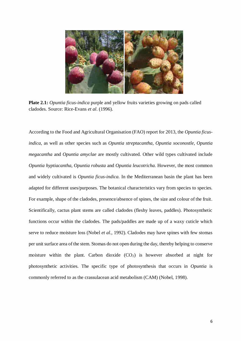
Plate 2.1: Opuntia ficus-indica purple and yellow fruits varieties growing on pads called
cladodes. Source: Rice-Evans et al. (1996).
According to the Food and Agricultural Organisation (FAO) report for 2013, the Opuntia ficus-
indica, as well as other species such as Opuntia streptacantha, Opuntia xoconostle, Opuntia
megacantha and Opuntia amyclae are mostly cultivated. Other wild types cultivated include
Opuntia hyptiacantha, Opuntia robusta and Opuntia leucotricha. However, the most common
and widely cultivated is Opuntia ficus-indica. In the Mediterranean basin the plant has been
adapted for different uses/purposes. The botanical characteristics vary from species to species.
For example, shape of the cladodes, presence/absence of spines, the size and colour of the fruit.
Scientifically, cactus plant stems are called cladodes (fleshy leaves, paddles). Photosynthetic
functions occur within the cladodes. The pads/paddles are made up of a waxy cuticle which
serve to reduce moisture loss (Nobel et al., 1992). Cladodes may have spines with few stomas
per unit surface area of the stem. Stomas do not open during the day, thereby helping to conserve
moisture within the plant. Carbon dioxide (CO2) is however absorbed at night for
photosynthetic activities. The specific type of photosynthesis that occurs in Opuntia is
commonly referred to as the crassulacean acid metabolism (CAM) (Nobel, 1998).

7
Freshly harvested cactus pear cladodes (green parts of the plant) are used as vegetables. They
have high water content, little amounts of dietary fibre, calcium and proteins. Other antioxidant
constituents include beta-carotene and ascorbic acid (Betancourt-Domínguez et al., 2006).
Flours made from cactus cladodes have been used as food supplement, ingredients and
alternatives. The dried flours are microbiologically stable, thereby helping to greatly reduce
storage and transport, and highly concentrate nutrients (Torrecilla et al, 2005). Furthermore,
immature spineless Opuntia ficus-indica cladodes of 15±5cm in length were analysed by
López-Cervantes et al. (2011). In their study, cladodes were dried at 60°C to 80°C followed by
profiling for amino acid and other chemical components. The analyses showed no significant
difference in proximate content of the flours, which was attributed to the effect of drying
temperature. Nevertheless, at 80oC drying temperature, colour (green) intensity reduced as well
as water holding capacity. On the other hand, there was increased concentration of glutamic
acid and proline amino acids, while oleic, palmitic and linoleic fatty acids concentrations were
highest. The best drying temperature for flour production with regard to nutrient composition
and retention was 60°C. They surmised that the physiological effects of the cactus plant could
be attributed to the functional properties of its constituents (Feugang et al., 2006).
2.2.1. Opuntia plant: growth locations and general compound profile
Opuntia ficus-indica is a slow growing perennial shrub which can grow up to 3-5 m high. Some
arid growth locations for Opuntia species include Saudi Arabia, Egypt, Pakistan, Middle East,
Algeria, Iran, Libya, Mexico, South Africa, Spain (Feugang et al., 2006; Moussa-Ayoub et al.,
2014). The temperate regions where the plant may be found include Tunisia (Ayadi et al.,
2009), while tropical countries include Brazil (Paiva et al., 2011), Ethiopia (Chiteva and
Wairagu, 2013), Kenya (Kunyanga et al., 2014), and the Mediterranean country of Morocco
(Castellanos-Santiago and Yahia, 2008). Some Opuntia species and their locations of growth

8
are shown in Table 2.1, but most species are native to Mexico and the Americas. Some Opuntia
species have spread to some African and Asian regions and show significant biodiversity within
and across species (Caruso et al., 2010). This biodiversity determines the component profile of
each species, as well as other factors such as growth location, prevailing climatic conditions
and soil conditions, among others (Ammar et al., 2012).
Cactus (Opuntia) plants contain carotenoids, amino acids, vitamins, fibres and antioxidant
phenol components (Table 2.2). Its phytochemical compounds such as phenolic acids,
flavonoids and derivatives, sterols, esters, coumarins, terpenoids, alkaloids bring about several
health benefits such as hypoglycaemic (Paiz et al., 2010), and antioxidant activities (Osorio-
Esquivel et al., 2011). Opuntia also contains water-soluble nitrogenous betalain pigments
(Table 2.2) which are classified as betacyanins (red to purple coloured) and betaxanthins
(yellow and orange coloured) (Khan and Giridhar, 2015). Betalains serve as chemo-systematic
markers for members of the Caryophyllales family (Slimen et al., 2017). Esters, alkaloids,
phenolic acids, essential fatty acids (Ramadan and Mörsel, 2003b), polysaccharides, sugars can
also be found in Opuntia species (Benayad et al., 2014; Yeddes et al., 2014a,b) as well as
alkanes, carotenoids, amino acids, among others (Manzano et al., 2017). The profile of phenolic
and non-phenolic components (Table 2.2) reported to be present in Opuntia species plant parts
constantly evolve from one location to another (Butera et al., 2002). Therefore, it is important
to investigate the phenolic compound profiling of southern African Opuntia species.

9
Table 2.1: Selected Opuntia spp. and their locations
Species Locations
Opuntia basilaris
Southwest United States (US) and northwest Mexico
Opuntia chlorotica
Native to southwest US, and the Sonoran and
Mojave deserts
Opuntia engelmannii
Mexico
Opuntia ficus-indica
Originally in south-central Mexico; cultivated
in warm parts of the world for its edible fruit
Opuntia fragilis
Northern Great Plains and as far west as
British Columbia, also found in the southern
Great Plains
Opuntia humifusa,
Opuntia compressa var.
humifusa
Throughout the US, east of the Great Plains
and into southern Ontario
Opuntia leucotricha
Mountains of Central Mexico
Opuntia macrocentra Southwest US and Northern Mexico
Opuntia macrorhiza
Throughout the Great Plains except for the northernmost areas (not found in North
Dakota), and extending sporadically eastward as far as Kentucky.
Opuntia ficus-indica Spain, Italy
Opuntia microdasys Mexico (Hidalgo) Opuntia ficus-indica
(prickly and spineless), Opuntia stricta, Opuntia
macrorhiza, Opuntia microdasys
Algeria, Tunisia, Morocco, Kenya, Egypt, South Africa (Northern, Eastern and Southern
Africa) Opuntia dillenii,
Opuntia ficus-indica,
Opuntia humifusa India, Iran, Korea, Saudi Arabia
Opuntia santa-rita
Texas, Arizona and Northern Mexico
Opuntia stricta
Opuntia polyacantha
Opuntia phaeacantha
Opuntia lindheimeri,
Opuntia engelmanii var.
lindheimeri
Opuntia littoralis
Opuntia erinacea
Opuntia pusilla
Coastal regions
Compiled from Griffiths, 1915; MacMahon and Wagner, 1985; Stiling et al., 2000; Bobich and
Nobel, 2001; Van Sittert, 2002; Goettsch and Hernández, 2005.

10
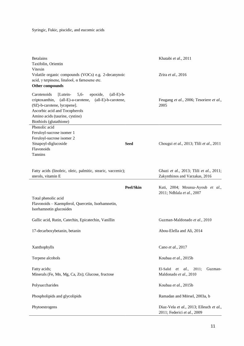
Table 2.2: Phenolic and non-phenolic compounds in Opuntia cactus plant parts
Major Compounds Cacti part References
Gallic acid
Quercetin 3-O-Rutinoside
Quercetin 7-O-Rutinoside
Kaempferol 3-O-Rutinoside
Quercetin 3-O-Glucoside
Isorhamnetin 3-O-Robinobioside
Isorhamnetin 3-O-Galactoside
Isorhamnetin 3-O-Rutinoside
Isorhamnetin 3-O-Glucoside
Flower
Ammar et al., 2015; De Leo et al.,
2010; Yeddes et al., 2014a, b
Isorhamnetin 7-O-rutinoside
Isorhamnetin 3-O-rhamnosyl 7-O-rutinoside
Kaempferol 3-O-arabinoside
Ammar et al., 2015; Benayad et al.,
2014; De Leo et al., 2010
Rhamnetin 3-O-rutinoside Merina et al., 2011
Other compounds
Terpenes: (limonene, linalool, germacrene D,
aromadendrene, squalene)
Esters and alkaloids (ethyl linoleate)
Sterols
Carboxylic acids (linoleic, palmitic, octanoic,
butanedioic, pentanedioic)
Phenolic acids: Hydroxycinnamic acid derivatives:
(5-Hydroxyferulic acid-rhamnosidehexoside; Caffeoyl
methoxycinnamoyl quinic acid; 1,4-Diferuloyl syringic
acid; 4-p-Coumaroyl caffeic acid; 1,5-Dicaffeoyl
ferulic acid; 1,4-Syringicferuloyl 4-coumaroyl caffeic
acid)
Ammar et al., 2012
Flavonoids
Rutin, Myricetin
Quercetin, Isorhamnetin
Kaempferol, Luteolin
Isorhamnetin glycosides
Kaempferol glucosides
Catechin
Whole fruit/pulp
Bensadón et al., 2010; Cha et al.,
2013; Fernández-López et al., 2010;
Mabrouki et al., 2015; Osorio-
Esquivel et al., 2011
Phyllocactin
Protocatechuic acid
Osorio-Esquivel et al., 2011;
Tesoriere et al., 2004
4-hydroxybenzoic acid,
Ferulic acid
Vanillic acid (also isovanillic acid)
Trans-coumaric and trans-cinnamic acids
Galati et al., 2003; Serra et al., 2013
11
Syringic, Fukic, piscidic, and eucomic acids
Betalains
Taxifolin, Orientin
Vitexin
Khatabi et al., 2011
Volatile organic compounds (VOCs) e.g. 2-decanynoic
acid, γ terpinene, linalool, α farnesene etc.
Zrira et al., 2016
Other compounds
Carotenoids [Lutein- 5,6- epoxide, (all-E)-b-
criptoxanthin, (all-E)-a-carotene, (all-E)-b-carotene,
(9Z)-b-carotene, lycopene].
Ascorbic acid and Tocopherols
Amino acids (taurine, cystine)
Biothiols (glutathione)
Feugang et al., 2006; Tesoriere et al.,
2005
Phenolic acid
Feruloyl-sucrose isomer 1
Feruloyl-sucrose isomer 2
Sinapoyl-diglucoside
Flavonoids
Tannins
Seed
Chougui et al., 2013; Tlili et al., 2011
Fatty acids (linoleic, oleic, palmitic, stearic, vaccenic);
sterols, vitamin E
Ghazi et al., 2013; Tlili et al., 2011;
Zakynthinos and Varzakas, 2016
Total phenolic acid
Flavonoids – Kaempferol, Quercetin, Isorhamnetin,
Isorhamnetin glucosides
Peel/Skin
Kuti, 2004; Moussa-Ayoub et al.,
2011; Ndhlala et al., 2007
Gallic acid, Rutin, Catechin, Epicatechin, Vanillin
Guzman-Maldonado et al., 2010
17-decarboxybetanin, betanin
Abou-Elella and Ali, 2014
Xanthophylls
Cano et al., 2017
Terpene alcohols
Koubaa et al., 2015b
Fatty acids;
Minerals (Fe, Mn, Mg, Ca, Zn); Glucose, fructose
El-Salid et al., 2011; Guzman-
Maldonado et al., 2010
Polysaccharides
Koubaa et al., 2015b
Phospholipids and glycolipids
Ramadan and Mörsel, 2003a, b
Phytoestrogens
Diaz-Vela et al., 2013; Elleuch et al.,
2011; Federici et al., 2009

12
Phenolic acids - Gallic acid, Coumaric acid, 3,4-
dihydroxybenzoic, 4-hydroxybenzoic; Ferulic, Salicylic,
Vanillic Syringic, Synaptic acids; Protocatechuic acid
etc.
Cladode
Bensadón et al., 2010; Gallegos-
Infante et al., 2009; Guevara-Figueroa
et al., 2010; Valente et al., 2010;
Wright and Setzer, 2014
Flavonoid - Isorhamnetin, Quercetin, Kaempferol,
Isoquercetin, Nicotiflorin, Rutin, Narcissin
Terpenoid volatiles (cis-linalool oxide, trans-linalool
oxide)
Avila-Nava et al., 2014; Gallegos-
Infante et al., 2009; Ginestra et al.,
2009; Guevara-Figueroa et al., 2010;
Fatty acids and tocopherols Chahdoura et al., 2014
Other components
Alkanes (heptadecane)
Ascorbic acid
β-carotene, Lutein
Avila-Nava et al., 2014; Betancourt-
Domínguez et al., 2006
Alpha-pyrones, opuntiol, opuntiosides
Qiu et al., 2007; Siddiqui et al., 2016
Pectic polysaccharides
Lignans
Panico et al., 2007;
Rocchetti et al., 2018
Flavonoids
Glycosides
Coumarins
Stem
Jiang et al., 2006; Manzano et al.,
2017
Terpenoids:
Triterpenes, Labdane-type diterpenes
de las Heras and Hortelano, 2009;
Jamal et al., 2009; Manzano et al.,
2017
Tannins
Polysaccharides
Fatty acids (azelaic acid, palmitelaidic acid)
Manzano et al., 2017
C29-5β-sterols (Taraxerol) Jiang et al., 2006
Reports from different areas have frequently shown differences in compound profile. For
example, Galati et al. (2003) carried out the analysis of fruit juice of prickly pear cultivars from
San Cono, Sicily and reported ferulic acid as the major phenolic acid present. Other compounds
detected include isorhamnetin derivatives like isorhamnetin-3-rutinoside, isorhamnetin-3-

13
glucoside, kaempferol-3-rutinoside (all flavonols) as well as rutin. In a study of Opuntia in
Tunisia, Yeddes et al. (2013) showed that flavonoid glycoside derivatives dominated the
phenolic compound profile of Opuntia fruit pulp and peel, and that they were mostly
isorhamnetin derivatives. They also indicated that more flavonoids were identified in the spiny
cultivars compared to the spineless varieties. Compared to the fruit pulp, prickly pear peels
contained more flavonoids. Prickly pear cladodes also contain biochemical compounds such as
glucose, galacturonic acid, calcium oxalate, eucomic and piscidic acids; and flavonoids such as
keampferol and isorhamnetin glycosides, isoquercitrin, quercetin and myricetin (Ginestra et al.,
2009). Besides the presence of betalains, Tesoriere et al. (2005) reported the presence of amino
acid taurine as well as tocopherols (α-tocopherols, β-tocopherols and γ-tocopherols),
carotenoids and thiols such as glutathione and cysteine (Table 2.2) in the pulp of red, yellow
and white Silician Opuntia ficus-indica cultivars. They, however, surmised that industrial
processing of the fruit pulps reduced the contents of cysteine, ascorbic acid, beta-carotene, and
glutathione. However, tocopherols, betalains and taurine appeared to be less affected by
industrial processing. Taurine is an amino acid also referred to as 2-aminoethanesulfonic acid
and is considered a cell protective compound and suspected to be involved in inflammatory
response modulation (McCarty, 1999). While considerable amounts of taurine were reported in
Opuntia cultivars from Mexico and South Africa (Tesoriere et al., 2005), the concentrations
were lower than those reported by Stintzing et al. (1999). However, Ali et al. (2014) analysed
the amino acid content of prickly pear fruits and observed that taurine was absent in the different
prickly cultivars sampled from the Near East, Italy and South Africa. The same was the case
for the commercially available juices which were purchased from the market. This led to their
summary that the relative presence and amount of taurine that has been reported in literature
for Opuntia fruits may have resulted from the confusion of this amino acid with γ-Aminobutyric
acid (GABA). This misrepresentation was linked to the analytical methods utilised. GABA and
proline constituted some of the most abundant amino acids in all the fruits tested.

14
Chiteva and Wairagu (2013) assessed the nutrient composition of prickly pear fruits from
Laikipia, Kenya and compared results with Egyptian, American and Ethiopian samples. They
reported no significant differences in moisture contents of fruits. Ascorbic acid content varied
with samples, that is, 13.7 mg/100 g for Egyptian samples and 5.17 mg/100 g for the Kenyan
samples. In agreement with Lee et al. (2002b), it was submitted that increased light intensity
during growing seasons contributed to the increase in the vitamin C contents of the prickly pear
fruits. Other contributory factors to higher vitamin C content in fruits include good storage, low
harvest temperatures, and reduced irrigation (Lee and Kader, 2000). They also recorded low
vitamin C content in physically damaged fruits (Lee and Kader, 2000). Results for proteins
ranged from 0.2 to 2.0%, but only very few fruits contained some proteins. Opuntia sugar
content as low as 0.11% and as high as 59.40% have also been reported for Kenyan samples
(El Samahy et al., 2006). Seasons also determine fruits sugar content (Chiteva and Wairagu,
2013). In light of the nutrient variations in cactus pears, it will be interesting to investigate fruits
from Opuntia species from sub Saharan Africa/South Africa, which have largely been
neglected.
2.3. Antioxidants: sources, mechanisms, extended benefits and purification
Free radicals or reactive oxygen species (ROS) are highly unstable molecules that can cause
oxidative stress especially when their presence in the body outweighs the concentration of
antioxidants present (Jerome-Morais et al., 2011). In an oxidatively stressed state, the
probability of developing non-communicable diseases (NCDs) such as cardiovascular and
respiratory diseases, diabetes and cancer, increases. Body inflammations or tissue trauma, and
external inducers such as pollution, smoking, ultraviolet (UV) exposure, industrial solvents,
ozone are also contributory factors that may lead to the generation of free radicals and then
oxidative stress. Free radicals seek to stabilise themselves by damaging other cells leading to

15
diseases and premature aging (Song et al., 2009; Wilson et al., 2017). The structure of a free
radical has at least one unpaired electron. The unpaired electrons are very reactive and cause
cellular damage and/or genetic changes. Some examples of free radicals include hydroxyl
(HO•), hydroperoxyl (HOO•), alkyloxyl (ROO•), superoxide anion (O2−•), nitric oxide (NO•),
thiyls (RS•) and disulfide anions (RSSR−•), and carbonate (CO3−•) group radicals (Ayala et
al., 2014). In order to prevent or inhibit the continuous production of free radicals, antioxidants
are required. Studies have shown that the probability of contracting NCDs may by reduced by
improving eating habits and lifestyles such as the increased consumption of antioxidant-rich
foods and exercise (Song et al., 2009; Wilson et al., 2017). While the body has its own innate
antioxidative defense systems, it may require additional supply of antioxidants to facilitate the
prevention or inhibition of ROS generating chain reactions and to halt the progression of
associated diseases. The extra antioxidants may be derived from antioxidant-rich diets or
supplements (Ayala et al., 2014; Wilson et al., 2017).
An antioxidant has been described as a substance that can inhibit the initiation or progression
of an oxidative process (Wilson et al., 2017). Antioxidants have been used successfully to
reduce degradation and loss of nutrients in foods during processing and storage. Antioxidants
may be natural or synthetic. Natural antioxidants imply those derived from natural sources such
as plants/foods and are usually naturally present as dietary constituents. Synthetic antioxidants
on the other hand are produced from non-natural processes or from chemosynthetic industrial
processes (Song et al., 2009). Endogenous antioxidants produced by the body also fall under
natural antioxidants. Flavonoids, phytoestrogens, polyphenols and catechins are examples of
natural plant-based antioxidants (Jerome-Morais, et al., 2011; Song et al., 2009). Plant sourced
antioxidants are also usually referred to as phytochemicals or phytonutrients (Nahar et al.,
2009). The demand for natural antioxidants has increased all over the world as synthetic
antioxidants cause unhealthy side effects. Globally there is also an increase in consumer

16
preference and sensitisation for clean labels/natural flavours and reduction in use of synthetic
food additives. In addition, natural antioxidants may have added benefits for health, and they
are also readily absorbed in vivo. Synthetic antioxidants/food preservatives are usually used to
mitigate oxidative food spoilage during processing or storage. They help in maintaining food
properties such freshness, nutrients and shelf life due to their high stability (Wilson et al., 2017).
However, some bring about nausea, as well as allergenic and carcinogenic effects and they may
have no added benefits to health (Embuscado, 2015).
Antioxidative compounds are a principal line of defence in protecting food quality from farm
to market and homes. There is an increase in consumer consciousness with respect to quality
and health and these affect the food choices. The farmers and producers of foods are therefore
challenged with supplying high-quality goods. Oxidation of foods which occur is a major bane
in tackling this challenge (Shahidi and Zhong, 2015). Oxidation in lipid-based foods affects
texture, toxicity, nutrient quality and flavour. Antioxidants are required to achieve extended
shelf-life for lipid-based foods such as animal fat, vegetable oils, nuts, processed meats, and
margarines among others (Shahidi and Zhong, 2015). Antioxidative compounds act by
inhibiting oxidative processes. Preservation of foods by added antioxidants is achieved through
this inhibitive process. Many nutraceuticals and dietary supplements boost and promote health
through the antioxidative action of their components (Shahidi and Zhong, 2015). Antioxidants
carry out their function using various modes of action.
Free radicals attack important macromolecules such as proteins, lipids and DNA, resulting in
cell destruction/damage and homeostatic imbalance. Targets of free radicals include all kinds
of molecules in the body (Huang et al., 2005; Lobo et al., 2010). Mechanisms of antioxidant
action include hydrogen or electron donation, peroxide decomposition, singlet oxygen
quenching, enzyme inhibition, synergism (co-antioxidants), metal-chelation and gene

17
expression regulation. Most of these functions fall under the chain-breaking and/or chain-
initiation quenching modes of action (Lobo et al., 2010; Moon and Shibamoto, 2009). The
antioxidant power and mechanism of action of antioxidant compounds vary from molecule to
molecule. Phenolic acids scavenge free radicals more than they chelate metals, while flavonoids
efficiently carry out their functions using both mechanisms. Also, as an added benefit, many
phenolics are not prone to attack by molecular oxygen (Brewer, 2011). Once free radical
generation is initiated, body cells protect themselves against oxidative stress using its cascade
of antioxidant enzymes within the body. Enzymatic and nonenzymatic antioxidants exist in the
intracellular and extracellular environment to detoxify ROS. Intracellularly, an enzyme like
superoxide dismutase is involved with the breakdown of the superoxide anion into oxygen and
hydrogen peroxide, while catalases facilitate the breakdown of hydrogen peroxide to water and
oxygen (Lobo et al., 2010). Also, glutathione, glutathione reductase, glutathione peroxidases,
and glutathione S-transferases which make up the glutathione enzyme system, catalyse the
decomposition of hydrogen peroxide and organic hydroperoxides (Nyanhongo et al., 2011).
Tocopherols and tocotrienols (Vitamin E), melatonin, uric acid, and ascorbic acid are examples
of non-enzymatic antioxidants. Lipophilic or hydrophilic endogenous antioxidants may act
through any of the mechanisms previously mentioned (Brewer, 2011; Lobo et al., 2010).
Hydrophilic antioxidant examples include thiols, uric acid, albumin and bilirubin. Ubiquinol
and tocopherol are lipophilic antioxidants. Certain de novo and repair antioxidants such as
proteinases, proteases, and peptidases also act as lines of defense to prevent oxidised proteins
accumulation in the body (Brewer, 2011). Glycosylases and nucleases which are known as
deoxyribonucleic acid (DNA) repair enzymes/antioxidants protect the DNA against oxidative
damage.
Before exogenous antioxidants can be used, however, they may need to be purified from their
sources for use in assays and various application. Antioxidants may be purified using

18
chromatographic techniques. In thin layer chromatography (TLC), substances are separated on
thin adsorbent plate layer based on polarity and affinity of the compounds for the stationary
adsorbent, usually silica gel. Following the purification of different components, several assays
can be carried out. Nevertheless, some tests can be performed during the chromatographic
isolation/separations (Adelakun et al., 2012a). Pure concentrates of bioactive compounds may
be obtained by utilising selective extraction methods depending on the target compound or
group of compounds. Some isolated compounds contribute more to the bioactivity of plant
formulations/extracts and only bioactivity assays for each component can determine which
compounds are more active (Prior et al., 2005). The degree to which a bioactive compound or
antioxidant can scavenge free radicals constitute the basis of some in vitro assays. Some of the
mechanisms for detecting antioxidant activity include hydrogen atom transfer (HAT), single
electron transfer (SET), reducing power, and metal chelation, among others (Leopoldini et al.,
2004; Mishra et al., 2012; Pinela et al., 2016). Antioxidant substances decrease or block the
oxidation reactions induced by free radicals (Costa et al., 2012). Ennouri et al. (2005) reported
that antiradical activity of cactus pear extracts was consistent with the presence of some
phenolic flavonoids such as quercetin and carotenoids, and several vitamins, among them
ascorbic acid. In addition, as regards Opuntia antioxidants/extracts roles, Galati et al. (2003)
demonstrated that rats treated with extracts from Opuntia ficus-indica peel and fruit were
protected against ethanol-induced gastric ulcer. Opuntia ficus-indica extracts have also shown
liver protective capabilities. Extracts reduced liver damage induced by carbon tetrachloride
(CTC)-induced poisoning when administered orally two hours after the toxic substance. Prior
to inducing toxicity in the liver, hepatoprotective effect was also demonstrated by application
of the juice consecutively for nine days (Livrea and Tesoriere, 2006). Prickly pear fruits have
also been applied in cancer treatment and prevention. Aqueous extract from Arizonan Opuntia
ficus-indica fruits successfully suppressed ovary tumors (Zou et al., 2005). The fruit extract
was injected intraperitoneal a day before the injection of tumor cells. Significant tumor

19
suppression and modulation of tumorigenic genes expression showed similar effect with that of
N-(4-hydroxyphenyl) retinamide (4-HPR), a known chemo-preventive agent. The same extract
effectively treated immortalised ovarian, cervical and bladder epithelial and cancer cells (Zou
et al., 2005). The extract initiated an increase in cancer cell growth inhibition, death and
affected cancerous cells cycle in a time and dose-dependent manner. Opuntia ficus-indica
methanol extract had also earlier been shown to have neuroprotective effects on radical-
mediated (superoxide and hydroxyl-) neural damage (Ha et al., 2003).
2.4. Biologically active compounds
Bioactive compounds occur in nature, have varying effect on living organisms and are
sometimes called nutraceuticals (Biesalski et al., 2009). The presence of natural bioactive
components in food and plants such as the Opuntia sp. provide added health functions while
providing basic nutrients. On the other hand, nutraceuticals may be packaged as dietary
supplements, isolated nutrients, genetically modified foods (GMFs), and also as processed
beverages and/or cereals (Biesalski et al., 2009). Moon and Shibamoto (2009) defined
biological antioxidant as any substance that, when present in low concentrations compared to
those of an oxidisable substrate, significantly inhibits oxidation of that substrate. A good
example of natural bioactive antioxidant compound is phenols. Phenolic compounds are able
to inhibit lipid peroxidation (Kristinova et al., 2009) and lipooxygenation in vivo. This is mainly
attributed to the reducing properties of the chemical structures that enable neutralisation or
sequestration of free radicals, as well as the chelation of transition metals (Moon and
Shibamoto, 2009).
Phenols are a group of bioactive substances with structures consisting one or more hydroxyl
groups (-OH) directly bonded to an aromatic ring. They are produced and found in plants and

20
microorganisms. The simplest of the class is phenol (carbolic acid - C6H5OH). Phenols may be
monophenolic or polyphenolic in nature depending on number of phenol units within each
molecule (Sant’Anna et al., 2013). Phenolic compounds contribute to sensory attributes such
as astringency, colour and bitterness of many foods (Sant’Anna et al., 2013). Phenolic
antioxidants are able to donate electrons or hydrogen which form the backbone of their
reductive capabilities and the basis for predictions on their free radical scavenging potential.
The structural chemistry of polyphenols is directly linked with their ability to scavenge free
activities (Rice-Evans et al., 1996). In addition, the inclination of polyphenols to chelate copper
and iron in metal chelation mechanisms facilitate their function as pre-emptive antioxidants
when they obstruct transition metal-catalysed free radical formation (Rice-Evans et al., 1996).
A polyphenol is regarded an antioxidant if when present in low concentration compared to the
oxidisable substrate it is able to prevent free radical-mediated oxidation. Secondly, after
scavenging, free radicals generated must be stable upon further oxidation (Rice-Evans et al.,
1996). A large number of phenolic compounds and antioxidants are derived from plant sources
such Opuntia spp.
Thousands of different phenolic compounds are ubiquitous in plant materials. In vegetables,
onions, cereals and Opuntia fruits, cladodes and peel by-product, phenolic acids, lignans and
flavonoids abound. Teas and fruits also contain major constituents of the human diet (Stahl et
al., 2002). However, prior to the absorption of phytochemicals like phenols which are trapped
within plant tissues, there is need for the application of processing methods that would serve to
release these valuable compounds for health and nutrition, and other potential applications
(Stahl et al., 2002). The type of phenolics targeted and their solubility in various solvents go a
long way to determine which processing/extractive methods are applied. Usually, water soluble
phenols have lower molar masses compared to the more insoluble high molecular weight
phenolic compounds (Saura-Calixto, 2012). During absorption in vivo, phenols bind with

21
themselves and proteins irreversibly, while direct or enzymatic oxidation of native phenols may
also occur during mastication or raw plant material preparation. High molecular weight
compounds may also be produced through several condensation reactions. Phenolic compounds
condensation may however be inhibited if a reducing agent like ascorbic acid is present.
Reducing agents are able to reduce the condensed compound to the original forms. A low pH
environment, as well as the presence of sulphur dioxide may also affect oxidation and
condensation reactions of phenols in vivo (Stahl et al., 2002).
A study by Spencer et al. (2000) demonstrated that phenolic dimers are more likely to be better
absorbed in the small intestine compared to higher oligomeric forms such as trimers or
tetramers. In other words, the higher the molecular size the lower the probability of being
absorbed in vivo. For example, high molecular weight procyanidin oligomers were reduced to
the native epicatechin monomer and other dimer/oligomeric units as well and were unstable in
acidic gastric juice ex vivo (Spencer et al., 2000). Although insignificant levels of oligomer
absorption across the intestine was observed, the significant levels of epicatechin released
suggested that higher molecular weight oligomers could ensure a constant and significant
supply of the monomeric and dimeric forms that can be absorbed. This may play a critical role
in the in vivo action of procyanidins (Lin et al., 1999). The probability of the phenols being
metabolised by native colon microflora subsequent to metabolites absorption, as well as biliary
excretion determine the rate and extent of phenolic compounds absorption (Lin et al., 1999).
Also, the time it takes for the compound to pass through the intestine and gastric region, and
lumen pH constitute biological factors that influence phenols absorption in vivo. In addition,
solubility, hydrophobicity, molecular weight and pKa are some other factors that can affect
phenolic compounds absorption in the small intestine (Stahl et al., 2002).

22
Phenolic compounds are regarded as primary antioxidants. They are able to delay the
progression of oxidation in lipids such that ketonic and aldehyde decomposition products are
not formed. The decomposition products give rise to the phenomenon known as rancidity which
is associated with lipid spoilage/denaturation (Alamed et al., 2009). Propyl gallate (PG),
butylated hydroxyanisole (BHA), tertiary-butylhydroquinone (TBHQ) and butylated
hydroxytoluene (BHT) are some of the approved synthetic phenolic antioxidants which may be
used in food products (Makahleh et al., 2015). However, reports of toxicity and other adverse
effects attributed to the use of synthetic antioxidants have raised global concerns and calls for
the use of natural alternatives to safeguard public health (Aremu et al., 2011). In addition,
besides scavenging of free radicals, phenolic antioxidants also act by reducing peroxides to the
stable hydroxyl derivatives or act as metal chelators (Frankel and Finley, 2008; Sadiq et al.,
2017). Food manufacturers are encouraged to use natural antioxidative ingredients in place of
synthetic antioxidants (Shahidi and Ambigaipalan, 2015). Naturally occurring, plant-sourced,
antioxidant compounds include tocopherols, flavonoids, lignans, phenolic acids, and poly-
functional organic acids and betalains (Shahidi and Ambigaipalan, 2015). Some of these natural
ingredients and compounds are found in Opuntia plant parts such as the cladode, fruit and peels
and show various biological activities. The biological activities of pure Opuntia bioactive
compounds are, however, low. Enzymatic modification could be an option for improving the
biological activity of Opuntia phenolics.
2.5. Opuntia cladode: compounds, fingerprinting and biological activities
Cladodes are the spiny or spineless paddle-like, oblong (up to 70-80 cm), thick and succulent
parts of the Opuntia plant with varying widths. They possess a waxy, water repellent epidermis
and are capable of photosynthesis and asexual reproduction. Small bristles (glochidia) may be
present with barbs in the areoles (Heuzé and Tran, 2017). Several studies have shown that

23
Opuntia cladodes compounds profile change with species type, post-harvest treatment,
environmental conditions and plant age (Contreras-Padilla et al., 2011; Astello-García et al.,
2015). Nevertheless, some studies have demonstrated that different cladode cultivars from
distinct locations have similar flavonol profiles which may be used in taxonomic
identification/classification of cultivars (Moussa-Ayoub et al., 2014). Only isorhamnetin
aglycon (m/z 315) was detected which was in partial agreement with results published by
Santos-Zea et al. (2011) and Ginestra et al. (2009) from cactus O. ficus-indica cladodes
collected from Mexico and Italy, respectively. Flavonoid glycosides such as isoquercitrin,
kaempferol-3-O-rutinoside, isorhamnetin-3-O-glucoside and isorhamnetin-3-O-rutinoside
were detected in wild and commercial O. ficus-indica cladodes cultivated in Mexico (same
location and close phylogenetic proximity), but no kaempferol was detected (Guevara-Figueroa
et al., 2010). Sugars, fatty acids, tocopherols and organic acids were reported in TunisianO.
microdasys and O. macrorhisa cladodes varieties (Chahdoura et al., 2014). Eucomic acid,
kaempferol 3-O-robinobioside-7-O-arabinofuranoside and isorhamnetin 3-O-rhamno-side-7-
O-(rhamnosyl-hexoside) dominated the phenol profile of O. ficus-indica (highly domesticated),
O. albi-carpa (intermediate domestication), and O. hyptiacantha and O. streptacantha
(wildest). Lesser amounts of quercetin 3-O-rhamnosyl-(1-2)-[rhamnosyl-(1-6)]-glucoside)
were reported in domesticated species, and kaempferol 3-O-arabinofuranoside in wild cultivars
(Astello-García et al., 2015). In other studies, wild cladode varieties had similar or higher
amounts of pigment compounds such as β-carotene, lutein and xanthophylls compared to
commercial ones (Jun et al., 2013).
Cladode extracts of O. ficus-indica have been shown to lower cholesterol levels and have
antiulcer and anti-inflammatory activities. Aqueous extracts also improved wound healing
(Laughton et al., 1991). Nopal extracts containing quercetin, isorhamnetin, kaempferol and
vitamin C also showed in vitro (IC50 of 0.8 mg/mL) and in vivo (20% increase in plasma

24
antioxidant activity after 3 days of subjects feeding) antioxidant activity (Avila-Nava et al.,
2014) and hepatoprotective activities (Zourgui et al., 2008). Antidiabetic and antioxidant
activities of cladode extracts have been attributed to significant amounts of polysaccharides, as
well as lignans, flavonols and phenolic acids in the cladode extracts (Valente et al., 2010;
Rocchetti et al., 2018). Opuntia ficus-indica nopal extracts rich in polysaccharides and
polyphenols were also able to decrease the hydroxyl radical induced oxidation of linoleic acid
and DNA (IC50 of 9.3 µg/mL, better than 3.2 µg/mL for ascorbic acid) (Lee et al., 2002a,b;
Panico et al., 2007). Mucilage, pectin and total pectic polysaccharide fractions of Opuntia ficus-
indica cladodes were also characterised and showed antioxidant activity (Bayar et al., 2016).
Cladode extracts radical scavenging abilities may be linked to presence of phenolic antioxidants
and vitamin content (Stintzing and Carle, 2005). Cladode cultivars have further demonstrated
useful anticarcinogenic selenium-enriched chemotherapeutic properties and could serve as food
providing advanced dietary selenopharmacology to help fight human diseases (Bañuelos et al.,
2012). Cladode extracts may find use as surfactant and natural antioxidants in food and
pharmaceutical industries.
Recently, Opuntia dillenii cladodes were shown to contain α-pyrones, and novel opuntiol and
opuntioside (opuntiol glucosides) compounds (Siddiqui et al., 2016). Opuntiol and opuntioside
have been successfully utilised in the reduction of pain, but opuntioside was a more effective
analgesic (IC50 = 26 ± 0.9 mg/kg). In future design of novel analgesics, both compounds may
serve as integral constituents (Siddiqui et al., 2016). O. humifisa cladode extract also protects
against UVB-induced skin degeneration achieved by reducing associated enzymes gene
expression (Park et al., 2017). In addition, azelaic acid and labdane-type diterpene were recently
detected in cactus cladodes (Manzano et al., 2017). Azelaic acid shows good antibacterial
activity (Sieber and Hegel, 2014). Diterpenes demonstrate broad biological effects (de las Heras
and Hortelano, 2009). Bioactivities may be linked to terpenoids possession of isoprene structure

25
with added elements such as oxygen and acetate units, as well as the branched, cyclic structure
and the presence of hydroxyl groups (Ciocan and Bara, 2007; Wu et al., 2016). It is interesting
also that under stressed growth conditions, Opuntia cladodes can biologically accumulate
compounds (Lallouche et al., 2015).
Algerian O. engelmanii and O. streptacantha cladode cultivars accumulated significant
amounts of proline and soluble sugars which ensured osmotic adjustment under saline stress
conditions (Lallouche et al., 2015). This phenomenon was also observed in other parts of the
cactus plant, but to lesser extent as salinity increased (Hadjadj et al., 2011; Lallouche et al.,
2015). This bioaccumulative potential may pave the way for the application of cladodes in
atmospheric pollution and bioremediation by heavy metal (lead) bioaccumulation (El Hayek et
al., 2017). Opuntia cladodes are also able to bioaccumulate inorganic constituents such as
magnesium oxide, calcium carbonate, calcium oxalate monohydrate, calcium-magnesium
bicarbonate, potassium chloride and potassium peroxydiphosphate [K4P2O8] using diffractive,
microscopic, and spectrometric techniques (Contreras-Padilla et al., 2011).
2.6. Opuntia fruit: compounds and biological activities
Fruits are the succulent, ellipsoid and edible part of the Opuntia plant. They are usually about
7 cm long but can have a wide range of colours which include green, white reddish, purple and
yellow (Heuzé and Tran, 2017).
Cactus pear fruits contain significant quantities of vitamin C and polyphenols compared to other
Opuntia plant parts such as the root, cladodes and seeds (Hahm et al., 2015). Cactus pear fruits
also contain significant quantities of minerals (Mabrouki et al., 2015), amino acids (Ammar et

26
al., 2012), flavonoids such as luteolin (Kıvrak et al., 2018), phenolic acids and betalains (Cha
et al., 2013; Osorio-Esquivel et al., 2011).
Opuntia fruit/pulp extracts possess antidiabetic, cardioprotective, neuroprotective, anti-
inflammatory, and hepatoprotective properties (Serra et al., 2013). Betanin in Opuntia ficus-
indica fruits was reported to reduce chronic myeloid leukaemia cell spread (K562), and cell
death was recorded at an IC50 of 40 mM betanin (Sreekanth et al., 2007). Cactus fruit ethyl
acetate extracts containing flavonoids, trans taxifolin, and dihydrokaempferol also suppressed
HeLa cervical carcinoma cell proliferation (at ≥ 100 µg/mL concentrations), while normal
human BJ fibroblasts were unaffected, which suggests potential application as intervention for
human cervical carcinoma management (Hahm et al., 2015). Taxifolin has been known to
repress prostate cancer cell growth and inhibit HCT116 colorectal cancer cell growth (Woo et
al., 2012; Zhang et al., 2013). Reduction in the viability of hepatic (78.9 - 79.2%), prostate
(61.2 - 75.2%) and colon (52.5 - 74.1%) cancer cells have been reported for prickly pear fruit
coloured (Chavez-Santoscoy et al., 2009). Fruit component compounds and antioxidant
properties also evolve with fruit ripening (Cayupán et al., 2011). Antioxidant activity have been
reported for purified Opuntia fruit polysaccharides (Zhong et al., 2010). The molar ratio of the
polysaccharide sugars differed from those reported earlier (Panico et al., 2007). The difference
was attributed to different soil types/conditions and climates. Domestication gradients may also
affect the content of bioactives and their biological activities.
Opuntia ficus-indica red, purple and orange-coloured fruits grown on agricultural drainage
sediment, were shown to possess improved and selenium-enriched chemotherapeutic properties
compared to those grown on normal soil (Bañuelos et al., 2012). Purple cultivars such as the
Mexican Opuntia robusta (semi-domesticated) showed the highest total betalain, phenol,
carotenoid and vitamin C contents, but with low antioxidant activity compared to other

27
varieties. Yellow commercial O. albi-carpa had no betalains, low total phenolic content and
low antioxidant activity (Yahia and Mondragon-Jacobo, 2011). However, in some studies
phenol content did not always correlate with good antioxidant activity (Chavez-Santoscoy et
al., 2009).
Reports on O. ficus-indica fruits have shown major variation in total phenolics content. Phenol
content of approximately 172 mg/kg (Chavez-Santoscoy et al., 2009) and 452 mg/kg (Díaz-
Medina et al., 2007) have been reported for juice from Opuntia spp. Higher levels of phenolic
content have also been recorded in juice (Galati et al., 2003) and fruit pulp (Moussa-Ayoub et
al., 2011). Moussa-Ayoub et al. (2011) also reported higher antioxidant activity for O.
macrorhisa pulp compared to whole O. ficus-indica fruits. In other studies, the antioxidant
activity and vitamin C content varied between different fruit samples. Purple Opuntia ficus-
indica fruit varieties usually showed the highest antiradical ability (Bañuelos et al., 2012).
However, Sumaya-Martínez et al. (2011) showed that antioxidant activities of differently
coloured fruit cultivars (white and yellow) did not show great variance but had reduced
activities compared to the purple and red variants. On the contrary, red cactus pears from
Mexico had the highest ascorbic acid content/antioxidant activity compared to the purple
variety (Stintzing et al., 2005; Abdel-Hameed et al., 2014). Nevertheless, the presence of
betaxanthin in coloured cultivars was; purple > red > yellow > white pear (Stintzing et al.,
2005).
In another study, El Kharrassi et al. (2016) showed that Opuntia fruit skin colour did not
correlate with ascorbic acid concentration. The differences in these results were attributed to
genotype, growth location and environmental contributors affecting the vitamin C content of
the Opuntia cultivars. However, in Opuntia species earlier studied, ascorbic acid concentrations
were highest in red fruits than in yellow fruits (Kuti, 2004). Also, O. streptacantha (red skinned

28
cactus pear) carotenoids content was lower (14.6 mg/g fresh weight) than O. stricta var. stricta
(yellow-skinned cactus pear) (23.7 mg/g fresh weight) (Kuti, 2004). Current research has shown
that it is possible to concentrate pigments (betalains) and other bioactive fruit components
during ripening, compared to the very immature and under ripe cultivars days after anthesis
(Pinedo-Espinoza et al., 2017). In addition, a novel, neutral, and water-soluble (1→4)-α-d-
glucan polysaccharide from Opuntia ficus-indica aqueous fruit extracts was reported by Ishurd
et al. (2010). The polysaccharide was identified by gel-permeation and anion-exchange
chromatographic techniques.
In general, the biological capacities and nutraceutical benefits of Opuntia fruit species may be
due to the synergistic effects of betalains, flavonoids and other biologically active components
present (Stintzing et al., 2005). The highlighted applications suggest that Opuntia fruit/extracts
have potential for industrial application in the food, agricultural and nutraceutical industries.
2.7. Opuntia peel/skin: compounds and biological activities
Peels make up the rinds of the prickly pear fruit. They are often discarded and regarded as a by-
product (Ramadan and Mörsel, 2003b). The generation of high value food products with
improved nutritional value through food processing methods such as fermentation applied to
agro-industrial by-products continues to evolve as an economically viable option (Diaz-Vela et
al., 2013). Utilisation of food by-products as food or additives to produce functional foods with
improved health benefits is expanding. Fruit peels are a good source of fibre for prebiotic
preparations and antioxidants (Diaz-Vela et al., 2013). Studies have shown that non-digestible
oligosaccharides from plant by-products with prebiotic potential can undergo fermentative
processes involving lactic acid bacteria (LAB) and bifidobacteria to produce new compounds,
with enhanced health benefits (Diaz-Vela et al., 2013).

29
Opuntia peels contain fatty acids, vitamins and antioxidants (Cerezal and Duarte, 2005).
Antioxidant components identified include oleuro, pyrogallol, benzoic, 3-OH tyrosol, ellagic,
chorogenic, protocatechuic acid, epicatechin, gallic acid and showed 62.14% inhibition of the
DPPH radical. In vivo study involving hamster diet supplementation with peel extracts
increased cholesterol excretionand lowered liver cholesterol levels. Mechanisms of extracts
hypocholesterolemic activities include inhibition of hepatic HMG-CoA activity, interference
with cholesterol absorption in the intestine and bile acid trafficking and antioxidant activity.
These mechanisms work synergistically for coronary heart disease prevention (del Socorro
Santos et al., 2017). Fermented flours Opuntia peels with higher fibre and carbohydrate content
also showed significantly higher antioxidant activities (89% inhibition of ABTS and 2.6 trolox
equivalent antioxidant capacity value) compared to pineapple peel flours (69% ABTS radical
inhibition and 1.5 trolox equivalent antioxidant capacity (Diaz-Vela et al., 2013).
Opuntia ficus-indica peels contain considerable amounts of neutral glycolipids and
phospholipids (Ramadan and Mörsel, 2003b). The presence of xantophylls [(all-E)-lutein, (all-
E)-violaxanthin and (all-E)-zeaxanthin], hydrocarbon carotenes (belonging to two types of
oxygenated carotenoid derivatives); and chlorophyll (Cano et al., 2017) have also been
identified. Flavonoid glycosides dominate the flavonoid profile of cactus peels (Ginestra et al.,
2009). Spineless fruit cultivars contain more flavonols than the spiny varieties. Prickly pear
peels however contain higher level of flavonoids when compared to the pulp (Yeddes et al.,
2013). Polysaccharides have also been reported in Opuntia stricta peel extracts and exhibited
significant antioxidant activity. It showed 94% DDPH inhibition at 50 mg/ml concentration and
an IC50 of 6.5 mg/ml (Koubaa et al., 2015a). Other bioactive components include
phytoestrogens (Diaz-Vela et al., 2013) and terpene alcohols (Koubaa et al., 2015b).

30
Anticarcinogenic and antihypertensive activities have also been reported for Opuntia peels
(Mulaudzi et al., 2017). Opuntia ficus-indica fruit peel extracts exhibited anticancer activity in
Ehrlich Ascites Carcinoma Cells (EACC). Their potency and total phenolic content were
significantly related, with two purified compound fractions having cancer cell killing efficiency
of 51.5-76.0 dead cells/100 μg/ml (Abou-Elella and Ali, 2014). By induction of
hyperpolarisation in cell membrane, O. ficus-indica extract phenolic composition significantly
increased calcium levels within human Jurkat T-cell lines (Aires et al., 2004). The extent of
molecular branching, chemical structure and composition, presence of sugar moieties and
physicochemical attributes of these bioactive molecules are responsible for the biological
activities reported for Opuntia peel compounds/extracts (Koubaa et al., 2015a).
The components and bioactivities of peel extracts may depend on the extraction method.
Recently, ultrasound and pulsed electric field technologies have been successfully used to
achieve greater cell denaturation to facilitate better recovery of the intracellular coloured
compounds from red prickly pear fruit peels (Koubaa et al., 2016). At higher temperatures,
more phenolic compounds are extracted, but with a reduction in antioxidant activity. In some
studies, optimal extraction methods for retention of phenols and antioxidant activities was
achieved at 80°C with 45% ethanol after 2 hours of extraction (Han et al., 2016). The
physicochemical properties of peel fibres can be exploited in the food industry to improve food
product parameters such as shelf-life, sensory attributes, staling, and viscosity (Anwar and
Sallam, 2016). Peels are cheap and readily available, and these factors enhance the potential for
commercialisation (Anwar and Sallam, 2016).). Margarines containing peel extracts resisted
oxidation better than vitamin E margarine and physicochemical and microbiological properties
of the margarine were unaffected (Chougui et al., 2015).

31
2.8. Effect of processing conditions on compound profile and bioactivities
Processing techniques applied to foods matrices are integral factors that determine the
bioavailability of nutrients and bioactive compounds and bioactivities. The impact of
processing technologies on expression of bioactive compounds vary widely and is not a simple
cause and effect relationship and remains complex to date (Cilla et al., 2018). However, the
major aim of food processing should be to achieve preservation of functional and nutritional
characteristics of the foods being processed. Bioactive compounds in Opuntia plant may better
assert their benefits to health when they are more bioavailable in vitro and in vivo.
Cladode processing conditions affect the amount and types of compounds present. It has been
shown that a combination of 45oC and 3 ms−1 air flow were optimal for the preservation of
phenol, flavonoid, ascorbic acid and β-carotene (Medina-Torres et al., 2011). Boiling processes
affected compound preservation of vitamins and sugars whereas lipids, proteins and fibre were
well preserved. Decreased antioxidant activity (38.9 and 31.6 trolox equivalent for raw and
boiled samples, respectively) was linked to phytochemical losses due to processing
technologies (Ramírez-Moreno et al., 2013). Jaramillo-Flores et al. (2003) showed 84%
retention of cladode carotenoids after heat processing. However, in another study, reduced
amounts of carotenoids were reported (Ayadi et al., 2009). Lactic acid bacteria (LAB)
fermentation of cladode pulp by Lactobacillus brevis have also been shown to improve
biological properties of the cladode pulp. This enhancement of bioactivity was confirmed using
Caco-2/TC7 cells in which fermented pulp demonstrated significant inhibition of related
prostaglandin production (Filannino et al., 2016).
Many studies on Opuntia plant parts and other plants have used methods such as
milling/grinding to break down food matrices into smaller particle sizes in order to achieve
32
improved expression of compounds and nutrients (Medina-Torres et al., 2011; Minekus et al.,
2014). Following milling of plant material, other processing methods may be applied depending
on the required end-product. Thermal treatment such as steaming, boiling, roasting, microwave,
have been used to modify food structure and for preservation purposes but with the attendant
loss of compounds/nutrients and bioactivities. These effects, however, vary depending on the
intensity of the heat applied (Cilla et al., 2018; Wang and Bohn, 2012). In addition, it has been
recommended that in the preparatory stages for food/plant materials, freeze-drying or oven-
drying of plant materials for short periods at temperatures lower than 50oC may produce more
satisfactory results with respect to bioactive compounds expression, yields and biological
activities (Torres et al., 2010). Freeze-drying minimises loss of functional and nutritional
properties of foodstuffs through better retention of phytonutrient and phytochemical properties.
Phytochemical losses increase when oven drying is carried out for longer periods or at higher
temperatures (Reyes et al., 2011). It was therefore of interest to investigate the effect of freeze-
drying and oven-drying on extract yields, biological activities and phenolic compound profile.
2.9. Techniques for the extraction of compounds from Opuntia residue
Pomaces/residues have been ignored as source of viable phytochemicals which are usually not
released from plant matrices (Durazzo et al., 2016; Gonzales et al., 2015). Therefore,
extraction/separation techniques have been developed and utilised to characterise essentially
valuable components from Opuntia residues which are not released from conventional
extractive processes. Separation by adsorption using synthetic resins is usually applied for
polyphenols recovery from plant crude extracts. This process is valuable in the food industry
(Soto et al., 2011). Low operation costs is one of its advantages as well as high adsorption
capacities for different classes of compounds and simplicity in handling (Soto et al., 2011).
Antioxidant and antiproliferative compounds like polyphenols and anthocyanins have been

33
recovered from apples and cherries through this process (Bobe et al., 2006; Schaefer et al.,
2006).
The use of alkaline hydrogen peroxide treatment (Vilela et al., 2016); acid and alkaline
hydrolysis and/or sonication; butanolysis and phloroglucinolysis (Arranz et al., 2009; Tarascou
et al., 2010) have been efficient in the release of residue/fibrebound antioxidants
(macromolecular antioxidants - MA), also known as antioxidant dietary fibres (ADF) (Durazzo
et al., 2016). Flavonols undergo degradation (to protocatechuic acid) when subjected to acidic
hydrolysis (Moussa-Ayoub et al., 2011), and the extent of degradation depends on the
concentration of acid utilised, nature of flavonol or plant material as well as hydrolytic
temperature and treatment time (Häkkinen and Törrönen, 2000; Tura and Robards, 2002).
Enzymatic hydrolysis has however been shown to be milder and more sensitive and yielded
significant amounts of quercetin flavonol, and kaempferol compared to acidic hydrolysis. It
also released undegraded sugars from flavonol glycosides to yield its corresponding aglycon
(Bilyk et al., 1984).
2.9.1. Opuntia macromolecular antioxidant (MA) biological activities
Macromolecular antioxidants (MA) are high molecular weight antioxidant compounds when
compared with extractable phenols, and exhibit significant biological activites (Arranz et al.,
2009; Cardador-Martínez et al., 2011). They are made up of hydrolysable polyphenols, tannins
(hydrolysable and condensed tannins) (Cardador-Martínez et al., 2011), single phenolic acids
and polymeric proanthocyanidins (Pérez-Jiménez and Saura-Calixto, 2015). MA are usually
determined in non-extractable polyphenol (NEPP) and non-extractable proanthocyanidins
(NEPAC) and expressed in acidified methanol and HCl/butanol (butanolysis) fractions,
respectively. The depolymerised hydrolysates are then utilised in quantitative assays (Durazzo

34
et al., 2016). In vivo, MAs are hypothesised to occur mainly in the colon where microbes
ferment them to produce absorbable metabolites. MA exhibit different health-related properties
which include antiproliferative, antioxidant, and gene expression modification (Tarascou et al.,
2010). MA have been reported to constitute up to 50-100% of some fruits studied, and showed
remarkable biological activities (Arranz et al., 2009; Tarascou et al., 2010).
Natural anticancer ingredients (ferulic acid, isorhamnetin derivatives, betalains) have been
isolated from Opuntia fruit residues after hydroalcoholic solvents extraction and separation
with microporous resins (Amberlite XAD16 resin) (Serra et al., 2013). Juice residues from O.
robusta and O. ficus-indica wild fruits from Portugal contained useful compounds which were
responsible for inhibition of human colon carcinoma HT29 cell growth (ED50 of 4.6 - 6 mg/mL
after 72 h of proliferation). Residue extracts resulted in an increase in reactive oxygen species
(ROS) in carcinoma cells, indicating that the prooxidant effect of constituent compounds may
induce cancer cell death (Serra et al., 2013). The combination of an extractive and resin
concentration methods can be used to concentrate bioactive compounds for potential
application in pharmarceutical and food industries. The antiproliferative effect was, however,
slightly lower than those described by Hahm et al. (2015) using highly fractionated O. humifusa
extracts (hexane, ethyl acetate extracts and water partitioned fractions) in a more sensitive
cancer cell line (human glioblastoma cell line-U87MG cells). Compared to doxorubicin (a
known anticancer compound), earlier studies on Opuntia extracts showed reduced inhibition of
cancer cell proliferation. Nevertheless, a combination of these extracts with doxorubicin could
improve anticancer efficacy and reduce chemoresistance (Serra et al., 2013).
Research has reported high antioxidant activity of MA extracts (52.22 ± 1.07 μmol trolox
equivalent/g) even in the absence of some cladode MA compounds such as hydrolysable tannins
and proanthocyanidins (Bensadón et al., 2010). Hydrolysed cladode extracts were more

35
effective radical scavengers than the unhydrolysed fractions (Avila-Nava et al., 2014). Acid
hydrolysis released polyphenols bound to or associated with the insoluble fibre as glycosides
(Avila-Nava et al., 2014; Ginestra et al., 2009). Greek cactus pear fruit seed residue extract also
showed good antioxidant activity (69.5 - 95.1 mmol/kg) (Zakynthinos and Varzakas, 2016).
Other biological/gastrointestinal activities of antioxidants from non-Opuntia residues include
antiinflammatory activity of grape proanthocyanidins (Tomás-Barberán and Andrés-Lacueva,
2012), and pomegranate ellagitannins (Larrosa et al., 2010). Non-Opuntia residue antioxidants
also have beneficial microbial interactions [Cocoa flavanols (Tzounis et al., 2008)]; and
antioxidant effects, that is, they inhibit oxidation of cholesterol and lipids, and proteins and
vitamins (Tomás-Barberán and Andrés-Lacueva, 2012). They also interact with α-glucosidase,
lipase and alpha-amylase enzymes (Tomás-Barberán and Andrés-Lacueva, 2012). Biological
activities of Opuntia MAs could have potential applications as pill and capsule supplements;
components of cosmetics, food ingredients (prebiotics) and drug excipients. More research into
MAs may provide new data on novel antioxidant compounds and their applications (Amoo et
al., 2014; Aremu et al., 2014; Pérez-Jiménez et al., 2013). MA analyses and quantification in
more Opuntia/plant species are still required in the compilation of a broader database. This
would be useful in guiding future biological and nutritional research (Arranz et al., 2009).
2.10. Potential side effects of Opuntia species extracts/compounds
Little information is available on Opuntia spp. toxicology and on adverse side effects of its
compounds and/or extracts. However, from literature, a low colonic obstruction has been
attributed to the consumption of Opuntia ficus-indica seeds (Kleiner et al., 2002). Nausea, mild
diarrhoea, increased stool volume and frequency, headaches and abdominal bloating, have also
been reported in books on traditional folk medicine and case reports (De Smet, 2002; Gagnier
et al., 2006). These effects were, however, not corroborated with results from scientific

36
research. Although some health care professionals believe that herbal medicines are relatively
safe because they are “natural”, there is little supporting data to back such an assumpt ion.
Although herbal mixtures may contain certain contaminants from any stage of the
extraction/production process, side effects may also be linked to plant components and human
factors (intrinsic) and non-plant related contaminant factors (extrinsic) (Gagnier et al., 2006).
There are possible indications of extreme sensitivity to any component of Opuntia as individual
pharmacokinetics vary widely. Dermatitis has been reported to be a common adverse reaction
to prickly pears (Pawar et al., 2017). The presence of opportunistic pathogens/microorganisms
or microbial toxins could also affect the content of active components in herbal mixtures and
result in adverse health effects. Therefore, plant-derived herbal interventions need to be treated
with care, or as containing potentially toxic chemical compounds until proven otherwise. Some
of these toxic chemical hazards have been listed in the Maryland National Library of Medicine
(NLM) database for harmful substances (Osuna-Martínez et al., 2014).
2.11. Opuntia plant compounds and the search for new antimicrobial compounds
The rise of new infectious diseases and increased bacterial resistance to available antimicrobials
have necessitated plant-related research channelled towards identifying novel compounds with
antimicrobial activity (Aremu et al., 2011; Silver and Bostian, 1993; Valtierra-Rodríguez et al.,
2010). Antimicrobial compounds belong in a range of phenolic and non-phenolic classes such
as betalains, polyphenols and phenolic acids (caffeic, cinnamic, catechol), quinones, flavones,
flavonoids, flavonols, tannins, coumarins, lectins and polypeptides, alkaloids, terpenoids,
essential oils, polyamines (spermidine), isothiocyanates, thiosulfinates, glucosides,
polyacetylenes and acetylene compounds (Ciocan and Bara, 2007; Tapiero et al., 2002). The

37
antimicrobial activities of Opuntia species extracts are attributed to the presence of quite a
number of these compounds.
Bactericidal activity has been reported for Cardon Blanco (O. streptacantha), Jalpa and Real
de Catorce (O. ficus-indica) nopal cacti cultivars against Campylobacter jejuni (1.1 - 12.5
mg/ml), Clostridium perfringens (0.8–16 mg/ml) and Vibrio cholera (4.4 - 30 mg/ml). The
activities were partly ascribed to the significant amounts of total flavonoids and phenols present
in the cultivars (Sánchez et al., 2014). Opuntia ficus-indica extracts in a wide array of solvents
have shown activity against different bacterial strains such as Staphylococcus aureus,
Pseudomonas aeruginosa, Escherichia coli, Salmonella spp., Klebsiella pneumoniae,
Citrobacter freundii and Streptococcus pneumoniae (Shafiei et al., 2013; Wasnik and Tumane,
2016), and against Bacillus subtilis (Gnanakalai and Gopal, 2016). Terpenoids, glycosides,
saponins, alkaloids and flavonoids were identified in the extracts. Acetone extract compared to
n-hexane and petroleum ether extracts showed better antimicrobial activity (Wasnik and
Tumane, 2016), while aqueous extracts of both stem and fruit (Gnanakalai and Gopal, 2016)
showed the least antimicrobial activity which could be attributed to poor solubility of bioactive
components in extraction solvents.
Opuntia flower extracts also showed in vitro antibacterial activity against P. aeruginosa,
Staphylococcus aureus, B. subtilis, and E. coli, and antifungal activity against Candida
lipolytica and Aspergillus niger. Most important was the antibacterial activity which make the
extracts potentially suitable for food industry applications, for example, as food additives or
preservatives (Ennouri et al., 2005). Extracts of Opuntia matudae containing betalains inhibited
the growth rate of E. coli O157:H7 (Hayek and Ibrahim, 2012). In addition, Ammar et al. (2015)
reported marked antibacterial action (zone of inhibition 25±1.8 mm) of Opuntia flower
methanol extract on Listeria monocytogenes. Recently, first report of a 28.3 kDa protein with

38
RNase activity purified from Opuntia cladode aqueous extract demonstrated antiviral action
against cucumber and tobacco mosaic viruses (CMV and TMV) (MIC of 40 mg/mL). Antiviral
activity was linked to interaction of the protein with RNase activity with viral nucleic acid
(Rasoulpour et al., 2017).
Broad spectrum antimicrobial activity of betalain-rich extracts against Bacillus cereus, L.
monocytogenes, Proteus vulgaris, Yersinia enterocolitica and Enterobacter aerogenes, as well
as against Rhizoctonia solani and Candida albicans yeasts (125 - 250 mg/ml), Fusarium
oxysporum and Aspergillus flavus moulds (500 mg/ml), have also been reported (Tenore et al.,
2012). The broad-spectrum activity of the extracts was attributed to the adverse effect of
bioactive components on microbial cell membrane integrity, function and structure
(Canadanovic-Brunet et al., 2011). Opuntia stricta essential oil extracts containing mainly
thymol (42.7%) also showed antimicrobial activity against important food and environment
borne opportunistic bacterial pathogens such as E. coli, B. cereus, P. aeruginosa (1.25–2.5
mg/ml MICs) and yeast such as C. albicans (2.5 mg/ml MIC). The extracts of the Opuntia plant
therefore have potential for commercialisation as novel drugs for use in antimicrobial therapy
(Moosazadeh et al., 2014).
Most plant compounds with antimicrobial properties are aromatic organic molecules (Ciocan
and Bara, 2007). Phenols and flavonoids are aromatic, hydroxylated biologically active
compounds. Presence of either phenols or aldehyde compounds in extracts reportedly show
highest antibacterial activities compared to those containing terpene alcohols (Koubaa et al.,
2015a; Naveed et al., 2013). The number and site of the hydroxyl groups on the aromatic
molecules determine antimicrobial action. Usually the more hydroxyl groups present, the better
the antimicrobial effect. Antimicrobial quinone compounds bind to extracellular and soluble
proteins and disrupt the integrity of microbial cell membranes/wall to result in static or cidal

39
effects. Similar antimicrobial mechanism is observed in coumarins where benzene and α-
pyrone rings are bound together. Higher antimicrobial action was reported for polymeric
phenols (with multiple number of hydroxyl groups) such as hydrolysable and condensed tannins
which act by binding cell walls, inactivating microbial adhesins, enzymes, cell envelope and
transport proteins, thereby preventing growth and protease activity (Ciocan and Bara, 2007).
Some of the methods used for ascertaining the mechanism of action of a potential antimicrobial
agent include measurement of cell conductivity, bacterial rate of kill (time-kill) and cell
integrity assays. Transmission electron microscopy (TEM) and scanning electron microscopy
(SEM) techniques are also been utilised to give a visual observation of cells after treatment with
the potential antimicrobial compounds. SEM and TEM confirm changes to cell morphology,
cell wall and cell membrane of treated microbial cells compared to untreated cells (Sadiq et al.,
2017).
2.12. Laccases as biocatalysts in the synthesis of bioactive compounds and materials
Many phenolic compounds from Opuntia cladodes and fruits can serve as good substrates for
biocatalytic reactions. Enzymes may be utilised in an array of selective organic transformations,
including modification of natural compounds (Adrio and Demain, 2014; Su et al., 2018).
Enzymes have been used in many industries for centuries and can act on a wide range of natural
and artificial substrates. Enzymes are chemically selective of certain functional groups
(chemoselectivity) located in separate regions of a substrate (regioselectivity). These functions
are aided by the three-dimensional structure of enzymes (Shoda et al., 2016). Also, most
enzymes are composed of L-amino acid units which make them chiral catalysts. In other words,
for asymmetric and selective utilisation, chemo- and regioselectivity properties make up a
central attribute and determine an enzyme’s ‘specificity’ (Carr, 2004). Enzymes are known to
catalyse reactions mostly in the aqueous phase. Generally, for biocatalytic applications, organic

40
solvents have more advantage over other solvents. This is because they have reduced water
activity and allow greater solubility of hydrophobic components; side reactions observed in
aqueous reactions are also reduced, there is reduced probability of microbial contamination.
Organic solvents also aid isolation/purification in which also enhances yields (Zaks, 2001). The
use of enzymes cannot be overemphasised in their biotechnological applications as biocatalysts.
The use of laccase is proposed for use in modifying Opuntia related compounds in this study.
Laccases are glycoproteins which belong to the oxidoreductase group of enzymes (Riva, 2006).
Laccases are copper-containing, p-diphenol dioxygen oxidoreductases which carry out one-
electron oxidation of appropriate substrate molecules such as aromatic or aliphatic amines, and
phenols to the corresponding reactive radical with the subsequent reduction of oxygen to water
(Kudanga et al., 2017; Kudanga and Le Roes-Hill, 2014). The mechanism of action of laccase
in synthesis of new compounds is shown in Fig. 2.1. Laccase-catalysed oxidative reactions
generate radicals from the monomeric substrate units. The radicals may then bind to achieve
stability and evolve into a new molecule (Fig. 2.1) or may turn towards degradative processes.
Polymers may also be formed if further coupling is allowed to continue. Radicals may evolve
in three distinct ways. First, oxidative coupling is the most common path where radical reactions
are largely uncontrolled with respect to structure of the desired/end oxidation product.
Therefore, optimising reaction parameters cannot be overemphasised if a definite molecule is
targeted. In order to achieve this, the frequent application of a short reaction time and defined
molar concentrations (Hollmann and Arends, 2012), substrate engineering (Mita et al., 2002),
and use of co-solvent (Mita et al., 2004) are sometimes utilised to obtain defined products.
Laccases may also catalyse cross-coupling reactions as shown in Fig 1. This may result in new
and interesting structural conformations in the end products (Pezzella et al., 2015). Therefore,
laccases have been used as a catalyst in organic synthesis for example in drug manufacture and
related industries (Adrio and Demain, 2014; Mayer and Staples, 2002; Su et al., 2018).

41
Figure 2.1: Mechanism of action of laccase-catalysed oxidation of substrate to form radicals,
b radicals undergo oxidative coupling to produce dimers, c further coupling results in the
formation of polymers through polymerisation, and d coupling with a non-laccase substrate to
form cross-coupling products.
Source: Abdel-Mohsen et al. (2014) and De Regil and Sandoval (2013)
High molecular weight polyphenols have been reported to exhibit enhanced biological
properties with longer circulation time in vivo (Silva et al., 2011). The oxidative coupling of
flavonoids has been used to produce polymers with improved antioxidant capacity. Kurisawa
et al. (2003) employed laccase to catalyse the oxidative polymerisation of catechin. When
compared with the monomeric catechin, the synthesised polymer showed enhanced ability to
scavenge superoxide. Jeon et al. (2012) also reported a mixture of dimer, trimers and tetramers
from a catechin monomer, and these oligomers showed higher anti-glycation and superoxide
dimustase (SOD) activities. Oligomers that are insoluble in water have been produced from free
laccase, while linear and water-soluble oligomers were derived using a conjugated laccase and
endowed with high antioxidative action (Jadhav and Singhal, (2014). In an earlier study,
oligomers derived from quercetin and kaempferol monomers also showed enhanced antioxidant
activity (Desentis-Mendoza et al., 2006).

42
In other and more recent applications of laccase, phenolic compounds like flavonoids are
oxidatively grafted onto polymers in order to modify/impart new properties to the polymers
(Pezzella et al., 2015). Polymer terminals contain functional moieties which make it possible
for low molecular weight molecules to be coupled to them. The cross-linking may occur either
in the side or main functional chains (Kobayashi and Makino, 2009). Using this approach,
laccases have been used in the modification and improvement of properties of fibres (cotton
and wool) (Hossain et al., 2010). In a previous study, Silva et al. (2011) used laccase from
Myceliophthora thermophila to oxidise flavonoids in flax fibres, and covalently bound the
quinones produced to chitosan amino groups. The antibacterial activity of the grafted linen was
then determined. Chitosan-treated linens where active against E. coli, while S. aureus showed
susceptibility to both catechin and chitosan treated samples. Similarly, laccase has been used to
impart antibacterial activity on lignocellulose materials (Widsten et al., 2010).
The use of laccase in the modification of materials to produce more active end products has
great prospect in various industries (Pezzella et al., 2015). In addition, many penicillin and
cephalosporin substituents are potential natural substrates for laccase. Hence, laccases has
potential to catalyse the production of therapeutics and pharmaceuticals such as antibiotics and
anticancer agents/compounds. The use of enzymes to modify compounds with the aim of
increasing antioxidant properties and other biological activities is gaining a lot of research
interest in biotechnology (Kudanga et al., 2017).
2.13. Structure-activity relationship (SAR) in phenolic compounds
In determining the SAR of phenolic compounds, Cai et al. (2006) tested different groups of
phenolic compounds and found that polymeric tannins showed the best activity compared to
most quinones, lignans and isoflavones. The study showed that the number and position of

43
hydroxyl groups, as well as the presence of other substituents greatly determine the antioxidant
capacity in phenolic compounds. In other words, differences in molecular structure also
translated into differences in radical scavenging activity which was linked to different
hydroxylation patterns, presence and/or absence of methoxylation and glycosylation. The
presence of ortho-dihydroxy groups conferred high activity on all phenols tested. Where
hydroxyl groups were absent no antioxidant activity was recorded, for example in isoflavone
and flavanone (Cai et al., 2006). Benzoic and cinnamic acids also had no hydroxyl group and
no antioxidant activity (Cai et al., 2006). Monohydroxybenzoates showed activity because of
the presebce of a hydroxyl group (-OH). Vanillic (4-OH, 3-OCH3) and syringic acids (4-OH,
3,5-OCH3) showed even better activity than the monohydroxybenzoates that had no methoxy
groups. The dihydroxylated protocatechuic acid also demonstrated a lower activity compared
to syringic acid which indicated that the presence of methoxy groups increases the ability of the
molecules to donate hydrogen and scavenge radicals. However, in the low-density lipoprotein
(LDL) system, the hydroxydimethoxy acids had reduced antioxidant capacity compared to the
dihydroxy acids (Natella et al., 1999).
Cardio-protective effects of flavonoid and other phenolic antioxidants arise from their
inhibitory action on lipid peroxidation. They are also able to impair reactive oxygen species
(ROS) pathways and chelate redox active metals (Tsao, 2010). Flavonoids have different
mechanisms of action. On the other hand, one of the most pronounced features of anthocyanins
(flavonoid polyphenols) is their substantial antioxidant capacity. However, as result of their
vast structural variations, their bioactivity varies widely (Cai et al., 2006). Glycosylation, on
the other hand, can reduce free radical scavenging capacity, but enhances molecule stability
(Konczak et al., 2005). Quite a number of studies have aimed at deriving a common substitution
and antioxidant activity hierarchy for flavonoid compounds. Data from these studies have
helped scientists make reliable predictions on SARs, as well as improve our general

44
understanding of flavonoids bioactivities. All additional advancements and progress in this
research area could help in the improvement of nutraceuticals and semisynthetic analogues
which have little or no side effect and retain significant biological capacities (Heim et al., 2002).
In the case of high molecular weight dimers/oligomers generated from laccase-catalysed
reaction, different radical linkages are possible. In a study by Adelakun et al. (2012b), β-β and
β-5 linked dimers were produced using ferulic acid as the monomeric substrate unit. The β-5
dimers showed better enhancement in antioxidant activity compared to β-β dimers. The
presence of carboxylic acid group with an adjacent unsaturated C–C double bond, which can
provide additional sites for free radical attack. The increase in the number of electron-donating
groups on the β-5 dimer, was reported to be responsible for the enhancement in antioxidant
activity (Srinivasan et al., 2007). In another work which involved the use of a 2,6-
dimethoxyphenol (2,6-DMP) as substrate, a symmetrical C–C-linked dimer, 3,3′,5,5′-
tetramethoxy biphenyl-4,4-diol, was formed (Fig. 2.2). The antioxidant activity of the dimer
doubled compared to the starting substrate, 2,6-DMP (Adelakun et al., 2012a). The increased
in activity was mainly due to the increase in functional groups with electron donating capacity
(Matsuura and Ohkatsu, 2000), the reduction in the O-H bond dissociation energy, and
increased stability of radical due to resonance delocalisation (Sánchez-Moreno et al., 1998).

45
Figure 2.2: Proposed reaction mechanism for the homomolecular coupling of 2,6-DMP to
produce the C–Cdimer (3,3′,5,5′-tetramethoxy biphenyl-4,4′-diol) (Adelakun et al. 2012a)
The tendency of an antioxidant/compound to release electrons is also dependent on bond
dissociation enthalpies (BDEs) of active/functional groups within a compound. In other words,
antioxidants containing functional groups with lower BDE would show better antioxidant
capacity because they more readily release electrons to radical species (Szymusiak and
Zielinski, 2003). A lower antioxidant activity was reported for the β-β linked ferulic acid dimers
compared to the ferulic acid substrate (Adelakun et al., 2012b). This observation was consistent
with earlier findings where bis-ferulic acid (β-β dimers) had a higher BDE (85.76 kcal/mol)
than ferulic acid (84.70 kcal/ mol) (Murakami et al., 2005). The determination of BDE varies
with compounds and experimental conditions. For these reasons, research which have focused
on phenolic compounds BDE have published contrasting results (Klein and Lukeš, 2006;
Szymusiak and Zielinski, 2003). In general, phenolics are mostly used as antioxidants possibly
due to the fact that hydroxyl groups have lower BDE compared to alkyl and aniline groups
(Bendary et al., 2013).
Antioxidant activity is also affected by hydrophobicity, especially in a multicellular
hydrophobic environment (Ishige et al., 2001), as well as an antioxidant’s ability to produce

46
stable radicals that can inhibit the progression of oxidative chain reactions (Alov et al., 2015).
At sites where free radicals are generated, hydrophobicity increases the bioavailability of the
antioxidant (Ishige et al., 2001). For example, hydrophobic antioxidants would effectively
scavenge free radicals generated from lipid peroxidation on the lipid bilayer because of their
lipophilic properties (Lu et al., 2006). On the other hand, radical stability arises from the
resonance delocalisation of lone electrons into the aromatic ring and absence of groups prone
to attack by oxygen (Flora, 2009; Shahidi and Naczk, 2004). Large groups on the ortho
positions of the aromatic ring have been shown to help stabilise antioxidant radicals (Shahidi
and Naczk 2004), but they may also reduce antioxidant activity by steric masking of the
phenolic hydroxyl group (Murakami et al., 2005). In other words, an ideal antioxidant would
consist of a balance of hydroxyl groups that ensure the free radical scavenging ability of the
antioxidant, the presence of electron-donating groups that help stabilise the radical after
donation of electrons, and hydrophobic groups that ensure the bioavailability of the antioxidant
in multicellular systems. Research that focused on improving the hydrophilicity of silybin
resulted in its compromised antioxidant activity in lipophilic environments (Gažák et al., 2010;
Gažák et al., 2004). These studies emphasised the fact that hydrophobicity is an integral factor
if antioxidants are to function effectively in cellular environment.
2.14. Conclusion
The relevance of plant extract compounds profiling cannot be overemphasised. It remains
significant for the discovery of new bioactive compounds, especially in unexplored plant
species or in different geographical locations and climate. Improving bioactivities remains a
challenge for the scientists working with phenolic compounds. Enzymatic modification of plant
compounds using an enzyme such as laccase can be an option for the production of new
compounds with improved biological activities.

47
2.15. Research hypotheses, aim and objectives
2.15.1. Research hypotheses
1. Southern African Opuntia cladode, fruit and peel extracts will show good biological
activities and a different phytochemical profile.
Compound profile of plants have been shown to be affected by biotic and abiotic elicitors (Khan
and Giridhar, 2015) and may vary depending on the geographical location (Nascimento et al.,
2011).
2. Laccase-catalysed modification of selected phenolic compounds will produce higher
molecular weight products with improved antioxidant and antimicrobial activity.
Phenolic dimers have been reported to possess increased biological activity compared to the
monomeric forms (for example, hydroxytyrosol dimer) (Zwane et al., 2012). It is expected that
oligomeric forms would show improved bioactivities. Oligomerisation usually result in
increased number of hydroxyl groups thereby providing more sites to react with free radicals,
as well as an increase in electron donating groups (Kudanga et al., 2017).
2.15.2. Research aim
The overall aim was to characterise southern African Opuntia extracts and enzymatically
modify selected phenolic compounds for the production of new bioactive compounds with
enhanced antioxidant and/or antimicrobial activities.
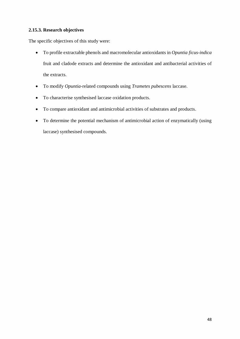
48
2.15.3. Research objectives
The specific objectives of this study were:
To profile extractable phenols and macromolecular antioxidants in Opuntia ficus-indica
fruit and cladode extracts and determine the antioxidant and antibacterial activities of
the extracts.
To modify Opuntia-related compounds using Trametes pubescens laccase.
To characterise synthesised laccase oxidation products.
To compare antioxidant and antimicrobial activities of substrates and products.
To determine the potential mechanism of antimicrobial action of enzymatically (using
laccase) synthesised compounds.

49
CHAPTER THREE
Extractable and macromolecular antioxidants of Opuntia ficus-indica cladodes:
Phytochemical profiling, antioxidant and antibacterial activities
Abstract
Macromolecular antioxidant (MA) fractions and their antioxidant and antimicrobial activities
are largely ignored from most conventional extractive processes compared to the well-known
extractable polyphenol fractions. This study investigated subtropical spineless cladode MA
extracts in comparison with their extractable fractions; as well as the effect of freeze- and oven-
drying methods on extract yields and bioactivities. Cladode extractable phenol (EP) and MA
fractions antioxidant capacities, antimicrobial potential against multi-drug resistant bacterial
cultures, and phenolic content were evaluated. LC/MS was used to analyse/profile extract
components. Freeze-dried samples from all the extracts showed better antioxidant and
antimicrobial activities than the oven-dried samples. Antibacterial and antioxidant activities
were generally higher in hydrolysed (MA) than in unhydrolysed (extractable) fractions. DPPH
radical scavenging activity (RSA) EC50 values correlated with phenol contents (R2=0.9609).
LC/MS extract profiles showed the presence of phenolic acids, flavonoid derivatives and other
unidentified compounds. Isovitexin 7-O-xyloside-2"-O-glucoside, polyhydroxypregnane
glycoside and neohancoside C were identified for the first time in Opuntia cladode extracts.
This study showed that cladode EP and MA extracts have potential as rich antioxidant sources
and can be key to the discovery of new antimicrobials.

50
3.1. Introduction
The cactus pear (Opuntia spp.) plant has a high genetic diversity with a wide variety of the plant
grown worldwide. Opuntia ficus-indica is the most common and main commercial variety, with
spines or no spines/thorns (Vigueras and Portillo, 2001). Globally, the demand for natural
antioxidants has increased due to safety concerns attributed to the use of synthetic antioxidants.
For example, synthetic antioxidants have been shown to be allergenic, carcinogenic, and cause
nausea, DNA and sperm abnormalities (Wojcik et al., 2010). Synthetic antioxidants may also
not contribute additional nutritional benefits (Satyanarayana et al., 2014). On the other hand,
natural antioxidants are readily assimilated by the body and show little or no adverse effect
(Wojcik et al., 2010). In addition, there is also the alarming trend of multi-drug resistance to
available antimicrobials (Valtierra-Rodríguez et al., 2010). The safety concerns with synthetic
antioxidants and increasing antimicrobial resistance trends have necessitated scientific studies
directed towards the search for novel, but safe bioactive molecules in plant extracts with
antioxidant (Aremu et al., 2011) and antimicrobial properties (Valtierra-Rodríguez et al., 2010).
Opuntia nopals (cladodes) have been used in traditional folk medicine (Flores et al., 1999).
Some reported health benefits of nopal extractable polyphenol (EP) extracts include
anticarcinogenic, antihypertensive (Livrea and Tesoriere, 2006) and antioxidative health effects
(Avila-Nava et al., 2014). The presence of insoluble and soluble fibres (1–2%), as well as
biologically active phytochemicals such as polyphenols and flavonoids (Avila-Nava et al.,
2014; Stintzing and Carle, 2005) is responsible for nopal health benefits. Ascorbic acid, a
natural antioxidant, is also found in considerable quantities in Opuntia nopals/cladodes
(Stintzing and Carle, 2005). However, the bulk of information on biological activity of cactus
pear cladodes comes from the extractable phenol fractions (Livrea and Tesoriere, 2006), while

51
much less information is available on macromolecular antioxidant (MA) fractions even though
there is evidence that both contain biologically active molecules (Avila-Nava et al., 2014).
Hydrolysable polyphenols [macromolecular antioxidants (MAs)] isolated in residues are often
overlooked in common plant extractive operations (Saura-Calixto, 2012). MAs include
hydrolysable tannins, phenolic acids and simple phenols (Pérez-Jiménez et al., 2013). Apart
from their antioxidant activity, MA compounds contribute to the beneficial health properties
associated with dietary food fibre (Saura-Calixto, 2012).
Several studies have reported antimicrobial (Gnanakalai and Gopal, 2016; Sánchez et al., 2014)
and antioxidant (Albano et al., 2015; Galati et al., 2003) activities of extractable polyphenol
extracts from Opuntia spp. (Livrea and Teosriere, 2006). Studies have also shown that the
drying methods applied to plant materials and/or their extracts could affect extract yield and
biological functions (Reyes et al., 2011; Torres et al., 2010). However, little data are available
on the biological potentials/activities of cladode (O. ficus-indica) MA extracts (Bensadón et al.,
2010), and none from the subtropical region of southern Africa. Bioactive molecule distribution
and antioxidant properties in foodstuffs such as cactus pear fruit and cladodes may also be
affected by cultivar/genetic factors, agronomic practices, environmental and climatic conditions
(Moussa-Ayoub et al., 2014). Therefore, this study investigated the phenolic composition,
antioxidant and antimicrobial activities of subtropical spineless cladode MA extracts. The study
also reports on the phenolic content/profile, antimicrobial and antioxidant activities of
extractable phenol (EP) extracts for comparative purposes. The effect of drying methods on
extracts yield and bioactivities are also reported.
52
3.2. Materials and methods
3.2.1. Chemicals and reagents
1,1-Diphenyl-2-picrylhydrazyl (DPPH), TPTZ [3(2,4,6-tripyridyl-s-triazine)], ascorbic acid,
and iron (III) chloride (FeCl3) were purchased from Sigma-Aldrich Chemical (Aston Manor,
Johannesburg, South Africa), while Folin-Ciocalteu phenol reagent and sodium carbonate were
purchased from Merck Chemical Supplies (Damstadt, Germany). All chemicals used, including
the solvents, were of analytical grade. Other chemicals (and culture media) unless otherwise
mentioned were obtained from Oxoid (UK) and Biolab-Merck (Modderfontein, South Africa).
3.2.2. Culture and maintenance of microorganisms
Pure cultures of the following American Type Culture Collection (ATCC) bacteria were used:
Gram-positive Bacillus cereus (ATCC 10876), Listeria monocytogenes (ATCC 19111),
Enterococcus faecalis (ATCC 29212), Staphylococcus aureus (ATCC 29213), methicillin
resistant Staphylococcus aureus (ATCC 33591); and Gram-negative Klebsiella pneumonia
(ATCC 700603), Pseudomonas aeruginosa (ATCC 27853), Escherichia coli (ATCC 25922),
Salmonella arizonae (ATCC 13314), Enterobacter cloacae (ATCC BAA1143), and
Acinetobacter baumannii (ATCC BAA1605). Bacterial stock cultures were kept on nutrient
agar slants of double strength and stored at 4oC. Overnight nutrient broth culture (18-24 h) of
each bacterium was prepared before use in experiments.

53
3.2.3. Plant material collection and preparation
Opuntia cladodes were manually harvested from a local orchard in Durban, Kwazulu-Natal
province, South Africa. The approximate length of nopal leaves used was 25-28 cm. Nopals
were washed in distilled water and disinfected using 80% ethanol, sliced (to facilitate drying)
and oven dried at 45-50oC for up to 72 h. Dehydrated cladodes/nopals were mechanically milled
in a blender and the powder stored in a closed container at 4oC. Separate wet milled fractions
were also freeze-dried at -80oC and 1 atm pressure for 24 h and thereafter stored in a closed
container at refrigeration temperature (4oC).
3.2.4. Extraction of cladode phenols/polyphenols
Dried plant material (5.0 g) was placed in separate conical flasks and shaken at 22-25oC
overnight in 50 mL of different extraction solvents [80% methanol (M), 80% ethanol (E),
acidified water (AQ), methanol:acetone:water (MAW), and acidified methanol (AM)].
Macerated extracts were filtered using Whatman No.1 filter paper, and the extraction solvents
evaporated in a rotary evaporator at 45°C and 60 rpm (Benayad et al., 2014).
Polyphenols were extracted with methanol, acetone and water (MAW) according to the method
of Avila-Nava et al. (2014). Dried samples were extracted with 50% (v/v) methanol at 22-25oC
for 1 h and filtered using Whatman No. 1 filter paper. The residue was extracted with 70% (v/v)
acetone under the same extraction conditions. The residue from this acetone extraction was re-
extracted in 10 mL of distilled water for 15 min. The resulting filtrates from freeze- and oven-
dried samples methanol, acetone and water (MAW) extractions were pooled separately and
evaporated to dryness. The freeze- and oven-dried acidified water (AQ) extract were also
obtained. The MAW and AQ extracts were separated into two portions (a portion for extractable

54
phenol, and another for acid hydrolysis). Separated portions of MAW and AQ were thereafter
subjected to acid hydrolysis for 3h using 5 mL of concentrated hydrochloric acid. The
unhydrolysed/extractable phenol and hydrolysed extracts fractions were then evaporated to
dryness. MAW and AQ extracts were hydrolysed because of their viscous nature.
Residue from the MAW extraction process described above was subjected to acid hydrolysis
(acid:methanol; 10:90, v/v) to obtain MA extract. A 1:10 (w/v) extraction was done with
shaking in a water bath at 85oC for 20 h (Durazzo et al., 2016). Samples were then centrifuged
(15 min, 25°C, 3000g) and the supernatants were used for further assays (total phenol and
hydrolysable polyphenol, antioxidant and antimicrobial). In order to remove the acidified
methanol solvent and concentrate MA extract, the supernatant was evaporated with subsequent
three cycle wash with acetone, methanol and acetone. A total of 15 extracts were obtained; 10
unhydrolysed and 5 hydrolysed extracts. The yield (%, w/w) of each extract was calculated
using the following equation:
Yield (%) = (w1 / w2) *100 (1)
where w1 = weight of the extract after solvent evaporation, w2 = weight of the plant powder.
3.2.5. Total phenolic content (TPC) of extracts
Total phenolic content of the cladode extracts (including hydrolysable tannins/polyphenols in
MA extract supernatant) was quantified according to a method described by Lee et al. (2002a).
Plant extracts were dissolved in methanol. Folin-Ciocalteu reagent (0.25 mL of 1:9 dilution)
and 0.05 mL of the extract were mixed, followed by 5 min incubation at room temperature.
Thereafter, 0.5 mL of 20% (w/v) sodium carbonate were added to the mixture and incubated

55
for 25 min in the dark at 25oC. The colour formed was read at 760 nm. The experiment was
performed in triplicate. A calibration curve was constructed (Appendix B), using gallic acid
(0.1-1.0 mg/mL) as standard, and total phenolic content of extracts expressed as milligram
gallic acid equivalents per gram of extract (mg GAE/g).
3.2.6. Determination of extracts antioxidant capacities
3.2.6.1. DPPH radical scavenging activity of extracts
In carrying out the DPPH assay, 50 μL of each extract (1-5 mg/mL) and 5 mL of 0.004% (w/v)
solution of DPPH (100 μM) were mixed, vortexed and incubated in the dark for 30 min at room
temperature. Then, absorbance of the mixtures was read with a UV-visible spectrophotometer
(Biochrom Libra S21, England) at 517 nm. The blank was 80% (v/v) methanol, and DPPH in
methanol was used as the negative control. Ascorbic acid was used as a positive control. The
percentage DPPH inhibition was calculated using the following formula:
I (%) = [(A0-A1)/A0] * 100 (2)
where A0 = absorbance of negative control, A1 = absorbance of the extract/standard. The
experiment was performed in triplicate. The percentage radical scavenging activity versus
extract concentration curve was plotted and the concentration of the sample that was required
for 50% radical scavenging activity was determined and expressed as the EC50 value. Lower
EC50 values indicated high antioxidant capacity.

56
3.2.6.2. Ferric reducing antioxidant power (FRAP) assay
The reducing power of 5 hydrolysed (MA, freeze- and oven-dried MAW, freeze- and oven-
dried AQ) and 10 unhydrolysed (five freeze-dried, five oven-dried) cladode extracts was
determined. Fresh FRAP reagent was prepared by mixing 25 mL of 300 mM acetate buffer pH
3.6, 2.5 mL of 10 mM TPTZ solution made in 40 mM HCl acid and 2.5 mL of 20 mM ferric
chloride solution. The reagent was incubated at 37°C for 15 min before use. The FRAP reagent
(2.85 mL) was mixed with 150 μL of a plant extract or standard. The mixture was incubated for
30 min in the dark. The coloured product (ferrous tripyridyltriazine complex) was measured at
593 nm. FRAP determination was performed in triplicate. The standard curve (Appendix B)
was linear between 100 and 900 μM FeSO4. Results were expressed in μM (Fe(II)/g dry mass.
3.2.7. Antibacterial activity of cladode extracts
3.2.7.1. Agar well diffusion assay
The agar well diffusion method was used to determine the antibacterial activity of the plant
extracts. A 100 µL of each standardised bacterium (0.5 McFarland turbidity standard) was
aseptically spread on petri dishes containing solidified Mueller Hinton agar. A sterile cork borer
was then used to make about 6 mm wells in the inoculated agar medium. Different extract
concentrations (80 µL of 25-300 mg/mL for EP and MA) were added to each well and plates
labelled accordingly for each bacterium and extract. Plates were then left on the bench for 1 h
for adequate diffusion of the extracts and incubated at 37°C for 24 h. Respective extract
resuspension solvents were used as negative controls. After incubation, the diameter of the zone
of inhibition around each well was measured to the nearest millimetre along two axes (i.e. 90°

57
to each other) and the mean of the two readings was calculated (Alabi et al., 2012).
Experimental determinations were done in triplicate.
3.2.7.2. Minimum inhibitory concentration (MIC) of extracts
MICs of extracts were determined using the broth microdilution method of Sen and Batra
(2013) in sterile 96-well polystyrene U-shaped microtiter plates (Corning, Costar Cambridge,
USA). Turbidity of 18-24 h old bacterial culture suspensions adjusted to a turbidity of 0.5
McFarland standard was used. Wells containing 80 μL of each hydrolysed and unhydrolysed
plant extract concentrations ranging from 25 to 300 mg/mL were inoculated with 10 μL of each
bacterial suspension. Microtitre plates were properly closed and incubated at 37oC for 24 h.
Thereafter optical density (OD) readings were taken at 600 nm using the microtitre plate reader
(Thermofisher-MultiskanGO, South Africa). Extracts in solvent and nutrient broth alone
(negative controls), and bacterial suspensions alone (positive growth controls) were also
prepared in separate wells. The experiment was done three times. The lowest concentration of
the extract that inhibited growth (reduction in OD) was recorded as the MIC.
3.2.8. Extracts profiling by LC-TOF/MS analysis
Samples were filtered through 0.2 mm syringe filters (VWR, Radnor, PA, USA), dried and
resuspended in methanol for analysis. LC-TOF/MS work was done at Stellenbosch University
Central Analytical Facility (CAF). Individual compounds were identified based on retention
time, UV spectra, and mass-to-charge ratio. Chromatographic separations were performed on a
Waters Synapt G2 (ESI probe injected into a stream of acetonitrile, ESI negative, Cone Voltage
15) and Dee inlet method, using a Waters Acquity HSS T3 (2.1×150mm 1.8µm) column
(Astello-García et al., 2015). Elution gradients were performed with solvent A (0.1 % formic

58
acid in water) and B (0.1 % formic acid in acetonitrile). The gradient was set up as follows:
98% A–0% A (20 min), 0% A–98% A (20–21 min), 98% A (21–23 min) and a flow rate of 0.5
mL/min was used. Complete spectral data were recorded in the range 200-600 nm. For
assignation of identity, the database mzCloud and relevant literature were consulted (Astello-
García et al., 2015).
3.2.9. Statistical analysis
Experimental data/results were presented as mean ± standard deviation (SD) following one-
way analysis of variance (ANOVA) and the differences between samples were determined by
Duncan’s Multiple Range test using Statistical Package for Social Sciences (SPSS) version 16.
A value of p<0.05 was regarded as statistically significant.
3.3. Results and discussion
3.3.1. Yield of cladode extracts
Yields for freeze-dried extracts (10.5-16.7%) were approximately 10 times higher than those
for oven-dried extracts (1.1-2.5%) (Table 3.1). The use of the freeze-drying method and
aqueous organic extraction solvents significantly increased extract yields per gram of dried
plant powder. Acidified water and pooled methanol: acetone: water extracts had the highest
freeze-dried yields. Results were lower compared with that of O. ficus-indica flowers, where a
percentage of 23.64% was obtained using 50% methanol as solvent (Alimi et al., 2011). Drying
for a long period of time (48-72 h) may have contributed to the significantly reduced extract
yields of oven-dried extracts. Freeze-drying or oven-drying for short periods at temperatures
lower than 50oC have been recommended from a previous study (Torres et al., 2010). Freeze-

59
drying minimises the impact on nutritional properties of foodstuffs and ensures the retention of
phytochemical properties (Reyes et al., 2011).
Table 3.1: Percentage yield of Opuntia ficus-indica cladode extracts
Extracts Methanol:
Acetone:
Water
(MAW)
Ethanol
(E)
Methanol
(M)
Acidified
methanol
(AM)
Acidified
water
(AQ)
Freeze-dried yield
(%, w/w)
Oven-dried (50oC)
yield (%, w/w)
14.9±0.2a
2.5±1.3b
10.5±1.2a
1.1±0.8b
14.7±0.6a
1.2±0.4b
12.3±0.4a
1.3±0.5b
16.7±1.3a
1.9±0.7b
Values are mean ± standard deviation (n=3).
Values with different superscripts (a, b) in the same column are significantly different as
shown by Duncan's multiple range test.
3.3.2. TPC and antioxidant activity of extracts
3.3.2.1. TPC of cladode extracts
Extractable polyphenols (EPs) have a low degree of polymerisation and can be extracted using
various extractive solvents. Total phenol content for unhydrolysed extracts ranged from 1.07-
10.46 mg GAE/g (Table 3.2). EP content was relatively low when compared with values in
common dietary foods (Saura-Calixto, 2010).The TPC of young nopals (O. ficus-indica) of
Mexican origin was recorded as being between 17 to 40 mg/g sample (Medina-Torres et al.,
2011). The significantly lower values reported in our study may be attributed to differences in
cladode age, climate, growth location, extraction solvent and quantification methods used
(Guevara-Figueroa et al., 2010; Kim et al., 2013). Freeze-dried ethanol and methanol extracts
had the highest TPC which may be due to the higher polarity of the extraction solvents. In the
freeze-dried extracts, AM extract showed good total phenol content (7.04 mg GAE/g),
compared to MAW (5.02 mg GAE/g) and AQ extracts (2.12 mg GAE/g) (Table 3.2). The low

60
phenol content in unhydrolysed MAW and AQ extracts may be due to their viscous nature
which did not allow the release of some phenolic constituents. Viscosity of extracts was reduced
by application of acid hydrolysis for better determination of phenol content. When applied in
our study it, resulted in a 1.4 to 8-fold increase in phenol content of hydrolysed MAW and AQ
extracts compared to the unhydrolysed extracts. In previous studies, a similar protocol
improved phenol content and bioactivities compared to unhydrolysed fractions (Avila-Nava et
al., 2014; Durazzo et al., 2016).
TPC of MA extract was relatively high (11.65 mgGAE/g) (Table 3.2). Acid hydrolysis may
have resulted in the release of polyphenols such polyhydroxylated and monohydroxylated
phenol compounds from the fibrous nopal extraction residues. However, while this indicated
the presence of hydrolysable polyphenols in sample, Bensadón et al. (2010) had reported the
presence of extractable phenols for cladode MA extract, but not the presence of hydrolysable
tannins using the Folin-Ciocalteau method. The variation may be attributed to the use of cladode
cultivars from different locations, as well as age and time of harvest (Castellanos-Santiago and
Yahia, 2008). Durazzo et al. (2016) also reported a high total polyphenol and antioxidant
activity for hydrolysable polyphenol fractions from Solina (Triticum aestivum L.) residue,
compared to the extractable polyphenol fraction.
3.3.2.2. In vitro antioxidant activity of cladode extracts
The antioxidant activities of unhydrolysed and hydrolysed (Table 3.2) extracts of nopal were
evaluated by the DPPH and FRAP methods. Both extract types exhibited antioxidant activity,
although the hydrolysed extracts were more effective than the unhydrolysed extracts. The
ethanolic extract had the least DPPH EC50 value (65.8 ± 0.05 mg/mL) of all unhydrolysed
extracts (Table 3.2). The range of EC50 values for hydrolysed extracts (12.34±0.10 - 21.64 ±

61
0.05 mg/mL), was lower than that of unhydrolysed extracts (65.8 ± 0.05 - 710.4 ± 0.01 mg/mL)
(Table 3.2). MA extract demonstrated the highest scavenging activity (12.34 ± 0.10mg/mL).
This showed that hydrolysed extracts were more effective DPPH scavengers. This effect may
be due to the digestion process during acid hydrolysis that released the polyphenols bound
within viscous extract matrix or associated with the insoluble fibres in the case of MA extract
(Avila-Nava et al., 2014; Ginestra et al., 2009; Williams et al., 2004). EC50 values were higher
compared to the ascorbic acid standard (1.1 mg/mL).
The release of polyphenols may be responsible for the improved antioxidant activity in the
hydrolysed cactus cladode extracts (Kim et al., 2013), and could also be linked with the phenol
types and the number and position of hydroxyl groups present (Gallegos-Infante et al., 2009).
The low antioxidant activity of unhydrolysed Opuntia fractions may be as a result of the
presence of high levels of less active monohydroxylated phenolic compounds (Gallegos-Infante
et al., 2009). The high EC50 values observed for unhydrolysed AQ and MAW may be attributed
to the viscous nature of the extracts which may have bound up phenol components, reducing
antioxidant activity (Avila-Nava et al., 2014). Hydrolysis served to release both extracts
consistency and phenol constituents (Avila-Nava et al., 2014). The application of hydrolysis
thereafter explained the lower EC50 values of hydrolysed AQ (21.02-21.64 mg/mL) and MAW
(18.39-19.11 mg/mL) extracts, compared to the unhydrolysed fractions (range from 206.4-
710.4 mg/mL) (Table 3.2). Avila-Nava et al. (2014) also reported lower EC50 values for
hydrolysed extractable phenol extracts compared to unhydrolysed extracts. Cactus cladode
extracts showed a moderate antioxidant activity which could be related, at least partially, to the
type and presence of phenolic compounds, phenolic acids and glycosides in the extracts
(Khomdram and Singh, 2011).

62
The lowest FRAP values were observed for oven-dried and unhydrolysed AQ and MAW
extracts at 31.42 μmol Fe (II)/g and 41.52 μmol Fe (II)/g respectively. The FRAP values,
however, increased to 31.44 μmol Fe (II)/g and 61.60 μmol Fe (II)/g for the oven-dried and
hydrolysed AQ and MAW extracts respectively (Table 3.2). This may be attributed to the
release of phenols as a result of digestion during acid hydrolysis (Durazzo et al., 2016). Freeze-
dried ethanolic extract showed the highest FRAP value of 94.67 ± 2.65μmol Fe (II)/g of all
unhydrolysed extracts. The hydrolysed MA extract, however, showed the highest antioxidant
activity at 96.20±1.22 μmol Fe (II)/g (Table 3.2). Similarly, high antioxidant activities have
been previously reported in nopal MA extracts (52.22 ± 1.07μmol trolox equivalent/g)
(Bensadón et al., 2010). It has been reported that Opuntia fruits and cladodes (Guevara-
Figueroa et al., 2010) contain flavonoids that are common in most fruits and vegetables (Kuti,
2004). The presence of isorhamnetin, kaempferol and other flavonoids has also been
qualitatively demonstrated in nopal extracts (Avila-Nava et al., 2014). The mechanism of action
for antioxidant compounds in extracts has been linked with their ability to donate hydrogen or
transfer electrons which halt/inhibit reactive oxygen species (ROS) generation (Bendary et al.,
2013). Generally, this study confirmed that hydrolysed extracts with considerable amounts of
total phenolics showed good antioxidant activity compared to unhydrolysed extracts. Acid
hydrolysis released phenols from bound extract matrix/residues which is consistent with the
work of Avila-Nava et al. (2014).
Phenol content and antioxidant activities of the extracts also decreased when the oven-drying
method was used compared to the freeze-drying method (Table 3.2). Freeze drying may have
helped to achieve significant preservation of cladode phytochemicals. Reyes et al. (2011) also
reported that there was no significant variation in antioxidant activity of freeze-dried blueberries
compared with that of fresh fruits. The freeze-drying method was also found to be less
aggressive than oven-drying methods in terms of retention of volatile and non-volatile

63
components (Torres et al., 2010). In addition, a significant relationship (R2=0.96) was observed
between total phenol content and antioxidant activities of all unhydrolysed extracts (Fig. 3).
This indicated that an increase in phenol content correlated with an increase in antioxidant
activity in unhydrolysed fractions. A similar pattern was observed in a previous study (Avila-
Nava et al., 2014).
Table 3.2: Total phenol and antioxidant values of hydrolysed and unhydrolysed Opuntia
nopal extracts
TPC of hydrolysed and unhydrolysed cladode extracts Unhydrolysed
Extracts
Methanol:
Acetone:
Water (MAW)
Ethanol
(E)
MethanoL
(M)
Acidified
methanol
(AM)
Acidified
water
(AQ)
Freeze-dried (mg
GAE/g)
Oven-dried (mg
GAE/g)
5.02±1.10d
2.74±0.15c
10.46±2.30j
9.86±1.52i
8.61±1.21h
7.23±2.49g
7.04±1.43f
6.86±0.40e
2.12±0.31b
1.07±0.07a
Hydrolysed
extracts
AQ.FH AQ.OH MAW.FH MAW.OH MA
Freeze and oven
dried (mgGAE/g)
14.79±1.11e
8.53±0.51b 6.84±0.05a
12.40±0.03d
11.65±0.31c
DPPH (EC50) values of nopal extracts
Unhydrolysed
Extracts
Methanol:
Acetone:
Water (MAW)
Ethanol
(E)
Methanol
(M)
Acidified
methanol
(AM)
Acidified
water
(AQ)
Freeze-dried yield
(EC50, mg/mL)
Oven-dried (50oC)
(EC50, mg/mL)
206.4±0.12g
310.3±0.03h
65.8±0.05a
73.3±0.02c
70.9±0.01b
87.1±0.10e
82.5±0.02d
98.9±0.02f
496.1±0.23i
710.4±0.01j
Hydrolysed
extracts
AQ.FH AQ.OH MAW.FH MAW.OH
DPPH
(EC50, mg/mL)
21.02±0.16d 21.64±0.05e
18.39±0.04b 19.11±0.02c 12.34±0.10a
Total antioxidant (FRAP) activity of cladode extracts
Freeze-dried (μmol
Fe (II)/g)
Oven-dried (μmol
Fe (II)/g)
49.68±1.60d
41.52±0.15c
94.67±2.65j
68.06±1.52h
87.17±2.33i
62.18±2.49f
67.17±2.49g
56.04±0.40e
39.19±0.40b
31.42±0.07a
Hydrolysed
extracts
AQ.FH AQ.OH MAW.FH MAW.OH MA
FRAP (μmol Fe
(II)/g)
39.51±0.51b 31.44±1.02a 49.73±1.11c 61.60±2.01d 96.20±1.22e
Different superscripted letters (a-j) indicate significant difference from one another (p˂0.05).
Values are mean± standard deviation of three replicate determinations; TPC = total phenol content; AQ.FH =
freeze-dried/hydrolysed acidified water extract; AQ.OH = oven-dried/hydrolysed acidified water extract;
MAW.FH = freeze-dried/hydrolysed methanol:acetone:water extract; MAW.OH = oven-dried/hydrolysed
methanol:acetone:water extract; MA= macromolecular antioxidant extract.

64
Figure 3: TPC correlation with EC50 (DPPH) values for all unhydrolysed cladode extracts
3.3.3. Antibacterial activity and MIC extracts
3.3.3.1. Antimicrobial activity of extracts
Extracts of Opuntia nopals showed antimicrobial activity against multi-drug resistant bacteria.
Antibacterial zones of inhibition ranged from 10.0-24.0 mm and 12.0-35.1 mm for
unhydrolysed and hydrolysed extracts, respectively (Table 3.3). Klebsiella pneumonia (20 mm)
and Listeria monocytogenes (24 mm) were strongly inhibited by ethanol and acidified methanol
extracts, respectively. Enterococcus faecalis was also strongly inhibited by methanol extract
(20 mm) (Table 3.3). E. faecalis and MRSA were significantly inhibited with the largest zones
at 34 mm and 35.1 mm, respectively, as observed from the MA extract (Table 3.3). For all the
selected bacterial cultures, the largest zones (26-35.1 mm) were observed with MA at 300
mg/ml concentrations. Much lower antimicrobial activities were observed in unhydrolysed
aqueous extracts compared to hydroalcoholic and hydrolysed cladode extracts.

65
Bari et al. (2012) demonstrated that methanol extracts of Opuntia monacantha cultivars showed
the best antimicrobial activities against selected microorganisms. Plant extracts and their
biologically active components (produced as secondary metabolites for plant defence systems
against infection) have been used extensively as natural antimicrobials (Srinivasan et al., 2001).
In this study, hydrolysed and hydroalcoholic extracts provided more powerful antimicrobial
activity compared to unhydrolysed extracts. Low antimicrobial action in unhydrolysed fractions
may be attributed to the type of solvent used, unreleased/bound phenols in extract matrices
(Avila-Nava et al., 2014) and/or inability of extracted compounds to diffuse into the
antibacterial assay medium (Kurek et al., 2011). On the other hand, the presence of more polar
compounds and other non-polar active residues in hydroalcoholic and hydrolysed extracts may
have contributed to the better antibacterial abilities observed in vitro. Comparable results
showing that hydroalcoholic extracts have the best antimicrobial activity have also been
reported by Preethi et al. (2010) with Holarrhena antidysenterica extracts. Seyydnejad et al.
(2010) also showed that the difference in antimicrobial activity between different alcoholic
extracts was due to the different compounds extracted.
Generally, Gram-positive bacterial cells possess peptidoglycan layers in their cell walls which
facilitate the penetration of antimicrobial agents. On the other hand, the diffusion of
hydrophobic compounds is restricted in Gram-negative bacteria because of the
lipopolysaccharide outer membrane which increases antimicrobial resistance. However, the
presence of hydrolysable polyphenols in MA extract and phenols in hydrolysed and
unhydrolysed extracts could have contributed to the antibacterial activity of extracts on both
Gram-positive and Gram-negative microorganisms in this study. Gallic, piscidic, eucomic,
ferulic and coumaric phenolic acids, as well as nicotiflorin, isorharmnetin 3-O-glucoside and
rutin flavonoids have been identified in nopals of Opuntia species. Most of these compounds
possess antimicrobial activities and are aromatic, hydroxylated bioactive compounds (Guevara-

66
Figueroa et al., 2010; Kurek et al., 2011) and saturated organic molecules (Ciocan and Bara,
2007). The site and number of hydroxyl groups in antimicrobial compounds determine
microbial toxicity/action (Ciocan and Bara, 2007). Some antimicrobials act by complexing with
extracellular and soluble proteins and affect microbial cell membrane and wall integrity causing
static or cidal effects. Others like coumarins with fused benzene and α-pyrone rings have similar
antimicrobial mechanisms. Tannins/polymeric phenols act by binding cell walls, inactivating
microbial enzymes and transport proteins, thereby preventing growth and protease activity
(Ciocan and Bara, 2007).
Freeze-dried extracts showed better antibacterial profiles which may be due to better retention
of phenolic compounds in extracts compared to oven-dried extracts (Torres et al., 2010).
Antibacterial activity did not always correlate with the total phenolic content of extracts as
observed with AM, AQ and MAW extracts (Table 3.3). Medina et al. (2006) also reported that
polyphenols content did not always correspond with antimicrobial activities. Poor antibacterial
activity has also been reported for Opuntia aqueous extracts and linked to poor solubility of
biologically active components (Gnanakalai and Gopal, 2016). Results of this study showed a
broader spectrum of antimicrobial activity for MA extract against bacteria of food and medical
importance compared to EP/unhydrolysed extracts. This may be linked to the presence and/or
high content of polyhydroxylated phenols in the MA extract (Ciocan and Bara, 2007). Nopal
extracts could be useful in the control of highly resistant foodborne bacteria (Salmonellae, L.
monocytogenes), as well as those of medical importance (P. aeruginosa, MRSA). However,
nontoxic techniques must be developed for extraction of active components before they can be
used for industrial applications.

67
3.3.3.2. MIC of cladode extracts
MICs for nopal extracts ranged from 50 to 300 mg/mL for unhydrolysed, and 25 to 250 mg/mL
for hydrolysed extracts (Table 3.3). MA extract had the lowest effective MICs between 25 and
50 mg/mL for all bacteria tested, while for other hydrolysed extracts, MICs ranged from 25 (for
K. pneumonia, A. baumannii) to 250 mg/mL (for methicillin-resistant S. aureus) (Table 3.3). A
similar but closer range of MICs (125-250 mg/mL) have been reported for betalain-rich Opuntia
extracts against B. cereus, L. monocytogenes, Proteus vulgaris and Enterobacter aerogenes
(Tenore et al., 2012). On the other hand, a lower MIC range (1.25-2.5 mg/ml) was reported for
Opuntia fruit extract against E. coli, B. cereus, P. aeruginosa (Moosazadeh et al., 2014) while
a 40 mg/mL MIC for Opuntia cladode extract against viruses (Rasoulpour et al., 2017) was also
recently demonstrated. Variation in MIC reports may be attributed to minor differences in
techniques applied such as concentration range used, length of incubation and inoculum size
used (Kalil et al., 2014; Moosazadeh et al., 2014). MA extracts from insoluble food
pomace/extraction residues have been reported to contain a considerable number of bioactive
compounds such as polyphenols (Saura-Calixto, 2010). The release of polyphenols has been
reported from fibrous matrices and residues during acid hydrolysis and positively impacted the
biological activities of extracts derived from them (Bensadón et al., 2010). Extract contents of
polyhydroxylated compounds, phenolic acids, flavonoid derivatives and other unidentified
compounds may be responsible for the broad-spectrum antimicrobial activity reported in this
study.

68
Table 3.3: Antibacterial inhibition zone and MIC of hydrolysed and unhydrolysed Opuntia
nopal extracts Microorganisms Unhydrolysed freeze-dried extracts [inhibition zone diameter (mm); and minimum inhibitory concentration
(MIC - mg/mL)]
A B C D E
Zone
(mm)
MIC Zone
(mm)
MIC Zone
(mm)
MIC Zone
(mm)
MIC Zone
(mm)
MIC
Klebsiella pneumonia
(G-)
Pseudomonas
aeruginosa (G-)
Escherichia coli (G-)
Bacillus cereus (G+)
Listeria
monocytogenes (G+)
Acinetobacter
baumannii (G-)
Enterobacter cloacae
(G-)
Enterococcus faecalis
(G+)
Staphylococcus aureus
(G+)
methicillin resistant
Staph. aureus (G+)
20.1±0.0a
16.0±0.2c
18.0±0.1b
18.1±0.1b
10.0±0.0d
-
-
-
18.0±0.2b
18.0±0.1b
100
100
100
150
200
-
-
-
100
200
-
-
-
-
18.1±0.0b
16.0±0.0c
16.2±0.1c
20.1±0.0a
-
-
-
-
-
-
100
50
50
100
-
-
-
14.0±0.1b
-
-
16.1±0.0a
-
-
-
12.2±0.0c
-
-
200
-
-
150
-
-
-
200
-
-
-
-
-
24.0±0.0a
14.1±0.0b
-
-
-
-
-
-
-
-
100
200
-
-
-
-
-
-
-
-
-
-
16.0±0.1a
-
-
-
-
-
-
-
-
-
200
-
-
-
Unhydrolysed oven-dried extracts
P. aeruginosa
E. coli
B. cereus
Salmonella arizonae
(G-)
A. baumannii
E. cloacae
E. faecalis
S. aureus
MRSA
-
-
20.0±0.1a
12.2±0.0c
-
-
-
18.1±0.1b
-
-
-
50
200
-
-
-
200
-
14.0±0.1b
-
20.0±0.1a
-
-
-
-
12.2±0.0c
-
150
-
100
-
-
-
-
150
-
-
-
16±0.1a
-
-
-
-
-
-
-
-
200
-
-
-
-
-
-
14.0±0.1b
12.0±0.1d
13.3±0.1c
12.2±0.0d
14.1±0.2b
12.0±0.1d
-
20.0±0.1a
10.1±0.0e
150
250
200
200
200
150
-
100
200
-
-
-
-
-
12.2±0.1b
-
-
13.2±0.1a
-
-
-
-
-
200
-
-
250
Hydrolysed freeze- and oven-dried extracts
AQ.FH AQ.OH MAW.F
H
MAW.O
H
MA
Zone
(mm)
MIC Zone
(mm)
MIC Zone
(mm)
MIC Zone
(mm)
MIC Zone
(mm)
MIC
K. pneumonia
P. aeruginosa
E. coli
B. cereus
S. arizonae
L. monocytogenes
A. baumannii
E. cloacae
E. faecalis
S. aureus
MRSA
19.0±0.1c
19.1±0.1c
19.0±0.0c
-
20.2±0.1b
-
17.0±0.1d
20.1±0.1b
22.0±0.0a
19.0±0.2c
20.0±0.1b
25
50
50
-
50
-
50
50
50
50
100
17.0±0.1c
17.0±0.3c
20.1±0.0a
-
-
-
-
20.0±0.1a
20.1±0.1a
18.2±0.0b
20.1±0.1a
100
100
50
-
-
-
-
50
100
100
150
16.0±0.1d
22.1±0.0a
18.1±0.0c
-
-
-
12.0±0.2e
18.0±0.0c
21.2±0.1a
16.1±0.2d
19.0±0.1b
150
100
100
-
-
-
25
100
50
100
100
13.1±0.0c
19.2±0.1a
18.1±0.1a
-
-
-
-
18.0±0.1a
17.2±0.1b
18.1±0.0a
18.0±0.1a
150
100
100
-
-
-
-
100
100
100
150
28.0±0.1g
32.1±0.1d
26.0±0.2h
34.0±0.1b
30.2±0.0e
32.0±0.0d
33.0±0.1c
29.2±0.0f
34.0±0.2b
31.1±0.1e
35.1±0.1a
50
25
50
25
25
25
25
25
25
25
25
MIC = minimum inhibitory concentration; G+ = Gram-positive; G- = Gram negative; A = ethanol extract; B =
methanol extract; C = methanol:acetone:water extract; D = acidified methanol extract; E = acidified water;

69
AQ.FH = freeze-dried/hydrolysed acidified water extract; AQ.OH = oven-dried/hydrolysed acidified water
extract; MAW.FH = freeze-dried/hydrolysed methanol:acetone:water extract; MAW.OH = oven-
dried/hydrolysed methanol:acetone:water extract; MA = macromolecular antioxidant extract; Values are mean of
three replicate determinations.
3.3.4. Mass spectral characteristics of profiled cladode extract compounds
LC/MS is a useful and sensitive method for derivation of elemental composition of ions and
accurate mass measurements which are useful for the discovery/recognition of unknown
compounds. Molecular ions mass data (elemental compositions and mass errors) were
processed using Microsoft Excel program. Phenolic compounds mass spectral data and
formulae were shown in Table 3.4. Compounds detected in the MA fraction included
polyhydroxylated and/or glycosylated flavonoid derivatives, alkanes or lipids and caffeic acid
derivatives. Flavonoid derivatives and phenolic acids were identified in the other extracts. The
errors obtained were between -5.6 and 4.6 ppm which fell within the generally accepted
threshold (Table 3.4). An acceptable limit for accurate measurement of mass is ≤ 5 ppm for the
verification of the elemental compositions (Chen et al., 2011). Almost all extracts contained
piscidic and eucomic acids, followed by flavonoid glucosides which were mainly present in
hydroalcoholic extracts (Table 3.5). The presence of these compounds with a total percentage
occurrence of 68.93% compared to other unidentified components which constituted about
31.07% may be responsible for the antioxidant and antibacterial activities recorded in this study.
Phenolic acids (eucomic, piscidic and coumaric acids) (Astello-García et al., 2015), as well as
glycosylated flavonoids, have been reported in Opuntia cladodes and exhibit in vitro biological
activities (Santos-Zea et al., 2011). However, this is the first report of isovitexin 7-O-xyloside-
2"-O-glucoside in Opuntia cladode EP extract, and pregnane glycoside and neohancoside C in
a cladode MA fraction. The pharmacological efficacy (anti-inflammatory and antioxidant
activities) of isovitexin in medicinal plants has been reported (Ganesan and Xu, 2017; He et al.,
2016). Isovitexin has been associated with rhizosphere soils rich in humic matter (Cesco et al.,

70
2012) and may have been bioaccumulated from the rich garden soil in which the cladodes were
grown. The flavonoid profile reported in this study could be used in southern African Opuntia
cladode cultivar identification. Research has shown that nopal varieties from different
regions/locations have different flavonoid types/profiles. This observation has proved to be
useful in classification and taxonomic identification of Opuntia cultivars (Moussa-Ayoub et al.,
2014). However, there is a need for more sensitive analytical method(s) to reduce the number
of unidentified compounds. Several new bioactive (neuroprotective) and structurally diverse
forms of pregnane glycosides have also been reported earlier but in Asian Cynanchum
otophyllum weed (Zhao et al., 2013). The novel phenol glycoside, neohancoside C, has been
reported, but in Chinese medicinal plants, Cynanchum inamoenum and C. hancockianum and
demonstrated antitumor activity (Konda et al., 1997; Wang et al., 2008). Therefore, cladode
extracts contain compounds that may be useful for functional food applications.

71
Table 3.4: Mass spectral properties of compounds in the Opuntia cladode extracts
EP extracts Retention
time (min)
M-H M-H
formula
ppm error
(˂5ppm)
UV Tentative
Identification
6.07
8.43
10.73
12.20
12.20
14.73
12.40
15.83
17.23
17.45 18.43
18.64
19.65
353.10
255.05
269.06
269.06
163.03
303.03
239.05
363.07
725.19
755.21 609.14
405.17
623.16
C13H21O11
C11H11O7
C12H13O7
C12H13O7
C9H7O3
C18H7O5
C11H11O6
C17H15O9
C32H37O19
C34H41O20 C27H29O16
C18H29O10
C28H31O16
0.6
0.4
1.5
1.5
-0.1
4.6
0.4
1.9
1.8
3.1 1.8
0.7
2.4
280
275
275
275
281
279
275
292
352
352 353
324
Unidentified
Piscidic acid
Unidentified
Unidentified
Coumaric acid
Unidentified
Eucomic acid
Unidentified
Isovitexin 7-O-xyloside-2"-O-
glucoside
Quercetin-3-rhamninoside Kaempferol-3,7-O-diglucoside
Unidentified
Isorhamnetin 3-O-glucoside 7-
O-rhamnoside
MA extract
4.10
4.81
5.62
9.61
10.80
10.99
15.60
16.81
17.32
415.12
429.13
197.05
425.22
439.23
473.12
719.53
733.55
735.59
C18H23O11
C19H25O11
C6H13O5S1
C19H37O8S
C20H39O8S
C20H25O13
-
-
-
-1.4
-3.3
-5.6
0.2
0.9
-0.4
-
-
-
-
-
-
-
-
-
280
280
280
Polyhydroxypregnane
glycoside Neohancoside C
Caffeic acid hydrate metabolite
Unidentified
Isorhamnetin derivative
Glycosyl flavanone
Straight chain alkane or lipid
Straight chain alkane or lipid
Straight chain alkane or lipid

72
Table 3.5: Identified and unidentified compounds in extractable polyphenol cladode extracts 353.11 255.05 269.11 269.07 163.04 303 239.05 363 725.19 609.16 405.18 623.16 755.20
Extract/
Compounds
UI. Piscidic
acid
UI. UI. p-coumaric
acid UI. Eucomic
acid
UI. Isovitexin
7-O-
xyloside-
2"-O-
glucoside
Kaempf
erol-3,7-
O-
diglucosi
de
UI. Isorhamnetin
3-O-glucoside
7-O-
rhamnoside
Quercetin
-3-
rhamnino
side
E.O - + - - + - + - + + + + -
E.F - + - - + - + - + + + + -
M.O - + - - + - + - + + + + -
M.F - + - - + - + - + + + + -
AM.O + + + - + - + - + + + + +
AM.F - + + - + - + - + + + + +
AQ.O - + - - - - + - - - - - -
AQ.F - + - - + - + - - - - + -
MAW.O - + - - + - + - + + + + +
MAW.F - + + - + - + - + + + + +
AQ.OH + + + + - + + + - - - - -
AQ.FH + + + + - + + + - - - - -
MAW.FH + + + + - + + - + + + + +
MAW.OH + + + + - - + - - - - - -
UI = unidentified; E.O = oven dried ethanol extract; E.F = freeze-dried ethanol extract; M.O = oven-dried methanol
extract; M.F = freeze-dried methanol extract; AM.O = oven-dried acidified methanol extract; AM.F = freeze-dried
acidified methanol extract; AQ.O = oven-dried acidified water; AQ.F = freeze-dried acidified water; MAW.O =
oven-dried methanol:acetone:water extract; MAW.F = freeze-dried methanol:acetone:water extract; AQ.OH = oven-dried/hydrolysed acidified water extract; AQ.FH = freeze-dried/hydrolysed acidified water extract;
MAW.FH = freeze-dried/hydrolysed methanol:acetone:water extract; MAW.OH = oven-dried/hydrolysed
methanol:acetone:water extract; MA = macromolecular antioxidant extract.
3.4. Conclusion
The phenolic, antioxidant and antimicrobial activities of extracts from freeze-dried Opuntia
ficus-indica cladodes were significantly better than those of oven-dried fractions. MA and other
hydrolysed extracts showed improved bioactivity profiles compared to the
unhydrolysed/extractable phenol fractions. Besides the identified compounds, unidentified
compounds also contribute to the biological activities of the extracts. Cladode extracts have
potential economic and health benefits and could be utilised as nutraceuticals and food
preservatives.

73
CHAPTER FOUR
Phenolic compound profile and biological activities of Southern African Opuntia ficus-
indica fruit pulp and peels
Abstract
Opuntia species have been used in traditional medicine for decades and research on Opuntia
plants and their by-product valorisation are also on the increase. This study investigated the
phenolic compound profiles and biological activities of Opuntia prickly pear fruit and peel
extracts and the effect of drying methods on extract yields and bioactivities. Ethanolic,
methanolic and hexane extracts of freeze-dried Opuntia peels showed better in vitro antioxidant
and antimicrobial activities compared to oven-dried peel and fruit pulp extracts. In most cases,
freeze-dried samples also showed better extract yields. Extracts antibacterial activity were
better against Gram-positive (inhibition zone range of 8.0-18.8 mm) compared to Gram-
negative (inhibition zone range of 10.0-14.0 mm) bacterial strains. Minimum inhibitory
concentrations (MICs) ranged from 2.50-18.75 mg/mL. The presence of pinellic acid is reported
for the first time in Opuntia fruit extracts. Phenolic acids, flavonols and flavonoid derivatives
were also identified. Opuntia peel and fruit pulp extracts were rich in biologically active
phenolic compounds and could have potential nutraceutical and food industry applications.
4.1. Introduction
The safety concerns associated with synthetic antioxidants and the increasing global trend in
antimicrobial resistance has necessitated investigations into new, but safe and natural bioactive
molecules in plant extracts with antioxidant (Zrira et al., 2016) and antimicrobial properties
(Koubaa et al., 2015b). Opuntia plant parts and by-products have recently been attracting a lot

74
of research interest and may be integral to the discovery of new and natural plant bioactive
compounds. The prickly pear (Opuntia spp.) contains biologically active compounds and has
found use in the management of non-communicable diseases such as diabetes (Tesoriere et al.,
2005). Most investigations have focussed on the fruit pulp as a source of bioactive compounds
(Zrira et al., 2016). The fruit peels have largely been ignored despite indications that they
contain significant amounts of bioactive compounds (Milán-Noris et al., 2016). Opuntia peels
make up 60% of the entire fruit but are usually not consumed by humans (Milán-Noris et al.,
2016). Therefore, Opuntia peel by-products are often disposed of after fruit consumption
(Ramadan and Mörsel, 2003a). The production of nutraceuticals from plant by-products such
as Opuntia peels using food processing techniques will continue to expand as a cheap and cost-
effective alternative (Shelembe et al., 2014; Torres et al., 2010). For example, Opuntia peels
can be used as food or functional food supplements to enhance their benefits for human/animal
health (Sepúlveda et al., 2018).
An important group of compounds associated with O. ficus-indica is polyphenols (Khatabi et
al., 2011) which possess antioxidative and antimicrobial properties. Several reports have
demonstrated a strong link between phenol content and antioxidant activity of extracts (Khatabi
et al., 2011; Kuti, 2004). Although studies have reported antimicrobial and antioxidant
activities of extractable polyphenol extracts from Opuntia spp. (Anwar and Sallam, 2016),
scientific data supporting plant by-product valorisation are on the increase. Most research data
on Opuntia fruits are generated from Mexico in the North American region (Castellanos-
Santiago and Yahia, 2008; Milán-Noris et al., 2016), the temperate (Yeddes et al., 2013) and
Mediterranean regions (Moussa-Ayoub et al., 2014; Zrira et al., 2016). In addition, most studies
have shown that processing methods (Torres et al., 2010) and extraction solvents used (Abou-
Elella and Ali, 2014) could affect yield, biological activity and phenolic compound profile of
extracts.

75
The Opuntia plant is most commonly used as fodder in southern Africa (Snyman et al., 2007),
and with regard to current knowledge as the time of this study, no data is available on the
phenolic compound profile of southern African Opuntia ficus-indica fruit pulp and peel by-
product extracts. Plant extract profiling increase the probability of new biomolecules discovery.
Novel biomolecules could also serve as potential substrates for biotechnological applications
(Kudanga et al., 2017). However, bioactive molecule distribution, as well as biological
properties of compounds in food materials such as Opuntia pear fruit and peels may be affected
by cultivar/genetic factors, agronomic practices, environmental and climatic conditions
(Moussa-Ayoub et al., 2014). Therefore, this study investigated the phenolic compound profile
and in vitro biological activities of subtropical Opuntia fruit and peel extracts. In addition, the
study reports the effect of drying methods and extraction solvent on the extract yield and
biological activities and phenolic compound types identified.
4.2. Materials and methods
All standards and reagents used in this study were obtained from Sigma-Aldrich (South Africa).
Other chemicals (and culture media) were obtained from Oxoid (UK) unless otherwise
mentioned. All solvents and chemicals used were of analytical grade.
4.2.1. Culture and maintenance of microorganisms
The same pure cultures of Gram-positive and Gram-negative bacteria that were listed in Chapter
three, subsection 3.2.2 were utilised. Bacterial stock cultures kept on agar slants and at 4oC
were used to prepare overnight broth cultures (18-24 h) for use in experimental determinations
(Sen and Batra, 2013).

76
4.2.2. Plant material collection and preparation
Opuntia fruits were manually harvested from a local orchard in Durban, Kwazulu-Natal
province, South Africa. Fruits were washed in distilled water and sanitised with 80% ethanol.
Fruit peels were removed, and pulp separated from seeds. Pulp and peels were cut into smaller
pieces (2×0.5 cm) and oven-dried at 45°C for 24 h. Dehydrated peels and fruit pulp were dry
milled in a blender and the powder (˂2 mm particle size) stored in a closed container at 4°C.
Separate wet milled pulp and peels were also freeze-dried for 24 h and thereafter stored in an
airtight container at 4°C.
4.2.3. Opuntia fruit and peel phenols extraction
Peel and fruit extraction were carried out according to Abou-Elella and Ali (2014) with some
modifications. Polar (ethanol, methanol, water) and non-polar (hexane) solvents were used. The
polar solvents were used in acidified form to facilitate release of phenolic compounds
(Shelembe et al., 2014). Dried plant materials (5.0 g) were mixed with 50 mL of different
extraction solvents [70% ethanol, acidified methanol, hexane, acidified water] in 250 mL flasks
and shaken at 22-25°C for 18-24 h. The mashed mixtures were filtered with Whatman No.1
filter paper, and filtrate solvents were evaporated in a rotary evaporator at 45°C and 60 rpm
(Abou-Elella and Ali, 2014). Extracts from acidified water were thereafter subjected to acid
hydrolysis for 3 h using 5 mL of concentrated HCl. The resulting extracts were thereafter
evaporated to dryness (oven dried). Acid hydrolysis was carried out to release the viscous/gel-
like constituency of extracts for better determination of phenolic profile and biological
activities. A total of sixteen extracts were obtained (four each from freeze-dried and oven-dried
peels, freeze-dried and oven-dried fruit pulp). The yield (%, w/w) of each extract was calculated
using the formula previously outlined in chapter three, subsection 3.2.4.

77
4.2.4. Total phenolic content (TPC) determinations
Phenolic content of Opuntia peel and pulp extracts were determined using the Folin-Ciocalteau
method according to Milán-Noris et al. (2016). The method followed was the same as that
described in chapter three, subsection 3.2.5. Total phenolic content of extracts expressed as
milligram gallic acid equivalent per gram of extract (mg GAE/g).
4.2.5. Determination of antioxidant capacity
4.2.5.1. DPPH radical scavenging activity of extracts
In carrying out the DPPH assay, 50 μL of each extract (1-5 mg/mL) and 5 mL of 0.004% (w/v)
solution of DPPH was mixed, vortexed and incubated in the dark for 30 min at room
temperature. Then, absorbance readings were taken and EC50 values per sample calculated as
described in chapter three, subsection 3.2.6.1.
4.2.5.2. Ferric reducing antioxidant power (FRAP) assay
The ferric reducing power of derived peel and pulp extracts was determined as described in
chapter three, subsection 3.2.6.2. The assay involved measurement of blue 2,4,6-tripyridyl-s-
triazine complex formed at 593 nm which indicted reduction of ferric (Fe3+) to ferrous (Fe2+)
ions at low pH. FRAP determination was performed in triplicate. Results were expressed in 𝜇M
(Fe (II)/g dry mass.

78
4.2.6. Antibacterial activity of fruit pulp and peel extracts
4.2.6.1. Agar well diffusion assay
The agar well diffusion method was used in antibacterial activity determination for pulp and
peel extracts. A similar protocol described in chapter three, subsection 3.2.7.1 was followed.
Respective extracts resuspension solvents were used as negative controls. After incubation, the
diameter of the zone of inhibition around each well was measured to the nearest mm. The
experiment was performed in triplicate.
4.2.6.2. Extracts MIC determination
MICs of extracts were determined using the broth microdilution method of Sen and Batra
(2013) in sterile 96-well polystyrene U-shaped microtiter plates (Corning, Costar Cambridge,
Mass., U.S.A.). The method followed was described in chapter three, subsection 3.2.7.2 with
optical density (OD) readings taken at 600 nm alongside negative and positive controls in
different wells. The experiment was replicated three times. The lowest concentration of the
extract that inhibited growth was recorded as the MIC.
4.2.7. Extract compound profiling by LC-ESI-TOF/MS analysis
Extract samples were filtered through 0.2 mm syringe filters (VWR, Radnor, PA, USA), dried
and resuspended in methanol for analysis. The method, chromatographic gradient/conditions
and mobile phases used were described in chapter three, subsection 3.2.8. Individual
compounds were identified based on retention time, UV spectra, and mass-to-charge ratio

79
(Yeddes et al., 2013). Assignation of probable identity was done after cross reference with
databases and literature consultation.
4.2.8. Statistical analysis
Results were reported as a mean ± standard deviation (SD). Data were subjected to one-way
analysis of variance (ANOVA) and sample differences were determined by Duncan’s Multiple
Range test using SPSS version 16. P values < 0.05 were regarded as significant.
4.3. Results and discussion
4.3.1. Opuntia fruit pulp and extract yields
The yields of fruit pulp extracts were in the range 0.64-40.76%, while peel extracts yield ranged
from 0.80-37.60%. Freeze-dried extract yields were higher and ranged from 0.64-40.76%
compared to 0.34-32.40% from oven dried extracts (Table 4.1). For the same extraction solvent
used, yields varied across plant material type and processing method applied. Ethanol (40.76%)
and water (37.6%) extracts had the highest yields of freeze-dried samples, while hexane extracts
generally had the lowest yields (0.34-0.8%).
Abou-Elella and Ali (2014) reported highest extract yield of 64.7% for Opuntia ficus-indica
ethanolic peel extract, followed by 50.7% yield for methanol peel extracts, and less for water
peel extract (36.12%). In this study, high polarity solvents showed high extract yields. A study
has shown that extract yields significantly correlate with extraction solvent polarity such that
high polarity solvents show better extraction yields (López-Vélez et al., 2003). Extraction

80
solvents allow dissolution of different solutes. Polar compounds such as polyphenols are more
likely to be expressed in solvents with high polarity index (PI) such as water, ethanol and
methanol. Polarity index measures the relative ability of a solvent to allow the expression of
polar solutes. In other words, a high yield/expression of polyphenols would be expected in a
high PI solvent compared to a non-polar solvent with low PI (Abou-Elella and Ali, 2014).
Phenols are usually better expressed in a mix of polar solvents such as aqueous ethanol and
methanol (Abou-Elella and Ali, 2014). On the other hand, less polar compounds would be better
expressed in low PI or non-polar solvents. Therefore, the solubility of phytochemical groups
and solutes in different solvents depends on the solute selectivity power and polarity of the
solvent (Abou-Elella and Ali, 2014). Hexane is a non-polar solvent used in the extraction of
phytosterols. A significantly low yield was obtained probably due to the low phytosterol content
of the Opuntia fruits, as well as the inability of hexane to allow expression of more polar solutes
due to its low polarity index (Abou-Elella and Ali, 2014). Also, drying temperature and drying
time affect extract yields (Torres et al., 2010). Although in this study oven drying was carried
out at relatively mild conditions of 45°C for 24 h which was not expected to significantly affect
yields (Reyes et al., 2011), yields were still lower than in freeze-dried samples. Freeze-drying
and temperatures ≤50°C have been reported to minimise phytochemical losses and give better
yields (Reyes et al., 2011).
Table 4.1: Yield of Opuntia ficus-indica fruit and peel extracts. Values expressed as %, w/w.
Extracts Peel/Freeze-
dried
Peel/Oven-
dried
Fruit
pulp/Freeze-
dried
Fruit
pulp/Oven-
dried
Ethanol
Acidified methanol
Hexane
Acidified water
25.47±1.26c
17.45±0.78c
0.80±0.34b
37.60±2.51a
18.61±1.11d
10.80±0.88d
1.14±0.62a
32.40±3.44b
40.76±1.61a
34.27±0.83a
0.64±0.22c
25.10±1.52d
29.96±1.01b
25.93±0.86b
0.34±0.08d
27.00±1.34c
Values are mean of three replicates ± standard deviation (n=3).
Values with different superscripts (a, b, c, d) in the same row are significantly different
(p˂0.05) as shown by Duncan's multiple range test.

81
4.3.2. TPC of Opuntia fruit pulp and peel extracts
The TPC of extracts generally decreased in the order ethanol ˃ methanol ˃ hexane ˃ water. The
least TPC was recorded for freeze- and oven-dried fruit pulp water extracts at 4.25 and 4.32 mg
GAE/g, respectively. The highest TPC was recorded in oven-dried peel ethanol and freeze-
dried peel methanol extracts at 17.59 and 16.51 mg GAE/g, respectively (Table 4.2).
O. ficus-indica fruits show significant variation in phenolic content. Variations in TPC of
extracts may be linked to the phenolic component type present in extracts, as well as differences
in processing methods and solvents used (Chavez-Santoscoy et al., 2009). Fruit maturity,
climate and quantification methodologies also affect TPC of plant extracts (Anwar and Sallam,
2016). Higher TPC were expressed with hydroalcoholic solvents possibly because
hydroalcoholic solvents have high PI and allow the expression of polar and non-polar
compounds (Abou-Elella and Ali, 2014).
4.3.3. Antioxidant activity of extracts
Hexane extracts generally showed the lowest EC50 of DPPH values (15.59-80.22 mg/mL) and
had the highest antioxidant activity. Freeze and oven-dried peel extracts generally had lower
EC50 values compared to freeze- and oven-dried fruit pulp extracts (Table 4.2). Freeze and
oven-dried ethanol and methanol pulp extracts showed the least ability to quench the DPPH
radical. These extracts showed the highest EC50 values ranging from 269.11-311.43 mg/mL
(Table 4.2).
A decrease in FRAP of extracts was observed to follow the sequence, hexane ˃ methanol ˃
ethanol ˃ water in most cases. Significant differences in antioxidant activities were observed
across same solvent and plant material/processing method applied. Highest FRAP was observed

82
for hexane at 192.49 and 175.44 μmol Fe (II)/g for oven-dried and freeze-dried peel extracts,
respectively, and least for water-extracted and oven-dried pulp extract at 15.49 μmol Fe (II)/g
(Table 4.2).
Polyphenols in Opuntia fruit (Kim et al., 2013) may have contributed to the antioxidant activity
of extracts reported in this study. Phenolic antioxidants in extracts can block oxidative reactions
caused by free radical generation through the ability to transfer hydrogen atoms or electrons,
which may be detected in antioxidant assays (Kim et al., 2013). Antioxidant activity values are
also a function of phenol types and site and number of hydroxyl groups present. Usually the
higher the number of hydroxyl groups, the better the antioxidant power (Gallegos-Infante et al.,
2009; Ravichandran et al., 2012). Polyhydroxylated flavonoids and phenolic acids occurred
frequently in our ethanolic and methanolic Opuntia extracts. However, the presence of
flavonoid glycosides in ethanolic and methanolic pulp and peel extracts may have contributed
to their reduced antioxidant activities (Table 4.2). Glycosylated polyphenols have reduced
ability to donate hydrogen and are less effective as antioxidants compared to their free aglycone
forms (Williams et al., 2004). Flavonoids have also been previously reported in Opuntia peels
(Koubaa et al. 2015a) and fruits (Hahm et al., 2015).
On the other hand, high TPC does not always translate into high bioactivity. Mexican Opuntia
robusta fruits with high phenol content showed low antioxidant activity, and yellow O. albi-
carpa fruits with reduced TPC demonstrated weak antioxidant activity (Yahia and Mondragon-
Jacobo, 2011). Extract biological activities may be attributed to a single extract component, or
the synergistic effect of several extract components (Castellanos-Santiago and Yahia, 2008).

83
Table 4.2: Total phenolic content and antioxidant activities of Opuntia ficus-indica fruit and
peel extracts.
Total phenol content (mg GAE/g) Extracts Peel/Freeze-
dried
Pulp/Freeze-
dried
Peel/Oven-
dried
Pulp/Oven-
dried
Ethanol
Acidified methanol
Hexane
Acidified water
15.13±0.01b
16.51±0.01a
10.99±0.07c
9.10±0.06d
14.83±0.10a
9.94±0.01b
8.61±0.01c
4.25±0.06d
17.59±0.02a
13.36±0.03b
8.35±0.03c
7.85±0.08d
14.39±0.02a
9.22±0.02b
5.24±0.01c
4.32±0.02d
DPPH (EC50, mg/mL) of extracts
Ethanol
Acidified methanol
Hexane
Acidified water
81.84±0.10b
58.99±0.07c
16.89±0.01d
145.14±0.32a
311.43±0.14a
301.87±0.62b
80.22±0.17d
132.28±0.10c
87.08±0.24b
48.11±0.23c
15.59±0.40d
131.05±0.07a
269.19±0.03b
307.94±0.64a
60.97±0.13d
131.26±0.12c
Total antioxidant [FRAP, μmol Fe (II)/g] activity of extracts
Ethanol
Acidified methanol
Hexane
Acidified water
60.96±0.09c
143.38±0.08b
175.44±0.07a
58.70±0.09d
18.42±0.01d
19.83±0.01c
137.65±0.02a
22.95±0.12b
93.74±0.18c
111.65±0.01b
192.49±0.44a
40.68±0.09d
27.01±0.15c
58.66±0.03b
71.24±0.33a
15.49±0.18d
Value are mean of three replicates ± standard deviation (n=3).
Values with different superscripts (a, b, c, d) in the same column are significantly different as
shown by Duncan's multiple range test p˂0.05.
4.3.4. Antimicrobial activity of pulp and peel extracts
The highest zone of inhibition (18.8 mm) was observed against Gram-positive MRSA for
methanolic peel/freeze-dried extract (Table 4.3). P. aeruginosa was the most inhibited of the
Gram-negative microorganisms with a 14.0 mm zone of inhibition observed when using
aqueous oven-dried extract. Other strains were also inhibited to varying degrees by peel and
pulp extracts (Table 4.3). Minimum inhibitory concentrations of the extracts were in the range
of 2.50-18.75 mg/mL (Table 4.3).
In most cases in this study, freeze and oven-dried hydroalcoholic extracts showed better
antimicrobial activity compared to aqueous extracts. This observation may be linked to the type

84
of compounds present as well as the type and polarity of solvents used (Abou-Elella and Ali,
2014). Ethanol and methanol allowed dissolution of an array of polyhydroxylated phenolic
acids, pinellic acid, flavonoids and unidentified components. Water showed reduced
antimicrobial activity compared to the alcoholic extracts. Usually, a good expression of phenols
may increase extract TPC and affect antimicrobial activities. Nevertheless, comparable results
showing that alcoholic extracts have the best antimicrobial activity have also been reported
(Preethi et al., 2010). This was mainly because aqueous alcoholic extracts contain polar and
non-polar solutes due to their high PI. Aqueous extracts containing phenolic acids and
unidentified compound species were effective in inhibiting Gram-negative A. baumannii and
P. aeruginosa growth (Table 4.3). Compound structure, type and prevailing concentration
around the cell may affect the ability to permeate microbial cells resulting in varied microbial
response to extract components (Ciocan and Bara, 2007). However, acid hydrolysis may have
released bound phenol compounds such as highly oxidised phenolic acids for better diffusion
into the assay medium (Kurek et al., 2011). Phenolic acids with high oxidation state have been
reported to be more inhibitory and toxic to microorganisms (Ciocan and Bara, 2007).
Antimicrobial toxicity may be attributed to the presence of piscidic, eucomic, caffeic, ferulic
and other phenolics observed in water, alcohol and hexane extracts. Varying degrees of extracts
biological activities were also observed in red beet extracts and were attributed to treatment
processes which affected phenol content (Ravichandran et al., 2012).
Generally, more Gram-positive than Gram-negative microorganisms were susceptible to
hydroalcoholic extracts. Gram-positive bacteria possess peptidoglycan cell wall which aid in
cell wall penetration, while the lipopolysaccharide cell wall of Gram-negatives restrict transport
across the membrane (Koubaa et al. 2015b). Broad-spectrum activity of Opuntia extracts has
been reported (Koubaa et al. 2015b). Antimicrobial compounds are mostly aromatic organic
molecules such as flavonoids and phenolic acids identified in our extracts. They act by

85
complexing with extracellular proteins, binding cell walls and inactivating enzymes (Ciocan
and Bara, 2007).
Hexane extracts with the third highest TPC after ethanol and methanol extracts showed good
antibacterial action in terms of MIC (Table 4.3). Studies have reported that TPC does not always
correspond with antimicrobial activities (Chavez-Santoscoy et al., 2009). The successful
transport of non-polar hexane extract constituents across bacterial cell membranes may have
contributed to the antibacterial activity. Extracts with good content of lipophilic compounds,
pinellic acid, other fatty acids and tocopherols have shown high biological activities (Fernandes
et al., 2010). Similar observations were made in studies where hydrophobic compounds showed
significant antioxidative and antimicrobial potentials (Celıkyurt, 2011).

86
Table 4.3: Antibacterial activity of Opuntia ficus-indica fruit and peel extracts.
Antibacterial activity
Extracts Strain Diameter of inhibition
zone (mm)
MIC (mg/mL)
Freeze-dried
peel/ethanol
Oven-dried
peel/ethanol
Freeze-dried
peel/acidified
methanol
Oven-dried peel/
acidified
methanol
Freeze-dried
peel/acidified
water
Freeze-dried
pulp/acidified
water
Oven-dried
peel/acidified
water
Freeze-dried
peel/Hexane
Oven-dried
peel/Hexane
Staphylococcus aureus (G+) Methicillin resistant Staph. aureus
(MRSA) (G+)
Bacillus cereus (G+)
Acinetobacter baumannii (G-)
MRSA
MRSA (G+)
S. aureus (G+)
B. cereus (G+)
Listeria monocytogenes (G+)
S. aureus (G+)
MRSA (G+)
E. coli (G-)
Pseudomonas aeruginosa (G-)
Acinetobacter baumannii (G-)
A. baumannii (G-)
P. aeruginosa (G-)
A. baumannii (G-)
B. cereus (G+)
Salmonella arizonae (G-)
S. aureus (G+)
15.1±0.1c 15.0±0.4c
11.3±0.1g
13.1±0.1e
13.0±0.2e
18.8±0.2a
15.3±0.1c
13.1±0.2e
12.1±0.2f
15.2±0.1c
16.3±0.3b
12.2±0.1f
11.1±0.1g
12.1±0.1f
11.0±0.2g
14.0±0.1d
11.1±0.1g
8.0±0.1i
10.0±0.1h
10.2±0.0h
9.38 18.75
9.38
9.38
9.38
3.13
3.13
6.25
6.25
9.38
9.38
18.75
18.75
6.25
9.38
12.5
12.5
5.00
2.50
2.50
Inhibition zones reported for hexane extracts at 20 mg/mL and other extracts at 300 mg/mL.
G+ = Gram-positive; G- = Gram negative. Values are mean of three replicates ± standard
deviation. Values with different superscripts (a-i) in the same column are significantly
different as shown by Duncan's multiple range test.
4.3.5. Mass, UV properties and occurrence of phenols in extracts
Mass and UV properties of probable phenolic compounds identified were shown in Table 4.4.
Compounds identified include phenolic acids, flavonoids and their derivatives (Tables 4.5 and
4.6). Piscidic acid had the highest percentage occurrence and relative occurrence of 93.75% and
8.43%, respectively. To our knowledge, pinellic acid with the second highest percentage and

87
relative occurrence of 75% and 6.75%, respectively, is reported in Opuntia fruits for the first
time. The occurrence of all unidentified compounds was 42.13% compared to 57.87% for those
identified (Table 4.5).
The biological activities herein reported for extracts may be attributed to the presence of
identified and unidentified compounds. Phenolic acids and flavonoids have been reported in
earlier studies on Opuntia peels (Koubaa et al., 2015b) and nopals (Guevara-Figueroa et al.,
2010). Piscidic, eucomic, ferulic, coumaric and caffeic acids, as well as rutin, taxifolin,
kaempferol, isorhamnetin and their glycosylated derivatives have been identified in Opuntia
fruit pulp and peels as recently reviewed (chapter 2, subsections 2.6 and 2.7). Pinellic acid,
which is reported for the first time, is linked with plant defence mechanisms and its antioxidant
and antimicrobial properties (Aghofack-Nguemezi and Schwab, 2013) could be exploited in
new functional food formulations following its purification from subtropical Opuntia extracts.
Medical and other industrial applications are also possible. The compound is a secondary
metabolite produced in plants in response to stressed environments. Levels of pinellic acid have
also been shown to increase with fruit maturity (Aghofack-Nguemezi and Schwab, 2013). Peel
extracts generally showed better in vitro bioactivities compared to pulp fractions which may be
due to the array of phenolic compounds identified. Osorio-Esquivel et al. (2011) also showed
that phenol compounds were present in significant amounts in peels compared to the fruit pulp.
The phenolic compound profile was also affected by the extraction solvent used and plant
material processing method applied. Flavonoids, phenolic acids and pinellic acid occurred
frequently in freeze- and oven-dried samples (Table 4.6). The qualitative presence of phenolic
compounds seemed not to be significantly affected by drying method. This comparable
observation was possibly due to the fact that oven-drying was done at a less aggressive
temperature of 45oC. Freeze-drying usually show better retention than oven drying especially
when oven drying is carried out above 50oC (Reyes et al., 2011; Torres et al., 2010).

88
Table 4.4: Mass and spectral properties of compounds from Opuntia pulp and peel extracts.
Retention
time
(min)
M-H M-H
Formula
ppm
error
(˂5ppm)
UV Probable compound identity
7.23
8.351
11.25
12.06
15.087
16.967
17.12 17.99
19.53
19.75
24.31
12.20
5.69
10.58
14.06
14.92
17.56
20.04
20.24 20.70
24.29
609.50
255.04
355.06
325.09
371.09
739.20
739.20 303.05
623.16
623.16
329.23
239.05
353.10
269.06
351.12
515.17
769.21
553.32
363.07 551.30
327.21
C27H29O16
C11H11O7
C15H15O10
C15H17O8
C16H19O10
C33H39O19
C33H39O19 C15H11O7
C28H31O16
C28H31O16
C18H33O5
C11H11O6
C13H21O11
C12H13O7
C14H23O10
C23H31O13
C34H41O20
C26H49O12
C17H15O9
C26H47O12
C18H31O5
0
-2.4
-3.1
-1.5
2.4
-0.9
0.3 0
-0.5
1.1
-1.2
-1.3
-3.1
-0.4
-2.3
-0.2
-2.3
0.7
-1.9 3.1
-1.8
265
275
312
282
280
265, 342
265, 342 289
254, 268
254, 352
367
275
265
275
312
266
253, 354
270
286 291
268
Rutin
Piscidic acid
Caffeic acid 4-O-glucuronide
p-Coumaric acid 4-O-glucoside
Dihydroferulic acid 4-O-glucuronide
Kaempferol 3-O-(2''-rhamnosyl-galactoside) 7-O-rhamnoside
Kaempferol 3-O-rhamnosyl-rhamnosyl-glucoside Taxifolin
Isorhamnetin 3-O-galactoside 7-O-rhamnoside
Isorhamnetin 3-O-glucoside 7-O-rhamnoside
Trihydroxy octadecenoic acid (pinellic acid)
Eucomic acid
Unidentified (not chlorogenic acid)
Unidentified
Unidentified
Unidentified
Unidentified
Unidentified
Unidentified Unidentified
Unidentified

89
Table 4.5: Pulp and peel extracts compounds and their occurrence (%)
aNumber of times a compound was identified divided by total number of extracts, multiplied
by 100. bNumber of times a compound was identified divided by number of times all compounds were
identified across all extracts, multiplied by 100.
Compounds aOccurrence
(%)
bRelative
occurrence
(%)
Rutin
Piscidic acid
Caffeic acid 4-O-glucuronide
p-Coumaric acid 4-O-glucoside
Dihydroferulic acid 4-O-glucuronide
Kaempferol 3-O-(2''-rhamnosyl-galactoside) 7-O-rhamnoside
Kaempferol 3-O-rhamnosyl-rhamnosyl-glucoside
Taxifolin
Isorhamnetin 3-O-galactoside 7-O-rhamnoside
Isorhamnetin 3-O-glucoside 7-O-rhamnoside
Trihydroxy octadecenoic acid (pinellic acid)
Eucomic acid
Total (identified)
*Unidentified (m/z 353.10)
*Unidentified (m/z 269.06)
*Unidentified (m/z 351.12)
*Unidentified (m/z 515.17)
*Unidentified (m/z 769.21)
*Unidentified (m/z 553.32)
*Unidentified (m/z 363.07)
*Unidentified (m/z 551.30)
*Unidentified (m/z 327.21)
Total % (unidentified)
Total % (all compounds)
25.00
93.75
37.50
37.50
50.00
56.25
50.00
50.00
43.75
50.00
75.00
75.00
-
25.00
75.00
50.00
50.00
50.00
50.00
43.75
50.00
75.00
-
-
2.25
8.43
3.37
3.37
4.49
5.07
4.49
4.49
3.93
4.49
6.75
6.75
57.87
2.25
6.75
4.49
4.49
4.49
4.49
3.93
4.49
6.75
42.13
100

90
Table 4.6: Compounds identified in Opuntia pulp and peel extracts Dried sample Freeze-dried peel Freeze-dried pulp Oven dried peel Oven dried pulp
Extract/
Compound
A1 A2 A3 A4 B1 B2 B3 B4 C1 C2 C3 C4 D1 D2 D3 D4
Rutin + + - - - - - - + + - - - - - -
Piscidic
Acid
+ + + + + + + + + + + + + + - +
Caffeic acid 4-
O-glucuronide
+ + - - + - - - + + - - + - - -
p-Coumaric acid 4-O-
glucoside
+ + - - - - - - + + - - + + - -
Dihydroferulic
acid
4-O-
glucuronide
+ + - - + + - - + + - - + + - -
Kaempferol 3-
O-
(2''-
rhamnosyl-
galactoside) 7-O-rhamnoside
+ + + - + + - - + + - - + + - -
Kaempferol
3-O-
rhamnosyl-
rhamnosyl-
glucoside
+ + - - + + - - + + - - + + - -
Taxifolin + + - - + + - - + + - - + + - -
Isorhamnetin
3-O-
galactoside
7-O-rhamnoside
+ + - - + - - - + + - - + + - -
Isorhamnetin
3-O-glucoside
7-O-
rhamnoside
+ + - - + + - - + + - - + + - -
Pinellic acid + + + + + + + + + + + + + + + +
Eucomic acid + + - + + + - + + + - + + + - +
Unidentified
m/z
269,
327,
351,
363,
515,
551, 553,
769
327,
351,
363,
515,
551,
553, 769
327 269,
353
269,
327,
351,
515,
551,
553, 769
269,
327,
351,
363,
515,
551, 553,
769
327 269,
353
327,
351,
515,
551,
553,
769
327,
351,
363,
515,
551,
553, 769
327 269,
353
269,
327,
351,
363,
515,
551, 553,
769
269,
327,
351,
363,
515,
551, 553,
769
327 269,
353
A-D = dried powder sample used in extraction; A = Freeze-dried peel, B = Freeze-dried pulp, C = oven dried
peel, D = oven dried pulp; 1-4 = extraction solvent used; 1 = ethanol extract, 2 = acidified methanol extract, 3 =
hexane extract, 4 = acidified water extract.

91
4.4. Conclusion
The plant material, processing method and extraction solvent affect yield, biological activity
and compound profile of Opuntia extracts. There was a wide variation in the occurrence of
phenolic compounds in the investigated extracts. However, the frequent occurrence of pinellic
acid is reported for the first time in Opuntia fruits and demonstrates potential for new
compound(s) discovery. The fruit and peel extracts could be utilised as nutraceuticals and have
potential for food and health applications. The unidentified compounds could also be
contributing to the biological activities of the extracts. However, more sensitive methods are
required in order to reduce the proportion of unidentified compounds.

92
CHAPTER FIVE
Enzymatic dimerisation of luteolin enhances antioxidant and antimicrobial activities
Abstract
Antimicrobial resistance and oxidative stress continue to be major challenges globally. The aim
of the current study was to enzymatically transform luteolin for the improvement of antioxidant
and antimicrobial activities, using the fungal laccase from Trametes pubescens. Laccase-
catalysed homocoupling reactions were carried out in a monophasic system with ethanol as
cosolvent. A new dimer (m/z 569) was produced and characterised for antioxidant and
antibacterial activity. The dimer showed a modest but significant (1.1 – 1.2×) in antioxidant
activity compared to luteolin as demonstrated by standard (DPPH, ABTS and FRAP)
antioxidant assays. The antibacterial activity of the dimer also improved as the minimum
inhibitory concentrations (MICs) recorded against Escherichia coli and methicillin resistant
Staphylococcus aureus (MRSA), were substantially reduced (approximately halved), when the
dimer was used instead of luteolin. A bactericidal mode of action (cell membrane disruption)
was evident from the cell integrity and time-kill assays and scanning electron microscopy
(SEM) images. These analyses showed loss of cell constituents, the formation of membrane
pores as well as cell fragmentations. The novel dimer with its enhanced biological activity may
be exploited in food, health and nutraceutical industries.

93
5.1. Introduction
There is an increasing trend of oxidative stress-related diseases as well as antibiotic resistance
related to excessive use of antibiotics. Oxidative stress arises from the overproduction of free
radicals and has been linked with non-communicable diseases (NCDs) such as cardiovascular
disease, type 2 diabetes, chronic lung disease and cancer (Barnham et al., 2004; Hofman, 2014).
Globally, there has been an upsurge in the incidence of NCDs (Mayosi et al., 2009). In southern
Africa and other developing countries, it was predicted that seven out of every ten deaths will
be attributed to NCDs by the year 2020 (UNDPI, 2011). Synthetic antioxidants have been used
to manage NCDs, but most have been reported to have unwanted side effects such as causing
sperm abnormalities, nausea, fatigue, cancer, asthma and degenerative diseases as result of their
accumulation in biological systems over time (Wojcik et al., 2010).
Multidrug resistance (MDR) in microbial strains such as Escherichia coli and methicillin
resistant Staphylococcus aureus (MRSA) occur because they are able to by-pass antimicrobial
agents efficacies through evasive mechanisms such as enzyme inactivation and drug ‘pump out’
systems (Ali et al., 2018). Some associated problems related with antimicrobial resistance
include malaise, diarrhoea, and disruption of the natural gut microflora (AHA, 2018), the re-
emergence of, and/or inability to easily cure formerly treatable infectious diseases such as
pneumonia, tuberculosis, salmonellosis, and other minor/self-limiting infections (Scarafile,
2016; Ventola, 2015). These problems lead to prolonged cost of treatments and hospital stays,
and an increase in morbidity and mortality rates (WHO, 2019).
Given the above global challenges, there is an increased world-wide demand for new, safe and
natural antimicrobial and antioxidant compounds (Aremu et al., 2011). Plant bioactive
compounds usually possess antioxidant (Zrira et al., 2016) and/or antimicrobial properties

94
(Koubaa et al., 2015b). Their addition to food and drug formulations would not only enhance
benefits to human and animal health, but also increase consumer acceptance (Shlar et al., 2017).
Recently studies have also focused on improving the biological activities of known plant
phenolics. This is because new microbe-sourced antimicrobials or their chemosynthetic variants
and new antioxidants are not being produced fast enough (Ahmed and Jambi, 2018; Aremu et
al., 2011). Therefore, research involving the biocatalytic enhancement of plant-based phenolic
compounds is increasing.
Laccase is one enzyme that is being investigated as a biocatalyst for the improvement of
biological activities of phenolic compounds (Kudanga et al., 2011). Laccase is a multi-copper
oxidase enzyme that catalyse the oxidation of a suitable substrate to its radical form with
concomitant reduction of molecular oxygen to water. The radicals formed can undergo several
non-enzymatic reactions including coupling to form homomolecular or heteromolecular
coupling products. The coupling reactions have found application in organic synthesis,
particularly in the synthesis of new antioxidants (Nemadziva et al., 2018) and other bioactive
compounds (Kudanga et al., 2011). The synthesised oligomeric products usually have improved
biological activities (Zwane et al., 2012). This is usually attributed to increase in the number of
hydroxyl groups from which hydrogen atoms are abstracted, and/or an increase in electron-
donating groups that help to stabilise the antioxidant after donating an electron (Kudanga et al.,
2017). Laccase catalysis may contribute to the production of green, novel compounds which do
not only have antioxidant properties but may also be active against drug resistant microbial
pathogens. Therefore, research interest in laccases as catalysts in organic synthesis is increasing
as summarised in a recent review article (Kudanga et al., 2017).
In the current study, we investigated laccase-catalysed biotransformation of luteolin, a bioactive
compound found in many plants, including Opuntia species. Antioxidant and antibacterial
95
activities of the organically synthesised compound were compared to the starting substrate
(luteolin) to evaluate enhancement of biological activities. In addition, the possible mode of
antibacterial action of the synthesised product using E. coli and MRSA as model pathogens was
investigated.
5.2. Materials and methods
5.2.1. Chemicals and reagents used
All chemicals used, including the solvents, were of analytical grade. Other chemicals (and
culture media) unless otherwise mentioned were obtained from Oxoid (UK) and Biolab-Merck
and Sigma-Aldrich (South Africa). The Trametes pubescens strain was obtained from the Cape
Peninsula University of Technology, South Africa and is currently deposited in the stock culture
collection in the Department of Biotechnology and Food Technology, Durban University of
Technology, Durban, South Africa.
5.2.2. Laccase production from T. pubescens
Laccase enzyme production was carried out in a Trametes-defined medium (TDM) with copper
induction on the third day of fermentation. The enzyme was harvested on the fifth day and
partially purified using ammonium sulphate precipitation. This was followed by the
determination of laccase activity (Childs and Bardsley, 1975) and protein concentration (Lowry
et al., 1951).

96
5.2.3. Determination of laccase enzyme activity
Laccase enzyme activity was done according to Childs and Bardsley (1975). Laccase activity
was determined spectrophotometrically by monitoring the oxidation of 2,2,-azinobis3-
ethylbenzthiazoline-6-sulfonic acid (ABTS ε420 = 36,000 M−1cm−1) as the substrate. The assay
mixture contained 0.330 ml ABTS (5 mM), 2.5 ml 100 mM sodium acetate buffer pH 5.0, and
0.17 ml laccase extract. Laccase activity was determined by monitoring the oxidation of ABTS
(1 mM) at 420 nm (ε = 36,000M−1 cm−1) in 100 mM acetate buffer (pH 5.0). One unit of laccase
activity was defined as the amount of enzyme required to oxidise 1 µmol of ABTS per minute
at 25oC.
5.2.4. Laccase protein concentration determination
Enzyme protein concentration was done using the Lowry assay (Lowry et al., 1951). Bovine
serum albumin (BSA - 1 mg/ml) solution was prepared by dissolving 10 mg BSA in 10 ml of
distilled water. Using this as a stock, various concentrations of BSA were used to generate a
standard curve (Appendix B). A 20 µl of aliquot sample was added in an Eppendorf tube
followed by 180 µl of distilled water. This was mixed with 1 ml of Lowry’s solution and
incubated at room temperature for 15 min. Thereafter 100 µl of a 1.0 N Folin’s phenol reagent
was added and incubation in the dark for 30 min, after which absorbance was measured at 660
nm.
5.2.5. Oxidation of luteolin by laccase
Oxidation of luteolin by laccase was carried out according to a previous method by Zwane et
al. (2012) with slight modifications. A monophasic system with ethanol as co-solvent was

97
employed in the biocatalytic oxidation reaction. The reaction mixture contained 50 mM
substrate and was carried out in sodium acetate buffer (100 mM, pH 5.0) and 50% ethanol (v/v)
at 37oC, with orbital shaking at 180 rpm. The reaction was started by adding 3.3 U of laccase.
An additional 3.3 U of the enzyme was added after 1 h to make up for enzyme inactivation by
the solvent. The reaction was monitored hourly using high-performance liquid chromatography
(HPLC) and thin layer chromatography (TLC). Reaction reached completion after 3 h.
TLC analysis was performed on aluminium–backed silica gel 60 F254 (Merck) plates using
toluene: ethyl acetate: formic acid (8:6:1, v/v/v) mobile phase. Reaction constituent profiles
were visualised by exposure to ultraviolet (UV) light at 254 nm. In carrying out HPLC analysis,
when miscible solvents were used, an equal volume of ice-cold absolute methanol was added
to the reaction mixtures. This served to precipitate the protein. The mixture was then allowed
to stand on ice for 20 min before centrifugation at 4oC for 15 min at 14,000 ×g. Supernatants
were then transferred into clean vials and analysed by HPLC. Reaction products separation was
done on a reversed phase column (5µm, 4.6×150 mm) using a linear gradient of 0.1% v/v formic
acid (solvent A) and acetonitrile (99.9%) (solvent B) at a flow rate of 0.5ml min−1, an injection
volume of 10µl and an oven temperature of 30oC. The gradient set up was as follows: 98% A–
0% A (20 min); 0% A–98% A (20–21 min); 98% A (21–23 min). Products were monitored and
detected at 254 nm.
5.2.6. Purification of reaction product
Column chromatography and preparative TLC were used for purification of luteolin oxidation
product using toluene: ethyl acetate: formic acid (8:6:0.5, v/v/v) as mobile phase. For TLC, the
compounds were visualised by exposure to UV light at 254 nm. Product yield was calculated
as a percentage of the initial amount of substrate (Adelakun et al., 2012b).

98
5.2.7. Characterisation of luteolin product by LCMS
Mass spectra (MS) of product were acquired in the negative mode using a Shimadzu LCMS-
2020 mass spectrometer (Shimadzu, Japan). An electrospray voltage was set to +3500V.
Nebuliser gas pressure at 35 psi with gas flow of 9 L min−1 was used. A linear LC elution
gradient of solvent A (0.1% formic acid) and solvent B (acetonitrile) was used for product
separation. The gradient was set up as follows: 98% A to 0% A (20 min), 0% A to 98% A (20–
21 min) and 98% A (21–23 min). Injection volume was 10 μL and flow rate set at 1 mL min-1
(Adelakun et al., 2012).
5.2.8. Antioxidant activity of luteolin and its dimer
5.2.8.1. The DPPH (2,2-Diphenyl-picryhydrazyl) assay
The test antioxidant compound in methanol was added to the DPPH solution (100 µM in
methanol). At 517 nm, the reduction in absorbance reading was monitored and measured. An
80% (v/v) methanol solution was used as blank, and DPPH in methanol was the negative
control. The percentage DPPH inhibition was determined using:
I (%) = [(A0-A1)/A0] * 100
where A0 = absorbance of negative control, A1 = absorbance of the compound/standard. The
experiment was performed in triplicate. The compound concentration required to quench or
scavenge 50% of the radical was determined and expressed as the EC50 value. Lower EC50
values were indicative of higher antioxidant capacity (Mahdi-Pour et al., 2012).

99
5.2.8.2. ABTS [(2,2´-Azinobis (3-ethylbenzthiazoline-6-sulfonic acid)] assay
An ABTS•+ radical working solution was prepared 12-16 h prior to its use in the assay. The
radical was produced by reacting 2.45 mM potassium persulfate and 7 mM of ABTS salt, and
the mixture was stored in the dark until the assay was performed. The absorbance of the ABTS•+
radical was adjusted with methanol to 0.70±0.002 at 734 nm. Test samples were prepared at
different concentrations mixed with the ABTS•+ and absorbances read after 30 min incubation
at 25°C (Re et al., 1999).
5.2.8.3. FRAP (ferric reducing antioxidant power) assay
A fresh FRAP reagent was made by mixing 2.5 mL of 20 mM ferric chloride (FeCl3), 25 mL
of 300 mM acetate buffer (pH 3.6) and 2.5 mL of 2,4,6-Tri(2-pyridyl)-s-triazine (TPTZ) in 40
mM HCl in a respective ratio of 1:10:1 (v/v/v). The mixture was incubated at 37°C for 15 min
before use. Thereafter, 2.85 mL of the reagent and 150 μL of the test compound at 2 mM
concentration were mixed. The mixture was incubated in the dark for 30 min and the absorbance
of the coloured complex formed was measured at 593 nm (Prabhavathi et al., 2016). The
experimental determinations were replicated three times.
5.2.9. Determination of antibacterial activity and mode of action
5.2.9.1. Antibacterial assay using the disc diffusion method
Antibacterial activity determination was carried out according to Zhao et al. (2015) using the
disc diffusion method due to small amounts of products purified and to optimize for
antibacterial effect. Luteolin and its product were impregnated on sterile filter paper discs at 10

100
mM concentration. A 100µl of each of E. coli and MRSA broth (0.5 McFarland bacterial
turbidity) was aseptically spread on petri dishes containing solidified Mueller Hinton agar.
Plates were labelled accordingly for each bacterium and compound. Thereafter, discs
impregnated with the test compounds were carefully placed on inoculated plates using sterile
forceps and the edges gently pressed down against the solidified agar surface. Plates were then
left for adequate diffusion, inverted and incubated at 37°C for 24 h. Thereafter, diameter of
zones of inhibition were measured and recorded in mm.
5.2.9.2. Minimum inhibitory concentration (MIC) assays
MIC determination was done using the microbroth dilution technique in microtitre plate wells
(Corning, Cambridge, U.S.A.) and 0.5 McFarland bacterial suspension standard (CLSI, 2000).
Negative (suspension broth and compounds in solvent) and positive (bacterial broth
suspensions) controls were included in separate wells. The experiment was replicated three
times. The lowest concentration of the compounds required to visibly inhibit growth was taken
as the MIC. Thereafter, an investigation of bacterial cell membrane integrity, time-kill assay
and scanning electron microscopy (SEM) analyses of product-treated and untreated bacterial
cells, were done to determine the potential mechanism of antibacterial action.
5.2.10. Bacterial cell wall integrity assay
The assessment of bacterial cell membrane integrity upon exposure to an antimicrobial
compound/agent was measured according to Sadiq et al. (2017), with minor modifications.
MRSA and E. coli cell cultures were centrifuged at 4000 x g for 15 min. Thereafter, a three
times wash of sedimented bacterial cells in phosphate buffer solution (100 mM, pH 7.2) was
done followed by the re-suspension of cells in the same phosphate buffer solution. Culture
101
suspensions in buffer were thereafter incubated with MIC and 2 x MIC concentrations of the
product (controls were left untreated) for 6 h with orbital shaking at 37oC. Cell suspensions
were then collected and centrifuged at 11000 x g for 5 min. Supernatants were used for protein
content determination using the Lowry assay method (Lowry et al., 1951). The amount of
cellular constituents in supernatants was measured at an absorbance of 260 nm using a UV-
visible spectrophotometer (Biochrom LibraS21, England).
5.2.11. Bacterial time-kill assay
The time-kill curve analysis was used to determine the bacteriostatic or bactericidal effect of
the luteolin oxidation product according to NCCLS macro-dilution guideline for the
standardised time-kill assay (Sim et al., 2014). Microbial suspensions of E. coli and MRSA
were prepared in the logarithmic growth phase and diluted to the final inoculum density
required for the assay. A 0.5 mL inoculum density (approximately 106 - 108 cfu/mL) was added
to 0.5 mL of test and control broth experiments to arrive at final concentrations of MIC and
2×MIC. The experimental set up was then orbitally shaken at 120 rpm and 37°C. An aliquot
(100 μL) was taken at hourly intervals for the determination of colony forming unit per mL
(cfu/mL) using the plate count method. If the original inoculum size was reduced by ˂3 log
cfu/mL at the lowest concentration, the test compound was regarded as having a bacteriostatic
effect. On the other hand, if the original inoculum size was reduced by ≥3 log cfu/mL at the
lowest concentration, the test compounds was said to have a bactericidal effect. The experiment
was performed in triplicate.

102
5.2.12. SEM observation of bacterial cells
The changes in morphology of bacterial cells treated with the oxidation product were observed
by SEM microscopy. E. coli and MRSA were incubated in nutrient broth for 10 h at 37oC,
followed by cell treatment at the respective MIC and 2×MIC concentrations of the oxidation
product (control cultures were left untreated). This was followed by incubation at 37oC for 6 h.
Thereafter, the cells were pelleted by centrifugation for 10 min at 1500 x g. Pelleted cells were
then subjected to three washes in in 100 mM PBS (pH 7.2) and resuspended in the same buffer.
A 40 µL aliquot of the bacterial cell suspension was then spread on a slide and allowed to dry.
Dried cells on the slide were then embedded with gradient ethanol washes. Sputter coating of
samples was done twice using gold particles in vacuum. Coating was followed by cell
examination under Field Electron Gun-SEM (FEGSEM) (Hitachi S-3400N, Tokyo, Japan)
(Sadiq et al. 2017).
5.2.13. Statistical analysis
Unless otherwise stated, experiments were carried out in triplicate and results expressed as a
mean ± standard deviation (SD). Duncan’s Multiple Range test in SPSS version 16 was used to
determine differences between samples. P values less than 0.05 were considered as significant.
5.3. Results and discussion
5.3.1. Laccase production from Trametes pubescens
Ammonium sulphate precipitation was utilised in the partial purification of T. pubescens fungal
laccase following separation of fungal biomass and other precipitates. Laccase activity of the

103
crude filtrate was carried out, as well as specific activity and protein content determination. The
BSA standard curve for protein concentration determination showed good correlation (R2
=0.9981) (Appendix B). The same curve was used to determine the protein concentration of
partially purified (50% and 80%) fractions. As shown in Table 5.1, laccase enzyme recovery
yield of only 4.74% was obtained with 50% ammonium sulphate precipitation compared to
21.45% yield from 80% ammonium sulphate purification. Increase in total activity (14.4U to
65.2U) and specific activity (25.71 U/mg to 40.75 U/mg) of the partially purified enzyme was
also observed as ammonium sulphate salt concentration was increased from 50 to 80% (Table
5.1). Similar higher laccase enzyme recovery yield have been reported at higher stages (70-
80%) of partial purification using ammonium sulphate salt. Adelakun et al. (2012a) recorded a
77.39% yield at 70% ammonium sulphate purification stage. Batch to batch variation is
however common and widespread and enzyme recovery rates are dependent on various factors
such as the strain used, presence or absence of inducers, content of fermentation medium,
fermentation length and method used, and growth conditions during production (Kumar et al.,
2016; Birhanli and Yeşilada, 2017).
Table 5.1: Partial purification table for laccase production Purification
Method
Vol.
(ml)
Start
activity
(U)
Start
protein
(mg)
Pellet
activity
(U)
Pellet
protein
(mg)
Tot.
activity
(U)
Tot.
protein
(mg)
Specific
Activity
(U/mg)
Recovery
(%)
Purification
(fold)
Filtrate 1600 1.90 0.091 - - 3040 14.3 21.11 80 1
50%
Ammonium
sulphate
10 1.9 0.091 1.44 0.056 14.4 0.56 25.71 4.74 1.22
80% Ammonium
sulphate
10 19 0.091 6.52 0.160 65.2 1.60 40.75 21.45 1.93

104
5.3.2. Characterisation of oxidation product by TLC and LCMS
Laccase oxidation reactions using luteolin as the starting substrate yielded an oxidation product
(Plate 5.1). The dimeric product retention factor (Rf) was lower (0.24) compared to that of the
substrate (0.50) (Plate 5.1). This indicated that the product is more polar than the substrate
because it is more tightly bound to the silica gel (Block et al., 2016). An increase in the
oxidation product polarity also suggested that an increase in hydroxylation in the dimerised
product may have occurred. A compound’s Rf value may vary and is dependent on factors such
as the kind of solvent used in making the mobile phase, their mixing ratios and polarities, as
well as nature of compounds being separated (Scientific, 2014). Laccase-catalysed oxidation of
luteolin (tR=12.98 min) used as the starting substrate produced one major oxidation product
(LP) (tR=9.13 min) (Fig. 5.1). Mass spectra of the major product showed m/z of 569 (M=570)
in negative mode (Fig. 5.2) which indicates that a dimer was formed from luteolin (m/z 285).
The hydroxyl group on the benzene ring of the luteolin compound is ortho- or para- directing
(Suparno et al., 2005). The coupling of unpaired electrons may yield C-C linked dimers and/or
ether linked dimers as proposed in Figs. 5.3 and 5.4, respectively. The respective reaction
mechanisms are proposed as shown in Fig. 5.5 and 5.6. Molecules with a free C-5 position
usually dimerise through 5−5 (C-C) and 4-O-5 (C-O-C) linkages (Boerjan et al., 2003).
However, ether linkages usually require higher heat of formation than C-C linkages (Valencia
and Marinez, 2005) and they usually have lower activities (Matsuura and Ohkatsu, 2000) due
to participation of the hydroxyl group in the linkage formation. Therefore, it is likely that the
dimer formed was a C-C linked dimer as proposed in Fig. 5.3. The C-C dimer has also been the
most frequently produced dimer in similar research (Gavezzotti et al., 2014; Zwane et al., 2012

105
Plate 5.1: TLC plate showing luteolin product (LP) and substrate (S) lanes
Figure 5.1: HPLC Chromatogram of luteolin substrate and its product (LP)
Figure 5.2: Mass spectrum of luteolin (m/z 285) and its dimeric product (m/z 569) in negative
mode
Luteolin (substrate) Rf= 0.50
Product (LP) Rf= 0.24
100 150 200 250 300 350 400 450 500 550 600 650 700 750 800 850 900 950 m/z0.0
1.0
2.0
3.0
4.0
5.0
6.0
Inten. (x1,000,000)
285
571
280857137 331 607399 721669545211 751451 470 810 910 932 989
100 150 200 250 300 350 400 450 500 550 600 650 700 750 800 850 900 950 m/z0.0
1.0
2.0
3.0
4.0
5.0
6.0
7.0Inten. (x1,000,000)
569
280
605137 158 667359 716 906391 987433225 855 948481284 811771551
Luteolin substrate m/z
peak
Luteolin product (dimer) m/z
peak
LP
SubstrateMIC
Datafile Name:LP_Christi.lcdSample Name:LP_ChristiSample ID:NX18062041
0.0 2.5 5.0 7.5 10.0 12.5 15.0 17.5 20.0 22.5 25.0 27.5 min
0
25
50
75
100
125
150
175
200
225
250
275
300
325
350
375
mAU
0.0 2.5 5.0 7.5 10.0 12.5 15.0 17.5 20.0 22.5 25.0 27.5 min
0
1000000
2000000
3000000
4000000
5000000
6000000
7000000
8000000
uVData1:L_Christi.lcd PDA Ch1:254nm,4nm(1.00)
Luteolin substrate peak
LP

106
Figure 5.3: Proposed structure of the C-C linked luteolin dimer.
Figure 5.4: Proposed structure of the ether (C-O-C) linked luteolin dimer.
Figure 5.5: Proposed reaction pathway for the formation of the C-C linked luteolin dimer.
Figure 5.6: Proposed reaction pathway for the ether (C-O-C) linked luteolin dimer.

107
A low luteolin dimer yield was obtained (14.42±1.32%). This could be due to a combination of
polymerisation reactions or non-specific side reactions which are usually associated with
laccase catalysed radical-radical coupling reactions (Kudanga et al., 2017). Variations in yield
may also be linked to the kind of solvent or co-solvent used in biocatalysis (Adelakun et al.,
2012a). Other factors which affect product yield include the type of substrate and its structure,
as well as the enzyme’s affinity for the substrate (Kudanga et al., 2017).
5.3.3. Antioxidant activity assays
Antioxidant activity of luteolin and its product are shown in Table 5.2. Product LP showed a
modest but significant increase in hydrogen donating ability as measured by the DPPH and
ABTS assays, compared to the substrate. Similarly, the ferric reducing antioxidant power
improved 1.2-fold compared to substrate (Table 5.2). The dimerisation and/or oligomerisation
of substrate molecules has almost always been associated with an enhancement of biological
activity which is ascribed to the increase in number of hydroxyl (OH) groups in the new product
(De Pinedo et al., 2007; Adelakun et al. 2012b; Kudanga et al., 2017).
Table 5.2: Antioxidant activity of luteolin and its products
Compound
Molecular
weight (g/mol)
EC50
DPPHc
(µM)
EC50
ABTSc
(µM)
FRAPd
(µM)
Luteolin 285 49.36±0.22a 15.69±0.23a 37.58±0.51b
Product
(LP)
569 45.22±0.11b 14.90±0.07b 43.67±0.24a
Values with different superscripts (a, b) in the same column are significantly different
(p<0.05). Results are mean ± standard deviation of three replicate determinations. c EC50 value is defined as the concentration (µM) of substrate that results in 50% loss of
DPPH• and ABTS• (Adelakun et al., 2012a) d Antioxidant ability to reduce Fe3+ complex to the ferrous Fe2+form (Saeed et al., 2007)

108
5.3.4. Antibacterial activity determination
The antibacterial activity of the luteolin dimer (LP) showed a 1.3-fold increase in the size of
zone of inhibition against E. coli and only 1.1-fold increase against MRSA, compared to the
substrate (Table 5.3). Similarly, a decrease in MICs against E. coli and MRSA strains, relative
to the substrate, was also observed (Table 5.3). Increased zones of inhibition and reduced MICs
for the product may also be a result of increased hydroxylation achieved during laccase-
catalysed coupling reactions (Ciocan and Bara, 2007). Gram-positive MRSA showed higher
susceptibility compared to Gram-negative E. coli. The presence of a peptidoglycan layer in
Gram-positive cell membrane may facilitate uptake of the compounds (Zhao et al., 2015). On
the other hand, the lipopolysaccharide layer in Gram-negative microorganisms obstructs
compounds transport across the cell membrane (Zorofchian Moghadamtousi et al., 2014).
A previous study reported that the higher the antioxidant potential, the better the probability of
a compound showing good antibacterial activity (Fazeli-Nasab et al., 2017). Polymeric phenol
compounds with multiple number of hydroxyl groups have been reported to show superior
antibacterial action (Ciocan and Bara, 2007; Hierholtzer et al., 2012). For example, Hierholtzer
et al. (2012) observed an improvement in antibacterial activity of phlorotannin (polymerised
form) compared to its monomeric phloroglucinol form. It is therefore not surprising that the
dimer showed better antibacterial activity.

109
Table 5.3: Antibacterial activity of luteolin and laccase-catalysed oxidation product (LP)
Luteolin Product (LP)
Inhibition zone
diameter (mm)
MIC (mM) Inhibition zone
diameter (mm)
MIC (mM)
Escherichia coli 5.25±0.25b 1.87 7.00±0.01a 0.94
methicillin
resistant
Staphylococcus
aureus (MRSA)
7.00±0.01a 0.94 7.50±0.14a 0.47
Results are mean ± standard deviation of three replicate determinations.
Values with different superscripts (a, b) in the same row are significantly different (p<0.05).
5.3.5. Integrity of bacterial cell membrane
Bacterial cell membrane disruption, stimulation of cell apoptotic pathways, cell lysis and
inhibition of cellular enzymes are some of the reported antimicrobial/bactericidal modes of
action of luteolin (Joung et al., 2016a,b; Tagousop et al., 2018). Therefore, in addition to
confirming the luteolin dimer antibacterial efficacy, we went further to determine the potential
mechanism of action of the new product. Results showed greater leakage of proteins and cell
constituents for MRSA compared to E. coli as compound concentration was increased (Table
5.4). This suggests damage to bacterial membranes after treatment with the dimer. The more
significant leakage of cell constituents in MRSA may be attributed to the nature of the Gram-
positive cell wall which facilitate better compound transport across the cell membrane (Zhao et
al., 2015). The presence of the lipopolysaccharide layer in Gram-negative E. coli cell wall may
have contributed to the reduced levels in loss of cell constituents compared to MRSA. The
lipopolysaccharide layer plays a major role in antimicrobial resistance in Gram-negative
microorganisms (Zhao et al., 2015; Sadiq et al., 2017). Nevertheless, the dimer was able to
compromise the structural integrity of the bacterial cell membranes and cause loss of cell
proteins and nucleic acids. A previous study has shown that phenolic compounds are able to
cause irreversible damage to microbial cell membranes (Borges et al., 2013).

110
Hydroxyl groups in oligomeric and polyphenolic compounds are able to bind cell wall proteins
and enzymes. This is achieved by the formation of unstable complexes that adversely affects
the crosslinking stage of cell wall synthesis (Shi et al., 2016). Thereafter, cell membrane
permeability increases due to disruption of cell wall synthesis. The increase in membrane
permeability also causes structural changes in the microbial cell membrane and facilitates the
leakage of cellular components (nucleic acid, glutamic acid) and influx of more of the
antimicrobial compound (Wu et al., 2016) which could eventually lead to cell death. Phenolic
compounds also act by binding to and inhibiting permease enzymes, denaturing bacterial
proteins and causing cell membrane lysis in the process. This, however, occurs at high
concentrations of the antimicrobial phenolic compound. The accumulation and increase of
hydrophobic phenolic groups in the lipid bilayer causes increased hyperpolarisation of the cell
membrane and may disrupt lipid–protein interaction and cell membrane potential (Wu et al.,
2016).
Table 5.4: Effect of luteolin product (LP) on bacterial cell membrane integrity
E. coli MRSA
Protein
(µg/ml)
Cell
Constituents
(OD260)
Protein
(µg/ml)
Cell
Constituents
(OD260)
Luteolin
product
2×MIC
MIC
Control
2.32±0.04a
0.84±.0.11b
0.04±.0.01c
0.017
0.008
0.002
5.23±0.14a
1.33±0.07b
0.01±0.01c
0.116
0.053
0.003
Results are means ± standard deviation of three replicate determinations.
Values with different superscripts (a, b, c) in the samecolumn are significantly different
(p<0.05).
5.3.6. Bacterial rate of kill with time (time-kill assay)
The kill rates of treated and untreated (control) bacterial cells were plotted against time. A
decrease in cfu/mL (8log to 3.2log) at MIC for E. coli (Fig. 5.7), and 8log to 3log reduction in

111
cfu/ml at MIC concentrations for MRSA (Fig. 5.8) was observed. The reduction in viable count
was greater than the 3log cfu/mL cut-off which suggests bactericidal activity (Barry et al.,
1999). The luteolin dimer showed bactericidal effect at both concentrations (MIC and 2×MIC)
and no viable cells were observed after 4 h. Cell growth in the untreated controls was increased
for both strains during the assay time. The kill rate was more pronounced in MRSA (Fig. 5.8)
compared to E. coli (Fig. 5.7) as more bacterial cells were killed within the first 1-3 h of
exposure. The cell death may be due to disruption of cell membrane and leakage of cellular
constituents as explained above or possibly inhibition of microbial enzymes (Joung et al.,
2016a,b). Differences in susceptibility of the strains could be due to differences in membrane
structure as also previously explained. The bactericidal activity of the dimer could find
application in food and medical industries.
Figure 5.7: Time-kill curve for bactericidal action of dimer (LP) against E. coli
Note: Test compound is considered bactericidal if inoculum size was reduced by ≥3 Log
cfu/mL (Barry et al., 1999)

112
Figure 5.8: Time-kill curve for bactericidal action of dimer (LP) against MRSA
Note: Test compound is considered bactericidal if original inoculum was reduced by ≥3 Log
cfu/mL (Barry et al., 1999).
5.3.7. SEM observations of cells
The antimicrobial mechanism of action of the luteolin dimer was further confirmed by SEM
imaging of treated and untreated bacterial cells. Cell morphology and membrane integrity
changes were pronounced in both E. coli and MRSA cells after treatment with the dimer (Fig.
5.9). The dimeric product showed a cell membrane disruptive effect on both tested pathogens.
Many treated cells were deformed, pitted, broken or fragmented, which indicate strong
bactericidal action. These observations support findings from cell integrity assays and may be
responsible for the loss of proteins and other cell constituents. Some studies have also shown
similar deformations and breakages in cell membrane after treatment with phenolic extracts or
purified phenolic antimicrobial compounds (Diao et al., 2014; Sadiq et al., 2017). Resistant
strains of E. coli and MRSA have food and medical relevance (Ali et al., 2018). Therefore, the
luteolin dimer could have potential for application in food preservation, as well as in novel drug
development.

113
Figure 5.9: SEM images of bacterial cells treated with the luteolin dimer (LP). (a) untreated E.
coli cells (control), (b) E coli cells treated with the MIC and (c) E coli cells treated with 2×MIC
concentrations, d) untreated MRSA cells, (e) MRSA cells treated with MIC and (f) MRSA cells
treated with 2×MIC.
5.4. Conclusion
This study reported for the first time the production of a new dimer from the biocatalytic action
of laccase on luteolin. The generated dimer showed improved antioxidant and antimicrobial
capacity compared to the starting substrate. A bactericidal mode of action of the dimer was
demonstrated. The synthesised dimer may find application in food, pharmaceutical and medical
industries.

114
CHAPTER SIX
Laccase-mediated modification of isorhamnetin improves antioxidant and antibacterial
activities
Abstract
Enzymatic modification of phenolic molecules to higher molecular weight compounds is a
promising approach for improving biological activity. In the current study the Trametes
pubescens laccase was used to modify isorhamnetin (m/z 316) as a way of improving
antioxidant and antibacterial activities. Using a monophasic system with ethanol as co-solvent,
laccase-catalysed oxidation of isorhamnetin produced a dimer, IP1 (m/z 629, yield 28.8%) and
an unidentified product, IP2 (m/z 457, yield 21.8%). The dimer showed ~2× increase in
antioxidant capacity compared to isorhamnetin, as measured in the standard DPPH assay while
the unidentified product IP2 was less active than isorhamnetin. Listeria monocytogenes and
methicillin resistant Staphylococcus aureus (MRSA) showed susceptibility to IP1 (MIC was
~2× lower compared to isorhamnetin), while Pseudomonas aeruginosa was susceptible to IP2
(MIC ~2× lower compared to isorhamnetin). Both oxidation products showed a bactericidal
mode of cation; membrane destruction was evidenced by loss of cell constituents in cell
integrity assays, and by cell pores and cell fragmentations observed in scanning electron
microscope images. Results indicated that the dimer had improved antioxidant and antibacterial
activities and may be exploited for nutraceutical purposes and in other relevant industries. On
the other hand, the product IP2 may be useful in the treatment of Gram-negative infections
associated with P. aeruginosa.

115
6.1. Introduction
The threatening global challenge of antibiotic resistance and oxidative stress have necessitated
an increase in research aimed at discovering novel bioactive compounds in plant extracts with
antioxidant (Zrira et al., 2016) and antimicrobial properties (Koubaa et al., 2015b). Bioactive
compounds which originate from plants are diverse, usually possess a broad spectrum of
biological activities and enjoy high consumer acceptance (Shlar et al., 2017). Therefore, natural
antimicrobial and antioxidant substances are in high demand globally (Aremu et al., 2011).
Increase in demand for natural antioxidant compounds has also been necessitated by adverse
effects attributed to the use of synthetic antioxidants such as allergenicity, carcinogenicity,
sperm and DNA abnormalities (Wojcik et al., 2010). At the same time, the increasing trend in
antimicrobial resistance (AMR) has led to an increase in the search for alternatives to existing
antibiotics.
Interestingly, phenolic compounds which exhibit antimicrobial and antioxidant properties are
abundant in plants/nature (Sofi et al., 2016). However, most monomeric phenols have low
antioxidant capacity, with some having prooxidant activity (Matsuura and Ohkatsu, 2000),
short half-life in the body (Kudanga et al., 2011) and poor physico-chemical properties such as
low stability and low solubility (Hierholtzer et al., 2012). To overcome these shortcomings,
researchers are investigating the possibility of oligomerisation of phenolic molecules to higher
molecular weight compounds with improved biological activities.
Laccase is one enzyme that is being investigated as a biocatalyst for the synthesis of polymeric
or oligomeric phenolic molecules. Laccases (EC 1.10.3.2) are generally regarded as green
catalysts as they are capable of catalysing the oxidation of phenols and related compounds to
form radicals and produce water as the only by-product. The coupling of the radicals formed

116
result in the formation of oligomeric bioactive compounds. The oligomeric compounds usually
have higher activities (Kudanga et al., 2011) and sometimes better physico-chemical properties
such as improved solubility (Nemadziva et al., 2018). The improvement in bioactivities is
mainly attributed to the increase in the number of hydroxyl groups as an outcome of the repeated
monomeric units. Although peroxidases were initially used for coupling reactions, in recent
years, laccase have become the preferred catalyst. Unlike peroxidases, they do not require co-
factors such as hydrogen peroxide. Therefore, research interest in laccases as catalysts for
organic synthesis of bioactive compounds is increasing (Kudanga et al., 2017). However, the
multiplicity of phenolic compounds in nature means there is room for further investigations and
the possibility of producing new bioactive compounds.
The current study reports the synthesis of new bioactive compounds through laccase-catalysed
biotransformation of isorhamnetin. Antioxidant and antibacterial activities of the synthesised
compounds were measured respective activities of the substrate for comparison purposes. In
addition, an investigation on the possible mode of antibacterial action of the synthesised
products using L. monocytogenes (ATCC 19111), MRSA (ATCC 33591) and P. aeruginosa
(ATCC 27853) as model pathogens was carried out.
6.2. Materials and methods
Analytical grade chemicals and solvents were used in this study. Isorhamnetin was purchased
from Sigma Aldrich (South Africa). Unless otherwise stated, all other chemicals (and culture
media) were obtained from Oxoid (UK) and Biolab-Merck (Modderfontein, South Africa). The
Trametes pubescens strain was obtained from the Cape Peninsula University of Technology,
South Africa and is currently deposited in the stock culture collection in the Department of
Biotechnology and Food Technology, Durban University of Technology.

117
6.2.1. Laccase production from T. pubescens
T. pubescens fungal laccase was produced and followed by laccase enzyme activity (Childs and
Bardsley, 1975) and protein concentration (Lowry et al., 1951) determination. Protocols were
the same as described in chapter five, subsections 5.2.2 to 5.2.4.
6.2.2. Oxidation of isorhamnetin by laccase
A monophasic system with ethanol as co-solvent was used for the enzyme modification
reaction. A 50%, v/v buffer to solvent ratio was used. The reaction was carried out in sodium
acetate buffer in a 1:1 (v/v) mixture of acetate buffer (100 mM, pH 5.0) and ethanol, at 37oC
with orbital shaking at 180 rpm. The reaction was started by adding laccase (3.3 U). A further
3.3 U of the enzyme was added after the first and second hour to replenish the enzyme
inactivated by the solvent. Monitoring protocols by TLC and HPLC were earlier described in
chapter five, section 5.2.5. TLC analysis was performed on aluminium–backed silica gel 60
F254 (Merck) plates using toluene: dioxane: acetic acid (11:2.5:0.3, v/v/v) mobile phase, and
HPLC using the gradient set-up described in section 5.2.5.
6.2.3. Reaction products purification
The products of isorhamnetin oxidation were purified by preparative TLC using toluene:
dioxane: acetic acid (11:2.5:0.3, v/v/v) as mobile phase. In the purification by TLC, the
compounds were visualised by exposure to UV light at 254 nm. Products yield were calculated
as a percentage of the amount of initial substrate used in the reaction. The pure product fractions
were dehydrated using a rotary evaporator and washed sequentially with acetone, methanol and

118
then acetone again to remove the acetic acid. Products yield were thereafter calculated as a
percentage of the initial substrate mass which went into the reaction (Adelakun et al., 2012a).
6.2.4. Product characterisation by LCMS
The isorhamnetin oxidation products were characterised using the method described in chapter
five, subsection 5.2.7. Liquid chromatogram and mass spectra (MS) characteristics of products
were acquired in the negative mode using a Shimadzu LCMS-2020 mass spectrometer
(Shimadzu, Japan).
6.2.5. Antioxidant activity assays
Antioxidant assays were carried out as described by Mahdi-Pour et al. (2012), Re et al. (1999)
and Prabhavathi et al. (2016) with some modifications. The DPPH, ABTS and FRAP methods
used were earlier described in chapter five, subsections 5.2.8.1, 5.2.8.2 and 5.2.8.3,
respectively.
6.2.6. Determination of antibacterial activity
6.2.6.1. Antibacterial activity by disc diffusion assay
Antibacterial activity was assayed using the disc diffusion test followed by determination of the
minimum inhibitory concentration (MIC) (Zhao et al., 2015). Isorhamnetin and its products
were impregnated on sterile filter paper discs at 10 mM concentration. A 100 µl of each 0.5
McFarland bacterial turbidity of L. monocytogenes and MRSA (for isorhamnetin dimer product
1) and P. aeruginosa (for isorhamnetin product 2) was aseptically spread on petri dishes

119
containing solidified Mueller Hinton agar. The choice of microorganisms to use for each
product was made after a susceptibility screening exercise across various bacteria. Plates were
labelled accordingly for each bacterium and compound. Thereafter, discs impregnated with the
test compounds were carefully placed on inoculated plates using sterile forceps. Plates were
then left for adequate diffusion, inverted and incubated at 37°C for 24 h. Thereafter the diameter
of zones of inhibition were measured and recorded in mm. A similar method was described in
chapter five section 5.2.9, subsection 5.2.9.1.
6.2.6.2. Minimum inhibitory concentration (MIC) determination
MIC was determined by the microbroth dilution technique in microtitre plate wells (Corning,
Costar, U.S.A.) using 0.5 McFarland bacterial suspension standards (CLSI, 2000). Cultures
were incubated with the respective test compound and plates were incubated at 37oC for 18-24
h. Negative (suspension broth and compounds in solvent) and positive (bacterial broth
suspensions only) controls were included in separate wells. The experiment was done three
times. The lowest concentration of the compounds required to visibly inhibit growth was taken
as the MIC. A similar method was described in chapter five section 5.2.9, subsection 5.2.9.2.
Following determination of antibacterial activity, integrity of bacterial cell membrane, time-kill
assay and scanning electron microscopy (SEM) analyses of treated and untreated bacterial cells
were carried out to determine the potential mechanism of antibacterial action of the oxidation
products.
6.2.7. Determination of products antibacterial mechanism of action
The potential mechanism of action of the isorhamnetin oxidation products were determined
using the bacterial cell membrane integrity assay (described in subsection 5.2.10), time-kill

120
assay (described in subsection 5.2.11), and SEM analysis of treated and untreated cells
(described in subsection 5.2.12) of treated and untreated bacterial cells using antibiotic resistant
typed strains of L. moncytogenes, MRSA and P. aeruginosa.
6.2.8. Statistical analysis
Unless otherwise stated, experiments were performed in triplicate and results were expressed
as mean ± standard deviation (SD). Duncan’s Multiple Range test in SPSS version 16 was used
as a posthoc test to separate the mean values. P values < 0.05 were regarded as significant.
6.3. Results and discussion
6.3.1. Characterisation of oxidation products by TLC and LCMS
Isorhamnetin (used as the monomeric substrate), was successfully oxidised using T. pubescens
laccase in 50% ethanol reaction with 100 mM sodium acetate buffer (pH 5) to yield two
oxidation products (Plate 6.1). A 50% ethanol ratio to buffer was chosen based on an earlier
study by Zwane et al. (2012). Product 1 (IP1) retention factor (Rf) was lower (0.16) compared
to that of the substrate (0.36), and substrate Rf lower compared to product 2 (IP2) (0.59). This
indicated that IP1 is more polar and more strongly interacted with the adsorbent (silica gel)
probably due to an increase in number of hydroxyl groups compared to product 2 which is a
less polar product (Block et al., 2016). A compound’s Rf value may vary and is dependent on
factors such as the type of mobile phase used, type of solvent used in making the mobile phase
and their respective ratios and polarities, type and structure of compounds being separated, as
well as a compound affinity for the adsorbent (Block et al., 2016).

121
HPLC analysis showed that laccase oxidation of isorhamnetin (tR=14.78) resulted in the
production of two products IP1 (tR=10.67 min) and IP2 (tR=13.68 min) (Fig. 6.1). The retention
times indicate that the products were more polar than the substrate possibly as a result of an
increase in the number of hydroxyl groups as larger molecular weight compounds were formed
(Block et al., 2016). IP1 was more polar than IP2 indicating that it may be carrying more
hydroxyl groups. LC-MS analysis in negative mode showed a m/z of 629 (M=630) for IP1
which indicated formation of a dimer isorhamnetin (m/z 315; M=316). On the other hand, a m/z
of 457 was observed for IP2 (Fig. 6.2) indicating a lower molecular weight which supports the
assumption that it may be carrying fewer hydroxyl groups (hence a lower polarity) compared
to IP1).
The hydroxyl group on the aromatic ring is ortho- or para directing in the predicted IP1 dimer
structures (Figs. 6.3 and 6.4) (Suparno et al., 2005; Schultz et al., 2001) and molecules with a
free C-5 position usually dimerise through 5−5 (C-C) and 4-O-5 (C-O-C). Therefore, the
proposed dimeric products of oxidative dimerisation of isorhamnetin are shown in Fig. 6.3 (C-
C dimer) and Fig. 6.4 (C-O-C-linked dimer). The corresponding reaction mechanisms are
proposed in Fig. 6.5 and 6.6, respectively. However, the C-C linkage has a lower heat for
formation compared to the C-O-C linkage (Valencia and Marinez, 2005). It is therefore
proposed that the dimer produce was the one linked through the more stable C-C bonds (del
Río and Gutierrez, 2008) (Fig. 6.3). This is also supported by the significant improvement in
antioxidant activity; usually C-O-C linkages do not show a significant improvement in
biological activity because of the participation of a hydroxyl group in the formation of the
linkage. The second product, IP2, was probably formed through the coupling of an oxidative
cleavage product and isorhamnetin monomer as proposed in Fig. 6.7. The corresponding
reaction mechanism is proposed in Fig. 6.8. Therefore, there was no doubling of hydroxyl
groups, and participation of a hydroxyl group in the formation of an ether linkage could have

122
resulted in low antioxidant activity, as will be discussed in the following section. Nevertheless,
the presence of an added methoxy group and presence of unsaturation in an aromatic ring have
been shown to enhance other biological activities such as antimicrobial activity more than
antioxidant activity (Hierholtzer et al., 2012).
Plate 6.1: TLC plate showing products (IP1 and IP2) and substrate lanes
Figure 6.1: HPLC chromatogram of isorhamnetin and its oxidation products (IP1 and IP2)
IP2
Isorhamnetin substrate
IP1
Isorhamnetin
0.0 2.5 5.0 7.5 10.0 12.5 15.0 17.5 20.0 22.5 25.0 27.5 min
0
50000
100000
150000
200000
250000
300000
350000
400000
450000
500000
550000
600000
650000uV
Data4:IP1new .xt.lcd PDA Ch1:254nm,4nm(1.00)Data3:IP2new .xt.lcd PDA Ch1:254nm,4nm(1.00)
IP2
IP2

123
Figure 6.2: Mass spectra of (a) Isorhamnetin (m/z 315), (b) Product IP1 (dimer) (m/z 629)
and (c) Product IP2 (m/z 457). The analysis was carried out in negative mode.
Figure 6.3: Proposed structure of the C-C linked dimer (IP1), m/z 629 (M=630).
Figure 6.4: Proposed structure of C-O-C linked dimer (m/z 629, M=630).
100 200 300 400 500 600 700 800 900 1000 1100 m/z0.0
2.5
5.0
7.5
Inten.(x1,000,000)
280
315
583
631
91413 716383137 169 485 549 947753 985901819 1047 1115 1169
100 150 200 250 300 350 400 450 500 550 600 650 700 750 800 850 900 950 m/z0.0
0.5
1.0
1.5
2.0
Inten. (x100,000)
629
347
280 543137 197299 763446148 391 481 901827601 697676 987933879
100 150 200 250 300 350 400 450 500 550 600 650 700 750 800 850 900 950 m/z0.00
0.25
0.50
0.75
1.00
1.25
Inten. (x100,000)
457
280299
169519118 743375339 587 803615 939690 982195 871 892
a
b
c
Isorhamnetin m/z peak
Product2 (IP2) m/z peak
Product1 (IP1) m/z peak

124
Figure 6.5: Proposed mechanistic pathway for the production of the C-C dimer through
laccase catalysed dimerisation of isorhamnetin.
Figure 6.6: Proposed mechanistic pathway for the production of the ether linked dimer
through laccase catalysed dimerisation of isorhamnetin.
Figure 6.7: Proposed structure of IP2 (m/z 457, M = 458).
Figure 6.8: Proposed reaction mechanism for the laccase-catalysed production of IP2.

125
The yields of IP1 and IP2 were 28.8±1.20% and 21.8±0.91%, respectively (Table 6.1).
Although the yields are comparable to other laccase-catalysed reactions (Adelakun et al.,
2012b), they are generally low. This may be attributed to non-specific side reactions, radical
polymerisation instead of forming oligomers, and possibly inactivation of the enzyme by the
solvent (Kudanga et al., 2017). Yield may also be affected by the type and amount of solvent
used, and the ease of oxidation of the substrate structure (Adelakun et al., 2012a).
Table 6.1: Percentage yield of isorhamnetin products
Compounds
Derived
product mass
(mg)
Reaction mass
(mg)
Yield
(%)
Product 1 (IP1) 7.0 24.3 28.8±1.20a
Product 2 (IP2) 5.3 24.3 21.8±0.91b
Values with different superscripts (a, b) in the same column are significantly different
(p<0.05). Results are means ± standard deviation of three replicate determinations.
6.3.2. Antioxidant activity of oxidation products
The results of antioxidant assays are shown in Table 6.2. The dimer IP1 showed a 2-fold
significant increase in radical scavenging activity, compared to the substrate in the DPPH assay
(Table 6.2). However, IP2 showed a lower scavenging activity compared to the substrate. This
same trend in antioxidant activity was observed in the ABTS and FRAP assays (Table 6.2). The
enhancement of hydrogen-donating ability or radical scavenging ability of the dimer (IP1) may
be explained by the increase in hydroxylation after dimerisation (Zwane et al., 2012).

126
Table 6.2: Antioxidant activity of isorhamnetin and its products
Compound Molecular
weight (g/mol)
EC50
DPPHd
(µM)
EC50
ABTSe
(µM)
FRAPd
(µM)
Isorhamnetin 315 10.17±0.61b 11.05±1.01b 79.84±0.07b
Product 1 (IP1) 629 5.04±0.08c 9.63±0.44c 81.09±0.55a
Product 2 (IP2) 457 27.16±0.38a 18.21±0.56a 47.62±0.62c
Values with different superscripts (a, b, c) in the same column are significantly different
(p<0.05). Results are means ± standard deviation of three replicate determinations. d EC50 value is defined as the concentration (µM) of substrate that results in 50% loss of DPPH•
and ABTS• (Adelakun et al., 2012a) e Antioxidant ability to reduce Fe3+ complex tothe ferrous Fe2+form (Saeed et al., 2007)
6.3.3. Antibacterial activity determination
With the dimer IP1, a 1.3- and 1.2-fold increase in the diameter of inhibition zones against L.
monocytogenes and MRSA, respectively, was observed when compared to isorhamnetin (Table
6.3). However, only a modest 1.1-fold increase in zone of inhibition against P. aeruginosa was
observed for IP2 when compared to isorhamnetin. It was also not active against L.
monocytogenes and MRSA at the highest concentration (20 mM) used. IP1 was not effective
against P. aeruginosa even at the highest concentration used. Reduced MICs for the products
compared to the substrate may be due to increase in hydroxyl groups in IP1. Usually, a high
number of hydroxyl groups or the presence of ortho hydroxylation increases the likelihood of
a better antimicrobial effect. An improvement in antimicrobial action has been reported for
polymeric phenol compounds that have several hydroxyl groups (Ciocan and Bara, 2007). For
example, Hierholtzer et al. (2012) observed improved bactericidal activity in phlorotannin
(polymerised form) compared to its monomeric phloroglucinol form. Enhancement of
antioxidant and antimicrobial properties in dimeric/oligomeric products have also been
attributed to an increase in electron-donating groups such hydroxy and methoxy (alkoxy)
groups. The higher sensitive of the Gram-positive microbes to IP1 may be linked to the

127
peptidoglycan nature of their cell membrane which facilitate cell wall penetration (Zhao et al.,
2015). On the other hand, Gram negative bacteria are usually more resistant because of the
presence of lipopolysaccharides in the outer membrane which obstruct the transport of
compounds across the cell membrane (Zorofchian Moghadamtousi et al., 2014). Nevertheless,
the higher efficacy of IP2 (Fig. 6.7) on Gram-negative P. aeruginosa compared to other test
bacteria was rather surprising. Gopalakrishnan et al. (2009) reported that at low concentrations,
chemically synthesised antimicrobial compounds having methoxy and methyl functional
groups at para positions of a phenyl ring showed strong antibacterial action on Gram-negative
P. aeruginosa and E. coli compared to some Gram-positive test bacteria used. However, further
research is necessary to fully understand the mechanism of action of IP2 and explain its
effectiveness against the Gram-negative P. aeruginosa.
An antimicrobial compound’s static or cidal action may be attributed to the binding of soluble
and extracellular proteins and/or disruption of cell membrane integrity (Ciocan and Bara, 2007).
Isorhamnetin brings about its bactericidal mode of action through the disruption of bacterial
cell division, cell membrane and cell lysis and inhibition of DNA binding activity (Jin et al.,
2013; Tagousop et al., 2018). Therefore, in addition to confirming the antibacterial efficacy of
isorhamnetin products, we went further to carry out assays to determine the potential
mechanism of action of the oxidation products.

128
Table 6.3: Antibacterial activity of isorhamnetin and its products
Isorhamnetin Product IP1 Product IP2
Inhibition
zone
diameter
(mm) at
10mM
MIC
(mM)
Inhibition
zone
diameter
(mm) at
10mM
MIC
(mM)
Inhibition
zone
diameter
(mm) at
10mM
MIC
Pseudomonas
aeruginosa
5.90±0.14b 1.25 0.00±0.00 - 6.45±0.07a 0.62
Listeria
monocytogenes
5.25±0.31b 1.25 6.55±0.07a 0.62 0.00±0.00 -
methicillin
resistant
Staphylococcus
aureus
(MRSA)
5.90±0.14b 2.50 7.20±0.23a 1.25 0.00±0.00 -
Results are means ± standard deviation of three replicate determinations.
Values with different superscripts (a, b) in the same row are significantly different (p<0.05).
Note: Where a ‘-’ is recorded there was no inhibition at the highest concentration (20 mM)
used.
6.3.4. Integrity of bacterial cell membrane
Leakage of proteins and cell constituents from L. monocytogenes and MRSA cells increased
with an increase in the concentration of IP1 used to treat the cells (Table 6.4). This was also the
case with IP2-treated P. aeruginosa cells (Table 6.4). Similar observations have been
demonstrated in other studies (Sadiq et al., 2017). Phenolic compounds (including high
molecular weight polyphenols) functional groups bind to cell wall proteins and enzymes by
forming unstable complexes, and inhibit the crosslinking stage of cell wall synthesis (Shi et al.,
2016). Inhibition of cell wall synthesis then results in increased cell membrane permeability.
The increase in membrane permeability causes structural alterations in the microbial
membrane, accelerates leakage of cellular components such as nucleic acid, glutamic acid and
protein derivatives, and facilitate the influx of more of the antimicrobial compound (Wu et al.,
2016). Usually, at high concentrations, phenols ten to bind to and inhibit permeases thereby

129
denaturing bacterial proteins and causing cell membrane lysis (Shi et al., 2016). The
accumulation and increase of hydrophobic phenolic groups in the lipid bilayer may also disrupt
cell membrane potential and lipid–protein interaction due to increased hyperpolarisation (Wu
et al., 2016).
Table 6.4: Effect of isorhamnetin products on bacterial cell membrane integrity
Listeria monocytogenes MRSA
Isorhamnetin
Products
Protein
(µg/ml)
Cell
constituents
(OD260)
Protein
(µg/ml)
Cell
constituents
(OD260)
Product 1
2xMIC
MIC
Control
37.38±0.24a
11.73±0.11b
3.08±0.02c
0.072
0.059
0.006
32.94±0.12a
18.62±0.23b
5.76±0.01c
0.110
0.090
0.002
Pseudomonas aeruginosa
Protein (µg/ml) Cell constituents (OD260)
Product 2
2xMIC
MIC
Control
13.68±0.04a
8.25±0.10b
2.43±0.02c
0.054
0.041
0.006
Results are means ± standard deviation of three replicate determinations.
Values with different superscripts (a, b, c) in the same column are significantly different
(p<0.05).
6.3.5. Bacterial rate of kill
Time-kill assays showed more than 5 log reduction in colony forming units of MRSA (from 6
log cfu/mL to 0.1 log cfu/mL) and L. monocytogenes (from 6.3 log cfu/mL to 0.4 log cfu/mL)
at respective MIC concentrations of IP1 within an hour (Fig. 6.9 and 6.10). IP2 reduced the
original number of P. aeruginosa cells from 7 log cfu/mL to 1log cfu/mL within the first two
hours of exposure (Fig. 6.11). This was a significant decrease in cell number, and for an
opportunistic Gram-negative pathogen with well-known antibiotic resistance. Observed
reductions from the original starting inoculum were greater than 3 log cfu/mL reduction
benchmark for bactericidal activity. This suggests a bactericidal model of action. Cell growth

130
in the absence of the compound (control) was consistent and exponential for all strains during
the assay time. The rate of kill was higher in L. monocytogenes than MRSA as almost all
bacterial cells were killed within the first two hours of exposure to MIC concentration of the
dimer IP1 (Fig. 6.9).
Oligomerisation and structural modification in our oxidation products could be responsible for
their bactericidal activity over a short period of time in time-kill assay (Olajuyigbe and
Afolayan, 2012). Bacterial growth inhibition and death may have been achieved by microbial
enzyme inhibition and cytoplasmic membrane destabilisation (Shi et al., 2016; Wu et al., 2016).
The variation in the compound killing kinetics for the different strains was expected. Although
both test strains were susceptible to IP1 and are Gram positive microbes with different cellular
morphologies (L. monocytogenes is a coccobacillus, while MRSA is a coccoid shaped
bacterium) (Lee and Kim, 2018), more important are the possible presence or absence of
intrinsic cellular resistance factors which may impact the killing rate in both microbes at varying
degrees (Kaur and Peterson, 2018). These mechanisms may be better developed in MRSA than
in L. monocytogenes. Several genes (mobile genetic elements such as mecA and aac6’ gene in
MRSA, gyrA in L. monocytogenes and a variety of other genes) contribute to the innate
resistance in all microorganisms mediated by the cell membrane and efflux systems (Kaur and
Peterson, 2018; Kumar and Schweizer, 2005). Listeria monocytogenes possesses a modified
antibiotic target (penicillin-binding proteins - PBP) which constitutes part of its intrinsic
resistance mechanisms to antimicrobials, but PBP bind poorly to antimicrobial compounds
(Cox and Wright, 2013). Microbes may also be able to modify antibiotic compounds through
the addition of acetyl, phosphate, or adenyl groups to make them less harmful (Kaur and
Peterson, 2018). Antibiotic degradation may also take place through enzymes that facilitate the
hydrolysis of the antimicrobial compounds, for example, β-lactamases in MRSA. Efflux pumps
remove antimicrobial compounds from within the microbial cell using energy from adenosine

131
triphosphate (ATP) (Kumar and Schweizer, 2005). The effect of antimicrobial agents on cells
may also be by-passed by cells producing additional antibiotic targets or subunits which mimic
the real targets and these targets are prone to being bound by the antibiotic agents instead (Kaur
and Peterson, 2018).
Figure 6.9: Time-kill curve for bactericidal action of IP1 against MRSA
Note: Test compound was considered bactericidal if original inoculum was reduced by ≥3
Log cfu/mL (Barry et al., 1999).
Figure 6.10: Time-kill curve for bactericidal action of IP1 against L. monocytogenes
Note: Test compound was considered bactericidal if original inoculum was reduced by ≥3 Log
cfu/mL (Barry et al., 1999).

132
Figure 6.11: Time-kill curve for bactericidal action of IP2 against P. aeruginosa
Note: Test compound was considered bactericidal if inoculum was reduced by ≥3 Log cfu/mL
(Barry et al., 1999).
6.3.6. SEM observation of bacterial cells
Following the initial antibacterial, cell integrity and time-kill assays, we went further to confirm
the potential antibacterial mechanism of action of our isorhamnetin novel products by SEM
visualisation of treated and untreated bacterial cells. Bases on the antimicrobial activities,
changes in cell surface morphology in MRSA and L. monocytogenes were assessed after
treatment with the dimerised product (IP1) (Fig. 6.12). Similarly, changes in cell morphology
of P. aeruginosa were evaluated after treatment with IP2 (Fig. 6.13). The changes were
compared with untreated (control) cells. Results showed disruption of cell membrane surface,
dips/pores, deformation, cell fragmentations (cell breakage leading to irregularity in cell
lengths) and loss of cell constituents (Figs. 6.12 and 6.13). Findings were consistent with results
of cell integrity assays. This suggests that the loss of cell constituents could have been a result
of successful penetration and transport of products across the cell membrane and into the cells.
Deformations and other changes in cell morphology and membrane integrity have been reported
upon exposure of bacterial cells to phenolic extracts (Zhao et al., 2015) and phenolic
compounds (Sim et al., 2014) with antimicrobial activity. The bactericidal effect of both

133
products may be attributed to their ability to circumvent resistant mechanisms such as the
presence of resistance genes and the pump-out efflux system resulting in penetration of the
bacterial cell wall and cell membrane (Wu et al., 2016).
Figure 6.12: SEM images of bacterial cells treated with the dimer (IP1) (a) untreated MRSA
cells (control), (b) MRSA cells treated with MIC and (c) MRSA cells treated with 2xMIC
concentrations, (d) untreated L. monocytogenes cells (control), (e) L. monocytogenes cells
treated with MIC and (f) L. monocytogenes cells treated with 2×MIC.
Figure 6.13: SEM images of P. aeruginosa cells treated with product (IP2) (a) untreated P.
aeruginosa cells (control), (b) P. aeruginosa cells treated with MIC and (c) P. aeruginosa
cells treated with 2×MIC.

134
6.4. Conclusion
The biocatalytic oxidation of isorhamnetin by the fungal laccase from T. pubescens resulted in
the production of two main products IP1 (a dimer) and IP2 (unidentified). The dimeric product
showed improved antioxidant and antimicrobial capacities compared to the starting substrate.
However, antimicrobial activity varied with microorganisms tested in the case of IP2. The
product IP2 was more active against Gram negative P. aeruginosa than the IP1 dimer. A
bactericidal mode of action of both products was demonstrated. Biologically synthesised
products may find application in the food industry, as well as in the pharmaceutical and medical
industries.

135
CHAPTER SEVEN
GENERAL DISCUSSION
In this thesis, Opuntia cladode, fruit pulp and peel extract profiling was used to determine
phenolic compound types in different extracts. The effect of extraction solvents and plant
processing methods such as freeze-drying and oven-drying, on phenolic composition,
antioxidant and antibacterial activities was also studied. Thereafter, laccase was used to modify
two selected Opuntia-related phenolic substrates after initial screening to determine their
suitability as good laccase substrates. The first part of this section discusses the effect of
processing methods on yield, phenolic profile, antioxidant and antibacterial activities of
Opuntia cladode extracts (Chapter 3). This was followed by a focus on the effect of plant
material dehydration methods on yield, phenolic profile, antioxidant and antibacterial activities
of Opuntia fruit pulp and peel extracts (Chapter 4). A third research chapter focuses on laccase
application in the enzymatic modification of luteolin, an Opuntia-related flavonoid compound,
for improving biological activities (Chapter 5). The final experimental chapter reported laccase
catalysis in homocoupling reactions using isorhamnetin, another selected Opuntia phenolic
compound, as a substrate (Chapter 6).
The phenolic compositions of Opuntia cladode extracts (Chapter 3) and Opuntia fruits and by-
products (Chapter 4) were determined as potential sources of new and natural phenolic
compounds. Extract yields were affected by plant material, drying method applied and
extraction solvent used. In most cases, freeze-dried extracts produced better phenolic content
and activities than oven-dried fractions (Torres et al., 2010). Generally, extracts demonstrated
varying degrees of antioxidant and antibacterial activities depending on their phenolic
composition. However, in a few cases phenolic content did not directly correlate with biological

136
activities. Macromolecular antioxidant (MA) extracts, however, showed the best biological
activity profile, followed by the hydroalcoholic and hexane fractions (Chapters 3 and 4).
Sample hydrolysis and use of alcoholic solvents allowed good expression of phenolic plant
constituents. Eucomic acid, coumaric acid, piscidic acid, quercetin, isorhamnetin and
kaempferol, were identified in the cladodes (Chapter 3), while rutin and taxifolin flavonoids,
caffeic acid, piscidic acid, coumaric acid, ferulic acid and eucomic acid were identified in the
fruits (Chapter 4). The relevance of extract profiling cannot be overemphasised in the discovery
of novel and natural bioactive compounds. Opuntia extract profiling in this study led to the
first-time report of isovitexin 7-O-xyloside-2"-O-glucoside, polyhydroxypregnane glycoside
and neohancoside C in Opuntia cladodes (Chapter 3), and pinellic acid in Opuntia fruits
(Chapter 4). The observations agreed with the first research hypothesis which stated that
southern African Opuntia cladodes and fruits would show a different phytochemical profile,
and presence of new bioactive compounds as nutritional/phytochemical profiles have been
shown to vary with differences in geographical location and climatic conditions (Lallouche et
al., 2015). The new compounds could serve as fingerprint molecules for the identification and
taxonomic classification of southern African Opuntia cladodes and fruits.
Given the increased consumer awareness of adverse effects attributed to the use of synthetic
antioxidants and antimicrobials in various industries, there is an increase in global demand for
health promoting natural components/additives/supplements in food and other industries
(Aremu et al., 2011). New microbial sources of antimicrobials are not readily available, and
synthetic antioxidants cause adverse health effects (Wojcik et al., 2010). Therefore, the
utilisation of biotechnological tools and systems to produce novel molecules with improved
biological capabilities from known natural compounds is a current trend in research. It is also
worthy noting the reported application of crosslinking enzymes such as laccase for improving

137
activities of monomeric phenols through coupling reactions (Nemadziva et al., 2018; Kudanga
et al., 2017).
In the light of the foregoing, the ability of laccase to form novel products from phenoxy radicals
generated from oxidation of Opuntia-related monomeric substrates in optimised reaction
systems was investigated (Chapters 5 and 6). Laccases are known to oxidise a wide array of
phenolic and non-phenolic compounds (Riva, 2006; Kudanga et al., 2017). Thus, they are
important biocatalysts in organic synthesis. Although yields obtained in this study (luteolin
dimer 14.42±1.32%, chapter 5; isorhamnetin dimer 28.8±1.20%, chapter 6, and unidentified
product IP2, 21.8±0.91%, chapter 6), were comparative to other studies (Adelakun et al.,
2012a,b; Gavezzotti et al., 2014), the yields were still low. The non-specificity of laccase
oxidation reactions may cause low product yields. Free radical generation is important and must
occur prior to their crosslinking or coupling. The oxidation reaction stage is difficult to control
and usually result in a mixture of products (Kudanga et al., 2017). Uncontrolled polymerisation
also results in a loss of target dimeric or oligomeric products in organic synthesis. Although the
use of organic co-solvents reduces polymerisation, it is not completely eliminated. The use of
organic solvents also compromises enzyme activity (Klibanov, 2001). A major limitation of
organic solvents arises from the fact that water is fundamentally required by enzymes for
conformational flexibility and catalysis activity. Therefore, the loss of this essential water that
is bound to the surface of the enzyme in organic solvent systems results in lessened enzyme
activity and conformational flexibility (Saha et al., 2010; Klibanov, 2001).
The enhancement of antioxidant activity of molecules is usually the main aim when laccase
coupling reactions are carried out. In this study, the possibility of improving antibacterial
activity and potential antimicrobial mechanism of action of the new oxidation products were
also investigated. The luteolin and isorhamnetin dimers showed improved antioxidant and

138
antimicrobial activities compared to their corresponding substrates. The antioxidant and/or
antimicrobial activity of molecules has been reported to improve with an increase in the number
of hydroxyl groups after oligomerisation (Cai et al., 2006; Kudanga et al., 2017). Dimerised
products showed improved antioxidant and antibacterial activity probably due to the doubling
of hydroxyl groups in the C-C coupled products. The enhancement in antioxidant and
antimicrobial activities in the dimeric products indicated that the products were most likely
formed through a C-C linkage of the monomeric phenolic compounds. According to earlier
research, C-C linked dimers/products show better improvement in biological activity compared
to ether (C-O-C) linked products (Adelakun et al., 2012a,b). In ether linkages, some hydroxyl
groups are lost as they participate in ether linkage formation. The predicted structures showing
the doubling of hydroxyl groups which is consistent with the improvement of activities were
shown in chapters 5 and 6.
The mode of antimicrobial action was elucidated using scanning electron microscopy (SEM)
observation, bacterial cell integrity and time-kill assays. Bactericidal action was determined
based on a ≥3Log reduction in cfu/mL of initial inoculum size used at beginning of time-kill
assay (Barry et al., 1999). Furthermore, cell integrity tests showed loss of cell constituents with
increasing product concentration, while SEM observations showed formation of dips/pores/pits
on the cellular membrane surfaces and also showed bacterial cell fragmentations. Cell integrity
assays and SEM observations indicated that oxidation products were active in the destruction
of bacterial cell membranes. The improvement in biological activities in the synthesised
oligomers/products indicated that laccase-catalysed modification of phenolics could be a viable
option for improving activities. The enzymatic biotransformation of readily available and
natural plant compounds to derive new molecules with enhanced biological capabilities has
great prospects for industries such as food, medical and pharmaceutical industries (Pezzella et
al., 2015).

139
CHAPTER EIGHT
CONCLUSION AND RECOMMENDATIONS
8.1. General conclusion
There has been an increase in research activities on the profiling of phenolic extracts for
potential discovery of new and natural bioactive compounds. This is due to the increase of
incidences of multidrug resistant microbial strains, and adverse effects associated with the use
of synthetic antioxidants. Natural molecules are believed to be better absorbed in vivo, and
research has also been channelled towards improving the biological activities of natural
molecules. This study investigated the compound profile of southern African Opuntia cladodes,
fruit pulp and peel. In addition, the T. pubescens laccase was used to modify selected
compounds (luteolin and isorhamnetin) as a way of improving biological activities of the
compounds.
Below is a brief summary of the key findings.
The Opuntia plant consists of a wide variety of phenolic compounds, most of which are yet to
be reported. The type of compounds expressed was dependent on the choice of extraction
solvent. Phenolic compound profile, antioxidant and antibacterial activities were better in
macromolecular antioxidant (MA) and hydroalcoholic extracts of cladodes, fruits and peels
compared to aqueous fractions. Higher biological activities were recorded for MA and other
hydrolysed extracts compared to the unhydrolysed/extractable polyphenol (EP) fractions. Both
EP and MA extracts showed the presence of compounds (isovitexin 7-O-xyloside-2"-O-
glucoside, polyhydroxypregnane glycoside, neohancoside C and pinellic acid) that had not been

140
previously reported to be associated with the Opuntia plant. Also, freeze-dried Opuntia extracts
demonstrated better total phenolic content and bioactivities than oven-dried extracts. However,
in some cases extracts phenolic content did not directly correlate with biological activities.
T. pubescens laccase crosslinked phenolics in homocoupling biocatalytic reactions with
relatively good yields. The laccase-mediated oxidation of luteolin and isorhamnetin resulted
mainly in dimeric products of the oxidised substrates. The dimers showed improved antioxidant
and antibacterial activities, compared to the substrates. However, the antioxidant activity of the
unidientified isorhamnetin product 2 (IP2 at m/z 457, M=458) was lower compared to the
substrate. All three synthesised products showed bactericidal activity as demonstrated by time
kill assays and SEM images of treated bacterial cells. While studies have proposed the use of
flavonoid compounds in combination therapy for reducing antimicrobial resistance, the use of
biotechnological enzymes in the modification of flavonoids and other compounds for
improvement of antibacterial activities is hereby proposed in addition.
Overall, this study has demonstrated that Opuntia extract phenolic compound profiling can lead
to the discovery of new compounds especially in environments that have not been previously
investigated. The study has also shown that laccase modification of Opuntia phenolics can lead
to the production of new products with improved biological activities. This approach could be
a viable alternative for the production of potent, novel bioactive compounds from known natural
bioactive molecules.
8.2. Recommendations
Future work may look at further characterisation of extracts using more sophisticated and
sensitive methods since many extract compounds remained unidentified. The new Opuntia

141
compounds identified for the first time may be subjected to preliminary small-scale laccase
oxidation reactions to determine their viability as good laccase substrates. Laccase
heterocoupling of some of these natural compounds may also be attempted to produce hybrid
antioxidants and antimicrobials. Future studies may also be focused on gaining a better
understanding of reaction engineering protocols to improve yields through elucidation of
thermodynamic mechanisms.
Additional work on other potential biological activities of the laccase modified products may
give interesting findings. The use of molecular docking techniques as well as structure-activity
analysis of new antimicrobial compounds/drugs may also produce interesting findings on
antimicrobial mechanisms of action. The monitoring of intracellular pathways could also be
studied to add to current knowledge on resistance and susceptibility patterns and mechanisms.
The stepwise combination of tyrosinase, another biotechnological enzyme, with laccase may
also yield new phenolic compounds with new structures that contribute to enhancement of
biological activities. Finally, nuclear magnetic resosnance (NMR) analysis could be further
attempted to confirm or otherwise, the predicted structures.

142
References
Abdel-Hameed, E.-S. S., Nagaty, M. A., Salman, M. S. and Bazaid, S. A. (2014).
Phytochemicals, nutritionals and antioxidant properties of two prickly pear cactus
cultivars (Opuntia ficus indica Mill.) growing in Taif, KSA. Food Chemistry, 160:31-38.
Abdel-Mohsen, H. T., Conrad, J. and Beifuss, U. (2014). Laccase-catalysed synthesis of
catechol thioethers by reaction of catechols with thiols using air as an oxidant. Green
Chemistry, 16:90-95.
Abou-Elella, F. M. and Ali, R. F. M. (2014). Antioxidant and anticancer activities of different
constituents extracted from egyptian prickly pear cactus (Opuntia ficus-indica) peel.
Biochemistry and Analytical Biochemistry, 3:158.
Adelakun, O. E., Kudanga, T., Green, I. R., Le Roes-Hill, M. and Burton, S. G. (2012a).
Enzymatic modification of 2,6-dimethoxyphenol for the synthesis of dimers with high
antioxidant capacity. Process Biochemistry, 47:1926-1932.
Adelakun, O.E., Kudanga, T., Parker, A., Green, I.R., le Roes-Hill, M. and Burton, S.G.
(2012b). Laccase-catalysed dimerisation of ferulic acid amplifies antioxidant activity.
Journal of Molecular Catalysis B: Enzymatic, 74:29-35.
Adrio, J. L. and Demain, A. L. (2014). Microbial Enzymes: Tools for Biotechnological
Processes. Biomolecules, 4:117-139.
Aghofack-Nguemezi, J. and Schwab, W. (2013). Spatiotemporal changes in the content and
metabolism of 9, 12, 13-trihydroxy-10 (E)-octadecenoic acid in tomato (Solanum
Lycopersicum L. CV Balkonsar) fruits. Journal of Science and Technology, 33:12-22.
Ahmed, Y. M. and Jambi, I. J. (2018). A novel antioxidant and antimicrobial compound
produced by Bacillus firmicutes. Biomedical Research, 29:2246-2253.
Aires, V., Adote, S., Hichami, A., Moutairou, K., Boustani, E. S. and Khan, N. A. (2004).
Modulation of intracellular calcium concentrations and T cell activation by prickly pear
polyphenols. Molecular and Cellular Biochemistry, 260:103-110.

143
Alabi, O. A., Haruna, M. T., Anokwuru, C. P., Jegede, T., Abia, H., Okegbe, V. U. and
Esan, B. E. (2012). Comparative studies on antimicrobial properties of extracts of fresh
and dried leaves of Carica papaya (L) on clinical bacterial and fungal isolates. Advances
in Applied Science Research, 3:3107-3114.
Alamed, J., Chaiyasit, W., McClements, D. J. and Decker, E. A. (2009). Relationship
between free radical scavenging and antioxidant activity in foods. Journal of Agricultural
and Food Chemistry, 57:2969-2976.
Albano, C., Negro, C., Tommasi, N., Gerardi, C., Mita, G., Miceli, A., Luigi De Bellis and
Blando, F. (2015). Betalains, phenols and antioxidant capacity in cactus pear [Opuntia
ficus-indica (L.) Mill.] fruits from Apulia (South Italy) genotypes. Antioxidants, 4:269-
280.
Ali, H. S. M., Al-Khalifa, A. S. and Brückner, H. (2014). Taurine is absent from amino
components in fruits of Opuntia ficus-indica. SpringerPlus, 3:663.
Ali, J., Rafiq, Q. A. and Ratcliffe, E. (2018). Antimicrobial resistance mechanisms and
potential synthetic treatments. Future Science OA, 4:p.FSO290.
Alimi, H., Hfaiedh, N., Bouoni, Z., Sakly, M. and Ben Rhouma, K. (2011). Evaluation of
antioxidant and antiulcerogenic activities of Opuntia ficus-indica f. inermis flowers
extract in rats. Environmental Toxicology and Pharmacology, 32:406-416.
Alov, P., Tsakovska, I. and Pajeva, I. (2015). Computational studies of free radical-
scavenging properties of phenolic compounds. Current Topics in Medicine and
Chemistry, 15:85-104.
American Heart Association (AHA) 2018. Scientific sessions on epidemiology and
prevention - lifestyle and cardiometabolic health, New Orleans, LA.
Ammar, I., Bardaa, S., Mzid, M., Sahnoun, Z., Rebaii, T., Attia, H. and Ennouri, M.
(2015). Antioxidant, antibacterial and in vivo dermal wound healing effects of Opuntia
flower extracts. International Journal of Biological Macromolecules, 81:483-490.

144
Ammar, I., Ennouri, M., Khemakhem, B., Yangui, T. and Attia, H. (2012). Variation in
chemical composition and biological activities of two species of Opuntia flowers at four
stages of flowering. Industrial Crops and Products, 37:34-40.
Amoo, S. O., Aremu, A. O., Moyo, M., Szüová, L., Doležal, K. and Van Staden, J. (2014).
Physiological effects of a novel aromatic cytokinin analogue in micropropagated Aloe
arborescens and Harpagophytum procumbens. Plant Cell, Tissue and Organ Culture,
116:17-26.
Anwar, M. M. and Sallam, E. M. (2016). Utilisation of prickly pear peels to improve quality
of pan bread. Arab Journal of Nuclear Sciences and Applications, 49:151-163.
Aremu, A. O., Amoo, S. O., Ndhlala, A. R., Finnie, J. F. and Van Staden, J. (2011).
Antioxidant activity, acetylcholinesterase inhibition, iridoid content and mutagenic
evaluation of Leucosidea sericea. Food Chemistry and Toxicology, 49:1122-1128.
Aremu, A. O., Moyo, M., Amoo, S. O., Gruz, J., Šubrtová, M., Plíhalová, L. and Van
Staden, J. (2014). Effect of a novel aromatic cytokinin derivative on phytochemical
levels and antioxidant potential in greenhouse grown Merwilla plumbea. Plant Cell,
Tissue and Organ Culture, 119:501-509.
Arranz, S., Saura-Calixto, F., Shaha, S. and Kroon, P. A. (2009). High contents of
nonextractable polyphenols in fruits suggest that polyphenol contents of plant foods have
been underestimated. Journal of Agricultural and Food Chemistry, 57:7298-7303.
Astello-García, M. G., Cervantes, I., Nair, V., del Socorro Santos-Díaz, M., Reyes-Agüero,
A., Guéraud, F., Negre-Salvayre, A., Rossignol, M., Cisneros-Zevallos, L. and de la
Rosa, A. P. B. (2015). Chemical composition and phenolic compounds profile of
cladodes from Opuntia spp. cultivars with different domestication gradient. Journal of
Food Composition and Analysis, 43:119-130.
Avila-Nava, A., Calderón-Oliver, M., Medina-Campos, O. N., Zou, T., Gu, L., Torres, N.,
Tovar, A.R. and Pedraza-Chaverri, J. (2014). Extract of cactus (Opuntia ficus-indica)

145
cladodes scavenges reactive oxygen species in vitro and enhances plasma antioxidant
capacity in humans. Journal of Functional Foods, 10:13-24.
Ayadi, M. A., Abdelmaksoud, W., Ennouri M. and Attia, H. (2009). Cladodes from Opuntia
ficus indica as a source of dietary fibre: Effect on dough characteristics and cake making.
Industrial Crops and Products, 30:40-47.
Ayala, A., Muñoz, M. F. and Argüelles, S. (2014). Lipid peroxidation: production,
metabolism, and signaling mechanisms of malondialdehyde and 4-hydroxy-2-nonenal.
Oxidative Medicine and Cellular Longevity, 2014:1-33.
Bañuelos, G. S., Stushnoff, C., Walse, S. S., Zuber, T., Yang, S. I., Pickering, I. J. and
Freeman, J. L. (2012). Biofortified, selenium enriched, fruit and cladode from three
Opuntiacactus pear cultivars grown on agricultural drainage sediment for use in
nutraceutical foods. Food Chemistry, 135:9-16.
Bari, M. N., Zubair, M., Rizwan, K., Rasool, N., Bukhari, I. H., Akram, S., Bokhari, T.
H., Shahid, M., Hameed, M. and Ahmad, V. U. (2012). Biological activities of Opuntia
monacantha cladodes. Journal of the Chemical Society of Pakistan, 34:990-995.
Barnham, K. J., Masters, C. L. and Bush, A. I. (2004). Neurodegenerative diseases and
oxidative stress. Nature Reviews Drug Discovery, 3:205-214.
Barry, A. L., Craig, W. A., Nadler, H., Reller, L. B., Sanders, C. C. and Swenson, J. M.
(1999). Methods for determining bactericidal activity of antimicrobial agents: approved
guideline. NCCLS document M26-A, 19(18).
Bayar, N., Kriaa, M. and Kammoun, R. (2016). Extraction and characterisation of three
polysaccharides extracted from Opuntia ficus indica cladodes. International Journal of
Biological Macromolecules, 92:441-450.
Benayad, Z., Martinez-Villaluenga, C., Friasa, J., Gomez-Cordoves, C. and Es-Safi, N. E.
(2014). Phenolic composition, antioxidant and anti-inflammatory activities of extracts

146
from Moroccan Opuntia ficus-indica flowers obtained by different extraction methods.
Industrial Crops and Products, 62:412-420.
Bendary, E., Francis, R. R., Ali, H. M. G., Sarwat, M. I. and El Hady, S. (2013). Antioxidant
and structure–activity relationships (SARs) of some phenolic and anilines compounds.
Annals of Agricultural Sciences, 58:173-181.
Bensadón, S., Hervert-Hernández, D., Sáyago-Ayerdi, S. G. and Goñi, I. (2010). By-
products of Opuntia ficus-indica as a source of antioxidant dietary fibre. Plant Food for
Human Nutrition, 65:210-216.
Betancourt-Domínguez, M. A., Hernández-Pérez, T., García-Saucedo, P., Cruz-
Hernández, A. and Paredes-López, O. (2006). Physico-chemical changes in cladodes
(nopalitos) from cultivated and wild cacti (Opuntia spp.). Plant Foods for Human
Nutrition, 61:115-119.
Biesalski, H-K.,Dragsted, V.,Elmadfa, I., Grossklaus, M., Müller, M., Schrenk, D.,
Walter, P. and Weber, P. (2009). Bioactive compounds: Definition and assessment of
activity. Nutrition, 25:1202-1205.
Bilyk, A., Cooper, P. L. and Sapers, G. M. (1984). Varietal differences in distribution of
quercetin and kaempferol in onion (Allium cepa L.) tissue. Journal of Agricultural and
Food Chemistry, 32:274-276.
Birhanli, E. and Yeşilada, Ö. (2017). The effect of various inducers and their combinations
with copper on laccase production of Trametes versicolor pellets in a repeated-batch
process. Turkish Journal of Biology, 41:587-599.
Block, R. J., Durrum, E. L. and Zweig, G. (2016). A manual of paper chromatography and
paper electrophoresis. Elsevier.
Bobe, G., Wang, B., Seeram, N. P., Nair, M. G. and Bourquin, L. D. (2006). Dietary
anthocyanin-rich tart cherry extract inhibits intestinal tumorigenesis in APC (Min) mice

147
fed suboptimal levels of sulindac. Journal of Agricultural and Food Chemistry, 54:9322-
9328.
Bobich, E. G. and Nobel, P. S. (2001). Nobel: vegetative reproduction as related to
biomechanics, morphology and anatomy of four cholla cactus species in the sonoran
desert. Annals of Botany, 87:485-493.
Boerjan, W., Ralph, J. and Baucher, M. (2003). Lignin biosynthesis. Annual Review of Plant
Biology, 54:519-546.
Borges, A., Ferreira, C., Saavedra, M. J. and Simões, M. (2013). Antibacterial activity and
mode of action of ferulic and gallic acids against pathogenic bacteria. Microbial Drug
Resistance, 19:256-265.
Brewer, M. S. (2011). Natural antioxidants: sources, compounds, mechanisms of action, and
potential applications. Comprehensive Reviews in Food Science and Food Safety, 10:221-
247.
Butera, D., Tesoriere, L., di Gaudio, F., Bongiorno, A., Allegra, M., Pintaudi, A. M.,
Kohen, R. and Livrea, M. A. (2002). Antioxidant activities of Sicilian prickly pear
(Opuntia ficus indica) fruit extracts and reducing properties of its betalains: betanin and
indicaxanthin. Journal of Agricultural and Food Chemistry, 50:6895-6901.
Cai, Y., Sun, M., Xing, J., Luo, Q. and Corke, H. (2006). Structure-radical scavenging
activity relationships of phenolic compounds from traditional Chinese medicinal plants.
Life Sciences, 78:2872-2888.
Canadanovic-Brunet, J. M., Savatovic, S. S., Cetkovic, G. S., Vulic, J. J., Djilas, S. M. and
Markov, S. L. (2011). Antioxidant and antimicrobial activities of beet root pomace
extracts. Czech Journal of Food Sciences, 29:575-585.
Cano, M. P., Gómez-Maqueo, A., García-Cayuela, T. and Welti-Chanes, J. (2017).
Characterisation of carotenoid profile of Spanish sanguinos and verdal prickly pear
(Opuntia ficus-indica spp.) tissues. Food Chemistry, 237, 612-622.
148
Cardador-Martínez, A., Jiménez-Martínez, C. and Sandoval, G. (2011). Revalorisation of
cactus pear (Opuntia spp.) wastes as a source of antioxidants. Food Science and
Technology (Campinas), 31:782-788.
Carr, J. A. (2004). The utilisation of enzymes in the synthesis and modification of natural and
non-natural compounds: A chemo-enzymatic approach to enantiomerically pure
compounds. University of South Florida. Graduate Theses and Dissertations.
Caruso, M., Currò, S., Las Casas, G., La Malfa, S. and Gentile, A. (2010). Microsatellite
markers help to assess genetic diversity among Opuntia fiicus indica cultivated genotypes
and their relationship with related species. Plant Systems and Evolution, 290:85-97.
Castellanos-Santiago, E. and Yahia, E. M. (2008). Identification and quantification of
betalains from the fruits of 10 Mexican prickly pear cultivars by high-performance liquid
chromatography and electrospray ionisation mass spectrometry. Journal of Agricultural
and Food Chemistry, 56:5758-5764.
Cayupán, Y. S. C., Ochoa, M. J. and Nazareno, M. A. (2011). Health-promoting substances
and antioxidant properties of Opuntia sp. fruits. Changes in bioactive-compound contents
during ripening process. Food Chemistry, 126:514-519.
Celıkyurt, I. K. (2011). Thought-provoking molecules for drug discovery: antioxidants.
Pharmaceutica Analytica Acta, 4:S3-001.
Cerezal, P. and Duarte, G. (2005). Use of skin in the elaboration of concentrated products of
cactus pear (Opuntia ficus-indica (L.) Miller). Journal of the Professional Association for
Cactus Development, 7:61-83.
Cesco, S., Mimmo, T., Tonon, G., Tomasi, N., Pinton, R., Terzano, R., Neumann, G.,
Weisskopf, L., Renella, G., Landi, L. and Nannipieri, P. (2012). Plant-borne flavonoids
released into the rhizosphere: impact on soil bioactivities related to plant nutrition. A
review. Biology and Fertility of Soils, 48:123-149.

149
Cha, M. N., Jun, H. I., Lee, W. J., Kim, M. J., Kim, M. K. and Kim, Y. S. (2013). Chemical
composition and antioxidant activity of Korean cactus (Opuntia humifusa) fruit. Food
Science and Biotechnology, 22:523-529.
Chahdoura, H., João, C. M., Barros, L., Santos-Buelga, C., Ferreira, I. C. and Achour, L.
(2014). Phytochemical characterisation and antioxidant activity of Opuntia microdasys
(Lehm.) Pfeiff flowers in different stages of maturity. Journal of Functional Foods, 9:27-
37.
Chavez-Santoscoy, R. A., Gutierrez-Uribe, J. A. and Serna-Saldívar, S. O. (2009). Phenolic
composition, antioxidant capacity and in vitro cancer cell cytotoxicity of nine prickly pear
(Opuntiaspp.) juices. Plant Foods for Human Nutrition, 64:146-152.
Chen, X-F., Wu, H-T., Tan, G-G., Zhu, Z-Y. and Chai, Y-F. (2011). Liquid chromatography
coupled with time-of-flight and ion trap mass spectrometry for qualitative analysis of
herbal medicines. Journal of Pharmaceutical Analysis, 1:235-245.
Childs, R. E. and Bardsley, W. G. (1975). The steady-state kinetics of peroxidase with 2, 2′-
azino-di-(3-ethyl-benzthiazoline-6-sulphonic acid) as chromogen. Biochemical Journal,
145:93-103.
Chiteva, R. and Wairagu, N. (2013). Chemical and nutritional content of Opuntia ficus-indica
(L.). African Journal of Biotechnology, 12:3309-3312.
Chougui, N., Djerroud, N., Naraoui, F., Hadjal, S., Aliane, K., Zeroual, B. and Larbat, R.
(2015). Physicochemical properties and storage stability of margarine containing Opuntia
ficus-indica peel extract as antioxidant. Food Chemistry, 173:382-390.
Chougui, N., Tamendjari, A., Hamidj, W., Hallal, S., Barras, A., Richard, T. and Larbat,
R. (2013). Oil composition and characterisation of phenolic compounds of Opuntia ficus-
indica seeds. Food Chemistry, 139:796-803.
Cilla, A., Bosch, L., Barberá, R. and Alegría, A. (2018). Effect of processing on the
bioaccessibility of bioactive compounds–a review focusing on carotenoids, minerals,

150
ascorbic acid, tocopherols and polyphenols. Journal of Food Composition and Analysis,
68:3-15.
Ciocan, I. D. and Bara, I. I. (2007). Plant products as antimicrobial agents. Analele Stiintifice
ale Universitatii, AlexandruIoan Cuza, Sectiunea Genetiasi Biologie Moleculara, 8:151-
156.
Clinical and Laboratory Standards Institute (CLSI) 2000. Methods for Dilution
Antimicrobial Susceptibility Tests for Bacteria that Grow Aerobically. Approved
Standards, Clinical and Laboratory Standards Institute (CLSI) document M7-A5, Wayne,
Pa, USA.
Contreras-Padilla, M., Pe´rez-Torrero, E., Herna´ndez-Urbiola, M. I., Herna´ndez-
Quevedo, G., Del Real, A., Rivera-Mun˜oz, E. M. and Rodrı´guez-Garcı´a, M. E.
(2011). Evaluation of oxalates and calcium in nopal pads (Opuntia ficus-indica var.
Redonda) at different maturity stages. Journal of Food Composition and Analysis, 24:38-
43.
Costa, D. A., Oliveira, G. A. L., Sousa, D. P. and Freitas, R. M. (2012). Evaluation of the in
vitro antioxidant potential of the cyano-carvone compound. Journal of Basic and Applied
Pharmaceutical Sciences, 33:567-575.
Cox, G. and Wright, G. D. (2013). Intrinsic antibiotic resistance: mechanisms, origins,
challenges and solutions. International Journal of Medical Microbiology, 303:287-292.
de las Heras, B. and Hortelano, S. (2009). Molecular basis of the anti-inflammatory effects
of terpenoids. Inflammation and Allergy Drug Targets, 8:28-33.
De Leo, M., Abreu, M. B. D., Pawlowska, A. M., Cioni, P. L. and Braca, A. (2010). Profiling
the chemical content of Opuntia ficus-indica flowers by HPLC–PDA-ESI-MS and
GC/EIMS analyses. Phytochemistry Letters, 3:48-52.

151
De Pinedo, A., Penalver, P. and Morales, J. (2007). Synthesis and evaluation of new
phenolic-based antioxidants: Structure−activity relationship. Journal of Food Chemistry,
103:55-61.
De Smet, P. A. (2002). Herbal remedies. The New England Journal of Medicine, 347:2046-
2056.
de Regil, R. and Sandoval, G. (2013). Biocatalysis for biobased chemicals. Biomolecules,
3:812-847.
del Río, J. C. and Gutierrez, A. (2008). Rapid assessment of the lignin composition of
lignocellulosic materials commonly used for paper pulp manufacturing by analytical
pyrolysis. Proceedings of the 18th International Symposium on Analytical and Applied
Pyrolysis; Lanzarote, Spain, May 18−23, 2008; p 99.
del Socorro Santos, D. M., Barba de la Rosa, A. P., Héliès-Toussaint, C., Guéraud, F. and
Nègre-Salvayre, A. (2017). Opuntia spp.: characterisation and benefits in chronic
diseases. Oxidative Medicine and Cellular Longevity, 2017:1-18.
Desentis-Mendoza, R. M., Hernández-Sánchez, H., Moreno, A., Rojas del C. E., Chel-
Guerrero, L., Tamariz, J. and Jaramillo-Flores, M. E. (2006). Enzymatic
polymerisation of phenolic compounds using laccase and tyrosinase from Ustilago
maydis. Biomacromolecules, 7:1845-1854.
Diao, W. R., Hu, Q. P., Zhang, H. and Xu, J. G. (2014). Chemical composition, antibacterial
activity and mechanism of action of essential oil from seeds of fennel (Foeniculum
vulgare Mill.). Food Control, 35:109-116.
Díaz-Medina, E. M., Rodríguez-Rodríguez, E. M. and Díaz-Romero, C. (2007). Chemical
characterisation of Opuntia dillenii and Opuntia ficus indica fruits. Food Chemistry,
103:38-45.
Diaz-Vela, J., Totosaus, A., Cruz‐Guerrero, A. E. and Pérez‐Chabela, M. (2013). In vitro
evaluation of the fermentation of added‐value agroindustrial by‐products: cactus pear

152
(Opuntia ficus‐indica L.) peel and pineapple (Ananas comosus) peel as functional
ingredients. International Journal of Food Science and Technology, 48:1460-1467.
Durazzo, A., Casale, G., Melini, V., Maiani, G.and Acquistucci, R. (2016). Total polyphenol
content and antioxidant properties of solina (Triticum aestivum l.) and derivatives thereof.
Italian Journal of Food Science, 28:221-229.
El Hayek, E., El Samrani, A., Lartiges, B., Kazpard, V. and Aigouy, T. (2017). Lead
bioaccumulation in Opuntia ficus-indica following foliar or root exposure to lead-bearing
apatite. Environmental Pollution, 220:779-787.
El Kharrassi, Y., Mazri, M. A., Benyahia, H., Benaouda, H. and Nasser, B. (2016). Fruit
and juice characteristics of 30 accessions of two cactus pear species (Opuntia ficus indica
and Opuntia megacantha) from different regions of Morocco. LWT- Food Science and
Technology, 65:610-617.
Elleuch, M., Bedigian, D., Roiseux, O., Besbes, S., Blecker, C. and Attia, H. (2011). Dietary
fibre and fibre-rich by-products of food processing: Characterisation, technological
functionality and commercial applications: A review. Food Chemistry, 124:411-421.
El-Salid, N. M., Nagib, A. I., Rahman, Z. A. and Deraz, S. F. (2011). Prickly pear [Opuntia
ficus-indica (L.) Mill] peels - chemical composition, nutritional value, and protective
effects on liver and kidney functions and cholesterol in rats. Functional Plant Science and
Biotechnology, 5:30-35.
El Samahy, S. K., Abd-El-Hady, E. A., Habiba, R. A. and Moussa, T. E. (2006). Chemical
and rheological characteristics of orange-yellow cactus pear pulp from Egypt. Journal of
the Professional Association for Cactus Development, 8:39-51.
Embuscado, M. E. (2015). Spices and herbs: natural sources of antioxidants-a mini review.
Journal of Functional Foods, 18:811-819.
Ennouri, M., Evelyne, B., Laurence, M. and Hamadi, A. (2005). Fatty acid composition and
rheological behaviour of prickly pear seed oils. Food Chemistry, 93:431-7.

153
Fazeli-Nasab, B., Rahnama, M. and Mazarei, A. (2017). Correlation between antioxidant
activity and antibacterial activity of nine medicinal plant extracts. Journal of Mazandaran
University of Medical Science, 27:63-78.
Federici, F., Fava, F., Kalogerakisc, N. and Mantzavinos, D. (2009). Valorisation of
agroindustrial by-products, effluents and waste: concept, opportunities and the case of
olive mill wastewaters. Journal of Chemical Technology and Biotechnology, 84:895-900.
Felker, P. and Inglese, P. (2003). Short term and long-term research need for Opuntia ficus-
indica (L.) Mill. utilisation in arid areas. Journal of Professional Association of Cactus
Development, 5:131-151.
Fernandes, Â. S., Barros, L., Carvalho, A. M. R. and Ferreira, I. C. F. R. (2010). Lipophilic
and hydrophilic antioxidants, lipid peroxidation inhibition and radical scavenging activity
of two Lamiaceae food plants. European Journal of Lipid Science and Technology,
112:1115-1121.
Fernández-López, J. A., Almela, L., Obón, J. M. and Castellar, R. (2010). Determination
of antioxidant constituents in cactus pear fruits. Plant Foods for Human. Nutrition,
65:253-259.
Feugang, M. J., Konarski, P., Zou, D., Stintzing, F. C. and Zou, C. (2006). Nutritional and
medicinal use of cactus pear (Opuntia spp.) cladodes and fruits. Frontiers in Bioscience,
11:2574-2589.
Filannino, P., Cavoski, I., Thlien, N., Vincentini, O., De Angelis, M., Silano, M., Gobbetti,
M. and Di Cagno, R. (2016). Lactic acid fermentation of cactus cladodes (Opuntia ficus-
indica L.) generates flavonoid derivatives with antioxidant and anti-inflammatory
properties. PloS One, 11, p.e0152575.
Flora, S. J. S (2009). Structural, chemical and biological aspects of antioxidants for strategies
against metal and metalloid exposure. Oxidative Medicine and Cell Longevity, 2:191-206

154
Flores, H. E., Vivanco, J. M. and Loyola-Vargas, V. M. (1999). Radicle biochemistry: the
biology of root-specific metabolism. Trends in Plant Science, 4:220-226.
Food and Agricultural Organisation (FAO) 2001. Cactus (Opuntia spp.) as forage. Plant
Production and protection paper 169. Rome Italy. p. 91.
Food and Agricultural Organisation. (FAO) 2013. Agro-industrial utilisation of cactus pear.
Rome. 1-152.
Frankel, E. N. and Finley, J. W. (2008). How to standardise the multiplicity of methods to
evaluate natural antioxidants. Journal of Agricultural and Food Chemistry, 56:4901-
4908.
Gabremariam, T., Melaku, S. and Yami, A. (2006). Effect of different levels of Cactus
(Opuntia ficus indica) inclusion on feed intake, digestibility and body weight gain in tef
(Eragrostis tef) straw - based feeding of sheep. Animal Feed Technology, 131:43-52.
Gagnier, J. J., DeMelo, J., Boon, H., Rochon, P. and Bombardier, C. (2006). Quality of
reporting of randomised controlled trials of herbal medicine interventions. American
Journal of Medicine, 119:800.
Galati, E. M., Mondello, M. R., Giuffrida, D., Dugo, G., Miceli, N., Pergolizzi, S.and
Taviano, M. F. (2003). Chemical characterisation and biological effects of Sicilian
Opuntia ficus-indica [L.] Mill. fruit juice: antioxidant and antiulcerogenic activity.
Journal of Agricultural and Food Chemistry, 51:4903-4908.
Gallegos-Infante, J. A., Rocha-Guzman, N. E., Gonzalez-Laredo, R. F., Reynoso-
Camacho, R., Medina-Torres, L.and Cervantes-Cardozo, V. (2009). Effect of air flow
rate on the polyphenols content and antioxidant capacity of convective dried cactus pear
cladodes (Opuntia ficus-indica). International Journal of Food Science Nutrition, 60:80-
87.
Gandia-Herrero, F., Escribano, J. and Garia-Carmona, F. (2016). Biological activities of
plant pigments betalains. Critical Reviews in Food Science and Nutrition, 56:937-945.

155
Ganesan, K. and Xu, B. (2017). Molecular targets of vitexin and isovitexin in cancer therapy:
a critical review. Annals of the New York Academy of Sciences, 1401:102-113.
Gavezzotti, P., Vavříková, E., Valentová, K., Fronza, G., Kudanga, T., Kuzma, M., Riva,
S., Biedermann, D. and Křen, V. (2014). Enzymatic oxidative dimerisation of silymarin
flavonolignans. Journal of Molecular Catalysis B: Enzymatic, 109:24-30.
Gažák, R., Purchartová, K., Marhol, P., Živná, L., Sedmera, P., Valentová, K., Kato, N.,
Matsumura, H., Kaihatsu, K. and Kren, V. (2010). Antioxidant and antiviral activities
of silybin fatty acid conjugates. European Journal of Medicinal Chemistry, 45:1059-
1067.
Gažák, R., Svobodová, A., Psotová, J., Sedmera, P., Přikrylová, V., Walterová, D. and
Kren V. (2004). Oxidised derivatives of silybin and their antiradical and antioxidant
activity. Bioorganic and Medicinal Chemistry, 12:5677-5687
Ghazi, Z., Ramdani, M., Fauconnier, M. L., El Mahi, B. and Cheikh, R. (2013). Fatty acids
sterols and vitamin E composition of seed oil of Opuntia ficus indica and Opuntia dillenii
from Morocco. Journal of Materials and Environmental Science, 5:967-972.
Ginestra, G., Parker, M. L., Bennett, R. N., Robertson, J., Mandalari, G., Narbad, A., Lo
Curto, R. B., Bisignano, G., Faulds, C. B. and Waldron, K. W. (2009). Anatomical,
chemical, and biochemical characterisation of cladodes from prickly pear [Opuntia ficus-
indica (L.) Mill.]. Journal of Agricultural and Food Chemistry, 57:10323-10330.
Gnanakalai, K. and Gopal, R. (2016). In vitro antibacterial activities of Opuntia ficus-indica
stem and fruit extracts using disc diffusion method. International Journal of Current
Pharmaceutical Research, 8:68-69.
Goettsch, B. and Hernández, H. M. (2005). Beta diversity and similarity among cactus
assemblages in the Chihuahuan Desert. Journal of Arid Environments, 65:513-528.
Gopalakrishnan, M., Thanusu, J. and Kanagarajan, V. (2009). Design, synthesis, spectral
analysis and in vitro microbiological evaluation of 2-phenyl-3-(4, 6-diarylpyrimidin-2-yl)

156
thiazolidin-4-ones. Journal of Enzyme Inhibition and Medicinal Chemistry, 24:1088-
1094.
Gonzales, G. B., Raesb, K., Vanhouttea, H., Coelusa, S., Smagghec, G. and Van Camp, J.
(2015). Liquid chromatography–mass spectrometry coupled with multivariate analysis for
the characterisation and discrimination of extractable and non-extractable polyphenols
and glucosinolates from red cabbage and Brussels sprout waste streams. Journal of
Chromatography A, 1402:60-70.
Griffiths, D. (1915). Yields of native prickly pear in southern Texas. Bull 208. Washington
DC: USDA.
Guevara-Figueroa, T., Jiménez, I. H., Reyes-Escogido, M. L., Mortensen, A. G., Laursen,
B. B., Lin, L. W., De León-Rodríguez, A., Fomsgaard, I. S. and de la Rosa, A. P. B.
(2010). Proximate composition, phenolic acids, and flavonoids characterisation of
commercial and wild nopal (Opuntia spp.). Journal of Food Composition and Analysis,
23:525-532.
Guzman-Maldonado, S. H., Morales-Montelongo, A. L., Mondraǵon-Jacobo, C., Herrera-
Herńandez, G., Guevara-Lara, F. and Reynoso-Camacho, R. (2010).
Physicochemical, nutritional, and functional characterisation of fruits xoconostle
(Opuntia matudae) pears from Central-Mexico region. Journal of Food Science, 75:C485-
C492.
Ha, H. J., Kwon, Y. S., Park, S. M., Shin, T. K., Park, J. H., Kim, H. C., Kwon, M. S. and
Wie, M. B. (2003). Quercetin attenuates oxygen-glucose deprivation-and excitotoxin-
induced neurotoxicity in primary cortical cell cultures. Biological and Pharmaceutical
Bulletin, 26:544-549.
Hadjadj, S., Djerroudi. O. and Bissati, S. (2011). Etude comparative des mécanismes
biochimiques de tolerance au stresse salin de deux espèces d’Atriplex: Atriplexhalimus
L. et Atriplexcanescens (Purch) Nutt. Algerian Journal of Arid Environment, 1:3-10.

157
Hahm, S. W., Park, J., Oh, S. Y., Lee, C. W., Park, K. Y., Kim, H. and Son, Y. S. (2015).
Anticancer properties of extracts from Opuntia humifusa against human cervical
carcinoma cells. Journal of Medicinal Food, 18:31-44.
Häkkinen, S. H. and Törrönen, A. R. (2000). Content of flavonols and selected phenolic acids
in strawberries and Vaccinium species: influence of cultivar, cultivation site and
technique. Food Research International, 33:517-524.
Han, Y. L., Gao, J., Yin, Y. Y., Jin, Z. Y., Xu, X. M. and Chen, H. Q. (2016). Extraction
optimisation by response surface methodology of mucilage polysaccharide from the peel
of Opuntia dillenii haw fruits and their physicochemical properties. Carbohydrate
Polymers, 151:381-391.
Hayek, S. and Ibrahim, S. A. (2012). Antimicrobial activity of xoconostle pears (Opuntia
matudae) against Escherichia coli O157:H7 in laboratory medium. International Journal
of Microbiology, 2012:1-6.
He, M., Min, J. W., Kong, W. L., He, X. H., Li, J. X. and Peng, B. W. (2016). A review on
the pharmacological effects of vitexin and isovitexin. Fitoterapia, 115:74-85.
Heim, K. E., Tagliaferro, A. R. and Bobilya, D. J. (2002). Flavonoid antioxidants: chemistry,
metabolism and structure-activity relationships. Journal of Nutritional Biochemistry,
13:572-584.
Heuzé, V. and Tran G. (2017). Prickly pear (Opuntia ficus-indica). Feedipedia, a programme
by INRA, CIRAD, AFZ and FAO. https://www.feedipedia.org/node/120. Last updated on
November 28, 2017, 16:04.
Hierholtzer, A., Chatellard, L., Kierans, M., Akunna, J. C. and Collier, P. J. (2012). The
impact and mode of action of phenolic compounds extracted from brown seaweed on
mixed anaerobic microbial cultures. Journal of Applied Microbiology, 114:964-973.
Hofman, K., (2014). Guest Editorial: Non-communicable diseases in South Africa: A
challenge to economic development. South African Medical Journal, 104(10):647.

158
Hollmann, F. and Arends I. W. C. E. (2012). Enzyme initiated radical polymerisations.
Polymers, 4:759-793.
Hossain, Kh. M. G., Gonzalez, M. D., Juan, A. R. and Tzanov, T. (2010). Enzyme-mediated
coupling of a bi-functional phenolic compound onto wool to enhance its physical,
mechanical and functional properties. Enzyme and Microbial Technology, 46:326-330.
Huang, D., Ou, B. and Prior, R. L. (2005). The chemistry behind antioxidant capacity assays.
Journal of Agricultural and Food Chemistry, 53:1841-1856.
Ishige, K., Schubert, D. and Sagara, Y. (2001). Flavonoids protect neuronal cells from
oxidative stress by three distinct mechanisms. Free Radical Biology and Medicine,
30:433-446.
Ishurd, O., Zgheel, F., Elghazoun, M., Elmabruk, M., Kermagi, A., Kennedy, J. F. and
Knill, C. J. (2010). A novel (1→ 4)-α-d-glucan isolated from the fruits of Opuntia ficus
indica (L.) Miller. Carbohydrate Polymers, 82:848-853.
Jadhav, S. B. and Singhal, R. S. (2014). Laccase-gum Arabic conjugate for preparation of
water-soluble oligomer of catechin with enhanced antioxidant activity. Food Chemistry,
150:9-16.
Jamal, A. K., Yaacob, W. A. and Din, L. B. (2009). Triterpenes from the root bark of
Phyllanthus columnaris. Australian Journal of Basic and Applied Sciences, 3:1428-1431.
Jaramillo-Flores, M. E., González-Cruz, L., Cornejo-Mazón, M., Dorantes-Álvarez, L.,
Gutiérrez-López, G. F. and Hérnandez-Sánchez, H. (2003). Effect of thermal
treatment on the antioxidant activity and content of carotenoids and phenolic compounds
of cactus pear cladodes (Opuntia ficus-indica). Food Science and Technology
International, 9:271-278.
Jeon, J. R., Baldrian, P., Murugesan, K. and Chang, Y. S. (2012). Laccase‐catalysed
oxidations of naturally occurring phenols: from in vivo biosynthetic pathways to green
synthetic applications. Microbial Biotechnology, 5:318-332.

159
Jerome-Morais, A., Diamond, A. M. and Wright, M. E. (2011). Dietary supplements and
human health: for better or for worse? Molecular Nutrition and Food Research, 55:122-
135.
Jiang, J., Li, Y., Chen, Z., Min, Z. and Lou, F. (2006). Two novel C29-5β-sterols from the
stems of Opuntia dillenii. Steroids, 71:1073-1077.
Jin, J. Y., Choi, E. Y., Park, H. R., Choi, J. I., Choi, I. S. and Kim, S. J. (2013). Isorhamnetin
inhibits Prevotella intermedia lipopolysaccharide-induced production of interleukin-6 in
murine macrophages via anti-inflammatory heme oxygenase-1 induction and inhibition
of nuclear factor-κB and signal transducer and activator of transcription 1 activation.
Journal of Periodontal Research, 48:687-695.
Joung, D. K., Kang, O. H., Seo, Y.S., Zhou, T., Lee, Y. S., Han, S. H., Mun, S. H., Kong,
R., Song, H. J., Shin, D. W. and Kwon, D. Y. (2016a). Luteolin potentiates the effects
of aminoglycoside and β‑lactam antibiotics against methicillin‑resistant Staphylococcus
aureus in vitro. Experimental and Therapeutic Medicine, 11:2597-2601.
Joung, D. K., Lee, Y. S., Han, S. H., Lee, S. W., Cha, S. W., Mun, S. H., Kong, R., Kang,
O. H., Song, H. J., Shin, D. W. and Kwon, D. Y. (2016b). Potentiating activity of
luteolin on membrane permeabilizing agent and ATPase inhibitor against methicillin-
resistant Staphylococcus aureus. Asian Pacific Journal of Tropical Medicine, 9:19-22.
Jun, H. I., Cha, M. N., Yang, E. I., Choi, D. G. and Kim, Y. S. (2013). Physicochemical
properties and antioxidant activity of Korean cactus (Opuntia humifusa) cladodes.
Horticulture, Environment, and Biotechnology, 54:288-295.
Kalil, A. C., Van Schooneveld, T. C., Fey, P. D. and Rupp, M. E. (2014). Association
between vancomycin minimum inhibitory concentration and mortality among patients
with Staphylococcus aureus bloodstream infections: a systematic review and meta-
analysis. Journal of the American Medical Association, 312:1552-1564.

160
Kaur, P. and Peterson, E. (2018). Antibiotic resistance mechanisms in bacteria: relationships
between resistance determinants of antibiotic producers, environmental bacteria, and
clinical pathogens. Frontiers in Microbiology, 9:2928.
Khan, M. I. and Giridhar, P (2015). Plant betalains: Chemistry and biochemistry.
Phytochemistry, 117:267-295.
Khatabi, O., Hanine H., Elothmani D. and Hasib A. (2011). Extraction and determination of
polyphenols and betalain pigments in the Moroccan Prickly fruits (Opuntia ficus-indica).
Arabian Journal of Chemistry, 9: S278-S281.
Khomdram, S. D. and Singh, P. K. (2011). Polyphenolic compounds and free radical
scavenging activity in eight lamiaceae herbs of Manipur. Notulae Scientia Biologicae,
3:108-113.
Kim, J. H., Lee, H-J., Park, Y., Ra, K. S., Shin, K-S., Yu, K-W.and Suh, H. J. (2013).
Mucilage removal from cactus cladodes (Opuntia humifusa Raf.) by enzymatic treatment
to improve extraction efficiency and radical scavenging activity. LWT-Food Science and
Technology, 51:337-342.
Kıvrak, Ş., Kıvrak, İ. and Karababa, E. (2018). Analytical evaluation of phenolic
compounds and minerals of Opuntia robusta JC Wendl and Opuntia ficus-barbarica A.
Berger. International Journal of Food Properties, 21:229-241.
Klein, E. and Lukeš, V. (2006). Study of gas-phase O–H bond dissociation enthalpies and
ionisation potentials of substituted phenols—applicability of ab initio and DFT/B3LYP
methods. Journal of Chemical Physics, 330:515-525.
Kleiner, O., Cohen, Z. and Mares, A. J. (2002). Low colonic obstruction due to Opuntia ficus
indica seeds: The aftermath of enjoying delicious cactus fruits. Acta Paediatrica, 91:606-
607.
Klibanov, A. M. (2001). Improving enzymes by using them in organic solvents. Nature,
409:241-246.

161
Kobayashi, S. and Makino, A. (2009). Enzymatic polymer synthesis: an opportunity for green
polymer chemistry. Chemistry Reviews, 109:5288-5353.
Konczak, I., Terahara, N., Yoshimoto, M., Nakatani, M., Yoshinaga, M. and Yamakawa,
O. (2005). Regulating the composition of anthocyanins and phenolic acids in a sweet
potato cell culture towards production of polyphenolic complex with enhanced
physiological activity. Trends in Food Science and Technology, 16:377-88.
Konda, Y., Iwasaki, Y., Takahata, S., Arima, S., Toida, T., Kaji, E., Takeda, K. and
Harigaya, Y. (1997). Synthesis of a new phenol glycoside, neohancoside C from
Cynanchum hancockianum. Chemical and Pharmaceutical Bulletin, 45:626-630.
Koubaa, M., Barba, F. J., Grimi, N., Mhemdi, H., Koubaa, W., Boussetta, N. and
Vorobiev, E. (2016). Recovery of colorants from red prickly pear peels and pulps
enhanced by pulsed electric field and ultrasound. Innovative Food Science and Emerging
Technologies, 37:336-344.
Koubaa, M., Ktata, A., Barba, F. J., Grimi, N., Mhemdi, H., Bouaziz, F., Driss, D. and
Chaabouni, S. E. (2015a). Water-soluble polysaccharides from Opuntia stricta Haw fruit
peels: recovery, identification and evaluation of their antioxidant activities. International
Agrophysics, 29:299-306.
Koubaa, M., Ktata, A., Bouaziz, F., Driss, D., Ghorbel, R.E. and Chaabouni, S.E. (2015b).
Solvent extract from Opuntia stricta fruit peels: chemical composition and biological
activities. Free Radicals and Antioxidants, 5:52-59.
Kristinova, V., Mozuraityte, R., Storrø, I. and Rustad T. (2009). Antioxidant activity of
phenolic acids in lipid oxidation catalysed by different prooxidants. Journal of
Agricultural and Food Chemistry, 57:10377-10385.
Kudanga, T. and Le Roes-Hill, M. (2014). Laccase applications in biofuels production: current
status and future prospects. Applied Microbiology and Biotechnology, 98:6525-6542.

162
Kudanga, T., Nyanhongo, G. S., Guebitz, G. M. and Burton, S. (2011). Potential
applications of laccase-mediated coupling and grafting reactions: a review. Enzyme and
Microbial Technology, 48:195-208.
Kudanga, T., Nemadziva, B. and Le Roes-Hill, M. (2017). Laccase catalysis for the synthesis
of bioactive compounds. Applied Microbiology and Biotechnology, 101:13-33.
Kumar, A. and Schweizer, H. P. (2005). Bacterial resistance to antibiotics: active efflux and
reduced uptake. Advanced Drug Delivery Reviews, 57:1486-1513.
Kumar, R., Kaur, J., Jain, S. and Kumar, A. (2016). Optimisation of laccase production
from Aspergillus flavus by design of experiment technique: Partial purification and
characterisation. Journal of Genetic Engineering and Biotechnology, 14:125-131.
Kunyanga, C. N., Vellingiri, V. and Imungi, K. J. (2014). Nutritional quality, phytochemical
composition and health protective effects of an under-utilised prickly cactus fruit
(Opuntia stricta Haw.) collected from Kenya. African Journal of Food, Agriculture,
Nutrition and Development, 14:9561-9577.
Kurek, A., Grudniak, A. M., Kraczkiewicz-Dowjat, A. and Wolska, K. I. (2011). New
antibacterial therapeutics and strategies. Polish Journal of Microbiology, 60:3-12.
Kurisawa, M., Chung, J. E., Uyama, H. and Kobayashi, S. (2003). Laccase‐catalysed
synthesis and antioxidant property of poly (catechin). Macromolecular Bioscience, 3:758-
764.
Kuti, J. O. (2004). Antioxidant compounds from four Opuntia cactus pear fruit varieties. Food
Chemistry, 85:527-533.
Lallouche, B., Boutekrabt, A. and Hadjkouider, B. (2015). Effect of salt stress on
accumulation of proline and soluble sugars in cladodes and roots of two Opuntia species
existing in Algerian steppe. International Journal of Bioscience, 6:43-51.
Larrosa, M., González-Sarrías, A., Yáñez-Gascón, M. J., Selma, M. V., Azorín-Ortuño,
M., Toti, S. and Espín, J. C. (2010). Anti-inflammatory properties of a pomegranate

163
extract and its metabolite urolithin-A in a colitis rat model and the effect of colon
inflammation on phenolic metabolism. Journal of Nutritional Biochemistry, 21:717-725.
Laughton, M. J., Evans, P. J., Moroney, M. A., Hoult, J. R. and Halliwell, B. (1991).
Inhibition of mammalian 5-lipoxygenase and cyclo-oxygenase by flavonoids and
phenolic dietary additives. Relationship to antioxidant activity and to iron ion-reducing
ability. Biochemical Pharmacology, 42:1673-1681.
Lee, J-C., Kim, H-R., Kim, J. and Jang, Y-S. (2002a). Antioxidant property of an ethanol
extract of the stem of Opuntia ficus-indica var. saboten. Journal of Agricultural and Food
Chemistry, 50:6490-6496.
Lee, K. S. and Kader, A. A. (2000). Pre-harvest and Post-harvest factors influencing Vitamin
C content of horticultural crops. Postharvest Biology and Technology, 20:207-210.
Lee, M. J., Maliakal, P., Chen, L., Meng, X., Bondoc, F. Y., Prabhu, S., Lambert, G.,
Mohr, S. and Yang, C. S. (2002b). Pharmacokinetics of tea catechins after ingestion of
green tea and (−)-epigallocatechin-3-gallate by humans: formation of different
metabolitesand individual variability. Cancer Epidemiology, Biomarkers and Prevention,
11:1025-1032.
Lee, M. S. and Kim, Y. (2018). Effects of isorhamnetin on adipocyte mitochondrial biogenesis
and AMPK activation. Molecules, 23:1853.
Leopoldini, M., Marino, T., Russo, N. and Toscano, M. (2004). Antioxidant properties of
phenolic compounds: H-atom versus electron transfer mechanism. The Journal of
Physical Chemistry A, 108:4916-4922.
Lin, J. H., Chiba, M., Baillie, T. A. (1999). Is the role of the small intestine in first-pass
metabolism overemphasised? Pharmacology Reviews, 51:135-158.
Livrea, M. A. and Tesoriere, L. (2006). Health benefits and bioactive components of the fruits
from Opuntia ficus-indica [L.] Mill. Journal of the Professional Association for Cactus
Development, 73-90.

164
Lobo, V., Patil, A., Phatak, A. and Chandra, N. (2010). Free radicals, antioxidants and
functional foods: Impact on human health. Pharmacognosy reviews, 4:118-126.
López-Cervantes, J., Sánchez-Machado1, D. I., Campas-Baypoli1, O. N., Bueno-Solano,
C. (2011). Functional properties and proximate composition of cactus pear cladodes
flours. Ciência e Tecnologia de Alimento Campinas, 31:654-659.
López-Vélez, M., Martínez-Martínez, F. and Del Valle-Ribes, C. (2003). The study of
phenolic compounds as natural antioxidants in wine. Critical Review in Food Science
Nutrition, 43:233-244.
Lowry, O. H., Rosebrough, N. J., Farr, A. L. and Randall, R. J. (1951). Protein
measurement with the folin phenol reagent. Journal of Biological Chemistry, 193:265-
275.
Lu, Z., Nie, G., Belton, P. S., Tang, H. and Zhao, B. (2006). Structure–activity relationship
analysis of antioxidant ability and neuroprotective effect of gallic acid derivatives.
Neurochemistry International, 48:263-274.
Mabrouki, L., Zougari, B., Bendhifi, M. and Borgi, M. A. (2015). Evaluation of antioxidant
capacity, phenol and flavonoid contents of Opuntia streptacantha and Opuntia ficus
indica fruits pulp. Nature and Technology, 13:2-8.
MacMahon, J. and Wagner, F. (1985). The Mojave, Sonoran and Chihuahuan deserts of
North America. In M. Evenari, I. Noy-Meier, & D. Goodall (Vol. Eds.), Hot deserts and
arid shrublands, ecosystems of the world. 12A. Hot deserts and arid shrublands. (pp. 105–
202). Amsterdam: Elsevier.
Madrigal-Santillán, E., García-Melo, F., Morales-González, J. A., Vázquez-Alvarado, P.,
Muñoz-Juárez, S., Zuñiga-Pérez, C., Sumaya-Martínez, M. T., Madrigal-Buiaidar,
E. and Hernández-Ceruelos, A. (2013). Antioxidant and anticlastogenic capacity of
prickly pear juice. Nutrients, 5:4145-4158.

165
Mahdi-Pour, B., Jothy, S. L., Latha, L. Y., Chen, Y. and Sasidharan, S. (2012). Antioxidant
activity of methanol extracts of different parts of Lantana camara. Asian Pacific Journal
of Tropical Biomedicine, 2:960-965.
Makahleh, A., Saad, B. and Bari, M. F. (2015). Synthetic phenolics as antioxidants for food
preservation. In F. Shahidi (Ed.), Handbook of antioxidants for food preservation (pp. 51–
78). Cambridge, UK: Woodhead Publishing Ltd
Manhivi, V. E., Amonsou, E. O. and Kudanga, T. (2018). Laccase-mediated crosslinking of
gluten-free amadumbe flour improves rheological properties. Food Chemistry, 264:157-
163.
Manzano, P. I., Pesantes, O. G., Sarmiento, G. M., Chóez-Guaranda, I., Burbano, Z. C.,
Duran, G. M. and Villacres, M. C. (2017). Chemical analysis and normo-glycemic
effect of Opuntia ficus-indica. Chemia Analityczna, 1:19-27.
Martins, S., Mussatto, S. I., Martínez-Avila, G., Montañez-Saenz, J., Aguilar, C. N. and
Teixeira, J. A. (2011). Bioactive phenolic compounds: production and extraction by
solid-state fermentation. A review. Biotechnology Advances, 29:365-373.
Matsuura, T. and Ohkatsu, Y. (2000). Phenolic antioxidants: effect of o-benzyl substituents.
Polymer Degradation and Stability, 70:59-63.
Mayer, A. M. and Staples, R. C. (2002). Laccase: new functions for an old enzyme.
Phytochemistry, 60:551-65.
Mayosi, B. M., Flisher, A. J., Lalloo, U. G., Sitas, F., Tollman, S. M. and Bradshaw, D.
(2009). The burden of non-communicable diseases in South Africa. The Lancet, 374:934-
947.
McCarty, M. F. (1999). The reported clinical utility of taurine in ischemic disorders may
reflect a down-regulation of neutrophil activation and adhesion. Medical Hypotheses,
53:290-299.

166
Medina, E., de Castro, A., Romero, C. and Brenes, M. (2006). Comparison of the
concentrations of phenolic compounds in olive oils and other plant oils: correlation with
antimicrobial activity. Journal of Agricultural and Food Chemistry, 54:4954-4961.
Medina-Torres, L., Vernon-Carter, E. J., Gallegos-Infante, J. A., Rocha-Guzm´an, N. E.,
Herrera Valencia, E., Calderas, F. and Jim´enez-Alvarado, R. (2011). Study of the
antioxidant properties of extracts obtained from nopal cactus (Opuntia ficus-indica)
cladodes after convective drying. Journal of the Science of Food and Agriculture,
91:1001-1005.
Merina, A. J., Kesavan, D. and Sulochana, D. (2011). Isolation and antihyperglycemic
activity of flavonoid from flower petals of Opuntia stricta. Pharmaceutical Chemistry
Journal, 45:317-321.
Milán-Noris, A. K., Chavez-Santoscoy, R. A., Olmos-Nakamura, A., Gutiérrez-Uribe, J.
A. and Serna-Saldívar, S. O. (2016). An extract from prickly pear peel (Opuntia ficus-
indica) affects cholesterol excretion and hepatic cholesterol levels in hamsters fed
hyperlipidemic diets. Current Bioactive Compounds, 12:1-7.
Minekus, M., Alminger, M., Alvito, P., Ballance, S., Bohn, T. O. R. S. T. E. N., Bourlieu,
C., Carriere, F., Boutrou, R., Corredig, M., Dupont, D. and Dufour, C. (2014). A
standardised static in vitro digestion method suitable for food–an international consensus.
Food and Function, 5:1113-1124.
Mishra, K., Ojha, H. and Chaudhury, N. K. (2012). Estimation of antiradical properties of
antioxidants using DPPH assay: A critical review and results. Food Chemistry, 130:1036-
1043.
Mita, N., Tawaki, S., Uyama, H. and Kobayashi, S. (2002). Structural control in enzymatic
oxidative polymerisation of phenols with varying the solvent and substituent nature.
Chemistry Letters, 31:402-403.

167
Mita, N., Tawaki, S., Uyama, H. and Kobayashi, S. (2004). Precise structure control of
enzymatically synthesised polyphenols. Bulletin of the Chemical Society of Japan,
77:1523-1527.
Moon, J. K. and Shibamoto, T. (2009). Antioxidant assays for plant and food components.
Journal of Agricultural and Food Chemistry, 57:1655-1666.
Moosazadeh, E., Akhgar, M. R. and Kariminik, A. (2014). Chemical composition and
antimicrobial activity of Opuntia stricta F. essential oil. Journal of Biodiversity and
Environmental Sciences, 4:94-101.
Moussa-Ayoub, T. E., Abd El-Hady, El-S. A., Omran, H. T., El-Samahy, S. K., Kroh, L.
W. and Rohn, S. (2014). Influence of cultivar and origin on the flavonol profile of fruits
and cladodes from cactus Opuntia ficus-indica. Food Research International, 64:864-872.
Moussa-Ayoub, T. E., El-Samahy, S. K., Kroh, L. W. and Rohn, S. (2011). Identification
and quantification of flavonol aglycons in cactus pear (Opuntia ficus-indica) fruit using a
commercial pectinase and cellulase preparation. Food Chemistry, 124:1177-1184.
Moβhammer, M. R., Stintzing, F. C. and Carle, R. (2006). Cactus pear fruits (Opuntia spp.):
a review of processing technologies and current uses. Journal of the Professional
Association for Cactus Development, 8:1-25.
Mulaudzi, R. B., Tshikalange, T. E., Olowoyo, J. O., Amoo, S. O. and Du Plooy, C. P.,
(2017). Antimicrobial activity, cytotoxicity evaluation and heavy metal content of five
commonly used South African herbal mixtures. South African Journal of Botany,
112:314-318.
Murakami, Y., Ito, S., Atsumi, T. and Fujisawa, S. (2005). Theoretical prediction of the
relationship between phenol function and COX-2/AP-1 inhibition for ferulic acid-related
compounds. In vivo, 19:1039-1044.

168
Nahar, L., Ripa, F. A., Rokonuzzaman, Al-Bari, M. A. (2009). Investigation on antioxidant
activities of six indigenous plants of Bangladesh. Journal of Applied Sciences Research,
5:2285-2288.
Nascimento, J. C., Lage, L. F. O., Camargos, R. D. C., Amaral, J. C., Costa, L. M., Sousa,
A. N. and Oliveira, F. Q. (2011). Antioxidant determination activity by DPPH method
and assay for total flavonoids in leaves extracts of Bauhinia variegata L. The Brazilian
Journal of Pharmacognosy, 92:327-32.
Natella, F., Nardini, M., Felice, M. D. and Scaccini, C. (1999). Benzoic and cinnamic acid
derivatives as antioxidants: structure-activity relation. Journal of Agricultural and Food
Chemistry, 47:1453-1459.
Naveed, R., Hussain, I., Tawab, A., Tariq, M., Rahman, M., Hameed, S., Mahmood, M.S.,
Siddique, A. B. and Iqbal, M. (2013). Antimicrobial activity of the bioactive
components of essential oils from Pakistani spices against Salmonella and other multi-
drug resistant bacteria. BMC Complementary and Alternative Medicine, 13:265.
Ndhlala, A. R., Kasiyamhuru, A., Mupure, C., Chitindingu, K., Benhura, M. A. and
Muchuweti, M. (2007). Phenolic composition of Flacourtia indica, Opuntia megacantha
and Sclerocarya birrea. Food Chemistry, 103:82-87.
Nemadziva, B., Le Roes-Hill, M., Koorbanally, N. and Kudanga, T. (2018). Small laccase-
catalysed synthesis of a caffeic acid dimer with high antioxidant capacity. Process
Biochemistry, 69:99-105.
Nobel, P. S. (1998). The incomparable agaves and cacti (No. 04; QK495, A26, N62.). Ed.
Trillas, Mexico.
Nobel, P., Cavelier, J. and Andrade, J. L. (1992). Mucilage in cacti: its apoplastic
capacitance, associated solutes, and influence on tissue water relations. Journal of
Experimental Botany, 43:641-648.

169
Nyanhongo, G. S., Kudanga, T., Nugroho, P. E. and Gubitz, G. M. (2011). Enzymatic
polymer functionalisation: advances in laccase and peroxidase derived lignocellulose
functional polymers. Advances in Biochemical Engineering and Biotechnology, 125:47-
68.
Olajuyigbe, O. O. and Afolayan, A. J. (2012). In vitro antibacterial and time-kill assessment
of crudemethanolic stem bark extract of Acacia mearnsii De Wild against bacteria in
shigellosis. Molecules, 17:2103-2118.
Osorio-Esquivel, O., Álvarez, V. B., Dorantes-Álvarez, L. and Giusti, M. M. (2011).
Phenolics, betacyanins and antioxidant activity in Opuntia joconostle fruits. Food
Research International, 44:2160-2168.
Osuna-Martínez, U., Reyes-Esparza, J. and Rodríguez-Fragoso, L. (2014). Cactus
(Opuntia ficus-indica): a review on its antioxidants properties and potential
pharmacological use in chronic diseases. Natural Products Chemistry and Research,
2:153.
Paiva, P. M. G., de Souza, I. F. A. C., Costa, M. C. V. V., Santos, A. D. F. S. and Coelho,
L. C. B. B. (2016). Opuntia sp. cactus: biological characteristics, cultivation and
applications. Advances in Research, 7:1-14.
Paiva, P. M. G., Santana, G. M. S., Souza, I. F. A. C., Albuquerque, L. P., Agra-Neto, A.
C., Albuquerque, A. C., Luz, L. A., Napoleão, T. H. and Coelho, L. C. B. B. (2011).
Effect of lectins from Opuntia ficus indica cladodes and Moringa oleifera seeds on
survival of Nasutitermes corniger. International Biodeterioration and Biodegradation,
65:982-989.
Paiz, R. C., Juárez-Flores, B. I., Aguirre, R. J. R., Cárdenas, O. C., Reyes, A. J. A., García,
C. E. and Álvarez, F. G. (2010). Glucose-lowering effect of xoconostle (Opuntia
joconostle A. Web. Cactaceae) in diabetic rats. Journal of Medicinal Plants Research,
4:2326-2333.

170
Panico, A. M., Cardile, V., Garufi, F., Puglia, C., Bonina, F. and Ronsisvalle, G. (2007).
Effect of hyaluronic acid and polysaccharides from Opuntia ficus indica (L.) cladodes on
the metabolism of human chondrocyte cultures. Journal of Ethnopharmacology, 111:315-
321.
Park, K., Choi, H. S., Hong, Y. H., Jung, E. Y. and Suh, H. J. (2017). Cactus cladodes
(Opuntia humifusa) extract minimises the effects of UV irradiation on keratinocytes and
hairless mice. Pharmaceutical Biology, 55:1032-1040.
Pawar, A. V., Killedar, S. G. and Dhuri, V. G. (2017). Opuntia: medicinal plant.
International Journal of Advance Research, Ideas and Innovations in Technology, 3:148-
154.
Pérez-Jiménez, J. and Saura-Calixto, F. (2015). Macromolecular antioxidants or non-
extractable polyphenols in fruit and vegetables: Intake in four European countries. Food
Research International, 74:315-323.
Pérez-Jiménez, J., Diaz-Rubio, M. E. and Saura-Calixto, F. (2013). Non-extractable
polyphenols, a major dietary antioxidant: occurrence, metabolic fate and health effects.
Nutrition Research Reviews, 26:118-129.
Pezzella, C., Guarino, L. and Piscitelli, A. (2015). How to enjoy laccases. Cellular and
Molecular Life Sciences, 72:923-940.
Pinedo-Espinoza, J. M., Aguirre-Mancilla, C. L., Jiménez-Alvarado, R., Raya-Pérez, J.
C., Iturriaga, G., Ramírez-Pimentel, J. G. and Hernández-Fuentes, A. D. (2017).
Bioactive compounds and antioxidant activity evolution during the ripening process of 12
Opuntia spp. fruit accessions. Emirates Journal of Food and Agriculture, 29:138.
Pinela, J., Prietoa, M. A., Barreirod, M. F., Carvalhoa, A. M., Oliveirab, M. B. P. P.,
Vázqueze, J. A. and Ferreira, I. C. F. R. (2016). Optimisation of microwave-assisted
extraction of hydrophilic and lipophilic antioxidants from a surplus tomato crop by
response surface methodology. Food and Bioproducts Processing, 98:283-298.

171
Prabhavathi, R. M., Prasad, M. P. and Jayaramu, M. (2016). In-vitro antioxidant studies of
Cissus quadrangularis (L) extracts. European Journal of Experimental Biology, 6:1-6.
Preethi, R., Devanathan, V. V. and Loganathan, M. (2010). Antimicrobial and antioxidant
efficacy of some medicinal plants against food borne pathogens. Advances in Biological
Research, 4:122-125.
Prior, R. L., Wu, X. and Schaich, K. (2005). Standardised methods for the determination of
antioxidant capacity and phenolics in foods and dietary supplements. Journal of
Agricultural and Food Chemistry, 53:4290–4302.
Qiu, Y. K., Zhao, Y. Y., Doude, Q., Xu, B. X. and Liu, K. (2007). Two new alpha-pyrones
and other components from the cladodes of Opuntia dillenii. Archives of Pharmaceutical
Research, 30:665-669.
Ramadan, M. F. and Mörsel, J. T. (2003a). Oil cactus pear (Opuntia ficus-indica L.). Food
Chemistry, 82:339-345.
Ramadan, M. F. and Mörsel, J. T. (2003b). Recovered lipids from prickly pear [Opuntia
ficus-indica (L.) Mill] peel: a good source of polyunsaturated fatty acids, natural
antioxidant vitamins and sterols. Food Chemistry, 83:447-456.
Ramírez-Moreno, E., Córdoba-Díaz, D., de Cortes Sánchez-Mata, M., Díez-Marqués, C.
and Goñi, I. (2013). Effect of boiling on nutritional, antioxidant and physicochemical
characteristics in cladodes (Opuntia ficus indica). LWT-Food Science and Technology,
51:296-302.
Rasoulpour, R., Afsharifar, A., Isadpanah, K. and Aminlari, M. (2017). Purification and
characterisation of an antiviral protein from prickly pear (Opuntia ficus-indica (L.) Miller)
cladode. Crop Protection, 93:33-42.
Ravichandran, K., Ahmed, A. R., Knorr, D. and Smetanska, I. (2012). The effect of
different processing methods on phenolic acid content and antioxidant activity of red beet.
Food Research International, 48:16-20.

172
Re, R., Pellegrini, N., Proteggente, A., Pannala, A., Yang, M. and Rice-Evans, C. (1999).
Antioxidant activity applying an improved ABTS radical cation decolorisation assay.
Free Radical Biology and Medicine, 26:1231-1237.
Reyes, A., Evseev, A., Mahn, A., Bubnovich, V., Bustos, R. and Scheuermann, E. (2011).
Effect of operating conditions in freeze-drying on the nutritional properties of blueberries.
International Journal of Food Science Nutrition, 62:303-306.
Rice-Evans, C. A., Miller, N. J. and Paganga, G. (1996). Structure-antioxidant activity
relationships of flavonoids and phenolic acids - review. Free Radical Biology and
Medicine, 20:933-956.
Riva, S. (2006). Laccases: blue enzymes for green chemistry. Trends in Biotechnology, 24:219-
226.
Rocchetti, G., Pellizzoni, M., Montesano, D. and Lucini, L. (2018). Italian Opuntia ficus-
indica cladodes as rich source of bioactive compounds with health-promoting properties.
Foods, 7:1-12.
Rodriguez-García, M. E., de Lira, C., Hernández-Becerra, E., Cornejo-Villegad, M. A.,
Palacios-Fonseca, A. J., Rojas- Molina, I., Reynoso, R., Quintero, L. C., del Real, A.,
Zepeda, A. and Muňoz-Torres, C. (2007). Physicochemical characterisation of nopal
pads (Opuntia ficus indica) and dry vacuum nopal powder as a function of the maturation.
Plant Foods for Human Nutrition, 62:107-112
Sadiq, M. B., Tarning, J., Aye Cho, T. Z. and Anal, A. K. (2017). Antibacterial activities
and possible modes of action of Acacia nilotica (L.) Del. against multidrug-resistant
Escherichia coli and Salmonella. Molecules, 22:47.
Saeed, M., Deng, Y., Dai, R. and Yu, Y. (2007). Optimisation of conditionfor maximum
recovery of antioxidants from Cephalotaxus sinensis andtheir FRAP assay. In Complex
Medical Engineering, 2007. IEEE/ICME International Conference, 1664-1667.

173
Saha, B., Taylor, K. E., Bewtra, J. K. and Biswas, N. (2010). Laccase-catalysed removal of
phenol and benzenediols from wastewater. Journal of Hazardous, Toxic, and Radioactive
Waste, 15:13-20.
Sánchez, E., D´avila-Avi˜na, J., Castillo, S. L., Heredia, N., V´azquez-Alvarado, R. and
Garc´ıa, S. (2014). Antibacterial and antioxidant activities in extracts of fully-grown
cladodes of 8 cultivars of cactus pear. Journal of Food Science, 79:M659-M664.
Sánchez-Moreno, C., Larrauri, J. A. and Saura-Calixto, F. (1998). A procedure to measure
the antiradical efficiency of polyphenols. Journal of the Science of Food and Agriculture,
76:270-276.
Sant’Anna, V., Gurak, P. D., Marczak, L. D. F. and Tessaro, I. C. (2013). Tracking
bioactive compounds with colour changes in foods - A review. Dyes and Pigments,
98:601-608.
Santos-Zea, L., Gutierrez-Uribe, J. A. and Serna-Saldivar, S. O. (2011). Comparative
analyses of total phenols, antioxidant activity, and flavonol glycoside profile of cladode
flours from different varieties of Opuntia spp. Journal of Agricultural and Food
Chemistry, 59:7054-7061.
Satyanarayana, U., Kumar, A. N., Naidu, J. N. and Prasad, D. K. V. (2014). Antioxidant
supplementation for health-a boon or a bane? Journal of Dr. NTR University of Health
Science, 3:221-230.
Saura-Calixto, F. (2010). Dietary fibre as a carrier of dietary antioxidants: an essential
physiological function. Journal of Agricultural and Food Chemistry, 59:43-49.
Saura-Calixto, F. (2012). Concept and health-related properties of non-extractable
polyphenols: the missing dietary polyphenols. Journal of Agricultural and Food
Chemistry, 60:11195-11200.
Scarafile, G. (2016). Antibiotic resistance: current issues and future strategies. Reviews in
Health Care, 7:3-16.

174
Schaefer, S., Baum, M., Eisenbrand, G. and Janzowski, C. (2006). Modulation of oxidative
cell damage by reconstituted mixtures of phenolic apple juice extracts in human colon cell
lines. Molecular Nutrition and Food Research, 50:413-417.
Schultz, A., Jonas, U., Hammer, E. and Schauer, F. (2001). Dehalogenation of chlorinated
hydroxybiphenyls by fungal laccase. Applied. Environmental Microbiology, 67:4377-
4381.
Scientific, C. (2014). The theory of HPLC. Chromatographic parameters. e-Learning for the
Analytical Chemistry Community, LC/GC, Chromacademy, pp. 1-21.
Sen, A. and Batra, A. (2013). Evaluation of antimicrobial activity of different solvent extracts
of medicinal plant: Melia azedarach L. International Journal of Current Pharmaceutical
Research, 4:67-73.
Sepúlveda, L., Romaní, A., Aguilar, C. N. and Teixeira, J. (2018). Valorisation of pineapple
waste for the extraction of bioactive compounds and glycosides using autohydrolysis.
Innovative Food Science and Emerging Technologies, 47:38-45.
Serra, A. T., Poejo, J., Matias, A. A., Bronze, M. R. and Duarte, C. M. M. (2013).
Evaluation of Opuntia spp. derived products as antiproliferative agents in human colon
cancer cell line (HT29). Food Research International, 54:892-901.
Seyydnejad, S. M., Niknejad, M., Darabpoor, I. and Motamedi, H. (2010). Antibacterial
activity of hydroalcoholic extract of Callistemon citrinus and Albizialebbeck. American
Journal of Applied Science, 7:13-16.
Shafiei, S., Kariminik, A. and Hasanabadi, Z. (2013). Antimicrobial activity of methanol
extract of Opuntia stricta F. International Research Journal of Applied and Basic
Sciences, 7:907-910.
Shahidi, F. and Ambigaipalan, P. (2015). Phenolics and polyphenolics in foods, beverages
and spices: Antioxidant activity and health effects - A review. Journal of Functional
Foods, 18:820-897.

175
Shahidi, F. and Naczk, M. (2004). Antioxidant properties of food phenolics. In: Shahidi F,
Naczk M (eds) Phenolics in food and nutraceuticals. CRC Press, Boca Raton, pp. 403-
436.
Shahidi, F. and Zhong, Y. (2015). Measurement of antioxidant activity. Journal of Functional
Foods, 18:757-781.
Shelembe, J. S., Cromarty, D., Bester, M., Minnaar, A. and Duodu, K. G. (2014). Effect of
acidic condition on phenolic composition and antioxidant potential of aqueous extracts
from sorghum (Sorghum bicolor) bran. Journal of Food Biochemistry, 38, 110-118.
Shi, C., Zhang, X., Sun, Y., Yang, M., Song, K., Zheng, Z., Chen, Y., Liu, X., Jia, Z., Dong,
R. and Cui, L. (2016). Antimicrobial activity of ferulic acid against Cronobacter
sakazakii and possible mechanism of action. Foodborne Pathogens and Disease, 13:196-
204.
Shlar, I., Droby, S., Choudhary, R. and Rodov, V. (2017). The mode of antimicrobial action
of curcumin depends on the delivery system: monolithic nanoparticles vs. supramolecular
inclusion complex. Royal Society of Chemistry Advances, 7:42559-42569.
Shoda, S., Uyama, H., Kadokawa, J., Kimura, S. and Kobayashi, S. (2016). Enzymes as
green catalysts for precision macromolecular synthesis. Chemistry Reviews, 116:2307-
2413.
Siddiqui, F., Abidi, L., Poh, C. F., Naqvi, S., Faizi, S. and Farooq, A. D. (2016). Analgesic
potential of Opuntia dillenii and its compounds opuntiol and opuntioside against pain
models in mice. Records of Natural Products, 10:721.
Sieber, M. A. and Hegel, J. K. (2014). Azelaic acid: properties and mode of action. Skin
Pharmacology and Physiology, 27:9-17.
Silva, C., Matamá, T., Kim, S., Padrão, J., Prasetyo, E. N., Kudanga, T., Nyanhongo, G.
S., Guebitz, G. M., Casal, M. and Cavaco-Paulo, A. (2011). Antimicrobial and

176
antioxidant linen via laccase-assisted grafting. Reactive and Functional Polymers,
71:713-720.
Silver, L. L. and Bostian, K. A. (1993). Discovery and development of new antibiotics: the
problem of antibiotic resistance. Antimicrobial Agents Chemotherapy, 37:377-383.
Sim, J. H., Jamaludin, N. S., Khoo, C. H., Cheah, Y. K., Halim, S. N. B. A., Seng, H. L.
and Tiekink, E. R. (2014). In vitro antibacterial and time-kill evaluation of
phosphanegold (I) dithiocarbamates, R 3 PAu [S 2 CN (iPr) CH 2 CH 2 OH] for R= Ph,
Cy and Et, against a broad range of Gram-positive and Gram-negative bacteria. Gold
Bulletin, 47:225-236.
Slimen, I. B., Mabrouk, M., Hanène, C., Najar, T. and Abderrabba, M. (2017). LC-MS
analysis of phenolic acids, flavonoids and betanin from spineless Opuntia ficus-
indicafruits. Cell Biology, 5:17-28.
Snyman, H. A., Fouché, H. J., Avenant, P. L. and Ratsele, C. (2007). Frost Sensitivity of
Opuntia ficus-indica and O. robusta in a semiarid climate of South Africa. Journal of the
Professional Association of Cactus Development, 9:1-21.
Sofi, F. R., Raju, C. V., Lakshmisha, I. P. and Singh, R. R. (2016). Antioxidant and
antimicrobial properties of grape and papaya seed extracts and their application on the
preservation of Indian mackerel (Rastrelliger kanagurta) during ice storage. Journal of
Food Science Technology, 53:104-117.
Song, Y., Cook, N. R., Albert, C. M., Van Denburgh, M. and Manson, J. E. (2009). Effects
of vitamins C and E and beta-carotene on the risk of type 2 diabetes in women at high risk
of cardiovascular disease: a randomised controlled trial. American Journal of Clinical
Nutrition, 90:429-437.
Soto, M. L., Moure, A., Domingues, H. and Parajó, J. C. (2011). Recovery, concentration
and purification of phenolic compounds by adsorption: a review. Journal of Food
Engineering, 105:1-27.

177
Spellberg, B., Lewis, R. J., Boucher, H. W. and Brass, E. P. (2011). Design of clinical trials
of antibacterial agents for community-acquired bacterial pneumonia. Clinical
Investigation, 1:19-32.
Spencer, J. P., Chaudry, F., Pannala, A. S., Srai, S. K., Debnam, E. and Rice-Evans, C.
(2000). Decomposition of cocoa procyanidins in the gastric milieu. Biochemical and
Biophysical Research Communication, 272:236-241.
Sreekanth, D., Arunasree, M. K., Roy, K. R., Reddy, T. C., Reddy, G. V. and Reddanna,
P. (2007). Betanin, a betacyanin pigment purified from fruits of Opuntia ficus-indica
induces apoptosis in human chronic myeloid leukaemia cell line-K562. Phytomedicine,
4:739-746.
Srinivasan, D., Perumalsamy, L. P. and Sures, T. (2001). Antimicrobial activity of certain
Indian medicinal plants used in folkloric medicine. Journal of Ethnopharmacology,
94:217-222.
Srinivasan, M., Sudheer, A. R. and Menon, V. P. (2007). Ferulic acid: therapeutic potential
through its antioxidant property. Journal of Clinical and Biochemical Nutrition, 40:92-
100.
Stahl, W., van den Berg, H., Arthur, J., Bast, A., Dainty, J., Faulks, R. M., Gartner, C.,
Haenen, G., Hollman, P., Holst, B., Kelly, F. J., Polidori, M. C., Rice-Evans, C.,
Southon, S., van Vliet, T., Vina-Ribes, J., Williamson, G. and Astley, S. B. (2002).
Bioavailability and metabolism. Molecular Aspects of Medicine, 23:39-100.
Stiling, P., Rossi, A. and Gordon, D. (2000). The difficulties of single factor thinking in
restoration: replanting a rare cactus in the Florida Keys. Biological Conservation, 94:327-
333.
Stintzing, F. C. and Carle, R. (2005). Cactus stems (Opuntia spp.): a reviewon their chemistry,
technology, and uses. Molecular Nutrition and Food Research, 49:175-194.

178
Stintzing, F. C., Herbach, K. M., Mosshammer, M. R., Carle, R., Yi, W., Sellappan, S.,
Akoh, C., Bunch, R. and Felker, P. (2005). Colour, betalain pattern, and antioxidant
properties of cactus pear (Opuntia spp.) clones. Journal of Agricultural and Food
Chemistry, 53:442-451.
Stintzing, F. C., Schieber, A. and Carle, R. (1999). Amino acid composition and betaxanthin
formation in fruits from Opuntia ficus-indica. Planta Medicina, 65:632-635.
Su, J., Fu, J., Wang, Q., Silva, C. and Cavaco-Paulo, A. (2018). Laccase: a green catalyst for
the biosynthesis of polyphenols. Critical Reviews in Biotechnology, 38:294-307.
Sumaya-Martínez, M. T., Cruz-Jaime, S., Madrigal-Santillán, E., García-Paredes, J. D.,
Cariño-Cortés, R., Cruz-Cansino, N., Valadez-Vega, C., Martinez-Cardenas, L. and
Alanís-García, E. (2011). Betalain, acid ascorbic, phenolic contents and antioxidant
properties of purple, red, yellow and white cactus pears. International Journal of
Molecular Sciences, 12:6452-6468.
Suparno, O., Covington, A., Phillips, P. and Evans, C. (2005). An innovative new
application for waste phenolic compounds: Use of kraft lignin and naphthols in leather
tanning. Resource Conservation and Recycling, 45:114-127.
Szymusiak, H. and Zielinski, R. (2003). Bond dissociation enthalpy of phenolic antioxidants.
Polish Journal of Food, Nutrition and Science, 53:129-135
Tagousop, C. N., Ekom, S. E., Ngnokam, D. and Voutquenne-Nazabadioko, L. (2018).
Antimicrobial activities of flavonoid glycosides from Graptophyllum grandulosum and
their mechanism of antibacterial action. BMC Complementary and Alternative Medicine,
18:252.
Tapiero, H., Tew, K. D., Ba, G. N. and Mathe, G. (2002). Polyphenols: do they play a role
in the prevention of human pathologies? Biomedicine and Pharmacotherapy, 56:200-207.

179
Tarascou, I., Souquet, J. M., Mazauric, J. P., Carrillo, S., Coq, S., Canon, F., Fulcrand,
H. and Cheynier, V. (2010). The hidden face of food phenolic composition. Archives of
Biochemistry and Biophysics, 501:16-22.
Tenore, G. C., Novellino, E. and Basile, A. (2012). Nutraceutical potential and antioxidant
benefits of red pitaya (Hylocereus polyrhizus) extracts. Journal of Functional Foods,
4:129-136.
Tesoriere, L., Allegra, M., Butera, D. and Livrea, M. A. (2004). Absorption, excretion and
distribution of dietary antioxidant betalains in LDLs: potential health effects of betalains
in humans. The American Journal of Clinical Nutrition, 80:941-945.
Tesoriere, L., Fazzari, M., Allegra, M. and Livrea, M. A. (2005). Biothiols, taurine, and
lipid-soluble antioxidants in the edible pulp of Sicilian cactus pear (Opuntia ficus-indica)
fruits and changes of bioactive juice components upon industrial processing. Journal of
Agricultural and Food Chemistry, 53:7851-7855.
Tlili, N., Elguizani, T., Nasri, N., Khaldi, A. and Triki, S. (2011). Protein, lipid, aliphatic and
triterpenic alcohols content of caper seeds “Capparis spinosa”. Journal of the American
Oil Chemists' Society, 88:256-270.
Tomás-Barberán, F. A. and Andrés-Lacueva, C. (2012). Polyphenols and health: current
state and progress. Journal of Agricultural and Food Chemistry, 60:8773-8775.
Torrecilla, J. S., Aragón, M. J. and Palancar, C. M. (2005). Modeling the drying of a high-
moisture solid with an artificial neural network. Industrial and Engineering Chemistry
Research, 44:8057-8066.
Torres, C. de, Díaz-Marotoa, M. C., Hermosín-Gutiérrez, I. and Pérez-Coelloa, M. S.
(2010). Effect of freeze-drying and oven-drying on volatiles and phenolics composition
of grape skin. Analytica Chimica Acta, 660:177-182.
Tsao, R. (2010). Chemistry and biochemistry of dietary polyphenols. Nutrients, 2:1231-1246.

180
Tura, D. and Robards, K. (2002). Sample handling strategies for the determination of
biophenols in food and plants: A review. Journal of Chromatography A, 975:71-93.
Tzounis, X., Vulevic, J., Kuhnle, G. G., George, T., Leonczak, J., Gibson, G. R. and
Spencer, J. P. (2008). Flavanol monomer-induced changes to the human faecal
microflora. British Journal of Nutrition, 99:782-792.
United Nations Department of Public Information (UNDPI) 2011. Non-communicable
diseases deemed development challenge of 'epidemic proportions' in political declaration
adopted during landmark general assembly summit. GA/11138, 19 September 2011.
Available at http://www.un.org/News/Press/docs/2011/ga11138.doc.htm. Accessed April
16, 2019.
Valencia, P. and Marinez, L. (2005). Lignin in the restoration of the cultural heritage. Revista
colombiana de física., 37:106-109.
Valente, L. M. M., da Paixão, D., do Nascimento, A. C., dos Santos, P. F. P., Scheinvar, L.
A., Moura, M. R. L., Tinoco, L. W., Gomes, L. N. F. and da Silva, J. F. M. (2010).
Antiradical activity, nutritional potential and flavonoids of the cladodes of Opuntia
monacantha (Cactaceae). Food Chemistry, 123:1127-1131.
Valtierra-Rodríguez, D., Heredia, N. L., García, S. and Sánchez, E. (2010). Reduction of
Campylobacter jejuni and Campylobacter coli in poultry skin by fruit extracts. Journal of
Food Protection, 73:477-482.
Van Sittert, L. (2002). Our irrepressible fellow-colonist’: the biological invasion of prickly
pear (Opuntia ficus-indica) in the Eastern Cape c.1890–c.1910. Journal of Historical
Geography, 28:397-419.
Ventola, C. L. (2015). The antibiotic resistance crisis: part 1: causes and threats. Pharmacy
and Therapeutics, 40:277.

181
Vigueras, A. L. and Portillo, L. (2001). Uses of Opuntia species and the potential impact of
Cactoblastis cactorum (Lepidoptera: Pyralidae) in Mexico. Florida Entomology, 84:493-
498.
Vilela, W. F., Leão, D. P., Franca, A. S. and Oliveira, L. S. (2016). Effect of peroxide
treatment on functional and technological properties of fiber-rich powders based on spent
coffee grounds. International Journal of Food Engineering, 2:42-47.
Vulic´, J. J., C´ebovic´, T. N., Cˇanadanovic´-Brunet, J. M., C´etkovic´, G. S.,
Cˇanadanovic´, V. M., Djilas, S. M. and ˇSaponjac, V. T. T. (2014). In vivo and in vitro
antioxidant effects of beetroot pomace extracts. Journal of Functional Foods, 6:168-75.
Wang, L. and Bohn, T. (2012). Health-promoting food ingredients and functional food
processing. In Nutrition, Well-Being and Health. IntechOpen. Available at
https://www.intechopen.com/books/nutrition-well-being-and-health/health-promoting-
food-ingredients-development-and-processing. Pp 201-224.
Wang, L. Q., Shen, Y. M., Hu, J. M. and Zhou, J. (2008). A new C21 steroidal glycoside
from Cynanchum inamoenum Maxim. Loes. Journal of Asian Natural Products Research,
10:867-871.
Wasnik, D. D. and Tumane, P. M. (2016). In vitro antibacterial activity of Opuntia ficus-
indica l. (prickly pear) against multiple drug resistant (MDR) bacteria isolated from
clinical samples. World Journal of Pharmacy and Pharmaceutical Sciences, 5:996-1006.
Widsten, P., Heathcote, C., Kandelbauer, A., Guebitz, G., Nyanhongo, G. S., Prasetyo, E.
N. and Kudanga, T. (2010). Enzymatic surface functionalisation of lignocellulosic
materials with tannins for enhancing antibacterial properties. Process Biochemistry,
45:1072-1081.
Williams, R. J., Spencer, J. P. and Rice-Evans, C. (2004). Flavonoids: antioxidants or
signalling molecules? Free Radical Biology and Medicine, 36:838-849.
182
Wilson, D., Nash, P., Buttar, H., Griffiths, K., Singh, R., De Meester, F., Horiuchi, R. and
Takahashi, T. (2017). The role of food antioxidants, benefits of functional foods, and
influence of feeding habits on the health of the older person: an overview. Antioxidants,
6:1-20.
Wojcik, M., Burzynska-Pedziwiatr, I. and Wozniak, L. A. (2010). A review of natural and
synthetic antioxidants important for health and longevity. Current Medicinal Chemistry,
17:3262-3288.
Woo, Y., Shin, S. Y., Hyun, J., Lee, S. D., Lee, Y. H. and Lim, Y. (2012). Flavanones inhibit
the clonogenicity of HCT116 colorectal cancer cells. International Journal of Molecular
Medicine, 29:403-408.
World Health Organisation (WHO) 2019. Antibiotic resistance fact sheet.
https://www.who.int/news-room/fact-sheets/detail/antibiotic-resistance. Accessed 20
May 2019.
Wright, C. R. and Setzer, W. N. (2014). Chemical composition of volatiles from Opuntia
littoralis, Opuntia ficus-indica, and Opuntia prolifera growing on Catalina Island.
California. Natural Product Research, 28:208-211.
Wu, Y., Bai, J., Zhong, K., Huang, Y., Qi, H., Jiang, Y. and Gao, H. (2016). Antibacterial
activity and membrane-disruptive mechanism of 3-p-trans-coumaroyl-2-hydroxyquinic
acid, a novel phenolic compound from pine needles of Cedrus deodara, against
Staphylococcus aureus. Molecules, 21:1084.
Yahia, E. M. and Mondragon-Jacobo, C. (2011). Nutritional components and antioxidant
capacity of ten cultivars and lines of cactus pear fruit (Opuntia spp.). Food Research
International, 44:2311-2318.
Yeddes, N., Chérif, J. K. and Ayadi, M. T. (2014a). Comparative study of antioxidant power,
polyphenols, flavonoids and betacyanins of peel and pulp of three Tunisian Opuntia
forms. Pakistan Journal of Biological Sciences, 17:650-658.

183
Yeddes, N., Chérif, J. K., Guyot, S., Baron, A. and Trabelsi-Ayadi, M. (2014b). Phenolic
profile of Tunisian Opuntia ficus indica thornless form flowers via chromatographic and
spectral analysis by reversed phase-high performance liquid chromatography-UV-
photodiode array and electrospray ionisation-mass spectrometer. International Journal of
Food Properties, 17:741-751.
Yeddes, N., Chérif, J. K., Guyot, S., Sotin, H.and Ayadi, M. T. (2013). Comparative study
of antioxidant power, polyphenols, flavonoids and betacyanins of the peel and pulp of
three Tunisian Opuntia forms. Antioxidants, 2:37-51.
Zaks, A. (2001). Industrial biocatalysis. Current Opinions in Chemistry and Biology, 5:130-6.
Zakynthinos, G. and Varzakas, T. (2016). Lipid profile and antioxidant properties of selected
pear cactus (Opuntia ficus-indica) ecotypes from Southern Greece. Current Research in
Nutrition and Food Science, 4:54-57.
Zhang, Z. R., Al Zaharna, M., Wong, M. M., Chiu, S. K. and Cheung, H. Y. (2013).
Taxifolin enhances andrographolide-induced mitotic arrest and apoptosis in human
prostate cancer cells via spindle assembly checkpoint activation. PLoS One, 8:0054577.
Zhao, Y., Chen, M., Zhao, Z. and Yu, S. (2015). The antibiotic activity and mechanisms of
sugarcane (Saccharum officinarum L.) bagasse extract against food-borne pathogens.
Food chemistry, 185:112-118.
Zhao, Z. M., Sun, Z. H., Chen, M. H., Liao, Q., Tan, M., Zhang, X. W., Zhu, H. D., Pi, R.
B. and Yin, S. (2013). Neuroprotective polyhydroxypregnane glycosides from
Cynanchum otophyllum. Steroids, 78:1015-1020.
Zhong, X. K., Jin, X., Lai, F. Y., Lin, Q. S. and Jiang, J. G. (2010). Chemical analysis and
antioxidant activities in vitro of polysaccharide extracted from Opuntia ficus indica Mill.
cultivated in China. Carbohydrate Polymers, 82:722-727.

184
Zorofchian Moghadamtousi, S., Abdul Kadir, H., Hassandarvish, P., Tajik, H.,
Abubakar, S. and Zandi, K. (2014). A review on antibacterial, antiviral, and antifungal
activity of curcumin. Biomed Research International, 2014:12.
Zou, D. M., Brewer, M., Garcia, F., Feugang, J. M., Wang, J., Zang, R., Liu, H. and Zou
C. (2005). Cactus pear: a natural product in cancer chemoprevention. Nutrition Journal,
4:25-36.
Zourgui, L., Golli, E. E., Bouaziz, C., Bacha, H. and Hassen, W. (2008). Cactus (Opuntia
ficus-indica) cladodes prevent oxidative damage induced by the mycotoxin zearalenone
in Balb/C mice. Food and Chemical Toxicology, 46:1817-1824.
Zrira, S., Petretto, G. L., Saidi, B., Salaris, M. and Pintore, G. (2016). Volatile constituents
and polyphenol composition of Opuntia ficus-indica (L.) Mill from Morocco. Revue
Marocaine des Sciences Agronomiques et Vétérinaires, 4:5-11.
Zwane, R. E., Parker, A., Kudanga, T., Davids, L. M. and Burton, S. G. (2012). Novel,
biocatalytically produced hydroxytyrosol dimer protects against ultraviolet-induced cell
death in human immortalised keratinocytes. Journal of Agricultural and Food Chemistry,
60: 11509-11517.

185
APPENDIX A: Published papers

186
APPENDIX B
y = 0.0081x + 0.0013R² = 0.9981
0
0.05
0.1
0.15
0.2
0.25
0.3
0.35
0.4
0.45
0 10 20 30 40 50 60
Ab
sorb
ance
at
420
nm
Stanadrad BSA Concentration (µg)
y = 0.0817x + 0.0189R² = 0.9894
0
0.1
0.2
0.3
0.4
0.5
0.6
0.7
0.8
0.9
0 2 4 6 8 10 12
Ab
sorb
ance
at
760
nm
Gallic acid concentration (µg)

187
y = 0.0124x - 0.0149R² = 0.9976
-0.2
0
0.2
0.4
0.6
0.8
1
0 10 20 30 40 50 60 70 80
Ab
sorb
ance
at
593
nm
Iron Sulphate standard concentration (µg)