chris brown, m.d. eye specialty group, plc 2018 …€¦ · eye specialty group, plc 2018...
TRANSCRIPT
Chris Brown, M.D.
Eye Specialty Group, PLC
2018 Continuing Education Series

Disclaimer
I have no financial interests in this lecture
or any information discussed therein
Objectives
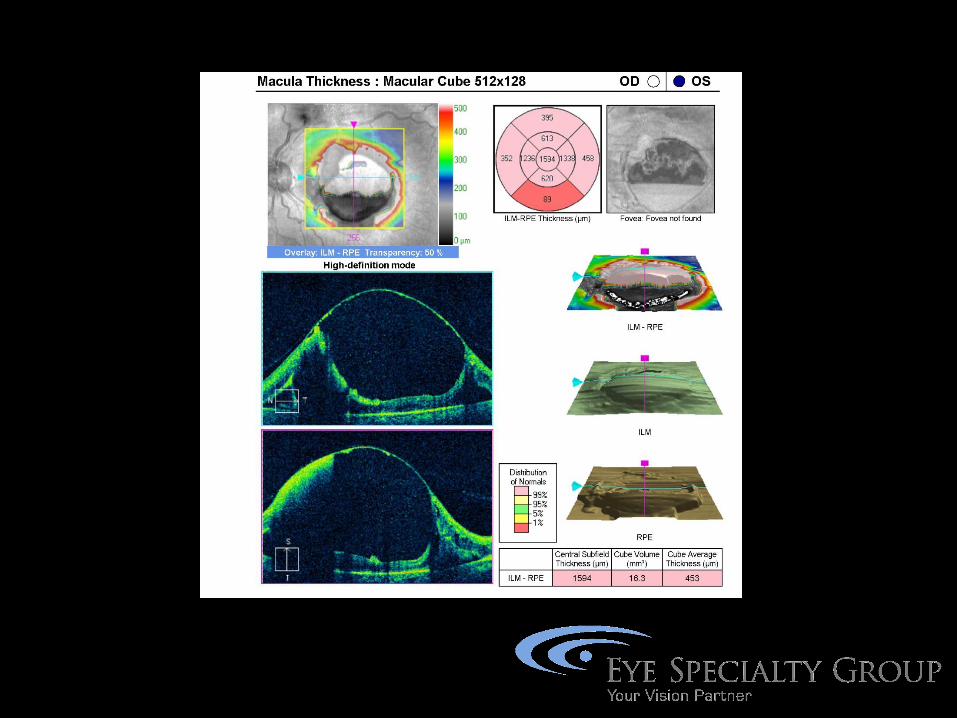
Fluorescein Angiogram
Dynamic imaging of the retina vs.
OCT/Fundus Photo snapshot
Retinal red flags
Warning signs for vision loss and mortality

Epidemiology
~1 per 10,000 outpatient visits in tertiary
eye centers
Mean age 60yo
M>F

Pathophysiology
Ophthalmic Artery: 1st branch of Internal
Carotid
15-20 Short posterior ciliary arteries from
ophthalmic artery supply choroid
○ Cilioretinal artery – derived from short ciliary
arteries, found in 15% of individuals
Central Retinal Artery (CRA): 1st branch
of Ophthalmic Artery
CRA = blood supply to inner retina

Pathophysiology
Arterial Occlusion:
Emboli Endogenous
○ Cholesterol (Hollenhorst) – ulcerated atheromatous plaques in the carotid
○ Calcific – derived from cardiac valves
○ Platelet/fibrin
○ Septic
○ Cardiac Tumor
○ Fat
○ Leukoemboli
○ Amniotic fluid

Pathophysiology
Emboli Exogenous
Talc/IVDA
Platelet – IV blood transfusion
Iatrogenic fragments – catheter tips
Nasal/periorbital steroids injections

Thrombosis – Acquired Coagulopathies
Prepapillary arterial loops
Nocturnal hypotension – poor perfusion
pressure of CRA
Vasospasm
Direct compression – trauma, retrobulbar
Elevated IOP
Initial Va is CF to LP
NLP is uncommon sugg. Choroidal
compromise
NLP without cherry-red spot, consider
ophthalmic artery occlusion
-Higher incidence of GCA
Emboli are visible in 25% of cases
Presence assoc. with increased CV disease
mortality
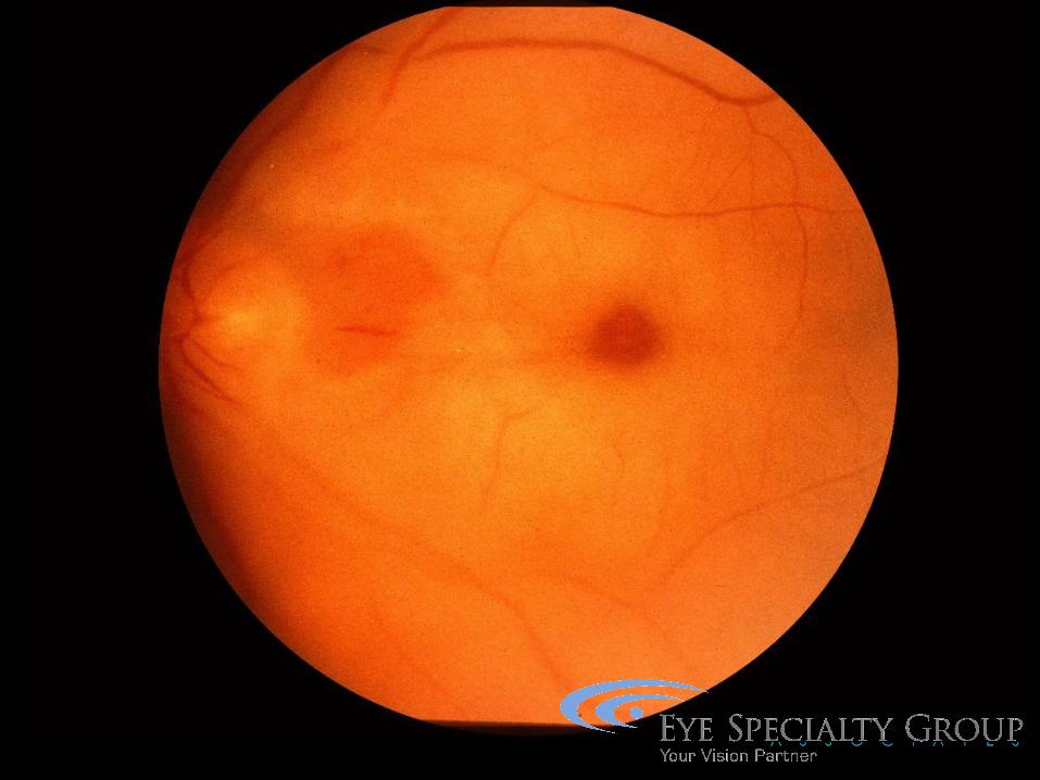
Central Retinal Artery Occlusion

A/C: Normal except APD
Fundus: Retinal whitening, cherry red
spot (no inner retinal neurons, intact blood
supply)
2-6 weeks subtle findings
Visual prognosis guarded, unless
cilioretinal artery includes fovea = 20/50
Central Retinal Artery Occlusion
Giant Cell Arteritis: 2-10%
Can become bilateral within hours/days
Headache, malaise, jaw claudication, nausea,
fever, etc
CBC, CRP, ESR, Fibrinogen
Most common cause of death: MI
9 yr mortality rate 56% compared to 17% in
age-matched controls
Higher risk of CVA

CRAO with Cilioretinal Artery Sparing
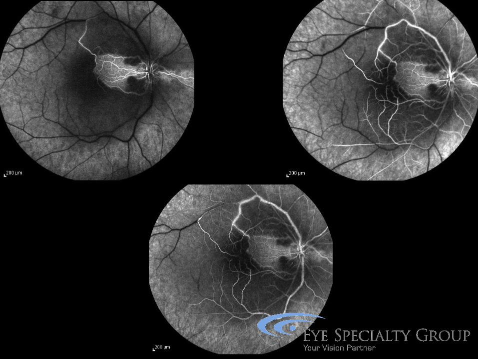

Cilioretinal Artery Occlusion
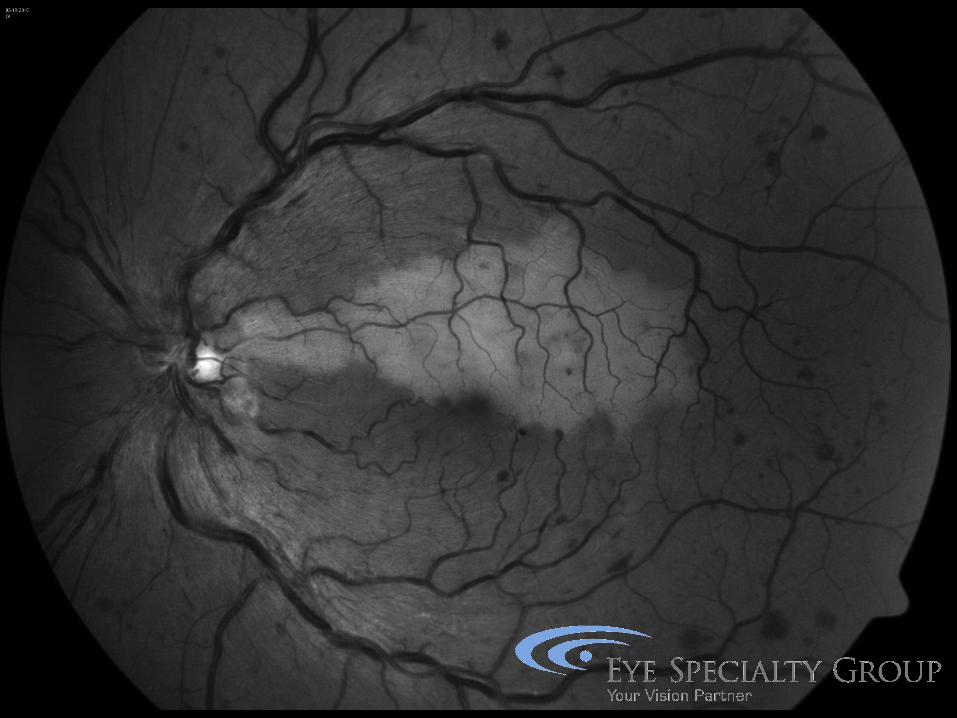
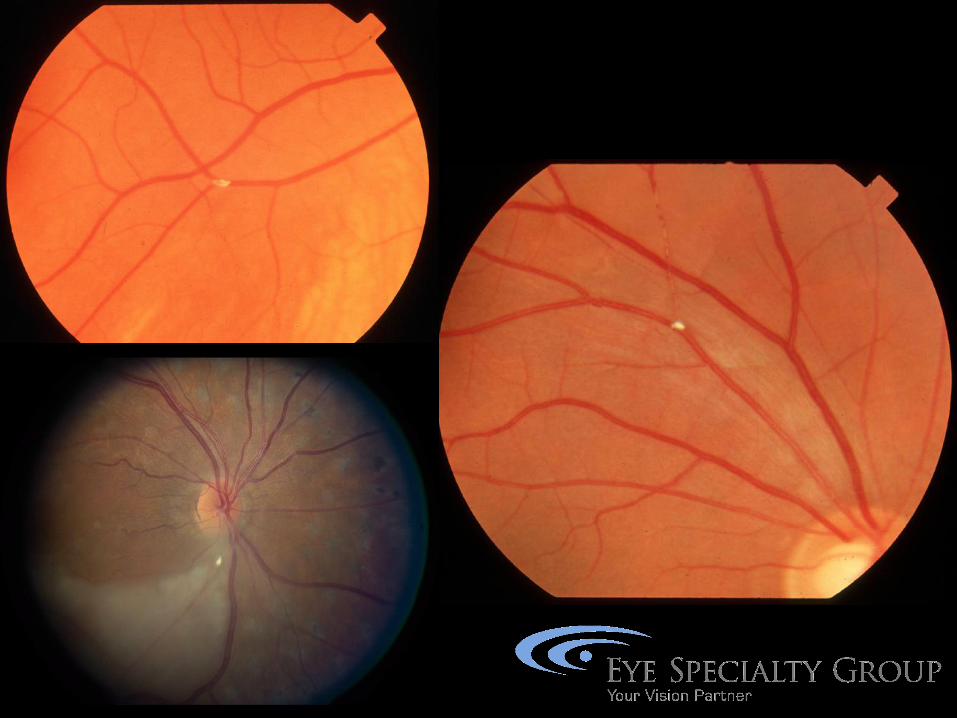
BRAO
Lower survivorship than CRAO
80% maintain 20/40 or better
Temporal vessels more often afflicted
GCA rare because vessels too small

15% of CRAO will develop NVI
NVD only occurs in about 2-3% of cases.
Only 1% of BRAO will develop NVI
IVFA:
Delayed arterial filling (most specific finding)
Delayed A-V transit (most sensitive finding)
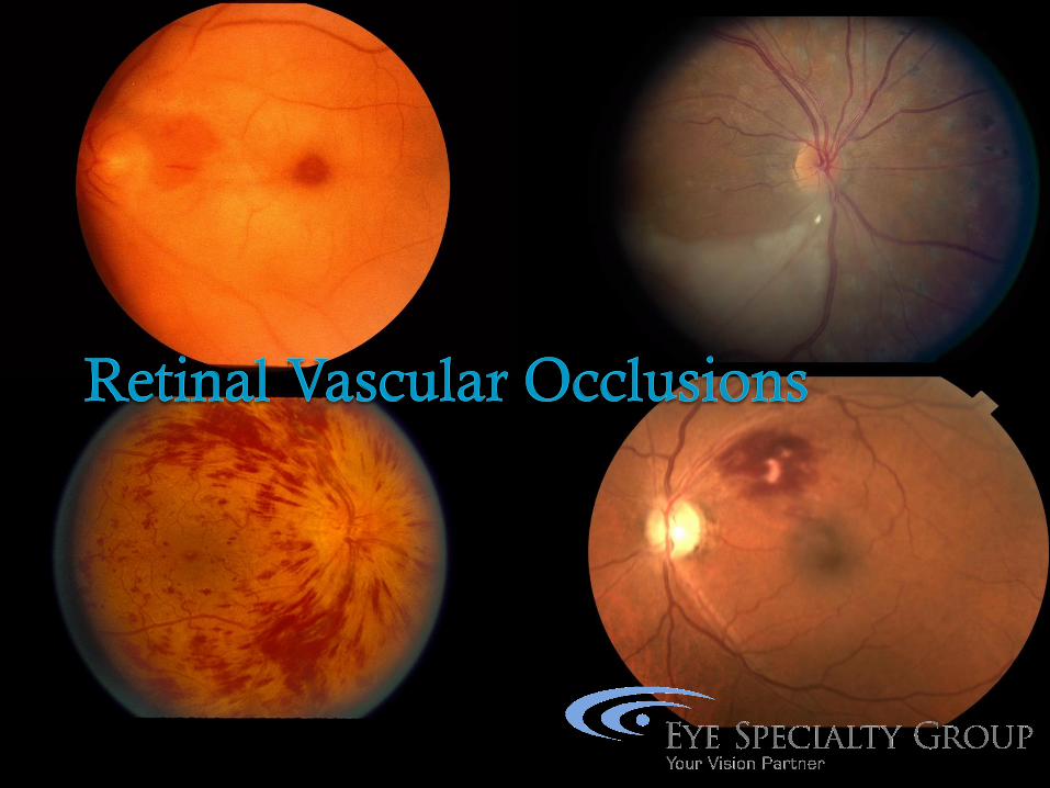
Retinal Artery Occlusion (RAO)
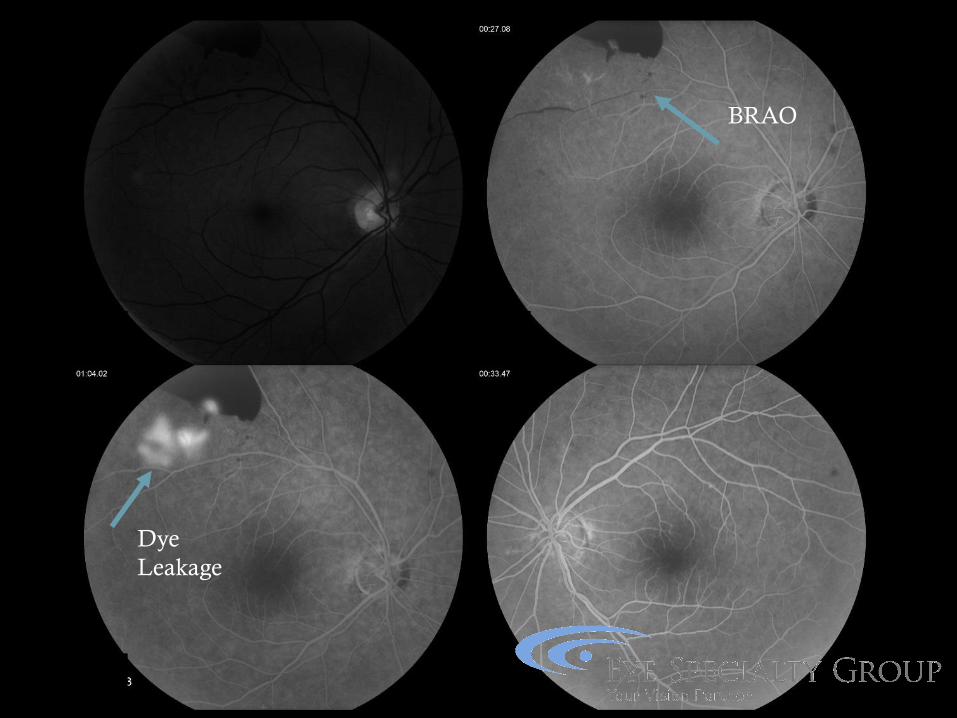
Dye
Leakage
BRAO
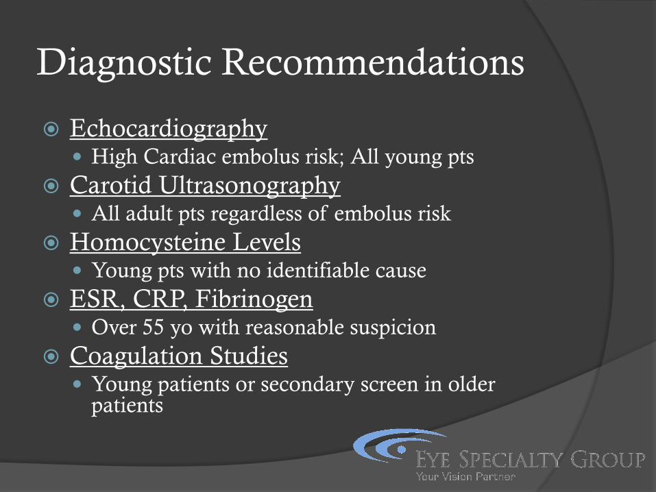
Diagnostic Recommendations
Echocardiography High Cardiac embolus risk; All young pts
Carotid Ultrasonography All adult pts regardless of embolus risk
Homocysteine Levels Young pts with no identifiable cause
ESR, CRP, Fibrinogen Over 55 yo with reasonable suspicion
Coagulation Studies Young patients or secondary screen in older
patients

Retinal Artery Occlusion (RAO)
Irreversible damage occurs at 90 minutes
Possible early treatment
(Estimated benefit ~1/4 Snellen line…)
Ocular massage
Breathing 95% O2/ 5% CO2
AC paracentesis/IOP lowering

Central Retinal Vein Occlusion
70% non-ischemic (low-risk NV)
Va >20/200
5-20% convert to ischemic
83% indeterminate convert over 4 months
34% perfused CRVO convert over 3 years
Ischemic
Va ~CF, +RAPD
>10CWS, >10 DD Non-perfusion
High-risk NVA/NVI
Low-risk NVE/NVD

Systemic Considerations
HTN, DM, Homocysteinuria
Hyperviscosity syndromes
(hypergammaglobulinemia, etc)
Hyperviscosity states (Malignancy,
Nephrotic syndrome, Chronic lung dz,
Syphilis, OCP)
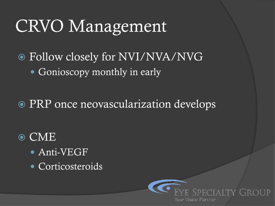
CRVO Management
Follow closely for NVI/NVA/NVG
Gonioscopy monthly in early
PRP once neovascularization develops
CME
Anti-VEGF
Corticosteroids

Central Retinal Vein Occlusion

Central Retinal Vein Occlusion
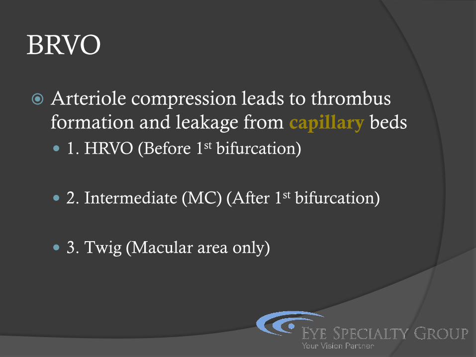
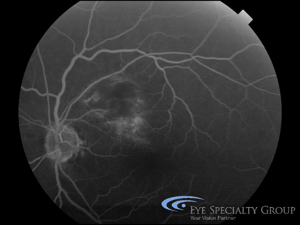
BRVO
Arteriole compression leads to thrombus
formation and leakage from capillary beds
1. HRVO (Before 1st bifurcation)
2. Intermediate (MC) (After 1st bifurcation)
3. Twig (Macular area only)
BRVO
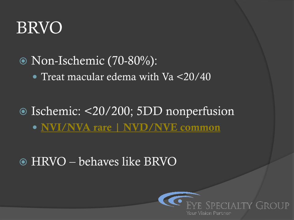
Non-Ischemic (70-80%):
Treat macular edema with Va <20/40
Ischemic: <20/200; 5DD nonperfusion
NVI/NVA rare | NVD/NVE common
HRVO – behaves like BRVO
BRVO Management
Follow monthly until heme resolves
Monitor q4-6 mos for next 3 years
50% Va >20/40
CME
Anti-VEGF
Laser
Steroid

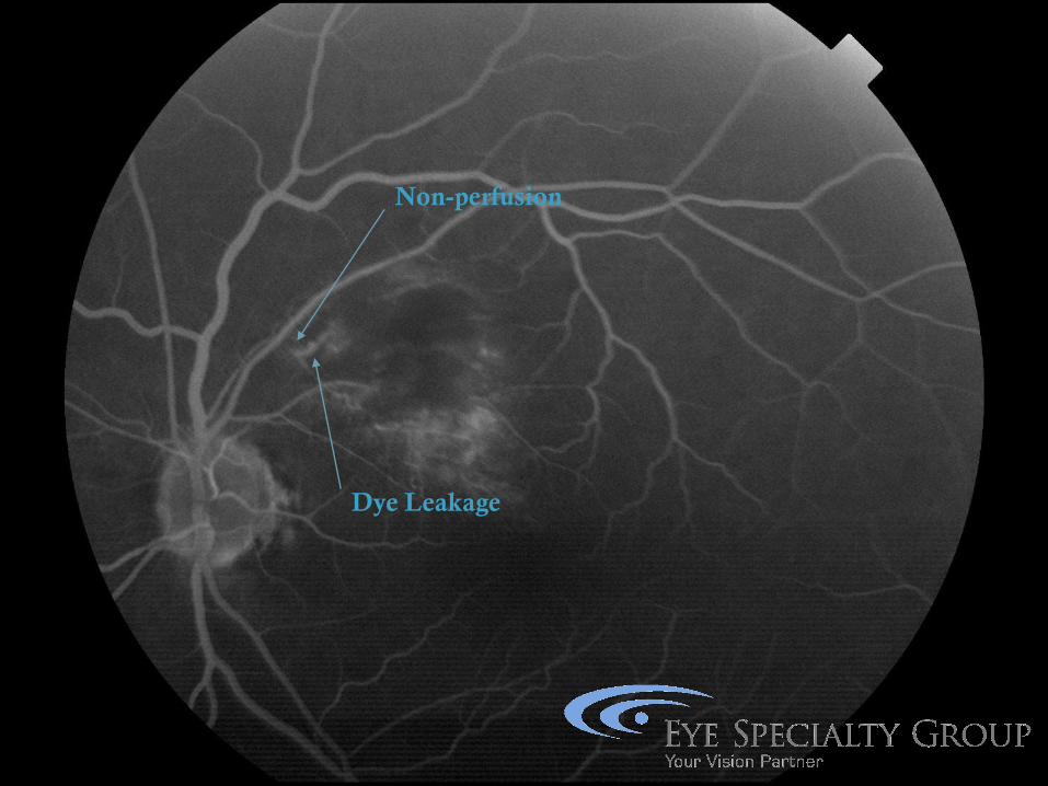
Non-perfusion
Dye Leakage
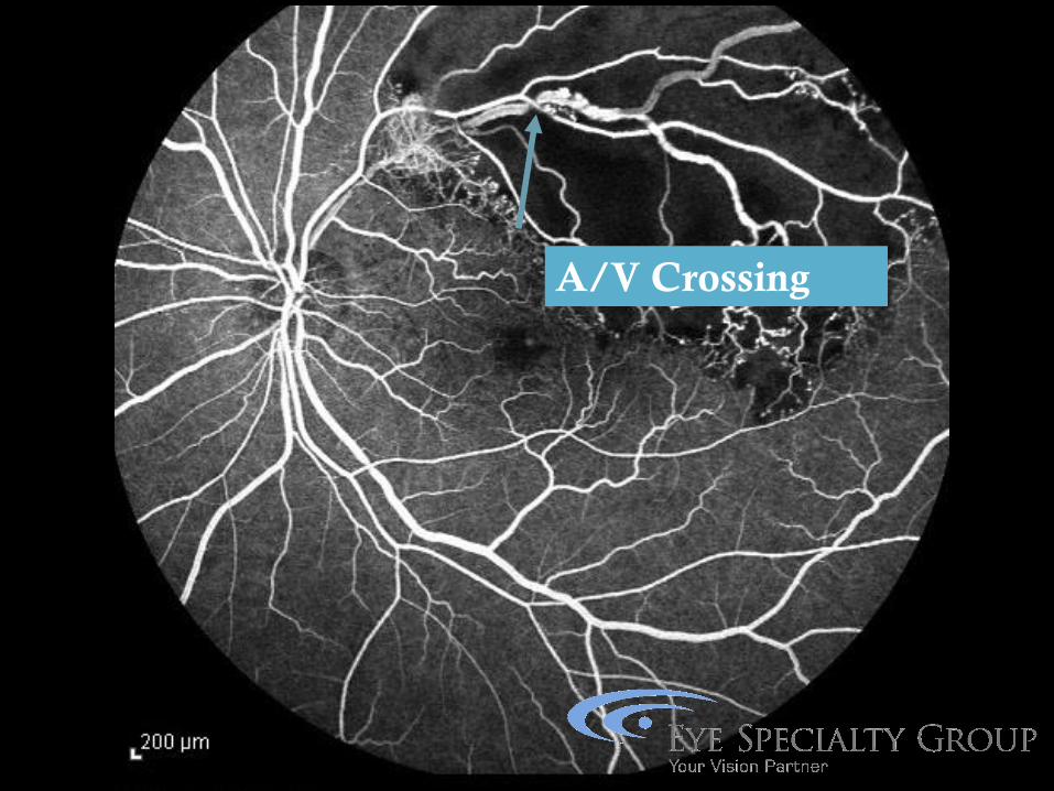
A/V Crossing


Combined Artery/Vein Occlusion
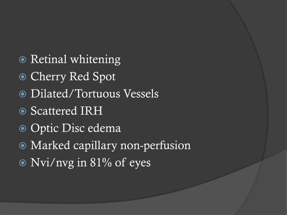
Retinal whitening
Cherry Red Spot
Dilated/Tortuous Vessels
Scattered IRH
Optic Disc edema
Marked capillary non-perfusion
Nvi/nvg in 81% of eyes
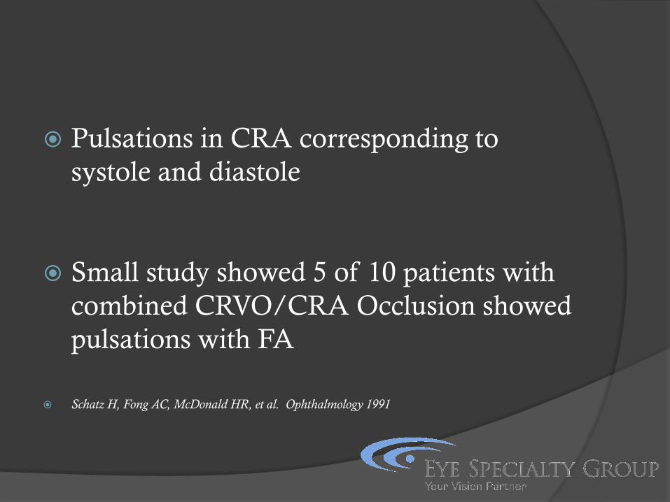
Pulsations in CRA corresponding to
systole and diastole
Small study showed 5 of 10 patients with
combined CRVO/CRA Occlusion showed
pulsations with FA
Schatz H, Fong AC, McDonald HR, et al. Ophthalmology 1991


Ocular Ischemic Syndrome
Symptoms Diminished vision in bright lights
Chronic Eye Pain
Progressive vision loss
Findings Dilated NON-tortuous veins
Mid-peripheral dot-blot heme
CME, CWS, +/- Cherry red spot
33% NVD/NVE
90% NVI and CF vision within 1 year
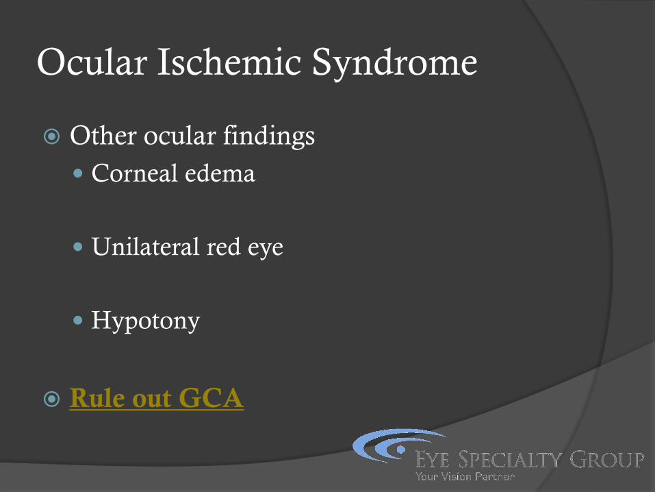
Ocular Ischemic Syndrome
Other ocular findings
Corneal edema
Unilateral red eye
Hypotony
Rule out GCA
OIS vs CRVO or DR
Vs. CRVO
CRVO has tortuousity
Patchy non-confluent heme in OIS
Vs. DR
DR is symmetric
Exudates rare in OIS

Diabetic Retinopathy
Importance of IVFA
Macular perfusion
○ Guides therapy (Laser vs. No Laser)
○ Guides prognosis
Peripheral non-perfusion
○ Chronic macular edema
○ Retinal neovascularization
○ NVI/NVA/NVG

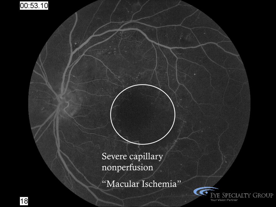
Severe capillary
nonperfusion
“Macular Ischemia”


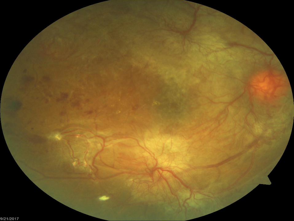


Retinal arteriole damage
Diffuse or focal vasospasm
Arteriosclerosis (thickening of the vessel wall)
Fibrinoid necrosis of the choroid
Hypertensive Retinopathy

Non-Perfusion
Dye Leakage

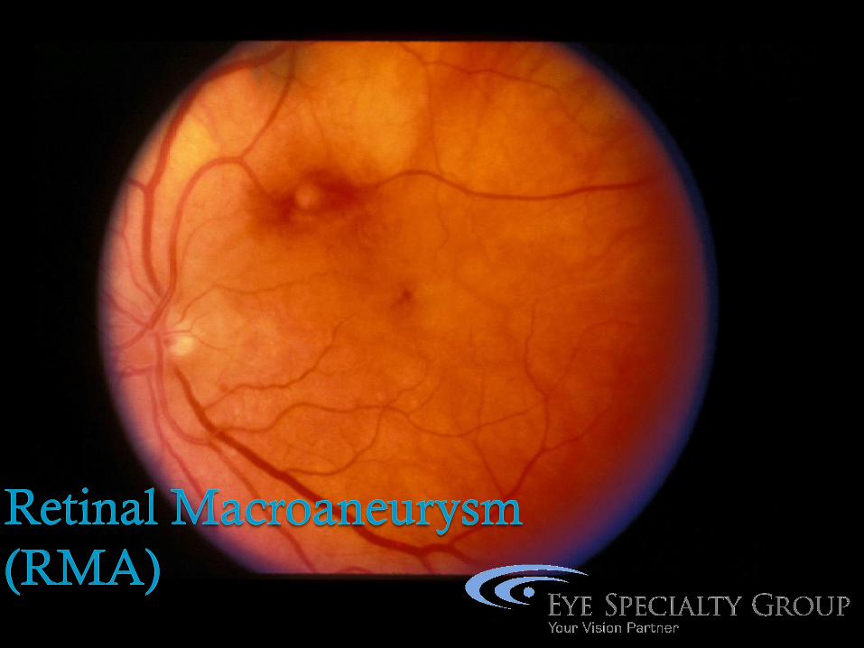

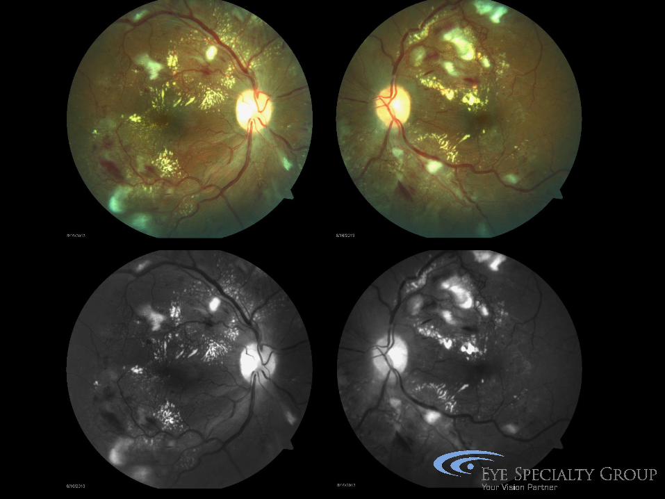
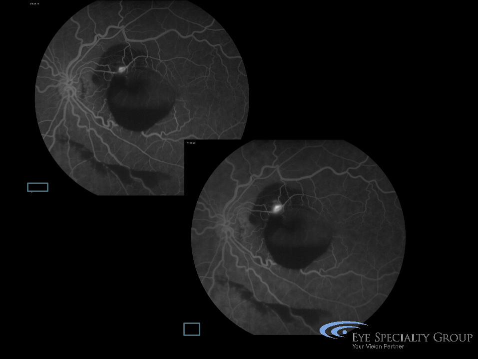
RMA
75% associated with HTN
Arise within 1st three bifurcations
Spontaneous thrombosis of aneurysm can
occur
Typically female >60yo
Bleeding can occur at all 3 levels
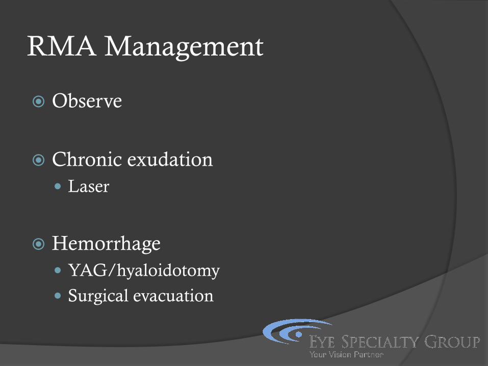
RMA Management
Observe
Chronic exudation
Laser
Hemorrhage
YAG/hyaloidotomy
Surgical evacuation

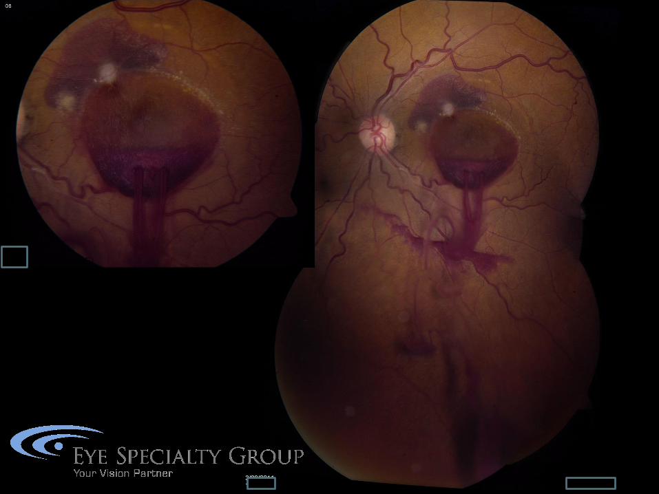
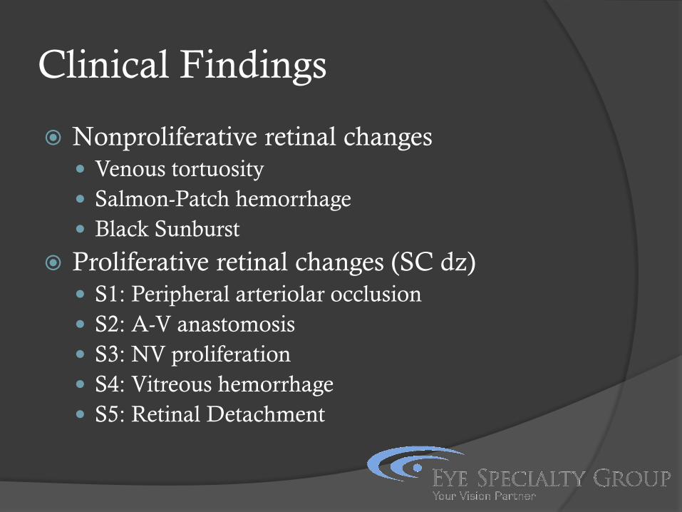
Clinical Findings
Nonproliferative retinal changes
Venous tortuosity
Salmon-Patch hemorrhage
Black Sunburst
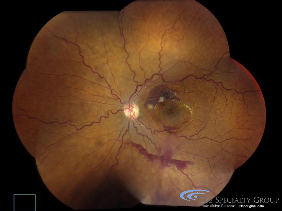
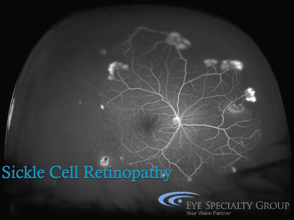
Proliferative retinal changes (SC dz)
S1: Peripheral arteriolar occlusion
S2: A-V anastomosis
S3: NV proliferation
S4: Vitreous hemorrhage
S5: Retinal Detachment

Sickle Cell Retinopathy

Sickle Cell Retinopathy
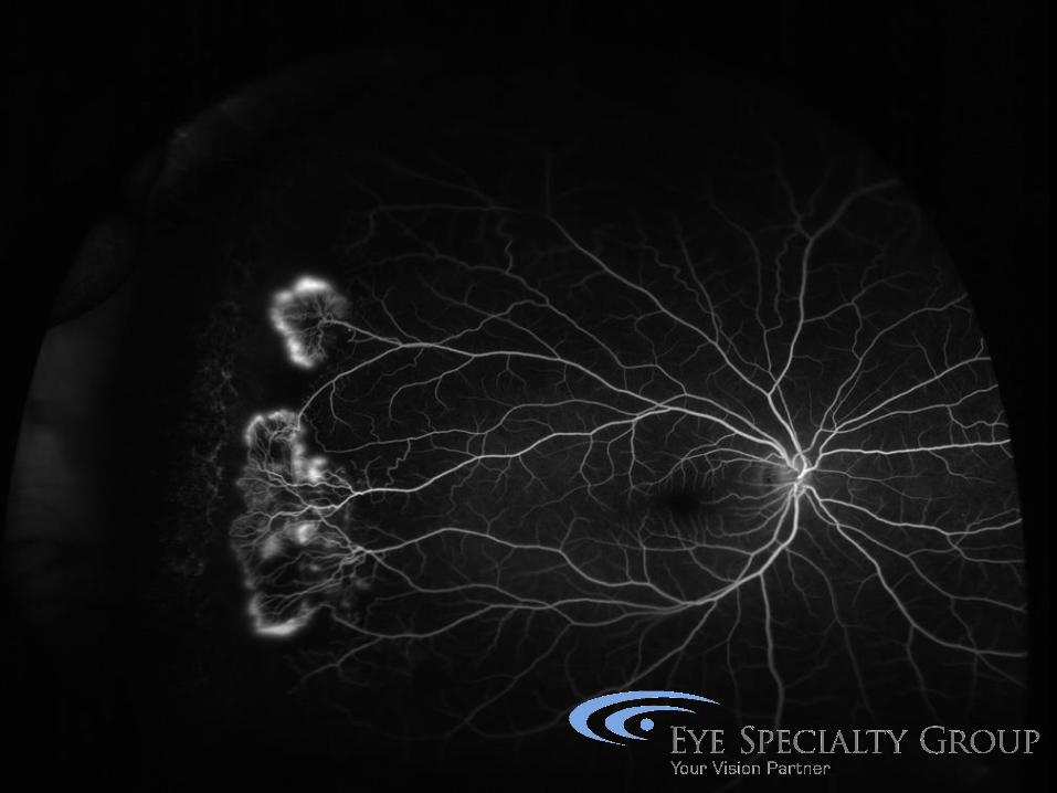
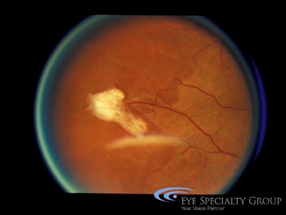
Sickle Cell Retinopathy

Management
Observation
Cryotherapy
Photocoagulation (Feeder vessel, Scatter)
Avoid Scleral Buckle – 71% ant segment
ischemia
References
http://www.djo.harvard.edu/
http://dro.hs.columbia.edu/
http://www.nyee.edu/digital-atlas-of-
ophthalmology.html
2015-2016 Basic and Clinical Science Course
(BCSC): Retina Section 12 (BCSC)
Schachat et al; Ryan’s Retina, Elsevier 2017
Yannuzzi; The Retinal Atlas, Saunders 2010