choroid plexus carcinoma — responses to chemotherapy alone in newly diagnosed young children
TRANSCRIPT

Journal of Neuro-Oncology 12: 69-74, 1992. © 1992 Kluwer Academic Publishers. Printed in the Netherlands.
Clinical Study
Choroid plexus carcinoma - responses to chemotherapy alone in newly diagnosed young children
Jeffrey Allen, 1 Jeffrey Wisoff, 1 Larry Helson, 2 Jennifer Pearce 3 and Edward Arenson 4 1 NYU Medical Center, New York, New York," 2 Westchester County Medical Center, Valhalla, New York; 3 Albany Medical Center, Albany, New York; 4 The Children's Hospital, Denver, Colorado, USA
Key words: choroid plexus carcinoma, chemotherapy, brain tumors
Abstract
Choroid plexus carcinoma (CPC) arising in the infant poses several treatment dilemmas. The tumor is often not totally resectable at presentation given its large size and tendency to invade adjacent brain. Because of its predisposition to regrow and metastasize, some form of postoperative cytotoxic therapy is required. Chemotherapy (CHT), as opposed to radiotherapy (RT), has a more desirable risk/benefit role in infants, since it is relatively sparing of late neurologic sequelae. Three young male children presented with large intraventricular CPC at 9, 18, and 27 months of age. One child had subarachnoid metastases at diagnosis and the other two had localized disease. Subtotal resections were accomplished and all three required VP shunts. Initial CHT consisted of four monthly courses of cisplatin (20 mg/m 2) and etoposide (100 mg/m2), both administered intravenously, daily, for five days. After four courses, two children had complete responses and one had stable disease. Additional CHT was given but one child developed a local recurrence and another diffuse CNS metastases. Both died with intratumoral hemorrhages at 5 and 57 months following diagnosis. The third child remains in continuous remission 46 months after diagnosis. None of the children received RT. Chemotherapy may permit long term deferral of RT. More aggressive CHT regimens should be explored in infants with CPC.
Introduction
Primary choroid plexus neoplasms are rare, consti- tuting 1-2% of cases in large pediatric institutional operative series [1]. They tend to arise in infants or young children with a median age of onset of 9 months. Over 70% of cases occur in children under 2 years of age. Approximately 70-80% of cases are histologically low grade neoplasms, i.e. choroid plexus papillomas (CPP), and amendable to cura- tive surgical expiration in most cases.
However, choroid plexus carcinoma (CPC) (20- 30% of cases) is a biologically more aggressive tumor and surgical debulking alone usually offers only temporary respite. This tumor tends not only
to be locally invasive but also to produce subarach- noid and intraventricular metastases. Institutional reviews have reported a median survival of 9 mos in this latter condition [2]. Because of the reluctance to use brain irradiation in infants, there exists a compelling rationale to explore the use of chemo- therapy alone, a modality which appears to be rela- tively sparing of late effects on the developing nerv- ous system. Chemotherapy may increase the resec- tability of 'unresectable' tumors as well as permit the deferral of radiotherapy. Radiotherapy has not been effective for long term control of CPC's [3].
We are reporting 3 recent cases managed during the period of 1986--88 who presented under 3 years of age with unresectable, intraventricular CPC's.
70
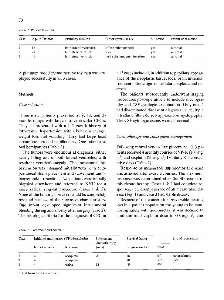
Table 1. Patient selection
Case Age at Dx mos Primilary location Tumor spread at Dx VP shunt Extent of resection
1 18 both lateral ventricles diffuse subarachnoid yes subtotal 2 27 left lateral ventricle none yes subtotal 3 9 left lateral ventricle local subependymal invasion yes subtotal
A platinum based chemotherapy regimen was em- ployed successfully in all 3 cases.
Methods
Case selection
Three male patients presented at 9, 18, and 27 months of age with large intraventricular CPC's. They all presented with a 1-2 month history of intracranial hypertension with a behavior change, weight loss and vomiting. They had large head circumferences and papilledema. One infant also had hemiparesis (Table 1).
The tumors were enormous at diagnosis, either nearly filling one or both lateral ventricles, with resultant ventriculomegaly. The intracranial hy- pertension was managed initially with ventriculo- peritoneal shunt placement and subsequent tumor biopsy and/or resection. Two patients were initially biopsied elsewhere and referred to NYU for a more radical surgical procedure (cases 1 & 3). None of the tumors, however, could be completely resected because of their invasive characteristics. One infant developed significant intratumoral bleeding during and shortly after surgery (case 2). The histologic criteria for the diagnosis of CPC in
all 3 cases included, in addition to papillary appear- ance of the neoplastic tissue, local brain invasion, frequent mitotic figures, cellular anaplasia and ne- crosis.
The patients subsequently underwent staging procedures post-operatively to include myelogra- phy and CSF cytologic examination. Only case 1 had disseminated disease at diagnosis i.e. multiple intradural filling defects apparent on myelography. The CSF cytologic exams were all normal.
Chemotherapy and subsequent management
Following central venous line placement, all 3 pa- tients received 4 monthly courses of VP-16 (100 mg/ m 2) and cisplatin (20 mg/m 2) IV, daily x 5 consec- utive days (Table 2).
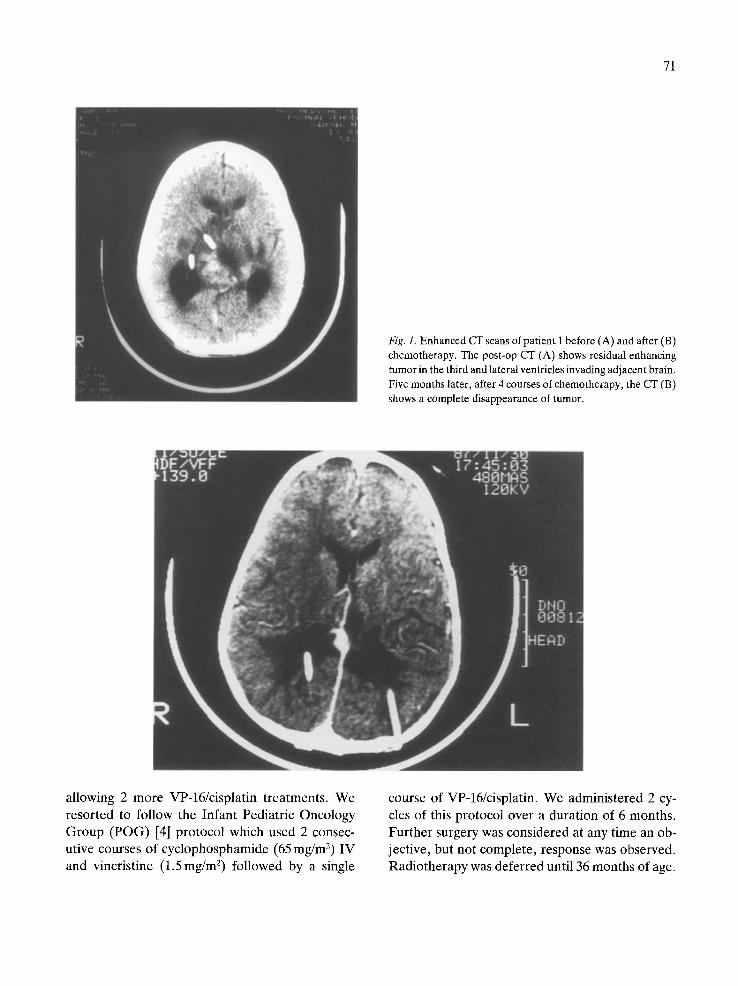
Response of measurable supratentorial disease was assessed after every 2 courses. The maximum response was determined after the 4th course of this chemotherapy. Cases 1 & 2 had complete re- sponses, i.e., disappearance of all measurable dis- ease (Fig. 1) and case 3 had stable disease.
Because of the concern for irreversible hearing loss in a patient population too young to be mon- itoring safely with audiometry, it was decided to limit the total cisplatin dose to 600mg/m 2, thus
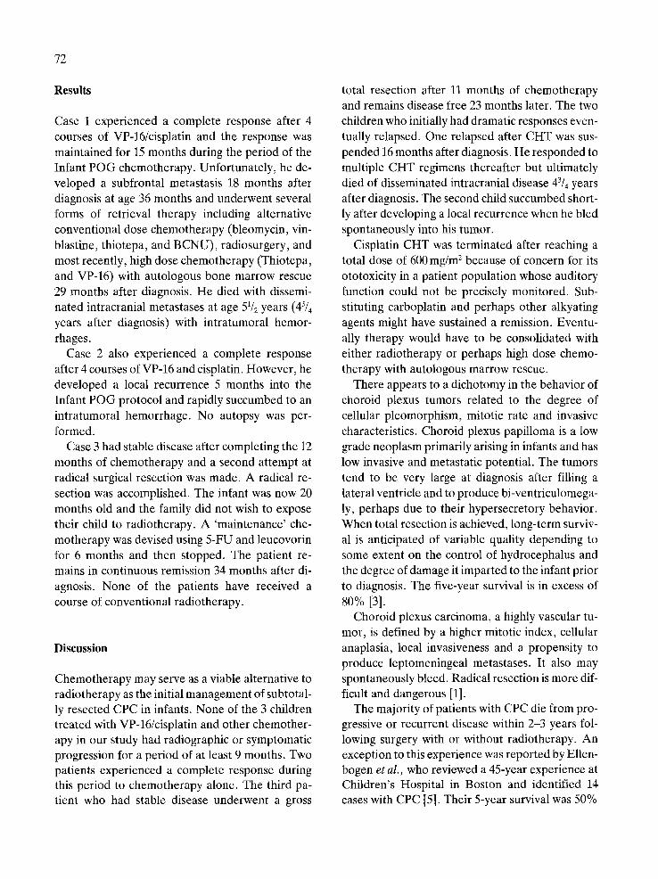
Table 2. Treatment and course
Case Initial chemotherapy (VP-16/cisplatin)
No. of courses Response
Subsequent chemotherapy (mos)
Survival (mos)
progression free total
Site of recurrence
1 4 complete 20 2 4 complete 5 3 4 stable 18
16 57 subarachnoid 10 1P local 9 46 +
a Died from local recurrence.
71
Fig. 1. Enhanced CT scans of patient 1 before (A) and after (B) chemotherapy. The post-op CT (A) shows residual enhancing tumor in the third and lateral ventricles invading adjacent brain. Five months later, after 4 courses of chemotherapy, the CT (B) shows a complete disappearance of tumor.
allowing 2 more VP-16/cisplatin treatments. We resorted to follow the Infant Pediatric Oncology Group (POG) [4] protocol which used 2 consec- utive courses of cyclophosphamide (65 mg/m 2) IV and vincristine (1 .5mg/m 2) followed by a single
course of VP-16/cisplatin. We administered 2 cy- cles of this protocol over a duration of 6 months. Further surgery was considered at any time an ob- jective, but not complete, response was observed. Radiotherapy was deferred until 36 months of age.
72
Results
Case 1 experienced a complete response after 4 courses of VP-16/cisplatin and the response was maintained for 15 months during the period of the Infant POG chemotherapy, Unfortunately, he de- veloped a subfrontal metastasis 18 months after diagnosis at age 36 months and underwent several forms of retrieval therapy including alternative conventional dose chemotherapy (bleomycin, vin- blastine, thiotepa, and BCNU), radiosurgery, and most recently, high dose chemotherapy (Thiotepa, and VP-16) with autologous bone marrow rescue 29 months after diagnosis. He died with dissemi- nated intracranial metastases at age 51/2 years (43/4 years after diagnosis) with intratumoral hemor- rhages.
Case 2 also experienced a complete response after 4 courses of VP-16 and cisplatin. However, he developed a local recurrence 5 months into the Infant POG protocol and rapidly succumbed to an intratumoral hemorrhage. No autopsy was per- formed.
Case 3 had stable disease after completing the 12 months of chemotherapy and a second attempt at radical surgical resection was made. A radical re- section was accomplished. The infant was now 20 months old and the family did not wish to expose their child to radiotherapy. A 'maintenance' che- motherapy was devised using 5-FU and leucovorin for 6 months and then stopped. The patient re- mains in continuous remission 34 months after di- agnosis. None of the patients have received a course of conventional radiotherapy.
Discussion
Chemotherapy may serve as a viable alternative to radiotherapy as the initial management of subtotal- ly resected CPC in infants. None of the 3 children treated with VP-16/cisplatin and other chemother- apy in our study had radiographic or symptomatic progression for a period of at least 9 months. Two patients experienced a complete response during this period to chemotherapy alone. The third pa- tient who had stable disease underwent a gross
total resection after 11 months of chemotherapy and remains disease free 23 months later. The two children who initially had dramatic responses even- tually relapsed. One relapsed after CHT was sus- pended 16 months after diagnosis. He responded to multiple CHT regimens thereafter but ultimately died of disseminated intracranial disease 43/4 years after diagnosis. The second child succumbed short- ly after developing a local recurrence when he bled spontaneously into his tumor.
Cisplatin CHT was terminated after reaching a total dose of 600 mg/m 2 because of concern for its ototoxicity in a patient population whose auditory function could not be precisely monitored. Sub- stituting carboplatin and perhaps other alkyating agents might have sustained a remission. Eventu- ally therapy would have to be consolidated with either radiotherapy or perhaps high dose chemo- therapy with autologous marrow rescue.
There appears to a dichotomy in the behavior of choroid plexus tumors related to the degree of cellular pleomorphism, mitotic rate and invasive characteristics. Choroid plexus papilloma is a low grade neoplasm primarily arising in infants and has low invasive and metastatic potential. The tumors tend to be very large at diagnosis after filling a lateral ventricle and to produce bi-ventriculomega- ly, perhaps due to their hypersecretory behavior. When total resection is achieved, long-term surviv- al is anticipated of variable quality depending to some extent on the control of hydrocephalus and the degree of damage it imparted to the infant prior to diagnosis. The five-year survival is in excess of 80% [31 .
Choroid plexus carcinoma, a highly vascular tu- mor, is defined by a higher mitotic index, cellular anaplasia, local invasiveness and a propensity to produce leptomeningeal metastases. It also may spontaneously bleed. Radical resection is more dif- ficult and dangerous [1].
The majority of patients with CPC die from pro- gressive or recurrent disease within 2-3 years fol- lowing surgery with or without radiotherapy. An exception to this experience was reported by Ellen- bogen et al., who reviewed a 45-year experience at Children's Hospital in Boston and identified 14 cases with CPC [5]. Their 5-year survival was 50%

and all their deaths occurred within 7 months of
diagnosis.
In 4 of these patients, when the tumor was initial- ly subtotally resected, a gross total resection was accomplished and none had a recurrence after a median period of follow-up of 8-9 years. Only one of these patients received post-op radiotherapy. One further patient was salvaged following initial management with subtotal resection and chemo- therapy at another institution followed by a gross total resection at Children's Hospital. No tumor has recurred after 3 years. Autopsies were per- formed in six of 7 patients. All 6 had had only partial tumor resections. Six of 7 died from local recurrences and one had, in addition, difusse sub- arachnoid metastases.
The authors concluded that CPC remains primi- rily a surgical disease and in half of cases surgery may be curative. This experience has not been duplicated by others primarily because of the tech- nical difficulties of performing total resections but also because the tumors appear to invade adjacent brain and/or spread to other parts of the craniospi- hal axis. The overall prognosis has been bleak. Some of the differences in outcome in institutional series may relate to the definition of CPC.
Several reports have documented the usefulness of chemotherapy in recurrent and newly diagnosed disease. Maria et al. were able to salvage one child with a local recurrence using cisplatin, bleomycin and vinblastine chemotherapy [6]. Duffner et al.
reported on 5 children treated on the Infant POG brain tumor protocol for 12-14 months [7]. One child had a partial response, 2 had stable disease. One had no measurable tumor following surgery. Three of these children remain progression free at 5 +, 15 + and 29 + months. Only one had died of recurrent disease. Another child with a recurrence was salvaged with radiotherapy.
Weitzman et al. treated 2 newly diagnosed in- fants, ages 1 and 24 months at diagnosis, with com- bination chemotherapy (carboplatin, ifosphamide, and VP-16). Significant responses were observed in both cases permitting eventual gross total resec- tions [8].
Thus, a number of multi-agent chemotherapy regimens have been employed successfully for in-
73
fants with recurrent and newly diagnosed CPC. We are of the view that all infants so diagnosed require
some form of cytotoxic therapy, and chemotherapy appears to have the least late effects. It is not clear how long chemotherapy should be given if a re- sponse or stable disease is achieved, but the dose- limiting toxicity, especially with cisplatin, may be achieved within 6-12 months depending on dose intensity. When maximal response is achieved and the disease has not metastasized, we recommend a second attempt at total resection. Radiotherapy may be postponed indefinitely as in our case # 3. Radiotherapy may be considered after 36 months and we prefer a regional rather than craniospinal therapy.
For patients who complete therapy prior to 36 months and have no measurable disease, we rec- ommend maintenance chemotherapy with drugs such as carboplatin, cyclophosphamide and vincris- tine. A similar approach could be used in patients with no measurable disease following initial sur- gery who are too young to be irradiated. Because the chemotherapy experience is so limited and an- ecdotal, it is important to continue to pursue clin- ical trials with other chemotherapy agents alone and in combination since CPC appears to be rela- tively sensitive to chemotherapy. It may be pos- sible to eliminate radiotherapy altogether with re- gimens involving high dose chemotherapy with au- tologous bone marrow rescue [9].
References
1. Boyd MC, Steinbock P: Choroid plexus tumors: problems in diagnosis and management. J Neurosurg 66: 800-805, 1987.
2. Pascual-Castroviejo I, Villarejo F, Perez-Higueras A, Mo- rales C, Pascual-Pascual S: Childhood choroid plexus neo- plasms. Eur J Pediatr 140: 51-56, 1983.
3. McGirr S, Eberson M, Scheithauer B, Quast L, Shaw E: Choroid plexus papillomas: long-term follow-up results in a surgically treated series. J Neurosurg 69: 843-849, 1988.
4. Duffner P, Cohen M: Treatment of brain tumors in babies and very young children. Pediat Neurosci 12: 304-310, 1986.
5. Ellenbogen R, Winston K, Kupsky W: Tumors of the choroid plexus in children. Neurosurg 25: 327-335, 1989.
6. Maria B, Graham M, Strauss L, Wharam M: Response of a recurrent choroid plexus tumor to combination chemother- apy. J Neurooncol 3(3): 259-262, 1985.

74
7. Duffner P, Cohen M, Horowitz M, Kun 1, et al.: The treat- ment of choroid plexus carcinoma in infancy with chemother- apy. Ann Neurol 26(3): 460, 1989.
8. Weitzman S, Greenberg M, Becker L, Hoffman H: Choroid plexus carcinoma - response to neoadjuvant combination chemotherapy. Ped Neurosci 14(3): 165, 1988.
9. Finlay J, August C, Packer R, et aL: High dose multi-agent chemotherapy followed by bone marrow 'rescue' for malig- nant astrocytomas of childhood and adolescence. J Neuro- Oncol 9: 23%248, 1990
Address for offprints: J.C. Allen, NYU Medical Center, 550 First avenue, New York, NY 10016, USA