chondroid syringoma of the periauricular area. a report of
TRANSCRIPT
Open Journal of Otolaryngology V3 . I1 . 2020 14
IntroductionChondroid syringoma (CS) is a benign neoplasm that belongs in the appendageal – sweat gland tumors.1 It was formerly known as a mixed tumor of the skin as it shows significant histological similarities with the mixed tumor of the salivary gland. 2,3,4 This lesion presents as a lump in the dermis or subdermis of middle-aged men and is usually encountered in the head.5 Most common sites are nose, upper lip and cheek. 2,5
Here we report a case of CS of a young woman aged 33 in a really uncommon site in the retroauricular area which is only the third case in the literature in
that area. We also present all CS encountered in the literature in the periauricular area.
Case Report
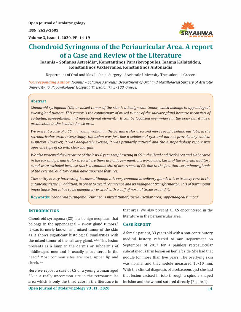
A female patient, 33 years old with a non-contributory medical history, referred to our Department on September of 2017 for a painless retroauricular subcutaneous firm lesion on her left side. She had that nodule for more than five years. The overlying skin was normal and that nodule measured 10x10 mm. With the clinical diagnosis of a sebaceous cyst she had that lesion excised in toto through a spindle shaped incision and the wound sutured directly (Figure 1).
Open Journal of Otolaryngology
ISSN: 2639-3603
Volume 3, Issue 1, 2020, PP: 14-19
Chondroid Syringoma of the Periauricular Area. A report of a Case and Review of the Literature
Ioannis – Sofianos Astreidis*, Konstantinos Paraskevopoulos, Ioanna Kalaitsidou, Konstantinos Vaxtsevanos, Konstantinos Antoniadis
Department of Oral and Maxillofacial Surgery of Aristotle University Thessaloniki, Greece.
*Corresponding Author: Ioannis – Sofianos Astreidis, Department of Oral and Maxillofacial Surgery of Aristotle University, ‘G. Papanikolaou’ Hospital, Thessaloniki, 57100, Greece.
AbstractChondroid syringoma (CS) or mixed tumor of the skin is a benign skin tumor, which belongs to appendageal, sweat gland tumors. This tumor is the counterpart of mixed tumor of the salivary gland because it consists of epithelial, myoepithelial and mesenchymal elements. It can be localized everywhere in the body but it has a predilection in the head and neck area.
We present a case of a CS in a young woman in the periauricular area and more specific behind ear lobe, in the retroauricular area. Interestingly, the lesion was just like a subdermal cyst and did not provoke any clinical suspicion. However, it was adequately excised, it was primarily sutured and the histopathology report was apocrine type of CS with clear margins.
We also reviewed the literature of the last 60 years emphasizing in CS in the Head and Neck Area and elaborated in the ear and periauricular area where there are only few mentions worldwide. Cases of the external auditory canal were excluded because this is a common site of occurrence of CS, due to the fact that ceruminous glands of the external auditory canal have apocrine features.
This entity is very interesting because although it is very common in salivary glands it is extremely rare in the cutaneous tissue. In addition, in order to avoid recurrence and its malignant transformation, it is of paramount importance that it has to be adequately excised with a cuff of normal tissue around it.
Keywords: ‘chondroid syringoma’, ’cutaneous mixed tumor’, ‘periauricular area’, ‘appendageal tumors’
Open Journal of Otolaryngology V3 . I1 . 202015
Chondroid Syringoma of the Periauricular Area. A report of a Case and Review of the Literature
the peripheral layer composed of myoepithelial cells and the inner layer by epithelial cells (Figure 3). Inside cystic areas contained eosinophilic spherules and in one site squamous differentiation was present. There
was not high rate of mitoses or sites of necrosis. The conclusion was an apocrine type of a mixed tumor of the skin. After two years follow-up there is no sign of recurrence.
Fig1. The wound in the 7th post operation day after suture removal. Because of the commonness of the lesion there is no pre operation image.
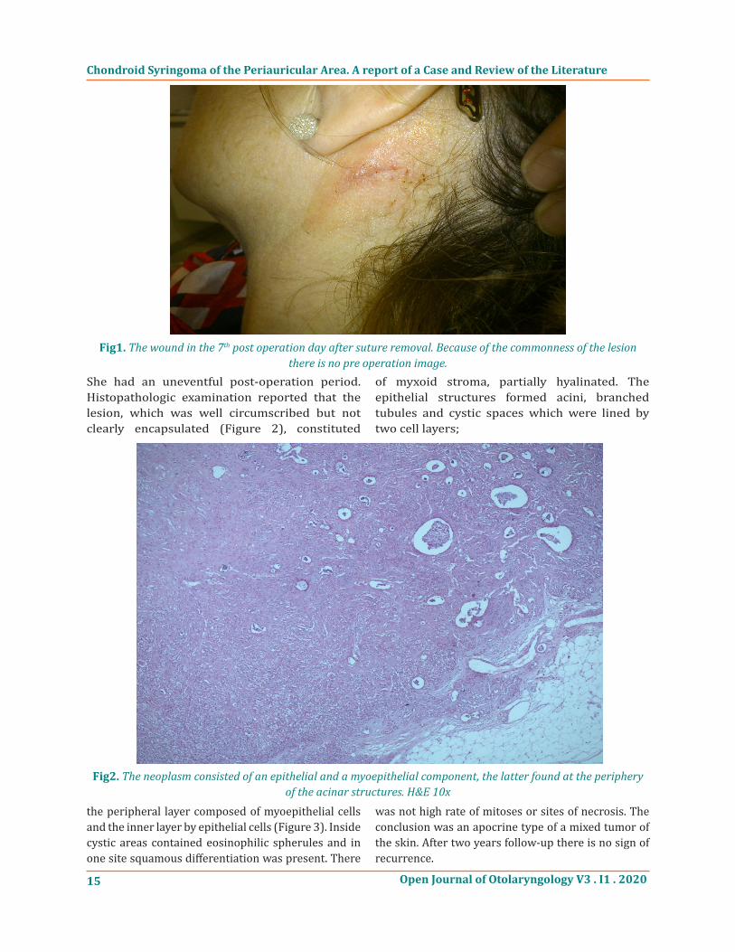
She had an uneventful post-operation period. Histopathologic examination reported that the lesion, which was well circumscribed but not clearly encapsulated (Figure 2), constituted
of myxoid stroma, partially hyalinated. The epithelial structures formed acini, branched tubules and cystic spaces which were lined by two cell layers;
Fig2. The neoplasm consisted of an epithelial and a myoepithelial component, the latter found at the periphery of the acinar structures. H&E 10x

Open Journal of Otolaryngology V3 . I1 . 2020 16
Chondroid Syringoma of the Periauricular Area. A report of a Case and Review of the Literature
Discussion
The first reports of mixed tumor of the skin was in 1859 by Billroth and in 1892 by Nasse, who were the first to note both epithelial and mesenchymal components of the neoplasm, resembling mixed tumors of the salivary glands. The term chondroid syringoma was given by Hirsch and Helwig in 1961 to emphasize the
fact that this benign tumor arises from sweat glands elements in a cartilage-like matrix.6
Searching in the literature we found 831 more cases of CS in the Head and Neck area since 1959. 156 out of them are not clearly specified where their exact site is. Table 1 reflects on our results of 676 cases in descending order of frequency, CS affects more often nose, upper lip and cheek.
Fig3. The neoplastic cells, forming acini, ducts and cysts, were surrounded by a myxoid stroma. The neoplasm was circumscribed, but non-encapsulated. H&E 40x
Table1. Location of the reported CS in the literature in descending order.
Area Number of Cases Nose 124Upper lip 104Cheek 100Scalp 77Forehead 51Eyelids 48External auditory canal 41Chin 34Eyebrow 34Auricle 18Neck 12Lower lip 10Nasolabial fold 8Lip (not specified) 5Orbit 3Medial canthus 2Preauricular 1Retroauricular 2 +1 (our case)Mandible 1
676Head (not specified) 156Clinically this neoplasm presents as a stationary or slow-growing, firm, painless subdermal lesion
measuring from 0,5-3 cm, which is covered with normal skin7 , like our case. Sometimes the lesion is

Open Journal of Otolaryngology V3 . I1 . 202017
Chondroid Syringoma of the Periauricular Area. A report of a Case and Review of the Literature
shiny or waxy and often associated with telangiectasia8, or even ulcerated without implying invasiveness or malignancy.7 Common sites of the neoplasms are nose, upper lip, cheek, scalp, forehead, chin, eyelids and ear but it can involve the trunk, the extremities and the scrotum as well.1,5 It presents in middle aged men with a mean age of 50 years.1 Male to female ratio ranges from 1,3/1 to 5/1.1 Due to the absence of typical clinical features it may be confused with any kind of dermal lesion or cyst like epidermal inclusion cyst, sebaceous cyst, neurofibroma or dermatofibroma.7 In this case we were convinced that this was a sebaceous cyst.
Histopathologically, CS is classified into the apocrine and eccrine variant.1, 2, 3, 4 Apocrine CS is a well-circumscribed lesion which is composed of a complex epithelial system of branching, elongated tubules and cystic spaces on a chondroid, myxoid, fibrous or adipocytic matrix.2, 3, 4, 8 This system is lined by a doubled cell layer. The outer layer consists of cuboidal myoepithelial cells and the inner of columnar cells manifesting secretion by decapitation.1, 3 Follicular and sebaceous differentiation is very often seen and
all these elements support the hypothesis that these tumors derive from the follico-sebaceous-apocrine unit.2, 3, 4 The eccrine CS is less common. Kazakov et al reported a case of eccrine CS in the retroauricular area in the largest case series reported until today consisting of 50 eccrine cases.3 Quite interesting is the fact the male to female ratio was 1:1, 65 for the eccrine CS.3 The epithelial component consists of non-branching, cord-like ductal structures which are lined by a mono-layer of cuboidal epithelial cells set in a myxohyaline and cartilage-like matrix.1, 2, 3 Yim et al reports that when immunohistochemical staining for the S-100 protein is negative and the lesion has a mixed character of glandular structures, then CS originates from apocrine glands located adjacent to the skin, so it is an apocrine type7. In our case, there was a double layered lining in a cartilaginous stroma and we concluded that this lesion was an apocrine type of a CS of the skin.
In Table 2 we show the periauricular cases pointing out that our case is only the third case of a retroauricular CS in the literature.
Table2. Location of the reported cases in the periauricular area.
Location Number ArticleAuricle 7 Mixed tumors of the skin of the salivary gland type - • Stout, A. P., 1959 (9)
External ear 1 Chondroid syringoma: mixed tumor of skin, salivary gland type. - Hirsch - Helwig 1961 6)
Earlobe 1 Pleomorphic adenoma of the auricle. - Ito 1982 (10)
Ear 1 A case of so- called mixed tumor grown in the skin of the ear - Yotsuyanagi – 1994 (11)
Ear 1 An immunohistochemical study of the apocrine type of cutaneous mixed tumors with special reference to their follicular and sebaceous differentiation. - Yamamoto 1999 (4)
Ear 1 Pleomorphic adenoma of the auricle. Nishimura 1999 (12)
Earlobe 1 Chondroid syringoma: a clinical and histological review of eight cases - Villalon 2006 (13)
Ear 3
Apocrine mixed tumor of the skin (“mixed tumor of the folliculosebaceous-apocrine complex”). Spectrum of differentiations and metaplastic changes in the epithelial, myoepithelial, and stromal components based on a histopathologic study of 244 cases. - Kazakov DV 2007 (2)
Earlobe 1 Pleomorphic adenoma in the auricle. - Yim 2009 (7) Earlobe 1 Chondroid syringoma of the ear lobule: a unique case - Omran – 2014 (14)
Preauricular 1 Subcutaneous pleomorphic adenomas in two different areas of the face. Tsukuno 2002 (15)
Retroauricular 1Cutaneous mixed tumor, eccrine variant: a clinicopathologic and immunohistochemical study of 50 cases, with emphasis on unusual histopathologic features - Kazakov DV – 2011 (3)
Retroauricular 1
Apocrine mixed tumor of the skin (“mixed tumor of the folliculosebaceous-apocrine complex”). Spectrum of differentiations and metaplastic changes in the epithelial, myoepithelial, and stromal components based on a histopathologic study of 244 cases. - Kazakov DV 2007 (2)
Retroauricular 1 Our case22

Open Journal of Otolaryngology V3 . I1 . 2020 18
Chondroid Syringoma of the Periauricular Area. A report of a Case and Review of the Literature
Cases of the external auditory canal were excluded because this is a common site of occurrence of CS, due to the existence of ceruminous glands of the external auditory canal which have apocrine features. In our research 41 cases of CS are reported in the external auditory canal alone making it the 7th commonest site of occurrence.
In the periauricular area, because of the proximity with the parotid gland Tsukuno et al suggests to have a CT or MRI check in cutaneous lumps just to exclude an association with the parotid gland especially when lesions are bigger9. The same is suggested when the lump is in the upper eyelid in proximity with the lacrimal gland. However, bigger researches are needed to draw safer conclusions. In 2009 Yim et al reported a case of a CS in the auricle. He reported that this was only the 4th case in the literature7. With a thorough research in the literature we found o total of 18 cases of CS in the auricle are reported until April 2020. Most of them are single case reports where its unsafe to draw conclusions.
On the other hand, there is a strong unanimity in the treatment of choice throughout the literature which is in toto excision including a cuff of normal tissue around the lesion without disrupting it. Otherwise, recurrence is quite possible.8, 9, 10 However, there are ambiguities on the role of preoperative FNA or incisional biopsy. Yim et al states that preoperative biopsies of any kind are contraindicated in any mixed tumor, as implantation of tumor cells may lead to recurrence.7
To conclude CS is a lesion that Head and Neck Surgeons must be aware as it presents in any site in the Head and Neck area. Due to its lack of clinical characteristics histopathologic examination is essential for an accurate diagnosis. A margin of normal tissue must be co-excised as a treatment of choice because recurrence or malignant transformation may exist. The key message of this article is to present firstly, an uncommon tumor of the skin, the Chondroid Syringoma in a very uncommon site for the first time as a case report and secondly, to highlight the significance of the excision with a cuff of normal tissue in order not to recur.
AcknowledgementsDr Pasteli Nikoleta is a consultant histopathologist at our Hospital who kindly provided the histopathological figures with their captions.
ReferencesElder De, Massi D, Scolyer R, Willemze R. [1] Appendageal tumours. In: Elder De, Massi D, Scolyer R, Willemze R, editors. WHO Classification of Tumours, Volume 11 IARC, Fourth Edition 2018. p. 193-3
Kazakov D, Belousova I, Bisceglia M, Calonje [2] E, Emberger M, Grayson W et al. Apocrine mixed tumor of the skin (‘‘mixed tumor of the folliculosebaceous-apocrine complex’’). Spectrum of differentiations and metaplastic changes in the epithelial, myoepithelial, and stromal components based on a histo pathologic study of 244 cases. J Am Acad Dermatol 2007; 57: 467-83.
Kazakov D, Kacerovska D, Hantschke M, Zelger [3] B, Kutzner H, Requena L et al. Cutaneous mixed tumor, eccrine variant: a clinicopathologic and immunohistochemical study of 50 cases, with emphasis on unusual histopathologic features. Am J Dermatopathol 2011;33:557-68.
Yamamoto O, Yasuda H. An immunohisto [4] chemical study of the apocrine type of cutaneous mixed tumors with special reference to their follicular and sebaceous differentiation. J Cutan Pathol 1999;26:232-41.
Paraskevopoulos K, Cheva A, Koloutsos G, [5] Matzarakis I, Vahtsevanos K. Chondroid Syringoma of the Medial Canthus. Case Rep Otolaryngol. 2014; 2014: 158527.
Hirsch P, Helwig E. Chondroid syringoma. Mixed [6] tumor of skin, salivary gland type. Archives of Dermatology 1961;84:835–847.
Yim M, Yoon W, Seo W, Kwon H, Jung N. [7] Pleomorphic adenoma in the auricle. J Craniofac Surg 2009; 20: 951-2
Misago N, Narisawa Y. Lipomatous apocrine mixed [8] tumor of the skin associated with chondroid and ossiferous stroma. J Dermatol 2006;33:380-2
Stout A.P., Gorman J.G. Mixed tumors of the skin [9] of the salivary gland type. Cancer, 1959; 12(3), 537-543
Ito A, Nakashima T, Kiltamura M. Pleomorphic [10] adenoma of the auricle. J Laryngol Otol 1982; 96(12): 1137-40
Open Journal of Otolaryngology V3 . I1 . 202019
Chondroid Syringoma of the Periauricular Area. A report of a Case and Review of the Literature
Yotsuyanagi T, Yokoi K, Sakuraba M. A case of so- [11] called mixed tumor grown in the skin of the ear. J. Jpn. Clin. Dermatol 2006;48:505
Nishimura S, Murofushi T, Sugasawa M. [12] Pleomorphic adenoma of the auricle. Eur Arch Otorhinolaryngol 1999;2561:22-4
Villalón G, Monteagudo C, Martín JM, Ramón D, [13] Alonso V, Jordá E. Chondroid syringoma: a clinical and histological review of eight cases. Actas Dermosifiliogr. 2006;97(9):573-577.
Al Omran Y, Mohamed R, Al Omran MK. Chondroid [14] syringoma of the ear lobule: a unique case. Ear Nose Throat J 2014:93(4-5):E 62-3
Tsukuno M, Nakamura A, Takai S, Kurihara K. [15] Subcutaneous pleomorphic adenomas in two different areas of the face. Scand J Plast Reconstr Surg Hand Surg. 2002;36:109-11.
Citation: Ioannis – Sofianos Astreidis, Konstantinos Paraskevopoulos, et al. Chondroid Syringoma of the Periauricular Area. A report of a Case and Review of the Literature. Open Journal of Otolaryngology. 2020; 3(1): 14-19.Copyright: © 2020 Ioannis – Sofianos Astreidis, Konstantinos Paraskevopoulos, et al. This is an open access article distributed under the Creative Commons Attribution License, which permits unrestricted use, distribution, and reproduction in any medium, provided the original work is properly cited.