chondrocytes in osteoarthritis through β
TRANSCRIPT

Page 1/15
Rock2 Regulates Proliferation and Differentiation ofChondrocytes in Osteoarthritis through β-cateninSignalingLiang Hao ( [email protected] )
Huashan Hospital Fudan University Department of Sports MedicineJun Chen
Huashan Hospital Fudan University Department of Sports MedicineXiliang Shang
Huashan Hospital Fudan University Department of Sports MedicineWu Yang
Huashan Hospital Fudan University Department of Sports MedicineShiyi Chen
Huashan Hospital Fudan University Department of Sports Medicine
Research Article
Keywords: Rock2, Osteoarthritis, Proliferation, Differentiation, β- catenin
Posted Date: September 15th, 2021
DOI: https://doi.org/10.21203/rs.3.rs-882633/v1
License: This work is licensed under a Creative Commons Attribution 4.0 International License. Read Full License

Page 2/15
AbstractBackground
Osteoarthritis (OA) adversely affects quality of life of elderly patients and is among hotspots andchallenges of current research efforts. However, mechanism of occurrence and development of OA hasnot been fully elucidated.
Methods
Through qRT-PCR and Western blot assays, the current study established that levels of Rock2, a keyprotein in Rho signaling pathway, were signi�cantly higher in OA cartilage. Furthermore, the current studyexplored effect of down-regulating Rock2 expression on growth and apoptosis of cartilage cells usingCCK-8, Edu and Flow cytometry assays. Alkaline phosphatase (ALP) and Alizarin Red S (ARS) assayswere then used to determine differentiation effects.
Results
Findings showed that expression of Rock2 is closely related to proliferation, apoptosis and differentiationof chondrocytes. Furthermore, the current study con�rmed that Rock2 affects growth and differentiationof chondrocytes by activating β-catenin signaling pathway.
Conclusion
the current study provided novel insights to targeted therapy of OA.
IntroductionPrevious studies aver that osteoarthritis (OA) is the most common degenerative joint disease[1], which ischaracterized by degradation of extracellular matrix (ECM) or progressive loss of cartilage[2, 3]. Withrising number of elderly population, number of patients with osteoarthritis is increasing, which adverselyaffect quality of life of elderly patients. In addition, more young people suffer from serious knee injuriesdue to excessive pressure caused by increasing antagonistic physical exercise[4]. However, mechanismof regulating OA pathogenesis remains unclear due to complicated combination of metabolic,mechanical, in�ammatory and genetic factors. Therefore, in addition to palliative pain control, there isurgent need for similar physical therapy or prosthetic joint replacement. However, there is no effectivedrug therapy target, necessitating search for effective molecular targets to provide more theoretical basisfor targeted treatment of OA. Cartilage tissue and cells play important roles in progress of osteoarthritis[5,6]. Several previous studies and clinical data show that cartilage cells are broken in patients withosteoarthritis, and synthesis of extracellular matrix leads to dysfunction of articular chondrocytes, which,in turn, promotes degradation of ECM. This eventually leads to loss of ECM and cartilage degradation[7].Therefore, elucidating molecular mechanisms of articular chondrocytes in progress of OA is key toprogress of OA treatment.

Page 3/15
Previous studies reported that Rho family related genes act as protein molecular switch and play keyroles in cellular signal transduction[8]. Rho kinase (Rock) is among most important downstream targeteffector proteins of Rho subfamily. Previous studies reported that Rock2 is involved in many cellularbiological activities, including cell morphogenesis, motility, cell division, proliferation, migration and celladhesion. Rock2 is activated by interaction with Rho /GTPase and regulates actin cytoskeleton throughsome molecular signaling pathways, thereby affecting cell growth and differentiation[9]. Previous studieshave also established that Rock2 plays important roles in smooth muscle and nerve regeneration as wellas in neuronal development[10]. Furthermore, recent studies have established that Rho signaling pathwayprotein plays important roles in progress of osteoarthritis[11], but its mechanism is still unclear. Previousstudies aver that Wnt/ β-catenin signaling pathway plays important roles in osteoblast maturation andbone formation[12, 13], which is the focus of OA research. Studies have shown that Wnt pathway isinhibited in normal cartilage, and its activation promotes OA and Wnt family to regulate bone mass. Inaddition, some previous studies have established that Wnt/β-catenin signal-induced chondrocytehypertrophy and differentiation, followed by OA aggravation and apoptosis due to β- catenin inactivationmay complicate normal bone development[14, 15]. However, mechanism of Wnt/β-catenin signalingpathway regulation in OA needs to be explored further.
The current study explored Rock2 expression in OA chondrocytes and its effects on proliferationand apoptosis of chondrocytes. In addition, the current study demonstrated role of Rock2 in OA andmechanism of Wnt/β-catenin signaling pathway in regulation of OA chondrocyte proliferation andapoptosis. The current study, therefore, provided new insights for OA treatment.
Materials And MethodsClinical samples
A total of 15 specimens were collected from patients who underwent total knee arthroplasty (TKA), withage distribution ranging between 58-75 years. In addition, a total of 15 normal cartilage specimens werecollected after traumatic fracture. All patients signed informed consent before sample collection. Allspecimens were stored at -80oC immediately after collection.
qRT-PCR
Total RNA from human OA cartilage and normal cartilage tissues were extracted based on TRIzolTM
reagent (Invitrogen, USA). TRT-PCR and real-time �uorescent quantitative PCR were then undertaken usingPrimeScript RT reagent kit and SYBR Green PCR kit (Takara, Japan), respectively.
Western Blot
Samples of human OA cartilage tissue, normal cartilage tissue and chondrocyte line were collected, fromwhich cell and tissue proteins were extracted using cell lysate and tissue extraction kit. After quantitativeanalysis using BCA method, protein buffer was added and boiled in boiling water for 10 min. Same

Page 4/15
amount of protein (40 μg) was separated using 10% SDS - PAGE gel. The �rst antibody including Rock2(ab-71598) was used to incubate overnight based on manufacturer's instructions, and second antibody ofcorresponding species was then used to incubate. Membrane was washed using 1 x TBST, and speci�cantibody interaction was then observed using ECL luminescent reagent. Expression of ROCK2 proteinwas �nally observed using chemiluminescence gel imaging analyzer.
CCK-8 assays
CCK-8 method was used to detect and analyze cell activity in treatment and control groups. 100μLchondrocytes suspension was dispensed in 96 well plate and preincubated for 24 hours (37℃, 5% CO2).Cells were then treated with inhibitor and plates were then incubated for 24, 48, and 72 h. 10μl CCK8solution was then added and incubated for 2 h. Absorbance of each well was determined at 450 nmusing Bio-Rad (USA) and mean absorbance of each well was then computed.
Edu Assay
Chondrocytes in logarithmic growth phase in cell culture bottle were digested with 0.25% trypsin andcentrifuged at 1000 rpm for 5 min to prepare single cell suspensions. Small amount of cell suspensionwas taken to cell counting plate, and cell concentration was computed after counting under microscope.Cells were seeded on 96 well plates, and 1× 10⁴ cells (1 cell per well) were cultured in 200μl completemedium for 24 h, following which medium was changed. After corresponding treatment, based on Edureagent instructions, each well containing 100μL Edu medium was incubated at 37℃ for 2 h. 200μL of4% paraformaldehyde was �xed for 30 min, and 100μL glycine was then added, followed by 100 μLApollo. 100μL 0.5% Triton-X was added, washed twice using PBS, and 100μL Hoechst 33342 wasdecolorized at room temperature, shaken for 30 min, and washed twice with PBS. Cell proliferation wasthen observed using �uorescence microscope.
Flow Cytometry (FCM) Assay
Chondrocytes (1x10⁵) in treatment and control groups were digested with 0.25% white trypsin. Aftercentrifugation at 500-1000 R/min for 5 min, culture medium was removed, medium was precooled,washed with PBS, centrifuged twice, and then centrifuged with 100µl. Cells were resuspended using BDBiosciences (USA) in apoptosis detection kit. 5μL annexin, v-pe and PI were incubated at roomtemperature for 15 min in dark, and 400mg/L were added. Flow cytometry was used to evaluateproportion of apoptosis in treatment and control groups.
Alkaline Phosphatase (ALP)Assay
Chondrocytes at 5 × density of 104/well were inoculated in lower chamber of 24 well Transwell plate.Chondrocytes in each group were treated and added to 200μl DMEM medium. ALP staining wasundertaken after 7 days using alkaline phosphatase staining kit.
Alizarin Red S (ARS) Assay

Page 5/15
After 21 days, cells were washed with PBS twice, �xed with 4% formaldehyde for 10 min, and stained withalizarin red for 30 min. Staining solution was removed, cells were washed, photographed and analyzed.
Statistical Analyses
All data were analyzed using SPPS 26.0 and Graphpad prism 8 softwares. Student’s t-test was used toanalyze differences between the two study groups. Differences between the two groups were comparedby one-way ANOVA. (P<0.05 was considered statistically signi�cant.
ResultsRock2 was over-expressed in OA cartilages
To explore expression of Rock2 in OA cartilage, the current study analyzed 15 OA artistic samples and 15normal artistic samples by qRT-PCR and Western blot. Findings showed that expression levels of Rock2protein and mRNA in OA artistic samples were signi�cantly increased (p< 0.05, Fig 1A-B).
Inhibition of Rock2 expression signi�cantly reduced proliferation of chondrocytes
To further clarify role of Rock2 in osteoarthritis, the current study �rst used Rock2 interference fragment(siRNA) and Rock2 inhibitor to downregulate expression of Rock2 in chondrocytes (Fig 2A-B, p<0.05.Findings of CCK8 and EdU assays showed that proliferation signi�cantly decreased in knockdown ofRock2 (Fig 2C-D). However, rate of apoptosis was signi�cantly increased (Fig 2E, p< 0.05). Furthermore,the current study established that cell cycle was blocked in G1 phase after down regulating expression ofRock2 in chondrocytes (Fig2F, p< 0.05).
Inhibition of Rock2 expression signi�cantly inhibited differentiation of chondrocytes
Effect of Rock2 expression on chondrocyte differentiation was further clari�ed in the current study and�ndings are shown in Fig 3A and B. Findings showed that differentiation was signi�cantly related todecrease in si-Rock2 cells, and expression of Runx2 and osterix protein also decreased.
Rock2 activated Wnt/β-catenin signaling pathway in OA chondrocytes
Findings of the current study indicated that Rock2 plays important roles in OA cartilages, althoughmechanism is unclear. Previous studies aver that Wnt/ β-catenin signaling pathway plays important rolesin progress of osteoarthritis[16]. The current study �rst used siRNA and Y27632 to reduce expression ofRock2 in chondrocytes, where expression of β- catenin,Gsk-3 and TCF4 was signi�cantly inhibited (Fig4A). Furthermore, the current study upregulated expression of Rock2 in chondrocytes and then inhibitedβ-catenin signaling pathway using xav939. Western blot was used to detect expression of β-catenin,TCF4, and GSK-3β proteins. (Fig 4b). Findings of CCK8 and EdU assays showed better inhibition effect,indicating that β-catenin signaling pathway attenuates effect of Rock2 on chondrocyte proliferation (Fig4C and D). Similarly, �ndings of �ow cytometry showed that cells were inhibited, an indication that β-

Page 6/15
catenin signaling pathway reduces effect of Rock2 on chondrocyte apoptosis and G1 phase tissue. Thecurrent study also examined effect β- catenin signaling pathway on cartilage differentiation. β-cateninsignaling pathway weakens effect of Rock2 on chondrocyte differentiation. These �ndings indicated thatRock2 activates β-catenin signaling pathway, further affecting proliferation, apoptosis and differentiationof chondrocytes.
DiscussionOsteoarthritis (OA) is a common degenerative joint disease, whose incidence rate increases with age.Main OA symptoms include joint pain, joint stiffness and reduced movement[17]. Statistics indicate thatabout 240 million people are affected by OA worldwide, accounting for about 10% of men and 18% ofwomen. Occurrence of OA leads to gradual decline of patient's function, which adversely affects qualityof life of patients[18, 19]. OA is characterized by articular cartilage destruction, subchondral bone lesionsand associated synovitis. OA is mainly caused by degeneration of articular cartilage. Maintenance ofchondrocytes is an important factor protecting whole cartilage[20, 21]. Epidemiology of OA is verycomplex. Previous studies reported that some risk factors related to occurrence and development of OAinclude aging, obesity, in�ammation, trauma, genetics, biology and biomechanics[22]. Current treatmentof osteoarthritis complements palliative pain control, physical therapy or prosthetic joint replacement.Effective drug therapy target for OA has not yet been developed. Therefore, there is urgent need elucidateeffective molecular targets to provide more theoretical basis for targeted therapy of osteoarthritis.
Ras homologous gene family member A (RhoA) is a small GTPase protein in Rho family. In human, RhoAis encoded by RhoA gene located on chromosome 3. It comprises effector domain, four exons,hypervariable region and CaX box motif. RhoA protein is expressed in all tissues including normal humantissues, embryonic tissues and stem cells[23]. RhoA is mainly located in plasma membrane andcytoplasm, and plays important roles in several cellular processes, including cell growth, transformationand cytoskeleton regulation, mainly formation of actin stress �bers and actin contractility. Rhoassociated protein kinase (rock) is downstream effector of RhoA, which exists in two isotypes; Rock1 andRock2[24, 25]. Rho/Rock signaling pathway is an important signal transduction system, which is closelyrelated to cell growth and differentiation. Previous studies established that RhoA/Rock signaling isinvolved in initiation and progression of OA through response to abnormal mechanical stimuli[26].Change of RhoA/Rock in articular chondrocytes is considerably a new marker for OA development.RhoA/Rock pathway plays important roles in OA development. Inhibition of RhoA/Rock pathway throughselective inhibitors bene�ts treatment of OA. Some successful in vivo experiments have demonstratedpotential value of RhoA/Rock pathway inhibition in treatment of OA. Therefore, there is need to furtherexplore biological characteristics of RhoA/Rock signaling pathway, develop strategies to targetRhoA/Rock in treatment of OA, and establish new therapies for OA in future. Findings of the current studyindicated that Rock2 mRNA and protein were signi�cantly overexpressed in OA cartilage compared withexpression in normal cartilage. Rock2 is often overexpressed in autoimmune diseases and in OA.Findings of the current study showed that proliferation of chondrocytes was signi�cantly inhibited afterdecrease in expression of Rock2. However, apoptosis was signi�cantly increased. Findings also showed

Page 7/15
that cells were arrested in G1 phase. The current study �ndings also con�rmed that down-regulation ofRock2 expression in chondrocytes reduced their differentiation ability. Moreover, �ndings of the currentstudy established that Rock2 knockdown inhibited propagation and induced apoptosis of OA cells, whichcan be utilized in development of effective anti-in�ammatory methods in autoimmune diseases beforethorough investigation on downstream molecular activities is undertaken.
Previous studies have shown that Wnt/ β-catenin signaling pathway plays key roles in development ofembryonic cartilage, development of postnatal cartilage, and growth of osteoblasts and osteoclasts[27].Findings of the current study showed that down-regulation of Rock2 expression, signi�cantly inhibited β-catenin, Tcf4 and GSK-3β expression. Furthermore, the current study undertook inhibition of Wntexpression in Rock2 overexpressing β-catenin. These �ndings indicated that overexpression of cateninsignaling pathway inhibited chondrocyte proliferation, apoptosis and differentiation. The current studycon�rmed that Rock2 activates Wnt by activating Wnt/β-catenin signaling pathway, thereby promotinggrowth and differentiation of chondrocytes.
ConclusionsThe current study established overexpression of Rock2 in OA cartilage and chondrocytes. Furthermore,knockdown of Rock2 inhibited proliferation and differentiation while promoting apoptosis of OAchondrocytes, indicating that siRock2 protects cartilage tissues. To the best of our knowledge, this is the�rst study that established that Rock2 regulates Wnt/β- catenin signaling pathway in OA, which providesnovel insights to OA treatment.
DeclarationsAcknowledgements
Not applicable
Authors’ contributions
All authors made substantial contribution to conception and design, acquisition of the data, or analysisand interpretation of the data; take part in drafting the article or revising it critically for importantintellectual content; gave �nal approval of the revision to be published; and agree to be accountable forall aspect of the work. The author(s) read and approved the �nal manuscript.
Funding
This study was supported by China Postdoctoral Science Foundation (No.2019M661371).
Availability of data and materials

Page 8/15
The analyzed data sets generated during the present study are available from the corresponding authoron reasonable request.
Declarations
Ethics approval and consent to participate
The present study was approved by the ethical review committee of Huashan Hospital of Fudanuniversity. Written informed consent was obtained from all enrolled patients.
Consent for publication
Not applicable.
Competing interests
The authors declare that they have no competing interests.
Author details
Department of Sports Medicine Huashan Hospital of Fudan University, 12 Middle Wulumuqi Road,Shanghai, 200040, People’s Republic of China.
References1. Park Y, Park S, Lee MY: The Relationship Between Pain and Quality of Life Among Adults WithKnee Osteoarthritis: The Mediating Effects of Lower Extremity Functional Status and Depression. OrthopNurs 2021, 40(2):73-80.
2. Kong J, Wang J, Gong X, Zheng X, Chen T: Punicalagin Inhibits Tert-Butyl Hydroperoxide-InducedApoptosis and Extracellular Matrix Degradation in Chondrocytes by Activating Autophagy andAmeliorates Murine Osteoarthritis. Drug Des Devel Ther 2020, 14:5521-5533.
3. Saklatvala J: Does decorin stabilize the extracellular matrix of articular cartilage and slow theprogression of osteoarthritis? Osteoarthritis Cartilage 2021.
4. Wluka AE, Yan MK, Lim KY, Hussain SM, Cicuttini FM: Does preoperative neuropathic-like pain andcentral sensitisation affect the post-operative outcome of knee joint replacement for osteoarthritis? Asystematic review and meta analysis. Osteoarthritis Cartilage 2020, 28(11):1403-1411.
5. Bacon K, LaValley MP, Jafarzadeh SR, Felson D: Does cartilage loss cause pain in osteoarthritisand if so, how much? Ann Rheum Dis 2020, 79(8):1105-1110.
6. Yang Y, Li P, Zhu S, Bi R: Comparison of early-stage changes of osteoarthritis in cartilage andsubchondral bone between two different rat models. PeerJ 2020, 8:e8934.

Page 9/15
7. Boyde A: The Bone Cartilage Interface and Osteoarthritis. Calcif Tissue Int 2021.
8. Onuma K, Sato Y, Okuyama H, Uematsu H, Homma K, Ohue M, Kondo J, Inoue M: Aberrantactivation of Rho/ROCK signaling in impaired polarity switching of colorectal micropapillary carcinoma.J Pathol 2021.
9. Ricker E, Verma A, Marullo R, Gupta S, Ye C, Pannellini T, Manni M, Tam W, Inghirami G, Elemento Oet al: Selective dysregulation of ROCK2 activity promotes aberrant transcriptional networks in ABC diffuselarge B-cell lymphoma. Sci Rep 2020, 10(1):13094.
10. Shimizu T, Fukumoto Y, Tanaka S, Satoh K, Ikeda S, Shimokawa H: Crucial role of ROCK2 invascular smooth muscle cells for hypoxia-induced pulmonary hypertension in mice. Arterioscler ThrombVasc Biol 2013, 33(12):2780-2791.
11. Zhu S, Liu H, Wu Y, Heng BC, Chen P, Liu H, Ouyang HW: Wnt and Rho GTPase signaling inosteoarthritis development and intervention: implications for diagnosis and therapy. Arthritis Res Ther2013, 15(4):217.
12. Huang Y, Jiang L, Yang H, Wu L, Xu N, Zhou X, Li J: Variations of Wnt/beta-catenin pathway-related genes in susceptibility to knee osteoarthritis: A three-centre case-control study. J Cell Mol Med2019, 23(12):8246-8257.
13. Wang Y, Fan X, Xing L, Tian F: Wnt signaling: a promising target for osteoarthritis therapy. CellCommun Signal 2019, 17(1):97.
14. Li W, Xiong Y, Chen W, Wu L: Wnt/beta-catenin signaling may induce senescence of chondrocytesin osteoarthritis. Exp Ther Med 2020, 20(3):2631-2638.
15. Zhu Z, Bai X, Wang H, Li X, Sun G, Zhang P: A study on the mechanism of Wnt inhibitory factor 1in osteoarthritis. Arch Med Sci 2020, 16(4):898-906.
16. Lories RJ, Monteagudo S: Review Article: Is Wnt Signaling an Attractive Target for the Treatmentof Osteoarthritis? Rheumatol Ther 2020, 7(2):259-270.
17. Bibel BM: Water Exercises for Osteoarthritis: The Effective Way To Reduce Pain and Stiffness,While Increasing Endurance and Strength. Library Journal 2007, 132(13):110-110.
18. Losina E, Walensky RP, Reichmann WM, Holt HL, Gerlovin H, Solomon DH, Jordan JM, Hunter DJ,Suter LG, Weinstein AM et al: Impact of obesity and knee osteoarthritis on morbidity and mortality in olderAmericans. Ann Intern Med 2011, 154(4):217-226.
19. Midgley J: Osteoarthritis and obesity; conservative management, multi-morbidity, surgery and theimplications of restricted access to knee or hip replacement: a literature review. Int J Orthop Trauma Nurs2021, 40:100840.

Page 10/15
20. Hu J, Zhou J, Wu J, Chen Q, Du W, Fu F, Yu H, Yao S, Jin H, Tong P et al: Loganin amelioratescartilage degeneration and osteoarthritis development in an osteoarthritis mouse model throughinhibition of NF-kappaB activity and pyroptosis in chondrocytes. J Ethnopharmacol 2020, 247:112261.
21. Li SH, Wu QF: MicroRNAs target on cartilage extracellular matrix degradation of kneeosteoarthritis. Eur Rev Med Pharmacol Sci 2021, 25(3):1185-1197.
22. Tan SHS, Tan BSW, Tham WYW, Lim AKS, Hui JH: The incidence and risk factors of osteoarthritisfollowing osteochondritis dissecans of the knees: a systematic review and meta-analysis. Knee SurgSports Traumatol Arthrosc 2020.
23. Wei YH, Liao SL, Wang SH, Wang CC, Yang CH: Simvastatin and ROCK Inhibitor Y-27632 InhibitMyo�broblast Differentiation of Graves' Ophthalmopathy-Derived Orbital Fibroblasts via RhoA-MediatedERK and p38 Signaling Pathways. Front Endocrinol (Lausanne) 2020, 11:607968.
24. Inaba H, Miao Q, Nakata T: Optogenetic control of small GTPases reveals RhoA mediatesintracellular calcium signaling. J Biol Chem 2021, 296:100290.
25. Lin Y, Lu S, Zhang J, Zheng Y: Structure of an inactive conformation of GTP-bound RhoA GTPase.Structure 2021, 29(6):553-563 e555.
26. Cheng C, Seen D, Zheng C, Zeng R, Li E: Role of Small GTPase RhoA in DNA Damage Response.Biomolecules 2021, 11(2).
27. Usami Y, Gunawardena AT, Iwamoto M, Enomoto-Iwamoto M: Wnt signaling in cartilagedevelopment and diseases: lessons from animal studies. Lab Invest 2016, 96(2):186-196.
Figures

Page 11/15
Figure 1
Rock2 was over-expressed in OA cartilages A -B: Western blot result showed that protein expression ofRock2 was increased in OA cartilages compared with in normal cartilages(**p<0.01); C, qRT-PCR resultshowed that mRNA expression of Rock2 was increased in OA cartilages compared with normalcartilages(*p<0.05).

Page 12/15
Figure 2
Inhibition of Rock2 expression signi�cantly reduced proliferation of chondrocytes AB Western blot andqRT-PCR results showed that Rock2 was successfully down-regulated in chondrocytes. B and C: Findingsof CCK8 and Edu showed that after down-regulating expression of Rock2 in chondrocytes, itsproliferation ability was signi�cantly weakened (*p<0.05).

Page 13/15
Figure 3
Inhibition of Rock2 expression promoted the rate of apoptosis of chondrocytes A -B: Flow cytometryassay results showed that after down-regulating expression of Rock2 in chondrocytes, apoptotic ratiowas signi�cantly increased, and cell cycle was blocked in G1 phase (*p<0.05).

Page 14/15
Figure 4
Inhibition of Rock2 expression signi�cantly inhibited differentiation of chondrocytes A: Quantitativeanalysis of alkaline phosphatase in different groups, B: Quantitative analysis of alizarin red staining indifferent groups C: Western blot analysis of osteogenic proteins (Runx2, ALP and Osterix).
Page 15/15
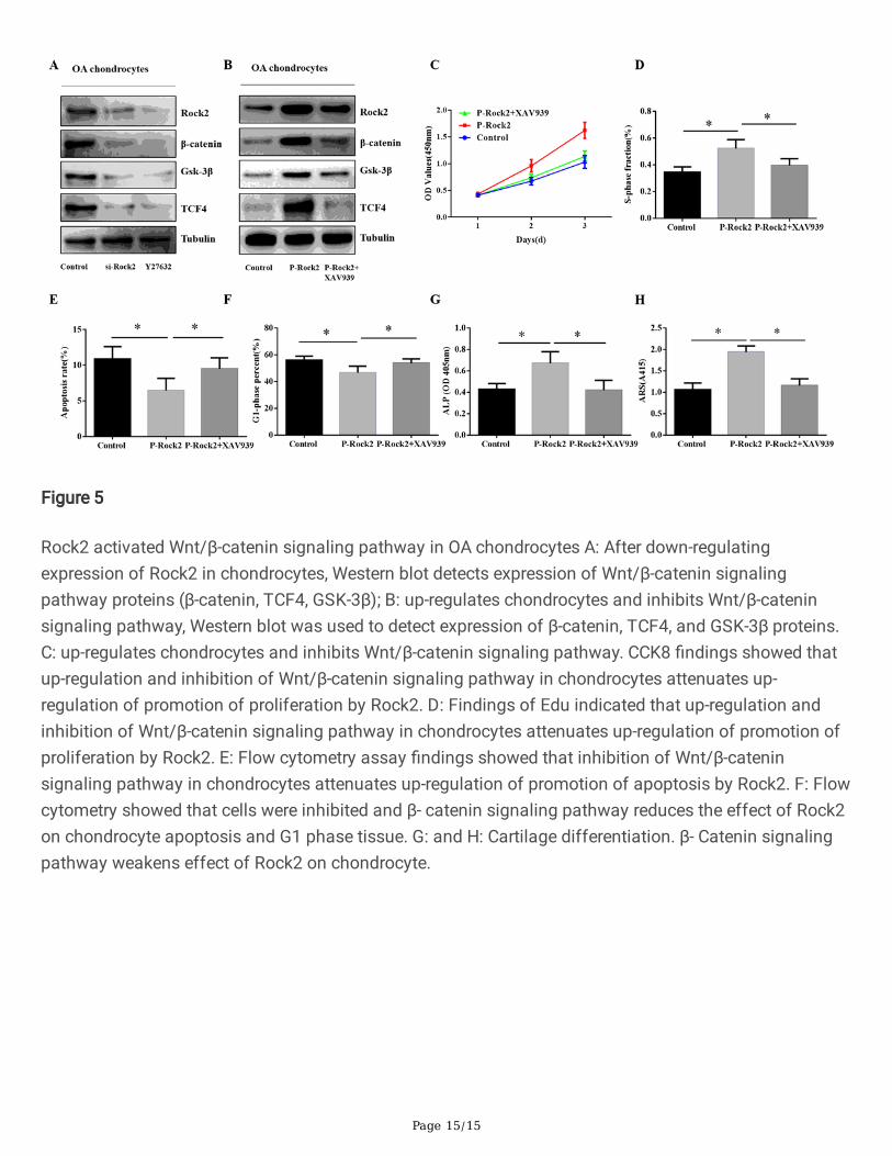
Figure 5
Rock2 activated Wnt/β-catenin signaling pathway in OA chondrocytes A: After down-regulatingexpression of Rock2 in chondrocytes, Western blot detects expression of Wnt/β-catenin signalingpathway proteins (β-catenin, TCF4, GSK-3β); B: up-regulates chondrocytes and inhibits Wnt/β-cateninsignaling pathway, Western blot was used to detect expression of β-catenin, TCF4, and GSK-3β proteins.C: up-regulates chondrocytes and inhibits Wnt/β-catenin signaling pathway. CCK8 �ndings showed thatup-regulation and inhibition of Wnt/β-catenin signaling pathway in chondrocytes attenuates up-regulation of promotion of proliferation by Rock2. D: Findings of Edu indicated that up-regulation andinhibition of Wnt/β-catenin signaling pathway in chondrocytes attenuates up-regulation of promotion ofproliferation by Rock2. E: Flow cytometry assay �ndings showed that inhibition of Wnt/β-cateninsignaling pathway in chondrocytes attenuates up-regulation of promotion of apoptosis by Rock2. F: Flowcytometry showed that cells were inhibited and β- catenin signaling pathway reduces the effect of Rock2on chondrocyte apoptosis and G1 phase tissue. G: and H: Cartilage differentiation. β- Catenin signalingpathway weakens effect of Rock2 on chondrocyte.