cholesteatoma - welcome to utmb health, the university … · cholesteatoma michael underbrink,...
TRANSCRIPT

Cholesteatoma
Michael Underbrink, M.D.
Faculty Advisor: Arun Gadre, M.D.
The University of Texas Medical Branch
Department of Otolaryngology
Grand Rounds Presentation
September 18, 2002

Introduction
Keratin-producing squamous epithelium in
the middle ear, mastoid or petrous apex
Johannes Müller (1838) coined the term
“a pearly tumor of fat…among sheets of
polyhedral cells”
Exhibits independent growth, replaces
mucosa, resorbs bone

Introduction
Histologically made up of :
Cystic content – anucleate keratin squames
Matrix – keratinizing squamous epithelium
Perimatrix – granulation tissue in contact with
bone (produces proteolytic enzymes)

Pathology
Molecular models
Preneoplastic transformation events
Defective wound-healing process
Collision between host inflammatory
response, normal middle ear epithelium, and
bacterial infection

Preneoplastic transformation
Hyperproliferative keratinocytes Increased proliferation
Decreased terminal differentiation
Expression of epithelial markers in the basal and suprabasal layers (cytokeratins –10,13,16, filaggrin, involucrin); confirm they arise from pars flaccida and overlying EAC skin
High expression of epidermal growth factor receptor, transforming growth factor
Upregulation of p53

Defective wound healing
Chronic inflammatory response around matrix
(granulation/perimatrix)
Infiltration of activated T-cells and macrophages
Production of cytokines (TGF,TNF,IL-1,IL-
2,FGF,PDGF)
Causes increased migration and invasion of
cholesteatoma epithelium and fibroblasts

Host response vs. bacteria
Bacterial related antigens producing host inflammatory response may stimulate the migrating epithelium’s uncoordinated proliferation
Granulation induces invasion of keratinocytes
Granulation – contains proteases, acid phosphatases, bone resorption proteins, osteoclast-activating factors, prostaglandins
Keratin implanted into mouse calvaria was shown by Chole, et. al., to activate osteoclasts and produce a localized inflammatory bone remodeling similar to cholesteatomas

Classification
Congenital
Acquired
Primary acquired (retraction pocket)
Secondary acquired

Pathogenesis
Congenital
Arise from embryonal rests of epithelial cells
Location (petrous pyramid, mastoid and middle ear
cleft)
Levenson criteria
White mass medial to normal TM
Normal pars flaccida and tensa
No history of otorrhea or perforations
No prior otologic procedures
Prior bouts of otitis media not grounds for exclusion

Congenital cholesteatoma

Pathogenesis
Primary acquired
Eustachian tube dysfunction
Poor aeration of the epitympanic space
Retraction of the pars flaccida
Normal migratory pattern altered
Accumulation of keratin, enlargement of sac
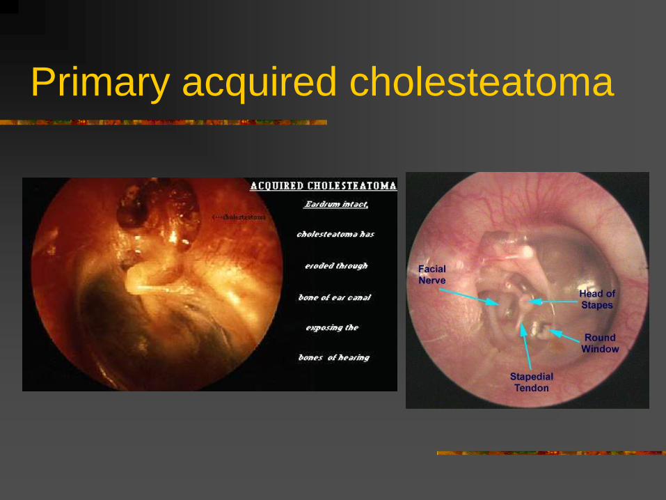
Primary acquired cholesteatoma
Pathogenesis
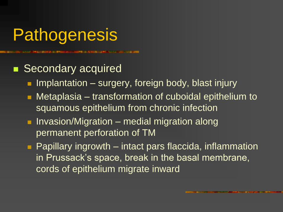
Secondary acquired
Implantation – surgery, foreign body, blast injury
Metaplasia – transformation of cuboidal epithelium to
squamous epithelium from chronic infection
Invasion/Migration – medial migration along
permanent perforation of TM
Papillary ingrowth – intact pars flaccida, inflammation
in Prussack’s space, break in the basal membrane,
cords of epithelium migrate inward

Anatomic Considerations
Mesotympanum
Facial recess
Sinus tympani
Hypotympanum
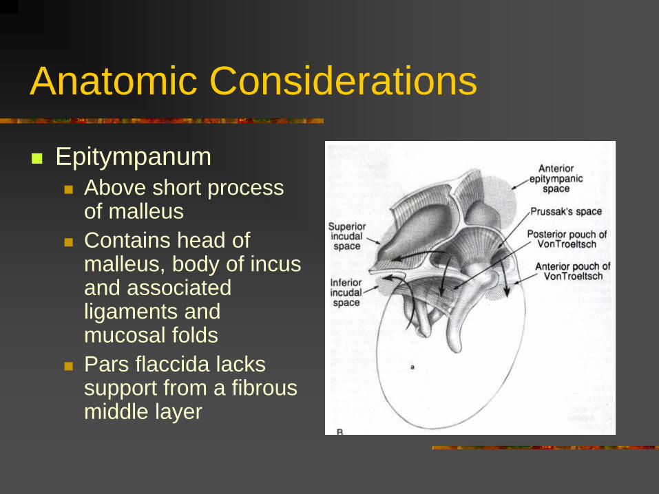
Epitympanum
Anatomic Considerations
Epitympanum Above short process
of malleus
Contains head of malleus, body of incus and associated ligaments and mucosal folds
Pars flaccida lacks support from a fibrous middle layer

Anatomic Considerations
Epitympanic cholesteatoma patterns of spread from Prussack’s space Posterior
epitympanum
Posterior mesotympanum
Anterior epitympanum

Cholesteatoma spread
Posterior epitympanum
– through superior
incudal space to
mastoid antrum
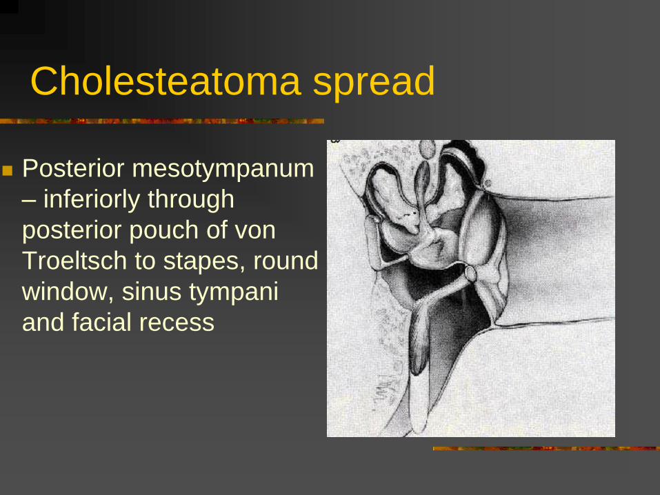
Cholesteatoma spread
Posterior mesotympanum
– inferiorly through
posterior pouch of von
Troeltsch to stapes, round
window, sinus tympani
and facial recess

Cholesteatoma spread
Anterior epitympanum
– anterior to head of
malleus, may gain
access to supratubal
recess via anterior
pouch of von
Troeltsch

Evaluation
History
Hearing loss, otorrhea, otalgia, tinnitus,
vertigo and nasal obstruction
Previous history of chronic otitis media,
tympanic membrane perforation or otologic
surgery
Progressive unilateral hearing loss with
chronic fetid otorrhea suspicious

Evaluation
Physical Examination
Otomicroscopy
Posterosuperior retraction pocket with squam
Granulation from diseased bone
Aural polyps
Pneumatic otoscopy – positive fistula
response suggests erosion into labyrinth
Cultures should be obtained in infected ears

Evaluation
Audiology – usually conductive loss, may
vary greatly; confirm with tuning forks
Imaging
CT temporal bone – definitely obtain for
revision cases, complications of chronic
suppurative otitis media, sensorineural
hearing loss, vestibular symptoms, other
complications of cholesteatoma

Management
Surgical disease with definite objectives:
Removal of disease for safe, dry ear
Restore or maintain functional capacity of ear, i.e.,
hearing
Maintain normal anatomy if possible
Management of complications takes priority
Each case treated individually according to
extent/location of disease
Preoperative counseling

Management
Medical
Aural toilet, local care,
patients with unacceptable anesthesia risks
Preventive
Tympanostomy tube for early retraction
pockets
Surgical exploration for persistence

Surgical Management
Canal-wall-down procedures
Intact-canal-wall procedure
Transcanal anterior atticotomy
Bondy modified radical procedure

Canal-wall-down procedures
Exteriorizing mastoid into external ear
canal by taking posterior canal wall down
Modified radical mastoidectomy – middle
ear space preserved
Radical mastoidectomy – middle ear
space eliminated, Eustachian tube orifice
obliterated

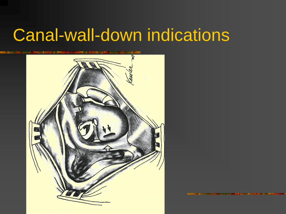
Canal-wall-down indications
Cholesteatoma in an only hearing ear
Significant erosion of posterior wall
Labyrinthine fistula
Limited access to epitympanum from
sclerotic mastoid
Recurrent cholesteatoma following ICW
surgery with ETD
Canal-wall-down indications

Canal-wall-down
Advantages
Residual disease easy to detect
Rare recurrence
Facial recess exteriorized
Disadvantages
Open cavity, lifetime maintenance
Longer healing time
Middle ear shallow (difficult OCR)
Water precautions necessary

Intact-canal wall procedure
Preservation of posterior wall
With/without posterior tympanotomy
2nd staged procedure (6 – 12 months)
Contraindications
Only hearing ears
Labyrinthine fistula
Long-standing ear disease, ETD

Intact-canal wall procedure

Intact-canal-wall
Advantages Rapid healing time
Easier long-term care
Hearing aids easier to fit
No water precautions
Disadvantages Technically more difficult
Recurrent disease possible
Staged operation often necessary
Residual disease harder to detect

Transcanal anterior atticotomy
Limited cholesteatoma (middle ear,
ossicular chain, epitympanum)
Endaural incision to raise flap
Removal of scutum around cholesteatoma
Aditus obliterated
Reconstruction of lateral attic wall optional
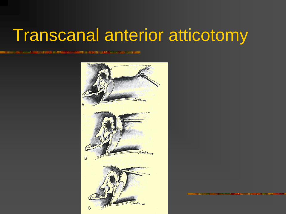
Transcanal anterior atticotomy

Bondy modified radical procedure
Attic and mastoid disease
Lateral to ossicles, not involving middle
ear space
Cholesteatoma marsupialized
Requires good Eustachian tube function
and intact pars tensa
Complications of cholesteatoma
Hearing loss
Labyrinthine fistula
Facial paralysis
Intracranial complications

Hearing loss
Conductive hearing loss common
Ossicular chain erosion – 30%
Severity of loss varies despite extent of
disease
SNHL – may indicate labyrinth involved
Surgical complication rates – 3% (can be
total hearing loss)

Labyrinthine fistula
Up to 10% of patients
Suspect with longstanding disease, SNHL,
induced vertigo
CT should be obtained
Most common structure – horizontal canal
Requires CWD mastoidectomy
Management of matrix overlying fistula
Facial paralysis
With cholesteatoma requires immediate
surgery
Rapid – infected cholesteatoma
Slow – chronic expansion of disease
CT localizes involved portion
Most common site – geniculate ganglion

Facial paralysis
Management
Mastoidectomy with facial recess approach
for horizontal and vertical segments
Middle cranial fossa for petrous apex
Remove cholesteatoma and infected debris
IV antibiotics and steroids helpful
Iatrogenic injury repaired immediately

Intracranial complications
Potentially life threatening
Periosteal abscess, lateral sinus thrombosis,
intracranial/epidural abscess, meningitis
Less than 1% of all patients
Suppurative otorrhea, chronic headache, pain,
fever – impending intracranial complication
Mental status changes, nuchal rigidity, cranial
neuropathies require neurosurgical consult

Conclusions
Exact mechanism of pathogenesis not clear
Knowledge of anatomy and function of middle ear
Careful initial evaluation
Primary goal of surgery: safe, dry ear
Surgical strategies vary
Complications can be life-threatening