cholangiocarcinoma patients with lymph node predicting
TRANSCRIPT

Page 1/22
Development and Validation of Nomograms forPredicting Prognosis in IntrahepaticCholangiocarcinoma Patients with Lymph NodeMetastasisxianmao shi
Guangxi Medical University Firth A�liated Hospital: First Peoples Hospital of Nanninghttps://orcid.org/0000-0002-6631-2475
Xing Sun Guangxi Medical University Firth A�liated Hospital: First Peoples Hospital of Nanning
Xin Qin Guangxi Medical University Firth A�liated Hospital: First Peoples Hospital of Nanning
Ze Su Guangxi Medical University Firth A�liated Hospital: First Peoples Hospital of Nanning
Zhaoshan Fang Guangxi Medical University Firth A�liated Hospital: First Peoples Hospital of Nanning
Yi Xiao ( [email protected] )Guangxi Medical University Firth A�liated Hospital: First Peoples Hospital of Nanning
Zhi Zhang Guangxi Medical University Firth A�liated Hospital: First Peoples Hospital of Nanning
Research
Keywords: Intrahepatic cholangiocarcinoma, Lymph node metastasis, Prognosis, Nomogram
Posted Date: September 20th, 2021
DOI: https://doi.org/10.21203/rs.3.rs-882564/v1
License: This work is licensed under a Creative Commons Attribution 4.0 International License. Read Full License

Page 2/22
AbstractBackground. Lymph node metastasis (LNM) is one of the common metastatic sites of in advanced-stageintrahepatic cholangiocarcinoma (ICC), and the prognosis of ICC patients with LNM is worse thanpatients without it. Our study aimed to identify the prognostic factors of ICC patients with LNM, anddevelop an effective nomogram to quantify the prognosis of ICC patients with LNM.
Methods. We retrospectively reviewed the data of ICC patients between 2010 and 2015 in theSurveillance, Epidemiology, and End Results (SEER) database. Univariate and multivariate logisticanalysis were used to determine the independent predictors for LNM in patients with ICC. Univariate andmultivariate Cox analyses were used to identify the independent prognostic factors for ICC patients withLNM. Finally, two nomograms for predicting overall survival (OS) and cause-speci�c survival (CSS) wereestablished, and the nomogram of predicting OS was evaluated by calibration curves, receiver operatingcharacteristic (ROC) curves, and decision curve analysis (DCA).
Results. A total of 1539 patients with ICC were enrolled into our analysis, including 381 cases (24.76%)with LNM at initial diagnosis and 1158 cases (75.24%) without it. The independent risk factors for LNMin newly diagnosed ICC patients are age, T stage, and tumor size. The independent prognostic factors forICC patients with LNM are grade, chemotherapy, and surgery of primary site. For the prognosticnomogram for OS, the AUCs of 6-, 12-, and 24-months were 0.809, 0.780, and 0.755 in the training set and0.806, 0.780, and 0.753 in the testing set, respectively. The calibration curves and decision curve analysisindicated the good performance of the nomogram.
Conclusions. The individualized nomogram could predict OS of ICC patients with LNM with goodperformance, which could be served as an effective tool for prognostic evaluation and individualtreatment strategies optimization in ICC patients with LNM, and clinical utility may bene�t for clinicaldecision-making.
IntroductionIntrahepatic cholangiocarcinoma (ICC) is rare, but it is the second most common primary liver tumor afterhepatocellular carcinoma. A research reported that ICC accounts for 10–20% of all primary liver tumors,and it also accounts for 8–10% of biliary tract cancers [1]. ICC has been regarded as aggressive, andcomplete resection offers the only potentially curative treatment, which can provides patients with ICC amedian overall survival (OS) of 14.4 to 38.8 months[2–7]. However, surgical resection can improve theprognosis of ICC patients, but it is estimated that 70% of ICC patients initially present with advanced-stage disease and considered unresectable[8]. Patients with clinically lymph node (LN) metastasis aredeem advanced-stage disease, and their prognosis is typically considered to be similar to unresectableICC patients. During a standard portal lymphadenectomy, the pericholedochal, periportal, and commonhepatic artery lymph nodes are all excised, and these are also the most common sites of lymph nodemetastasis. Several clinicopathological factors of tumor, including tumor number, lymph node metastasis

Page 3/22
and distant organ metastasis, have been regarded as risk factors of overall survival. Among theparameters studied, it is report that lymph node metastasis (LNM) is one of the most relevant factors[5, 9–
11].Lymph node (LN) metastasis is one of the major prognostic factors in ICC patients, with only less than20% of ICC patients with LNM surviving 5 or more years after surgical resection[5]. Lymph nodemetastasis strongly impacts the prognosis of ICC patients after surgical resection, so adjuvant strategy isneeded[12], including adjuvant chemotherapy and radiotherapy. Therefore, it is important to establishpredictive models for predicting the prognosis of ICC patients with LNM. Nomogram is a simple andmultivariable visualization tool in oncology research, which is used to predict and quantify the rate of theoutcome of individual patient [13]. The nomogram is also used to aid clinical decision-making andpromote the development of precision medicine. Therefore, based on the data from the Surveillance,Epidemiology, and End Results (SEER) database, we aimed to develop an individual nomogram forpredicting the overall survival (OS) of ICC patients with LNM.
Methods
Study populationOur data in this population-based study were retrieved from the US National Cancer Institute (NCI) openpublic database, the Surveillance, Epidemiology, and End Results (SEER) database. To identify thepatients diagnosed with intrahepatic cholangiocarcinoma (ICC) from 2010 to 2015, the detailedpopulation selection procedure was summarized in Fig. 1. The inclusion criteria in the present study werefollowing: (1) All patients were enrolled from 2010 to 2015; (2) All patients with a primary site of“intrahepatic bile duct”, with ICD-O-Histology/behavior codes of 8160/3 (cholangiocarcinoma) and withsingle primary intrahepatic cholangiocarcinoma. Cases diagnosed at autopsy or via death certi�cates,with unspeci�ed follow-up, with live, brain, lung and bone metastasis, and unknown lymph nodemetastasis information were excluded. Because our study used previously collected data from SEERdatabase, it was exempt from the ethical review of the ethics board of the Fifth A�liated Hospital ofGuangxi Medical University. SEER*Stat version 8.3.8 (https://seer.cancer.gov/seerstat/) (InformationManagement Service, Inc. Calverton, MD, USA) was used for case listing.
Statistical analysisAll statistical analysis in our present study was conducted with SPSS 25.0 and R software (version 4.0.4).The chi-square test was used to compare the variables between the training set and the testing set. In thepresent study, a p-value < 0.05(two sides) was considered as statistical signi�cance. Univariate logisticanalysis was applied to identify LNM-related factors. The variables with p value < 0.1 in the univariatelogistic analysis were included in the multivariate binary logistic regression analysis to determineindependent risk factors of LNM in initially diagnosed ICC patients. For prognostic factors, the univariateCox regression analysis was applied to identify prognostic variables. Then, signi�cant variables in the

Page 4/22
Results
Risk factors of LNM in ICC patientsIn 1539 ICC patients, 381 cases (24.76%) with LNM at initial diagnosis and 1158 cases (75.24%) withoutit. To identify LNM-related variables in ICC patients, the multivariate logistic regression analysis showedthat age (p = 0.002), T stage (p < 0.001), and tumor size (p < 0.001) were independent predictors for LNMin newly diagnosed ICC patients (Table 1).
univariate Cox regression analysis were incorporated into the multivariate Cox regression analysis, andthe independent prognostic factors of ICC with LNM were identi�ed.
The predictive and prognostic nomograms were developed by the “rms” package in R software based onthe independent prognostic factors. Meanwhile, the time-dependent receiver operating characteristic(ROC) curve for the prognostic nomogram was generated[14]. The area under the curve (AUC) was used toevaluate the discrimination of nomogram. Moreover, the calibration curves and decision curve analysis(DCA) curves were established for the nomogram[15]. Finally, according to the median of risk score, allpatients were divided into the high-risk and low-risk groups, and the survival curve with a log-rank testwas used to verify the prognostic value of the nomogram[16].

Page 5/22
Table 1Uni- and multivariable logistic analysis of characteristics of patients with ICC presenting with LNM at the
initial diagnosis (2010–2015)Variable Total with
LNMwithoutLNM
univariate multivariable
OR(95%) p OR(95%) p
patients 1539 381 1158
Age (year) <0.001 0.002
<65 704 204 500 Reference Reference
≥ 65 835 177 658 0.659(0.523–0.832)
<0.001 0.685(0.540–0.869)
0.002
Sex 0.208
Female 781 204 577 Reference
Male 758 177 581 0.862(0.683–1.087)
0.208
Race 0.362
White 1179 302 877 Reference
Black 145 31 114 0.790(0.520–1.199)
0.268
Other 215 48 167 0.835(0.590–1.180)
0.307
Grade 0.063 0.156
Grade I-II 423 96 327 Reference Reference
Grade III-IV 327 97 230 1.437(1.034–1.996)
0.031 1.389(0.993–1.943)
0.055
Unknown 789 188 601 1.066(0.805–1.410)
0.657 1.196(0.892–1.604)
0.231
T stage 0.001 <0.001
T1-T2 1153 260 893 Reference Reference
T3-T4 303 101 202 1.717(1.303–2.263)
<0.001 1.756(1.326–2.326)
<0.001
TX 83 20 63 1.090(0.647–1.837)
0.745 1.510(0.865–2.635)
0.147
Abbreviations: CI, con�dence interval; OR: odds ratio; ICC: intrahepatic cholangiocarcinom; LNM,lymph node metastasis.

Page 6/22
Variable Total withLNM
withoutLNM
univariate multivariable
Tumor size(cm)
468 84 384 <0.001 <0.001
<5 748 233 515 Reference Reference
≥ 5 323 64 259 2.068(1.560–2.743)
<0.001 1.981(1.487–2.639)
<0.001
Unknown 1.130(0.787–1.621)
0.509 1.020(0.696–1.495)
0.919
Surgery 0.016
NO 1012 270 742 Reference
YES 517 111 416 0.733(0.570–0.943)
0.016
Radiation 0.084
NO 1439 349 1090 Reference
YES 100 32 68 1.470(0.949–2.276)
0.084
Chemotherapy <0.001
NO 743 128 615 Reference
YES 796 253 543 2.239(1.758–2.851)
<0.001
Abbreviations: CI, con�dence interval; OR: odds ratio; ICC: intrahepatic cholangiocarcinom; LNM,lymph node metastasis.
Prognostic factors of patients with ICC presenting withLNM381 ICC patients with LNM were used to study the prognostic factors. Among 381 patients, the 204(53.5%) patients were male, 177(46.5%) patients female. And the race of 302 (79.3%) patients were White,31 (8.1%) patients were Africa American, and 48 (12.6%) patients were other. Meanwhile, 269 patientswere randomly divided into the training set, and the remaining 112 patients were incorporated into thetesting set. The chi-square test showed that there were no signi�cant differences between the training setand the testing set (Table 2).

Page 7/22
Table 2Clinical characteristics of patients in the training set and testing set
Variable Total Training set Validation set X2 p
patients 381 269 112
Age (year)
<65 204(53.5%) 148(55%) 56(50%) 0.801 0.371
≥ 65 177(46.5%) 121(45%) 56(50%)
Sex
Female 204(53.5%) 152(56.5%) 52(46.4%) 3.228 0.072
Male 177(46.5) 117(43.5%) 60(53.6%)
Race
White 302(79.3%) 208(77.3%) 94(83.9%) 3.059 0.217
Black 31(8.1%) 22(8.2%) 9(8.0%)
Other 48(12.6%) 39(14.5%) 9(8.0%)
Grade
Grade I-II 96(25.2%) 71(26.4%) 25(22.3%) 0.891 0.64
Grade III-IV 97(25.5%) 69(25.7%) 28(25.0%)
Unknown 188(49.3%) 129(48.0%) 59(52.7%)
T stage
T1-T2 260(68.2%) 186(69.1%) 74(66.1%) 0.367 0.832
T3-T4 101(26.5%) 69(25.7%) 32(28.6%)
TX 20(5.2%) 14(5.2%) 6(5.4%)
Tumor size (cm)
<5 84(22.0%) 57(21.2%) 27(24.1%) 0.664 0.718
≥ 5 233(61.2%) 168(62.5%) 65(58.0%)
Unknown 64(16.8%) 44(16.4%) 20(17.9%)
Surgery
NO 270(70.9%) 193(71.7%) 77(68.8%) 0.334 0.557
YES 111(29.1%) 76(28.3%) 35(31.3%)
Abbreviations:. *P values were calculated by chi-square test

Page 8/22
Variable Total Training set Validation set X2 p
Radiation
NO 349(91.6%) 246(91.4%) 103(92.0%) 0.027 0.869
YES 32(8.4%) 23(8.6%) 9(8.0%)
Chemotherapy
NO 128(33.6%) 84(31.2%) 44(39.3%) 2.302 0.129
YES 253(66.4%) 185(68.8%) 68(60.7%)
Abbreviations:. *P values were calculated by chi-square test
As shown in Table 3 and Table 4, after the univariate Cox analysis, age, grade, surgery, chemotherapy, andradiation were signi�cantly associated with OS and CSS, respectively. In the multivariate Cox analysis,grade(p<0.050), surgery (p<0.001), and chemotherapy(p<0.001) were identi�ed as independentprognostic factors of OS and CSS, respectively.

Page 9/22
Table 3Uni- and multivariate cox analysis of overall survival for ICC with LNM
Univariate Cox
analysis
Multivariate Cox analysis
Variable HR 0.950 P-value HR 0.950 P-value
Age (year) 0.001 0.241
<65 Reference Reference
≥ 65 1.531 1.179–1.989 0.001 1.179 0.896–1.551 0.241
Sex 0.080
Female Reference
Male 1.264 0.973–1.642 0.080
Race 0.667
White Reference
Black 1.234 0.776–1.963 0.374
Other 0.996 0.695–1.426 0.981
Grade <0.001 0.010
Grade I-II Reference Reference
Grade III-IV 1.771 1.211–2.590 0.003 1.801 1.230–2.638 0.003
Unknown 1.954 1.406–2.715 <0.001 1.423 1.003–2.020 0.048
T stage 0.884
T1-T2 Reference
T3-T4 0.927 0.686–1.252 0.62
TX 0.970 0.526–1.791 0.923
Tumor size (cm) 0.569
<5 Reference
≥ 5 1.007 0.722–1.404 0.968
Unknown 1.214 0.790–1.868 0.376
Surgery <0.001 <0.001
Abbreviations: CI, con�dence interval; HR, hazard ratio; ICC: intrahepatic cholangiocarcinom; LNM,lymph node metastasis.

Page 10/22
Univariate Cox
analysis
Multivariate Cox analysis
NO Reference
YES 0.475 0.346–0.652 <0.001 0.473 0.334–0.668 <0.001
Chemotherapy <0.001 <0.001
NO Reference
YES 0.357 0.270–0.473 <0.001 0.341 0.252–0.460 <0.001
Radiation 0.003 0.169
NO Reference Reference
YES 0.449 0.266–0.760 0.003 0.686 0.401–1.174 0.069
Abbreviations: CI, con�dence interval; HR, hazard ratio; ICC: intrahepatic cholangiocarcinom; LNM,lymph node metastasis.

Page 11/22
Table 4Uni- and multivariate cox analysis of cause-speci�c survival for ICC with LNM
Univariate Cox analysis Multivariate Cox
analysis
Variable HR 0.950 P-value HR 0.950 P-value
Age (year) 0.002 0.284
<65 Reference Reference
≥ 65 1.516 1.162–1.978 0.002 1.165 0.881–1.540 0.284
Sex 0.14
Female Reference
Male 1.222 0.937–1.595 0.14
Race 0.579
White Reference
Black 1.279 0.803–2.036 0.3
Other 1.002 0.695–1.443 0.993
Grade <0.001 0.011
Grade I-II Reference Reference
Grade III-IV 1.778 1.212–2.609 0.003 1.799 1.224–2.644 0.003
Unknown 1.916 1.373–2.674 <0.001 1.39 0.975–1.983 0.069
T stage 0.949
T1-T2 Reference
T3-T4 0.964 0.712–1.304 0.81
TX 0.921 0.485–1.750 0.803
Tumor size (cm) 0.659
<5 Reference
≥ 5 1.001 0.715–1.401 0.997
Unknown 1.182 0.762–1.834 0.455
Surgery <0.001 <0.001
Abbreviations: CI, con�dence interval; HR, hazard ratio; ICC: intrahepatic cholangiocarcinom; LNM,lymph node metastasis.

Page 12/22
Univariate Cox analysis Multivariate Cox
analysis
NO Reference Reference
YES 0.481 0.349–0.663 <0.001 0.476 0.335–0.675 <0.001
Chemotherapy <0.001 <0.001
NO Reference Reference
YES 0.359 0.270–0.478 <0.001 0.342 0.252–0.464 <0.001
Radiation 0.002 0.127
NO Reference Reference
YES 0.43 0.250–0.739 0.002 0.649 0.372–1.130 0.127
Abbreviations: CI, con�dence interval; HR, hazard ratio; ICC: intrahepatic cholangiocarcinom; LNM,lymph node metastasis.
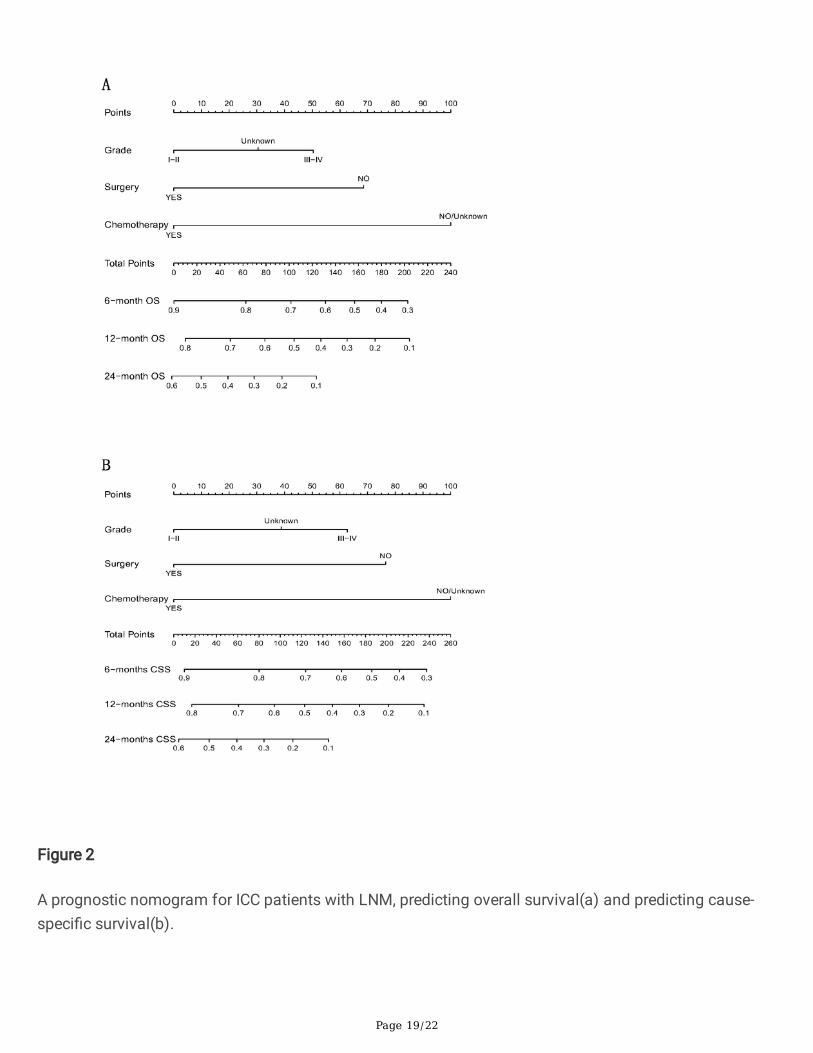
Prognostic nomogram for ICC patients with LNMTwo prognostic nomograms for predicting OS and CSS were established based on three independentprognostic factors(Fig. 2a and b). For the prognostic nomogram for OS, the AUCs of 6-, 12-, and 24-months were 0.809, 0.780, and 0.755 in the training set, respectively(Fig. 3a,b and c). In the testing set,the AUCs of 6-, 12-, and 24-months were 0.806, 0.780, and 0.753, respectively (Fig. 3d, e and f). Inaddition, in the training set, the Kaplan-Meier survival curve showed that ICC patients with LNM in thehigh-risk group have a worse prognosis than patients in the low-risk group (Fig. 4a). Similarly, in thetesting set, the OS of ICC patients with LNM in the high-risk group was also worse than patients in thelow-risk group (Fig. 4b).The calibration curves for the probability of 6-, 12-, and 24-months OS alsoindicated a good consistency between nomogram predicted OS and the actual outcome in the training set(Fig. 5a, b and c) and in testing set (Fig. 5d, e and f), respectively. In addition, the DCA curves showed thatthe nomogram had a good predictive e�ciency for OS of ICC patients with LNM in the training set(Fig. 6a,b and c) and in testing set (Fig. 6d, e and f), respectively.
Discussion

Page 13/22
Intrahepatic cholangiocarcinoma frequently metastasizes to regional lymph node, which be associatedwith tumor recurrence after curative-intent resection. The association between lymph node metastasisand prognosis of ICC patients has not been fully investigated and validated. In the current study, lymphnode metastasis was present in roughly 40% of ICC patients who underwent surgical resection. Evenseveral studies have reported an higher incidence rate of lymph node metastasis in patients with ICC, ashigh as 45–62%[17, 18].
Therefore, we attempted to identify valuable predictor for lymph node metastasis in patients with ICC, asthis information might be helpful in clinical practice. Several studies reported that tumor number andtumor diameter are signi�cant predictive factors for LN metastasis in ICC patients[19, 20].Our study foundthat age, T stage, and tumor size were the best-�t predictor for LN metastasis in ICC patients. In addition,poorly differentiated ICC was also associated with a higher rate of lymph node metastasis [20].
Lymph node metastasis was reported to be one of the most signi�cant prognostic factors for survivalfrom ICC patients[9, 21]. However, despite the prognostic relevance of the LN status, several investigatorsreported that lymph node dissection (LND) does not seem to improve the OS of patients with ICC[22,
23].Actually, a retrospective large cohort study suggested that adjuvant chemotherapy could improvesurvival of ICC patients with lymph node metastasis, or advanced-stage[24–28]. Despite making a progressin understanding the pathophysiology of ICC and the emergence of novel treatment options[29], liverresection remains the only potential cure for ICC patients, whose survival has not improved signi�cantlyin recent decades [5, 30, 31].
Our research showed that ICC patients with LNM with high grade, absence of chemotherapy, and absenceof surgery had unfavorable prognosis. Based on three independent prognostic factors of ICC patientswith LNM, an individual nomogram for predicting OS was established and evaluated. The resultsindicated that the nomogram can serve as an effective tool to identify high-risk patients. It is wellaccepted that tumor grade could explain some of the heterogeneity associated with the expected courseand clinical outcome in patients with various tumors[32, 33]. Tumor grade is a measure of the degree ofdifferentiation of the tumor and is related to the prognosis of patients with tumor. In the present study,tumor grade was also observed as an independent prognostic predictor for OS in ICC patients with LNM.As far as we know, it is the �rst study to build a nomogram for predicting OS in ICC patients with LNM.Our nomogram, incorporating easily accessible factors in clinical practice, enabled easy calculation ofindividualized OS probabilities for ICC patients with LNM. Moreover, the nomograms showed relativelyhigh accuracy with OS, and calibration curves display well-�tted in both the training set and testing set. Inaddition, high predictive accuracy does not mean that the predictive model has high clinical practicability[13, 15]. Decision curve analysis is one of the recommended methods in previous research on prognosticpredictive models. It can quantify the net bene�ts of predictive models according to the thresholdprobability, so as to evaluate the clinical practical value of a predictive model[34–36]. Therefore, weintroduced the decision curve analysis to examine the effectiveness of our nomogram in clinical practice.

Page 14/22
The decision curve analyses not only con�rmed the clinical validity of our nomogram for OS, but alsodemonstrated that our nomogram had better clinical application value in ICC patients with LNM.
However, our research also has some limitations. First, limited patients (N = 381) of ICC with LNM mayresult the possible error in our study. Second, the information collected in the SEER database was aboutthe ICC disease at the �rst diagnosis, which meant that the lymph node metastasis occur in the latterstage cannot be recorded. Third, this was a retrospective study in which patients’ selection bias existedinevitably, and the information about detailed treatment and progress after treatment was not available inthe SEER database. However, notwithstanding these limitations, our study is the �rst to use a nomogramcombined with clinically feasible variables including tumor grade, chemotherapy, and surgery of primarysite to predict the OS of ICC patients with LNM, which has clinical signi�cance for predicting prognosis ofICC patients with LNM.
ConclusionsOur study showed that age, T stage, and tumor size were the risk factors of LNM from ICC patients. As forICC patients with LNM, high grade, chemotherapy, and surgery of primary site were independentprognostic factors for OS and CSS. Two nomograms we created may be individual, convenient, and moreintuitive visual tools for prognostic prediction for ICC patients with LNM.
DeclarationsEthics approval and consent to participate
Not applicable.
Consent for publication
Not applicable
Competing interests
The authors declare that they have no competing interests.
Funding
Not applicable
Authors’ contributions
Xianmao Shi, Yi Xiao, and Zhi Zhang designed the study. Xianmao Shi and Xin Qin collected andanalyzed the data and drafted the manuscript. Xing Sun, Ze Su and Zhaoshan Fang revised themanuscript and contributed to data interpretation.

Page 15/22
Availability of data and materials
The datasets supporting the conclusions of this article are available in the SEER database athttps://seer.cancer.gov/seerstat/.
Acknowledgements
The authors gratefully acknowledge the efforts of the SEER Program for
providing high-quality open resources for researchers.
References1. Gupta A, Dixon E. Epidemiology and risk factors: intrahepatic cholangiocarcinoma. Hepatobiliary
Surg Nutr. 2017;6(2):101–4.
2. Bridgewater J, Galle PR, Khan SA, et al. Guidelines for the diagnosis and management of intrahepaticcholangiocarcinoma. J Hepatol. 2014;60(6):1268–89.
3. Ribero D, Pinna AD, Guglielmi A, et al. Surgical Approach for Long-term Survival of Patients WithIntrahepatic Cholangiocarcinoma: A Multi-institutional Analysis of 434 Patients. Arch Surg.2012;147(12):1107–13.
4. Farges O, Fuks D, Boleslawski E, et al. In�uence of surgical margins on outcome in patients withintrahepatic cholangiocarcinoma: a multicenter study by the AFC-IHCC-2009 study group. Ann Surg.2011. 254(5): 824 – 29; discussion 830.
5. de Jong MC, Nathan H, Sotiropoulos GC, et al. Intrahepatic cholangiocarcinoma: an internationalmulti-institutional analysis of prognostic factors and lymph node assessment. J Clin Oncol.2011;29(23):3140–5.
�. Uenishi T, Ariizumi S, Aoki T, et al. Proposal of a new staging system for mass-forming intrahepaticcholangiocarcinoma: a multicenter analysis by the Study Group for Hepatic Surgery of the JapaneseSociety of Hepato-Biliary-Pancreatic Surgery. J Hepatobiliary Pancreat Sci. 2014;21(7):499–508.
7. Luo X, Yuan L, Wang Y, Ge R, Sun Y, Wei G. Survival outcomes and prognostic factors of surgicaltherapy for all potentially resectable intrahepatic cholangiocarcinoma: a large single-center cohortstudy. J Gastrointest Surg. 2014;18(3):562–72.
�. Saha SK, Zhu AX, Fuchs CS, Brooks GA. Forty-Year Trends in Cholangiocarcinoma Incidence in theU.S.: Intrahepatic Disease on the Rise. Oncologist. 2016;21(5):594–9.
9. Guglielmi A, Ruzzenente A, Campagnaro T, et al. Patterns and prognostic signi�cance of lymph nodedissection for surgical treatment of perihilar and intrahepatic cholangiocarcinoma. J GastrointestSurg. 2013;17(11):1917–28.
10. Cho SY, Park SJ, Kim SH, et al. Survival analysis of intrahepatic cholangiocarcinoma after resection.Ann Surg Oncol. 2010;17(7):1823–30.
Page 16/22
11. Morine Y, Shimada M, Utsunomiya T, et al. Clinical impact of lymph node dissection in surgery forperipheral-type intrahepatic cholangiocarcinoma. Surg Today. 2012;42(2):147–51.
12. Maithel SK, Gamblin TC, Kamel I, Corona-Villalobos CP, Thomas M, Pawlik TM. Multidisciplinaryapproaches to intrahepatic cholangiocarcinoma. Cancer. 2013;119(22):3929–42.
13. Balachandran VP, Gonen M, Smith JJ, DeMatteo RP. Nomograms in oncology: more than meets theeye. Lancet Oncol. 2015;16(4):e173-80.
14. Heagerty PJ, Lumley T, Pepe MS. Time-dependent ROC curves for censored survival data and adiagnostic marker. Biometrics. 2000;56(2):337–44.
15. Vickers AJ, Elkin EB. Decision curve analysis: a novel method for evaluating prediction models. MedDecis Making. 2006;26(6):565–74.
1�. Ranstam J, Cook JA. Kaplan-Meier curve. Br J Surg. 2017;104(4):442.
17. Amini N, Ejaz A, Spolverato G, Maithel SK, Kim Y, Pawlik TM. Management of lymph nodes duringresection of hepatocellular carcinoma and intrahepatic cholangiocarcinoma: a systematic review. JGastrointest Surg. 2014;18(12):2136–48.
1�. Tsuji T, Hiraoka T, Kanemitsu K, Takamori H, Tanabe D, Tashiro S. Lymphatic spreading pattern ofintrahepatic cholangiocarcinoma. Surgery. 2001;129(4):401–7.
19. Marubashi S, Gotoh K, Takahashi H, et al. Prediction of the postoperative prognosis of intrahepaticcholangiocarcinoma (ICC): importance of preoperatively- determined anatomic invasion level andnumber of tumors. Dig Dis Sci. 2014;59(1):201–13.
20. Chen YX, Zeng ZC, Tang ZY, et al. Prediction of the lymph node status in patients with intrahepaticcholangiocarcinoma: analysis of 320 surgical cases. Front Oncol. 2011;1:42.
21. Yamamoto M, Ariizumi S. Surgical outcomes of intrahepatic cholangiocarcinoma. Surg Today.2011;41(7):896–902.
22. Kim DH, Choi DW, Choi SH, Heo JS, Kow AW. Is there a role for systematic hepatic pediclelymphadenectomy in intrahepatic cholangiocarcinoma? A review of 17 years of experience in atertiary institution. Surgery. 2015;157(4):666–75.
23. Zhou R, Lu D, Li W, et al. Is lymph node dissection necessary for resectable intrahepaticcholangiocarcinoma? A systematic review and meta-analysis. HPB (Oxford). 2019;21(7):784–92.
24. Morine Y, Shimada M. The value of systematic lymph node dissection for intrahepaticcholangiocarcinoma from the viewpoint of liver lymphatics. J Gastroenterol. 2015;50(9):913–27.
25. Sur MD, In H, Sharpe SM, et al. De�ning the Bene�t of Adjuvant Therapy Following Resection forIntrahepatic Cholangiocarcinoma. Ann Surg Oncol. 2015;22(7):2209–17.
2�. Miura JT, Johnston FM, Tsai S, et al. Chemotherapy for Surgically Resected IntrahepaticCholangiocarcinoma. Ann Surg Oncol. 2015;22(11):3716–23.
27. Wang ML, Ke ZY, Yin S, Liu CH, Huang Q. The effect of adjuvant chemotherapy in resectablecholangiocarcinoma: A meta-analysis and systematic review. Hepatobiliary Pancreat Dis Int.2019;18(2):110–6.

Page 17/22
2�. Lin YK, Hsieh MC, Wang WW, et al. Outcomes of adjuvant treatments for resectable intrahepaticcholangiocarcinoma: Chemotherapy alone, sequential chemoradiotherapy, or concurrentchemoradiotherapy. Radiother Oncol. 2018;128(3):575–83.
29. Shiao MS, Chiablaem K, Charoensawan V, Ngamphaiboon N, Jinawath N. Emergence of IntrahepaticCholangiocarcinoma: How High-Throughput Technologies Expedite the Solutions for a Rare CancerType. Front Genet. 2018;9:309.
30. Bagante F, Spolverato G, Weiss M, et al. Impact of Morphological Status on Long-Term OutcomeAmong Patients Undergoing Liver Surgery for Intrahepatic Cholangiocarcinoma. Ann Surg Oncol.2017;24(9):2491–501.
31. Okabayashi T, Yamamoto J, Kosuge T, et al. A new staging system for mass-forming intrahepaticcholangiocarcinoma: analysis of preoperative and postoperative variables. Cancer.2001;92(9):2374–83.
32. Greene FL, Sobin LH. The staging of cancer: a retrospective and prospective appraisal. CA Cancer JClin. 2008;58(3):180–90.
33. Rochefort MM, Ankeny JS, Kadera BE, et al. Impact of tumor grade on pancreatic cancer prognosis:validation of a novel TNMG staging system. Ann Surg Oncol. 2013;20(13):4322–9.
34. Wang ZX, Qiu MZ, Jiang YM, Zhou ZW, Li GX, Xu RH. Comparison of prognostic nomograms basedon different nodal staging systems in patients with resected gastric cancer. J Cancer. 2017;8(6):950–8.
35. Hijazi Z, Oldgren J, Lindbäck J, et al. The novel biomarker-based ABC (age, biomarkers, clinicalhistory)-bleeding risk score for patients with atrial �brillation: a derivation and validation study.Lancet. 2016;387(10035):2302–11.
3�. Dong F, Shen Y, Gao F, et al. Nomograms to Predict Individual Prognosis of Patients with PrimarySmall Cell Carcinoma of the Bladder. J Cancer. 2018;9(7):1152–64.
Figures

Page 18/22
Figure 1
Flowchart of the enrolled patients in the study according to inclusion and exclusion criterion.
Page 19/22
Figure 2
A prognostic nomogram for ICC patients with LNM, predicting overall survival(a) and predicting cause-speci�c survival(b).

Page 20/22
Figure 3
Receiver operating characteristic curves of 6-, 12-, and 24-months in the training set(a, b, and c); Receiveroperating characteristic curves of 6-, 12-, and 24-months in the testing set( c, d, and e);

Page 21/22
Figure 4
The Kaplan-Meier survival curve of the training set between high and low risk group(a); The Kaplan-Meiersurvival curve of the testing set between high and low risk group(b).
Figure 5
The calibration curves of the nomogram in the training set(a, b, and c); The calibration curves of thenomogram in the testing set(d, e, and f).

Page 22/22
Figure 6
The decision curve analysis of the nomogram in the training set(a, b, and c); The calibration curves of thenomogram in the testing set(d, e, and f).