chloride transport in the kidney: lessons from human ... · chloride transport in the kidney:...
TRANSCRIPT

Homer W. Smith Award Lecture
Chloride Transport in the Kidney: Lessons from HumanDisease and Knockout Mice
Thomas J. JentschZentrum fur Molekulare Neurobiologie (ZMNH), Universitat Hamburg, Hamburg, Germany
Knockout mouse models and human inherited diseases have provided important new insights into the physiologic role ofchloride transport by CLC Cl� channels and KCC K-Cl co-transporters. ClC-K/barrtin Cl� channels are important for renal saltreabsorption and possibly for acid secretion by intercalated cells. The endosomal ClC-5 protein is crucial for proximal tubularendocytosis. Its disruption in mice and patients with Dent’s disease leads to hypercalciuria and kidney stones through apathologic cascade that may be entirely explained by an impairment of endocytosis. KCC4 is important for recycling Cl� forthe basolateral anion exchanger in intercalated cells, as is evident from the renal tubular acidosis resulting from its knockout.Finally, both KCC3 and KCC4 are crucial for proximal tubular cell volume regulation.
J Am Soc Nephrol 16: 1549–1561, 2005. doi: 10.1681/ASN.2005020207
A lthough transepithelial transport of anions, for obvi-ous reasons, is quantitatively as important as cationtransport, it has received less attention. This is prob-
ably due in part to the influence of concepts from neurobiology,which stressed the importance of Na�, K�, and Ca2� for cellu-lar excitability but also to the overwhelming role of Na� ho-meostasis in the regulation of the extracellular volume and of BP.
Anions not only are passively dragged across epithelia by thetransepithelial voltage created by cation transport but also arethemselves transported by a plethora of different transportproteins. These include proteins that directly couple their trans-port to cations, anion exchangers, anion channels, and, as dis-covered recently, also Cl�/H� exchangers. Despite the comple-tion of several genome projects and the molecular identificationof an astounding variety of ion transport proteins, it seems thatmany transporters and channels have not yet been identified atthe molecular level. This is particularly true for the Cl� channelfield, where many currents and channels identified at the func-tional, biophysical level could not yet be correlated with aspecific gene product. For instance, we may still lack the rightclones for important channel classes such as swelling-activatedor Ca2�-activated Cl� channels.
Although the progress in molecular biology of ion channelsand transporters for some time seemed to displace classicalrenal physiologic techniques such as those used by Homer W.Smith to a second rank, the generation of genetic mouse modelscarrying targeted mutations in ion channel and transportergenes have bridged the gap to integrative physiology and willundoubtedly lead to a resurgence of classical methods of renalphysiology such as the perfusion of isolated tubules. The pa-
thologies observed in knockout (KO) mice and in human geneticdisease have led to invaluable and often unexpected insights intothe physiologic functions of specific transport proteins.
Here I review the renal aspects of our work on chloridetransport, which focuses on CLC Cl� channels (and transport-ers) and on electroneutral KCC K�/Cl� co-transporters. Thegeneration and analysis of mouse models have revealed thatthey play important roles in processes as diverse as salt reab-sorption, acid secretion, cell volume regulation, endocytosis,and lysosomal protein degradation, as well as kidney stones.
CLC Gene Family of Cl� Channels andTransporters
The CLC gene family, which was originally identified by theexpression cloning of a chloride channel (ClC-0) that is highlyexpressed in the electric organ of the marine ray Torpedo mar-morata (1), is present in all phylae from bacteria to humans.Mammals express nine distinct CLC genes (the gene CLCNXencodes the protein ClC-X). With the exception of the skeletalmuscle isoform ClC-1 (2), all of these genes are expressed tosome degree in kidney. None of these isoforms, however, iskidney specific. Human mutations in genetic disease and theanalysis of KO mice have shown that four of these (ClC-Ka,ClC-Kb, ClC-5, and ClC-7) have important impact on kidneyfunction.
Before addressing the renal functions of specific isoforms, itis useful to recall some basic features of these Cl� transportproteins. CLC channels are dimers, with each of the two sub-units having its own pore (“double-barreled channel”). Thiswas concluded from single-channel experiments on native Tor-pedo channels reconstituted into lipid bilayers (3) and convinc-ingly shown by studying WT-mutant ClC-0 concatemers (4,5)and ClC-0/ClC-2 heteromers (6). The dimeric structure of CLCproteins was confirmed impressively by the crystal structure ofbacterial CLC proteins (7–9), very strongly suggesting that thisarchitecture applies to all CLC proteins. In addition to ho-
Published online ahead of print. Publication date available at www.jasn.org.
Address correspondence to: Dr. Thomas J. Jentsch, Zentrum fur MolekulareNeurobiologie ZMNH, Universitat Hamburg, Falkenried 94, D-20246 Hamburg,Germany. Phone: 49-40-42803-4741; Fax: 49-40-42803-4839; E-mail: [email protected]
Copyright © 2005 by the American Society of Nephrology ISSN: 1046-6673/1606-1549
modimers, CLC proteins can form heterodimers, which may berestricted to members of the same homology branch (6,10,11).However, the physiologic relevance of heteromer formation iscurrently unclear. In addition, some CLC dimers may associatewith accessory �-subunits. The only known cases, so far, con-cern the two ClC-K isoforms, which associate with barttin (12).The crystal structure of bacterial CLC proteins showed a com-plicated architecture with 17 intramembrane but not necessar-ily transmembrane helices (7) and three distinct Cl� bindingsites (8). It also revealed a glutamate side chain that seemed toblock the access of extracellular Cl� to the narrowest part of thepore (7,8). There is very good evidence (8,13) that this gluta-mate plays a role in Cl�-dependent gating of CLC chloridechannels (14), as well as in the coupling of a countertransport ofH� to Cl� fluxes in the bacterial ClC-e1 protein (15).
Whereas many CLC proteins, such as the Torpedo channelClC-0 or the mammalian ClC-1, ClC-2, and ClC-Ka/barttin andClC-Kb/barttin, unambiguously function as Cl� channels, thebacterial protein ClC-e1, which was used for crystallizationstudies (7,8), surprisingly mediates an electrogenic 2Cl�/H�-exchange (15). Although mutating the glutamate that is alsoresponsible for chloride-dependent gating in CLC channelsabolished the H� coupling of anion fluxes, the detailed mech-anism of and the structural basis for the exchange activity arenot yet clear.
As ClC-6 and ClC-7, which are expressed in late endosomesand lysosomes, could not yet be expressed functionally in theplasma membrane (16), it is unclear whether they function asCl� channels or Cl�/H� exchangers. Upon heterologous ex-pression, a small fraction of the endosomal ClC-3, �4, and �5proteins reach the plasma membrane and yield Cl� currents(17–19). However, their very steep outward rectification pre-cluded a reliable determination of reversal potentials that couldbe used to differentiate Cl� channels from Cl�/H� exchangers(15). Experiments are undoubtedly under way to determinewhether some of these endosomal CLC proteins might serve aselectrogenic exchangers.
ClC-K/Barttin and Transepithelial TransportClC-Ka and ClC-Kb are two highly homologous isoforms (20)
(�90% identity at the protein level) that were first found in thekidney (hence the suffix K) (21) but are also found in epitheliaof the inner ear (12,22). As discussed in detail below, bothion-conducting ClC-K isoforms need the accessory �-subunitbarttin for functional expression (12).
The localization of both ClC-K isoforms on 1p36 (16) in closeproximity to each other (23) suggests a recent gene duplication.The high degree of similarity precluded the assignment ofspecies orthologs by sequence alone. Therefore, the rodent CLCisoforms were called ClC-K1 and -K2 (20,21,24,25) as opposedto the human ClC-Ka and -Kb (20). Fortunately, the comparisonof expression along the nephron and of functional characteris-tics suggests that ClC-K1 corresponds to ClC-Ka, and -K2 to Kb(20,23,26). Reverse transcription–PCR of dissected nephron seg-ments (20,21,27), in situ hybridization (28), and immunocyto-chemistry (12,24,29,30) were used to determine the expressionof either isoform along the nephron. As the two isoforms are
roughly 90% identical, generation of isoform-specific antibod-ies is difficult. The immunocytochemical investigation ofClC-K2 in a ClC-K1 KO mouse (31) and reporter genes drivenby ClC-K promoters in transgenic animals (32) provided im-portant additional insights. ClC-K1 (-Ka) is predominantly ex-pressed in the thin limb of Henle’s loop, whereas ClC-K2 (-Kb)is a basolateral channel in the thick ascending limb (TAL), thedistal convoluted tubule (DCT), connecting tubule (CNT), andintercalated cells (IC) of the collecting duct. It cannot be ex-cluded that segments that express ClC-K2 also express ClC-K1(Ka) to some degree. Whereas ClC-K2 (-Kb) is clearly restrictedto basolateral membranes of these epithelial cells, ClC-K1 wasreported to be in both basolateral and apical membranes byUchida et al. (25) but was detected only in basolateral mem-branes by Vandewalle et al. (30).
The crucial role of ClC-Kb in salt reabsorption became evi-dent when Simon et al. (23) showed that mutations in CLCNKB,the human gene encoding ClC-Kb, underlie Bartter syndrometype III. Other forms of Bartter syndrome are caused by muta-tions in the apical, reabsorptive isoform of the Na-K-2Cl co-transporter NKCC2 (Bartter I) (33), in the apical K� channelROMK (Kir1.1) (34), and in the Cl� channel �-subunit barttin(Bartter IV) (35). Bartter-like symptoms were also seen withcertain activating mutations in the extracellular Ca2�-sensingprotein, a G protein–coupled receptor (36,37). This last syn-drome may be classified as Bartter V.
These observations led to an integrated transport model forNaCl reabsorption in the TAL (Figure 1): Powered by the Na�
gradient generated by the basolateral (Na,K)-ATPase, the apicalNKCC2 accumulates K� and Cl� within the cell. Cl� leaves thecells through basolateral Cl� channels formed by ClC-Kb andits �-subunit barttin, whereas K� is recycled across the apicalmembrane through ROMK. The influence of the Ca2�-sensingreceptor is less clear and might involve an inhibition of apicalNKCC2 or ROMK (36). Mutations in NKCC2 and ROMK gen-erally lead to the severe antenatal Bartter syndrome, whereasthe clinical symptoms associated with ClC-Kb mutations arevariable and are mostly associated with the less severe “classi-cal” Bartter syndrome. As ClC-Kb is found not only in the TALbut also in more distal nephron segments, including the DCTand CNT, mutations in ClC-Kb are sometimes not associatedwith high or normal urinary Ca2�, as is typical for Barttersyndrome, but rather with hypocalciuria and hypomagnesemia(38–40), abnormalities typically found in the related Gitelmansyndrome. That syndrome is typically caused by mutations inthe thiazide-sensitive NaCl co-transporter (41) that is expressedin the DCT. That ClC-Kb mutations are generally not associatedwith the severe, antenatal form of Bartter syndrome suggeststhe presence of other basolateral Cl� exit pathways in the TAL.These might be provided by ClC-Ka/barttin or by the K-Clco-transporter KCC1 (42). However, this is pure speculation atthis point.
The function of ClC-K1 (human ClC-Ka) was elucidated in aKO mouse model (26). Their phenotype resembled diabetesinsipidus. The magnitude of the diffusion potential across iso-lated perfused TAL, a major site of ClC-K1 expression, wasdrastically changed in the KO (26,43) and had lost its sensitivity
1550 Journal of the American Society of Nephrology J Am Soc Nephrol 16: 1549–1561, 2005

to pH and the Cl� channel inhibitor NPPB (43). The lack of Cl�
permeability in this segment was suggested to lead to theobserved impaired accumulation of osmolytes (including NaCland urea) in the inner medulla (44) and, as a consequence, thediabetes insipidus–like phenotype of ClC-K1 KO mice.
With the enigmatic exception of rodent ClC-K1, which gavesmall currents when expressed alone in Xenopus oocytes (13,21),ClC-K proteins did not yield plasma membrane currents (20).Chimeras between human ClC-Kb and rat ClC-K1 indicatedthat a segment that included domains D9 to D12 (which in-cludes helices M through R that were revealed in the crystalstructure of bacterial CLC [7]) of ClC-K1 was needed for func-tional expression of the fusion protein (13).
The problem of expressing ClC-K channels was solved afterBSND was identified by Hildebrandt and coworkers (35) as thegene underlying Bartter syndrome type IV, which combinessevere renal salt loss with congenital deafness. This gene en-codes barttin, a small protein with two predicted transmem-brane domains close to its aminoterminus. Consistent withbarttin’s being an essential �-subunit of ClC-K channels, im-munocytochemistry revealed that it is co-expressed with ClC-Kin all renal membranes that express ClC-K1 or ClC-K2, i.e., thethin ascending limb and TAL of Henle’s loop, DCT, CNT, andIC of the collecting duct (12). As discussed below, it is alsoco-expressed with ClC-K in epithelial cells of the inner ear.
Co-expressing barttin with ClC-Ka or ClC-Kb in Xenopusoocytes led to significant anion currents with a Cl�Br�I selec-tivity (12). Although ClC-K1 yielded Cl� currents by itself,these currents were drastically increased by co-expressing bart-tin. Barttin enables or drastically increases the surface expres-sion of ClC-K proteins (12) and may also modify their biophys-ical properties as the sensitivity to external Ca2� (45). Currentsfrom ClC-K1 expressed by itself (13,21), as well as currents ofClC-Ka/barttin and ClC-Kb/barttin (12), are increased by ex-tracellular alkalinization or by raising extracellular Ca2�.Whether these properties are important for their physiologicfunction, however, is unclear. ClC-K/barttin currents showlittle evidence of voltage-dependent gating (12). It is interestingthat ClC-K proteins lack a highly conserved glutamate (13). Thecrystal structures of bacterial CLC suggested that its negativelycharged side chain might be responsible for the voltage- andchloride-dependent gating of CLC channels (7,8). Indeed,changing the valine at this position to glutamate introduceddrastic changes in voltage dependence into ClC-K1 (13).
Attempts have been made to compare native Cl� channels inthe DCT to ClC-K channels (46). However, this comparison didnot involve a full check of their biophysical “fingerprint,”which includes a Cl�I selectivity and a sensitivity to externalCa2� and pH. Indeed, the reported permeation properties forCl� and I� (46) would argue against such an identity.
The cytoplasmic, carboxyterminal “tail” of barttin displays apeptide sequence (PPYVRL) that resembles so-called “PY” mo-tifs as found in ClC-5 (47) or ENaC (48) or might represent atyrosine-based signal for endocytosis (YxxL). Compatible witheither notion, mutating the tyrosine to alanine increased cur-rents and surface expression in oocytes by a factor of roughly 2(12). PY motifs can bind to tryptophane-containing WW do-mains of ubiquitin ligases. Ubiquitination of the respectiveprotein then is a signal for their endocytosis. Although a recentreport suggested that Nedd4 might be involved in regulatingClC-K/barttin (49), we did not find such a regulatory relation-ship using dominant negative constructs of this and severalother WW domain–containing ubiquitin ligases (Estevez andJentsch, unpublished observations). It remains unknownwhether a downregulation of ClC-Kb/barttin by ubiquitinationor constitutive endocytosis plays a role in regulating Cl� reab-sorption in the kidney.
The functional investigation of several polymorphisms in thehuman CLCNKB gene identified a sequence change (T481S) thatled to a dramatic approximately seven-fold increase in ClC-Kbcurrents (50). Equivalent mutations had similar effects inClC-Ka but not in ClC-2 or ClC-5. The structural basis for theeffect of this mutation, that changes a residue between in-tramembrane helices O and P, is not clear. If ClC-Kb/barttinCl� channels were rate limiting for Cl� reabsorption, then thispolymorphism, which was found in 20% of the used controlpopulation, might increase salt retention and hence hyperten-sion. Indeed, a correlation between the presence of this alleleand slightly elevated BP was found in some cohorts (51). How-ever, these results should be repeated in other cohorts beforedrawing firm conclusions (52). A recent Japanese study did notfind a correlation between the T481S polymorphism and high
Figure 1. Model for NaCl reabsorption in the thick ascendinglimb of Henle’s loop (TAL). Powered by the Na� gradientestablished by the basolateral (Na,K)-ATPase, the apicalNKCC2 transports Na�, K�, and Cl� ions into the cell. K� isrecycled through apical ROMK (Kir1.1) K� channels, and Cl�
crosses the basolateral membrane through Cl� channels thatare heteromers of pore-forming ClC-Kb subunits and auxiliarybarttin subunits. Mutations in the genes encoding NKCC2,ROMK, ClC-Kb, and barttin cause Bartter syndrome I to IV.Certain activating mutations in the basolateral Ca2� sensingreceptor CaSR, a G protein–coupled receptor, underlie BartterV. The receptor might inhibit NKCC2, ROMK, or the (Na,K)-ATPase.
J Am Soc Nephrol 16: 1549–1561, 2005 Chloride Transport in the Kidney 1551
BP (53). The frequency of this polymorphism in that Japanesecohort, however, was low (approximately 3%).
In addition to renal epithelia, ClC-K proteins and barttin areco-expressed in epithelia of the inner ear (12). Expression islimited to basolateral membranes of marginal cells of the striavascularis and to dark cells of the vestibular organ. Both celltypes secrete potassium, thereby creating the high K� concen-tration of the scala media that is crucial for the function ofsensory hair cells. The transport model postulates that ClC-K/barttin channels are involved in recycling of Cl� for the baso-lateral NaK2Cl co-transporters NKCC1, a role resembling thatof the ROMK channel in recycling K� for NKCC2 in the TAL(Figure 1). Both ClC-K1 and ClC-K2 are expressed in the co-chlea (12). A co-expression of both isoforms in marginal cells,possibly as oligoheteromers, may easily explain that mutationsin the common �-subunit barttin cause deafness in Bartter IV(12,35) but that loss-of-function mutations in ClC-Kb in BartterIII cause renal salt loss without deafness (23). This model hasbeen confirmed by identifying a single pedigree with symp-toms of Bartter IV that did not carry mutations in barttin butthat carried a point mutation in ClC-Ka and a deletion inClC-Kb (54).
Thus, the deafness observed with a lack of barttin is due to aloss of both ClC-Ka and ClC-Kb. The loss of both channelsadditionally suggests that the renal phenotype in Bartter IVshould be more severe than in Bartter III and may includefeatures of diabetes insipidus as found in ClC-K1 KO mice (26).Indeed, Bartter IV patients present with very severe salt lossand growth retardation and often develop renal failure (55).
Finally, some considerations on a possible role of ClC-Kb/barttin in �IC of the collecting duct. ClC-K/barttin is found inits basolateral membranes (Figure 2, B and C), where it isco-expressed with the anion exchanger AE1 (12). �IC secreteacid into the tubular lumen via an apical H�-ATPase. Transportof acid equivalents over the basolateral membrane occursthrough AE1-mediated extrusion of bicarbonate in exchangefor Cl� (Figure 2C). This requires basolateral Cl� recycling,which may occur through ClC-K/barttin channels (similar tothe Cl� recycling for the NaK2Cl co-transporter in marginalcells of the stria vascularis). If ClC-K/barttin were rate limitingfor recycling, then one would expect a defect in acid secretion,i.e., distal renal tubular acidosis, with loss-of-function muta-tions in this channel. However, patients with Bartter syndromepresent with hypokalemic metabolic alkalosis, which is an in-
Figure 2. Role of KCC4 and ClC-Kb/barttin in �-intercalated cells (�IC). (A) Immunocytochemistry of a cortical collecting duct. �ICare identified by staining (in red) for the apical proton pump, which is located to a large degree in vesicles. Green stainingidentifies the K-Cl co-transporters KCC4 in the basolateral membrane. Reprinted from reference 56. (B) �IC also express ClC-Kchannels (in red) and barttin in their basolateral membranes. Reprinted from reference 12. (C) Model for proton secretion by �IC.Protons are secreted by a V-type H�-ATPase that is inserted from intracellular vesicles into the apical membrane. Protons arederived from the dissociation of carbonic acid. Bicarbonate ions leave the cell basolaterally through the Cl�/HCO3
� exchangerAE1. The exchange for Cl� requires basolateral Cl� recycling. There is experimental evidence that KCC4 plays a major role in thisrecycling process, as its disruption in mice led to renal tubular acidosis (56). The presence of ClC-K/barttin in the same membranesuggests that it may play a similar role.
1552 Journal of the American Society of Nephrology J Am Soc Nephrol 16: 1549–1561, 2005
direct consequence of the severe salt loss in the TAL. Thus,segment-specific gene disruption might be necessary to delin-eate a role of ClC-Kb/barttin in �IC. However, IC also expressKCC4 in their basolateral membranes (Figure 2, A and C) (56).As discussed below, the KO of this transporter indeed leads torenal tubular acidosis.
Some CLC pathologies may be inherited either as autosomalrecessive or as dominant traits. For instance, mutations in theskeletal muscle Cl� channel ClC-1 can cause autosomal reces-sive and dominant myotonia (57–60), and mutations in ClC-7underlie recessive malignant infantile osteopetrosis (61) as wellas autosomal dominant osteopetrosis (62). In contrast, no dom-inant form of Bartter syndrome has been described as yet.Because CLC channels and transporters function as dimers,certain mutations may lead to proteins that retain their abilityto assemble with their WT counterparts and thereby inhibittheir function. In the case of ClC-1, this almost always involvesa shift of their voltage-dependent activation to positive volt-ages, where the channel cannot contribute to the repolarizationof muscle action potentials (58). The dominant negative effectresults from a changed voltage dependence of the common gatethat acts on both pores in parallel (63). The structural basis forthe common gate remains poorly understood. However, it alsomight be influenced by the “gating glutamate” that is missingin ClC-K proteins and the side chain of which blocks the accessof anions to the pore in other CLC proteins. It therefore istempting to speculate that the absence of gating in ClC-Kchannels severely limits the propensity of random mutations tocause dominant negative effect. Another hypothesis to explainthe apparent absence of dominant Bartter might be the severityof the disease that is apparent already in infancy. This severitymight have prevented the spread of dominant negative allelesin the population.
ClC-5: An Endosomal CLC that Is Importantfor Renal Proximal Tubular Endocytosis
Whereas ClC-K/barttin channels are important for transepi-thelial transport, many other CLC proteins rather function inintracellular vesicles of the endosomal/lysosomal pathway.Among these vesicular CLC, ClC-5 is understood best. Thedisruption of ClC-5 results in impaired proximal tubular endo-cytosis (64) and in human Dent’s disease (65), an X-linkedhuman inherited kidney stone disorder.
ClC-3, -4, and -5 display approximately 80% sequence iden-tity and form their own branch of the CLC gene family. Theyare expressed in membranes of the endosomal/lysosomal path-way but can reach the plasma membrane to some degree uponheterologous expression. Whether this occurs in vivo is not yetclear. When present at the plasma membrane, they yieldstrongly outwardly rectifying anion currents that show onlyminor differences between isoforms (17–19). Currents weredetectable only in the positive voltage range. They displayed aNO3
��Cl��Br��I� conductance sequence and were inhib-ited by extracellular acidic pH. Their extreme outward rectifi-cation precludes reliable determinations of the reversal poten-tial. Therefore, one cannot exclude the possibility that thesecurrents are due to an electrogenic Cl�/H�-exchange activity
(15) instead of being mediated by a Cl� channel. The swelling-activated Cl� currents ascribed to ClC-3 (66) show very differ-ent biophysical characteristics and most likely represent endo-genous currents of the expression system, as indicated by ClC-3KO mice (67) and biophysical experiments (18,68).
ClC-5 but not ClC-3 or ClC-4 displays a PY motif between thetwo CBS domains in its carboxyterminus (47). PY motifs areknown to interact with WW domains of ubiquitin ligases. Apeptide corresponding to the PY motif of ClC-5 was shown tointeract with WW domains of the WWPII ubiquitin ligase invitro (69). Mutating this motif in ClC-5 led to an increase insurface expression and ClC-5 currents, as did the co-expressionof a dominant negative mutant of WWP2 that carried an inac-tivating mutation in the HECT ubiquitin ligase domain (47).Hence, the PY motif results in an increased rate of internaliza-tion of ClC-5 from the plasma membrane, an effect that is mostlikely triggered by ubiquitination (47). Indeed, ClC-5 can beubiquitinated (70). It is not yet clear whether ubiquitinationregulates ClC-5 function in vivo and, if so, whether it onlyinfluences the endocytosis from the plasma membrane or af-fects other intracellular transport steps.
ClC-5 is predominantly expressed in the kidney but is alsofound in other tissues, such as intestinal epithelia (19,71,72).In these tissues, it is present in vesicles of the endosomalpathway. In the kidney, expression is highest in the proximaltubule (PT) and in �IC and �IC of the collecting duct(71,73,74). In the PT, ClC-5 is present in apical endosomesand co-localizes with the H�-ATPase and proteins at earlytime points after endocytosis (71) (Figure 3, A and B). Itco-localized with endocytosed proteins and with the endo-somal marker protein rab5 in cultured cells (71). These data,together with the observation that a constant symptom ofDent’s disease is low molecular weight proteinuria (75),suggested a role of ClC-5 in endocytosis (71).
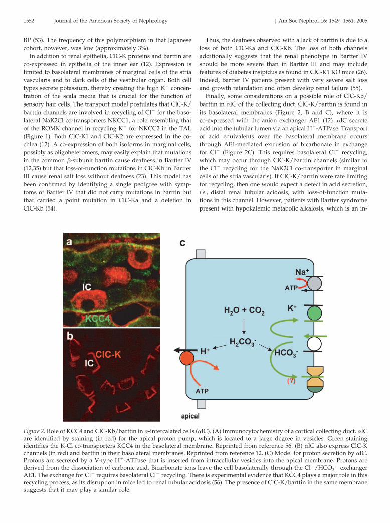
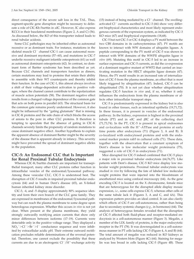
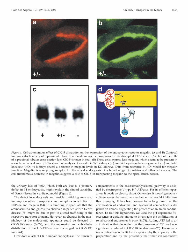
Mice disrupted for ClC-5 indeed confirmed that ClC-5 playsa major role in proximal tubular endocytosis (64,76,77). Likepatients with Dent’s disease, ClC-5 KO mice display low mo-lecular weight proteinuria. Proximal tubular endocytosis wasstudied in vivo by following the fate of labeled low molecularweight proteins that were injected into the bloodstream ofanesthetized mice using confocal microscopy (64). As the geneencoding ClC-5 is located on the X chromosome, female micethat are heterozygous for the disrupted allele display mosaicexpression, i.e., some cells express ClC-5, whereas other cells ofthe same tubule lack it (Figures 3, A and C, and 4A). Thisexpression pattern provides an ideal control. It can also clarifywhich effects of ClC-5 are cell-autonomous, rather than beingdue to secondary systemic changes (e.g., in hormones) (64). Theanalysis of heterozygous females revealed that the disruptionof ClC-5 affected both fluid-phase and receptor-mediated en-docytosis in a cell-autonomous manner (Figure 3). Megalin, amember of the LDL family of proteins, is the major endocytoticreceptor in the PT (78). It was downregulated in a cell-autono-mous manner in PT cells lacking ClC-5 (Figure 4, A and B) (64).The total amount of megalin was reduced in KO kidneys asanalyzed by Western blots (Figure 4C) (64). Staining for mega-lin was less broad in cells lacking ClC-5 (Figure 4B). These
J Am Soc Nephrol 16: 1549–1561, 2005 Chloride Transport in the Kidney 1553
results suggested that ClC-5 might play a role in recyclingmegalin back to the apical brush border membrane (Figure 4D)and may also explain the decreased amount of megalin in theurine of patients with Dent’s disease (64,76). The decrease ofmegalin (and of its co-receptor cubulin) was confirmed later byelectron microscopy and cell fractionation (79). The cell-auton-omous decrease in megalin (64) suggests that receptor-medi-ated endocytosis is more severely impaired by a lack of ClC-5than fluid-phase endocytosis (76).
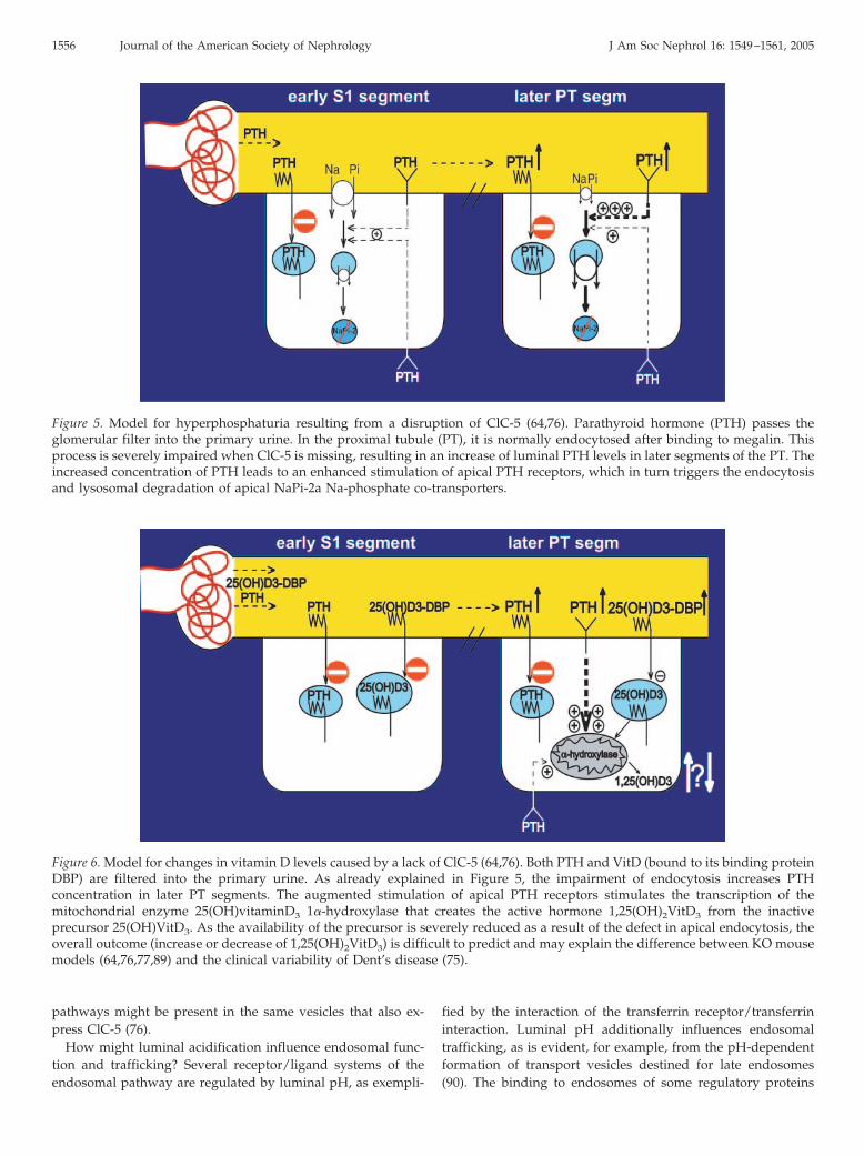
The disturbances in renal phosphate and calcium handlingand eventually the kidney stones in Dent’s disease might besecondary symptoms of the impaired renal tubular endocytosisand metabolism of calciotropic hormones (64,76). Parathyroidhormone (PTH) and VitD (bound to its binding protein) areendocytosed in the PT in a megalin-dependent manner (80,81).The defective endocytosis upon disruption of ClC-5 hence ispredicted to lead to a loss of PTH into the urine, as is indeedobserved in ClC-5 KO mice (64) and in patients with Dent’sdisease (82). The urinary loss of PTH is unlikely to have any
physiologic effect per se, as this peptide hormone would un-dergo lysosomal degradation after proximal tubular endocyto-sis. However, the decreased endocytosis will lead to increasedluminal concentration of PTH in later segments of the PT,which will result in an enhanced stimulation (64) of apical PTHreceptors in proximal tubular cells (Figure 5) (83,84). This, inturn, will stimulate the endocytosis of the apical Na-coupledphosphate transporter NaPi-2a (85), which is responsible forthe bulk of phosphate reabsorption in the PT. Consistent withthis hypothesis, immunocytochemistry revealed that NaPi-2awas found predominantly in intracellular vesicles of ClC-5 KOkidneys, whereas it was expressed prominently in the brushborder of WT PT (64). Although difficult to quantify, NaPi-2alocalization was not changed in early segments of the PT. Thisis expected as shortly after glomerular filtration, the luminalconcentration of PTH approximates that found in serum. Incontrast to the effect of ClC-5 disruption on endocytosis andmegalin localization, analysis of heterozygous females indi-cated that the change in localization of NaPi-2a was not cell-autonomous (64). This finding is consistent with the changes ofluminal PTH concentrations. The decrease in apical NaPi-2acan fully explain the hyperphosphaturia of ClC-5 mice (64) andof patients with Dent’s disease. One may argue, however, thatthe lack of ClC-5 might also impair the endocytosis of NaPi-2a.It therefore was essential to perform control experiments thatshowed that PTH-induced endocytosis of NaPi-2a was slowedbut not abolished in ClC-5 KO mice (64).
Another important effect of PTH on proximal tubular cells isthe transcriptional stimulation of 25(OH)vitaminD3 1�-hydrox-ylase (86,87), the mitochondrial enzyme that converts the inac-tive precursor 25(OH)-VitD3 into the active hormone1,25(OH)2-VitD3 (Figure 6). As predicted by increased luminalPTH concentrations (64), 25(OH)vitaminD3 1�-hydroxylasemRNA levels were markedly elevated in ClC-5 KO mice (76).The ensuing increase in enzymatic activity correlated with anincreased ratio of 1,25(OH)2-VitD3/25(OH)-VitD3 in the serumof KO mice (64,76). However, the absolute serum concentrationof the active hormone 1,25(OH)2-VitD3 was decreased as aconsequence of a severe loss of VitD together with its bindingprotein into the urine. This complex is normally endocytosed inthe PT in a megalin-dependent manner (81).
Thus, there are two opposing effects of the impaired en-docytosis in ClC-5 KO mice on serum 1,25(OH)2-VitD3 levels:The ensuing elevated luminal levels of PTH increase theamount of the enzyme that generates the active hormone1,25(OH)2-VitD3, whereas the decrease of apical endocytosisof 25(OH)-VitD3 severely limits the availability of its precur-sor. Because 1,25(OH)2-VitD3 stimulates intestinal Ca2� ab-sorption, an increase in its serum concentration might indi-rectly lead to hypercalciuria and kidney stones. Indeed, mostpatients with Dent’s disease have moderately elevated levelsof this hormone (75,88). In contrast, our ClC-5 KO mice havedecreased levels of 1,25(OH)2-VitD3 and do not display hy-percalciuria and kidney stones (64). An independent ClC-5KO mouse model, however, presents with hypercalciuria(77) and has elevated levels of 1,25(OH)2-VitD3 (89). Hence,the balance between the stimulation of 1�-hydroxylase and
Figure 3. Cell-autonomous effects of disrupting ClC-5 on recep-tor-mediated endocytosis (A and B) and fluid-phase endocyto-sis (C and D). Female mice that are heterozygous for the dis-rupted ClC-5 allele express ClC-5 in some cells of the proximaltubule, whereas others lack ClC-5, as shown in A and C byconfocal microscopy (ClC-5 is shown in red). The cells lackingClC-5 are indicated by the arrows. Endocytosis was investi-gated by injecting into mice lactoglobulin that was labeled witha fluorescent dye to test for receptor-mediated endocytosis (B)or FITC-dextrane as a fluid-phase marker (D). Kidneys werefixed by perfusion a few minutes after the tracer was injected.These experiments revealed a cell-autonomous defect in bothreceptor-mediated and fluid-phase endocytosis. Reprintedfrom reference 64.
1554 Journal of the American Society of Nephrology J Am Soc Nephrol 16: 1549–1561, 2005
the urinary loss of VitD, which both are due to a primarydefect in PT endocytosis, might explain the clinical variabilityof Dent’s disease in a unifying model (Figure 6).
The defect in endocytosis and vesicle trafficking may alsoimpinge on other transporters and receptors in addition toNaPi-2a and megalin (64). It is tempting to speculate that theaminoaciduria and glucosuria observed in patients with Dent’sdisease (75) might be due in part to altered trafficking of therespective transport proteins. However, no changes in the mor-phology of the endocytotic apparatus could be detected inClC-5 KO mice (64,79), and the expression and subcellulardistribution of the H�-ATPase was unchanged in ClC-5 KOmice (64).
How does a lack of ClC-5 impair endocytosis? The lumen of
compartments of the endosomal/lysosomal pathway is acidi-fied by electrogenic V-type H�-ATPases. For its efficient oper-ation, it needs an electric shunt. Otherwise, it would generate avoltage across the vesicular membrane that would inhibit fur-ther pumping. It has been known for a long time that theacidification of endosomal and lysosomal compartments de-pends on anions, suggesting the presence of an anion conduc-tance. To test this hypothesis, we used the pH-dependent flu-orescence of acridine orange to investigate the acidification ofrenal cortical endosomes in vitro (64,76). Adding ATP led to anacidification that depended on the presence of Cl� and wassignificantly reduced in ClC-5 KO endosomes (76). The remain-ing acidification in the KO was explained by the impurity of thepreparation and by the possibility that other ion-conductive
Figure 4. Cell-autonomous effect of ClC-5 disruption on the expression of the endocytotic receptor megalin. (A and B) Confocalimmunocytochemistry of a proximal tubule of a female mouse heterozygous for the disrupted ClC-5 allele. (A) Half of the cellsof a proximal tubular cross-section lack ClC-5 (shown in red). (B) These cells express less megalin, which seems to be present ina less broad apical area. (C) Western blot analysis of megalin in WT kidneys (�) and kidneys from heterozygous (�/�) and totalknockout (KO; �) kidneys reveal a decrease in megalin levels in KO kidneys. Data from reference 64. (D) Model for megalinfunction. Megalin is a recycling receptor for the apical endocytosis of a broad range of proteins and other substances. Thecell-autonomous decrease in megalin suggests a role of ClC-5 in transporting megalin to the apical brush border.
J Am Soc Nephrol 16: 1549–1561, 2005 Chloride Transport in the Kidney 1555
pathways might be present in the same vesicles that also ex-press ClC-5 (76).
How might luminal acidification influence endosomal func-tion and trafficking? Several receptor/ligand systems of theendosomal pathway are regulated by luminal pH, as exempli-
fied by the interaction of the transferrin receptor/transferrininteraction. Luminal pH additionally influences endosomaltrafficking, as is evident, for example, from the pH-dependentformation of transport vesicles destined for late endosomes(90). The binding to endosomes of some regulatory proteins
Figure 5. Model for hyperphosphaturia resulting from a disruption of ClC-5 (64,76). Parathyroid hormone (PTH) passes theglomerular filter into the primary urine. In the proximal tubule (PT), it is normally endocytosed after binding to megalin. Thisprocess is severely impaired when ClC-5 is missing, resulting in an increase of luminal PTH levels in later segments of the PT. Theincreased concentration of PTH leads to an enhanced stimulation of apical PTH receptors, which in turn triggers the endocytosisand lysosomal degradation of apical NaPi-2a Na-phosphate co-transporters.
Figure 6. Model for changes in vitamin D levels caused by a lack of ClC-5 (64,76). Both PTH and VitD (bound to its binding proteinDBP) are filtered into the primary urine. As already explained in Figure 5, the impairment of endocytosis increases PTHconcentration in later PT segments. The augmented stimulation of apical PTH receptors stimulates the transcription of themitochondrial enzyme 25(OH)vitaminD3 1�-hydroxylase that creates the active hormone 1,25(OH)2VitD3 from the inactiveprecursor 25(OH)VitD3. As the availability of the precursor is severely reduced as a result of the defect in apical endocytosis, theoverall outcome (increase or decrease of 1,25(OH)2VitD3) is difficult to predict and may explain the difference between KO mousemodels (64,76,77,89) and the clinical variability of Dent’s disease (75).
1556 Journal of the American Society of Nephrology J Am Soc Nephrol 16: 1549–1561, 2005

that are involved in vesicle transport, such as �COP (90) andARF6/ARNO (91), depends on the luminal pH of this compart-ment.
The role of ClC-5 in acidifying endosomes is similar to that ofother vesicular CLC. For instance, ClC-3 is involved in acidify-ing synaptic vesicles (67) and endosomes (92). ClC-7 is impor-tant for the acidification of the resorption lacuna of osteoclasts(61). Surprisingly, although ClC-7 is expressed on lysosomes,no change in steady-state lysosomal pH was detected (93). Onthe basis of more indirect evidence, ClC-4 has also been impli-cated in endosomal acidification (11,94). Although ClC-3, -4,and -7 all are expressed in the kidney, none of the respectiveKO has a defect in renal proximal tubular endocytosis (61,67)(Schaffer and Jentsch, unpublished observations for ClC-4 KOmice). However, the disruption of ClC-7 leads to lysosomalstorage disease in the central nervous system and in proximaltubular cells (93), supporting the importance of ClC-7 for theendosomal/lysosomal pathway.
In addition to the H�-ATPase, the sodium proton exchangerhas been suggested to acidify endosomes and to have a role inendocytosis (95,96). As endosomes have a high luminal [Na�]shortly after pinching off from the plasma membrane, the Na�
gradient across endosomal membranes would be suited to acid-ify their lumen. However, the PTH-stimulated endocytosis ofNHE3-containing vesicles was significantly slowed in ClC-5KO PT cells, demonstrating the predominant role of ClC-5 inendocytosis (64).
Renal Roles of KCC ElectroneutralPotassium-Chloride Co-Transporters
Electroneutral K-Cl co-transporters belong to the superfamilyof electroneutral cation-chloride co-transporters (which alsoincludes, e.g., NKCC1 and NKCC2) and are encoded by fourgenes (KCC1 through KCC4) (97). Whereas KCC1 is broadlyexpressed, KCC2 is almost exclusively expressed in neurons. Inaddition to KCC1, the kidney expresses KCC3 and KCC4. Bothlatter isoforms are also expressed in several other tissues. Thisincludes the brain for KCC3 but not for KCC4.
K-Cl co-transport has been implicated in transepithelialtransport, cell volume regulation, and the lowering of intracel-lular Cl� in neurons. To differentiate between the functions ofthe different isoforms, we disrupted the genes encoding KCC2,-3, and -4 in mice and analyzed their phenotypes (56,98,99).Mice lacking KCC2 died postnatally as a result of a disturbancein GABAergic neuronal inhibition that resulted from an in-crease in the intraneuronal Cl� concentration (99). The KO ofeither KCC3 (98) or KCC4 (56) resulted in deafness, which wastentatively attributed to an impairment of K� recycling in theinner ear. The disruption of KCC3 additionally led to severecentral and peripheral neurodegeneration (98), which corre-lated well with the pathology of human Anderman syndromethat is due to loss-of-function mutations in the human geneencoding KCC3 (100).
In addition to those phenotypes, both KCC3 and KCC4 KOmice displayed renal abnormalities. In the kidney, KCC3 wasdetected exclusively in basolateral membranes of the PT (98).KCC4 was co-expressed with KCC3 in these membranes and
was additionally found in IC of the collecting duct (Figure 2A)(56). As discussed above in the section on ClC-K/barttin Cl�
channels, acid-secreting �IC need to recycle Cl� ions that aretaken up through basolateral AE1 Cl�/HCO3
� exchangers(Figure 2C) (56). KCC4 may be better suited for this purposethan ClC-Kb/barttin Cl� channels, because the coupling to K�
will lead to lower intracellular Cl�. An inhibition of basolateralHCO3
� transport by impaired Cl� recycling should lead to lessH� secretion by the apical proton pump. KCC4 KO mice indeeddisplayed renal tubular acidosis, as indicated by an alkaline pHof the urine and a decrease in blood base excess (56). In supportof an impairment of basolateral Cl� exit, electron microprobeanalysis of intracellular electrolytes revealed an increase inintracellular Cl� concentration in IC (56). It will be interestingto test experimentally whether and to which extent recyclingthrough ClC-K/barttin channels takes place in these cells.
No overt renal phenotype was detected in KCC3 KO mice.These mice display hypertension (98), but the apparently ex-clusive expression of KCC3 in PT makes a renal origin of thisphenotype unlikely. Isolated perfused PT were used to assessthe role of KCC in cell volume regulation. The KO of KCC4 and,to a lesser extent, of KCC3 partially inhibited the regulatoryvolume decrease of PT cells (98). PT cells might need efficientmechanism for cell volume regulation as different loads ofsolutes that are reabsorbed by these cells (e.g., amino acids,glucose) might lead to osmotic changes. The disruption ofneither transporter, however, led to significant renal salt orwater loss. It remains to be seen whether KCC1, which may beexpressed in most nephron segments (42), plays a crucial role intransepithelial transport.
Summary and OutlookBridging the gap between genes and integrative physiology
by targeted gene disruption in mice, as well as by analyzinghuman inherited disease, has dramatically furthered our un-derstanding of renal physiology and pathology. Increasinglysophisticated approaches to gene targeting, combined with arevival of “classical” methods of renal physiology and withmorphologic techniques, will undoubtedly provide us withexciting new insights into the function of the kidney, a fasci-nating organ that serves as a paradigm for transport physiol-ogy.
AcknowledgmentsThe part of our work summarized here was supported by the Deut-
sche Forschungsgemeinschaft and the NGFN program of theBundesministerium fur Bildung und Forschung.
I thank my co-workers for their enthusiasm and hard work, withoutwhich our projects would not have flourished.
References1. Jentsch TJ, Steinmeyer K, Schwarz G: Primary structure of
Torpedo marmorata chloride channel isolated by expressioncloning in Xenopus oocytes. Nature 348: 510–514, 1990
2. Steinmeyer K, Ortland C, Jentsch TJ: Primary structure andfunctional expression of a developmentally regulated skel-etal muscle chloride channel. Nature 354: 301–304, 1991
J Am Soc Nephrol 16: 1549–1561, 2005 Chloride Transport in the Kidney 1557
3. Miller C: Open-state substructure of single chloride chan-nels from Torpedo electroplax. Philos Trans R Soc Lond B BiolSci 299: 401–411, 1982
4. Ludewig U, Pusch M, Jentsch TJ: Two physically distinctpores in the dimeric ClC-0 chloride channel. Nature 383:340–343, 1996
5. Middleton RE, Pheasant DJ, Miller C: Homodimeric archi-tecture of a ClC-type chloride ion channel. Nature 383:337–340, 1996
6. Weinreich F, Jentsch TJ: Pores formed by single subunits inmixed dimers of different CLC chloride channels. J BiolChem 276: 2347–2353, 2001
7. Dutzler R, Campbell EB, Cadene M, Chait BT, MacKinnonR: X-ray structure of a ClC chloride channel at 3.0 A revealsthe molecular basis of anion selectivity. Nature 415: 287–294, 2002
8. Dutzler R, Campbell EB, MacKinnon R: Gating the selec-tivity filter in ClC chloride channels. Science 300: 108–112,2003
9. Mindell JA, Maduke M, Miller C, Grigorieff N: Projectionstructure of a ClC-type chloride channel at 6.5 A resolu-tion. Nature 409: 219–223, 2001
10. Lorenz C, Pusch M, Jentsch TJ: Heteromultimeric CLCchloride channels with novel properties. Proc Natl Acad SciU S A 93: 13362–13366, 1996
11. Mohammad-Panah R, Harrison R, Dhani S, Ackerley C,Huan LJ, Wang Y, Bear CE: The chloride channel ClC-4contributes to endosomal acidification and trafficking.J Biol Chem 278: 29267–29277, 2003
12. Estevez R, Boettger T, Stein V, Birkenhager R, Otto M,Hildebrandt F, Jentsch TJ: Barttin is a Cl�-channel beta-subunit crucial for renal Cl�-reabsorption and inner earK�-secretion. Nature 414: 558–561, 2001
13. Waldegger S, Jentsch TJ: Functional and structural analysisof ClC-K chloride channels involved in renal disease. J BiolChem 275: 24527–24533, 2000
14. Pusch M, Ludewig U, Rehfeldt A, Jentsch TJ: Gating of thevoltage-dependent chloride channel ClC-0 by the per-meant anion. Nature 373: 527–531, 1995
15. Accardi A, Miller C: Secondary active transport mediatedby a prokaryotic homologue of ClC Cl� channels. Nature427: 803–807, 2004
16. Brandt S, Jentsch TJ: ClC-6 and ClC-7 are two novelbroadly expressed members of the CLC chloride channelfamily. FEBS Lett 377: 15–20, 1995
17. Friedrich T, Breiderhoff T, Jentsch TJ: Mutational analysisdemonstrates that ClC-4 and ClC-5 directly mediateplasma membrane currents. J Biol Chem 274: 896–902, 1999
18. Li X, Shimada K, Showalter LA, Weinman SA: Biophysicalproperties of ClC-3 differentiate it from swelling- activatedchloride channels in Chinese hamster ovary-K1 cells. J BiolChem 275: 35994–35998, 2000
19. Steinmeyer K, Schwappach B, Bens M, Vandewalle A,Jentsch TJ: Cloning and functional expression of rat CLC-5,a chloride channel related to kidney disease. J Biol Chem270: 31172–31177, 1995
20. Kieferle S, Fong P, Bens M, Vandewalle A, Jentsch TJ: Twohighly homologous members of the ClC chloride channelfamily in both rat and human kidney. Proc Natl Acad SciU S A 91: 6943–6947, 1994
21. Uchida S, Sasaki S, Furukawa T, Hiraoka M, Imai T, HirataY, Marumo F: Molecular cloning of a chloride channel that
is regulated by dehydration and expressed predominantlyin kidney medulla. J Biol Chem 268: 3821–3824, 1993 [pub-lished erratum appears in J Biol Chem 269: 19192, 1994]
22. Sage CL, Marcus DC: Immunolocalization of ClC-K chlo-ride channel in strial marginal cells and vestibular darkcells. Hear Res 160: 1–9, 2001
23. Simon DB, Bindra RS, Mansfield TA, Nelson-Williams C,Mendonca E, Stone R, Schurman S, Nayir A, Alpay H,Bakkaloglu A, Rodriguez-Soriano J, Morales JM, SanjadSA, Taylor CM, Pilz D, Brem A, Trachtman H, Griswold W,Richard GA, John E, Lifton RP: Mutations in the chloridechannel gene, CLCNKB, cause Bartter’s syndrome type III.Nat Genet 17: 171–178, 1997
24. Adachi S, Uchida S, Ito H, Hata M, Hiroe M, Marumo F,Sasaki S: Two isoforms of a chloride channel predomi-nantly expressed in thick ascending limb of Henle’s loopand collecting ducts of rat kidney. J Biol Chem 269: 17677–17683, 1994
25. Uchida S, Sasaki S, Nitta K, Uchida K, Horita S, Nihei H,Marumo F: Localization and functional characterization ofrat kidney-specific chloride channel, ClC-K1. J Clin Invest95: 104–113, 1995
26. Matsumura Y, Uchida S, Kondo Y, Miyazaki H, Ko SB,Hayama A, Morimoto T, Liu W, Arisawa M, Sasaki S,Marumo F: Overt nephrogenic diabetes insipidus in micelacking the CLC-K1 chloride channel. Nat Genet 21: 95–98,1999
27. Nissant A, Lourdel S, Baillet S, Paulais M, Marvao P,Teulon J, Imbert-Teboul M: Heterogeneous distribution ofchloride channels along the distal convoluted tubuleprobed by single-cell RT-PCR and patch clamp. Am JPhysiol Renal Physiol 287: F1233–F1243, 2004
28. Yoshikawa M, Uchida S, Yamauchi A, Miyai A, Tanaka Y,Sasaki S, Marumo F: Localization of rat CLC-K2 chloridechannel mRNA in the kidney. Am J Physiol 276: F552–F558,1999
29. Mejia R, Wade JB: Immunomorphometric study of rat renalinner medulla. Am J Physiol Renal Physiol 282: F553–F557,2002
30. Vandewalle A, Cluzeaud F, Bens M, Kieferle S, SteinmeyerK, Jentsch TJ: Localization and induction by dehydration ofClC-K chloride channels in the rat kidney. Am J Physiol 272:F678–F688, 1997
31. Kobayashi K, Uchida S, Mizutani S, Sasaki S, Marumo F:Intrarenal and cellular localization of CLC-K2 protein inthe mouse kidney. J Am Soc Nephrol 12: 1327–1334, 2001
32. Kobayashi K, Uchida S, Okamura HO, Marumo F, Sasaki S:Human CLC-KB gene promoter drives the EGFP expres-sion in the specific distal nephron segments and inner ear.J Am Soc Nephrol 13: 1992–1998, 2002
33. Simon DB, Karet FE, Hamdan JM, DiPietro A, Sanjad SA,Lifton RP: Bartter’s syndrome, hypokalaemic alkalosiswith hypercalciuria, is caused by mutations in the Na-K-2Cl cotransporter NKCC2. Nat Genet 13: 183–188, 1996
34. Simon DB, Karet FE, Rodriguez-Soriano J, Hamdan JH,DiPietro A, Trachtman H, Sanjad SA, Lifton RP: Geneticheterogeneity of Bartter’s syndrome revealed by mutationsin the K� channel, ROMK. Nat Genet 14: 152–156, 1996
35. Birkenhager R, Otto E, Schurmann MJ, Vollmer M, Ruf EM,Maier-Lutz I, Beekmann F, Fekete A, Omran H, FeldmannD, Milford DV, Jeck N, Konrad M, Landau D, KnoersNVAM, Antignac C, Sudbrack R, Kispert A, Hildebrandt F:
1558 Journal of the American Society of Nephrology J Am Soc Nephrol 16: 1549–1561, 2005
Mutation of BSND causes Bartter syndrome with sensori-neural deafness and kidney failure. Nat Genet 29: 310–314,2001
36. Vargas-Poussou R, Huang C, Hulin P, Houillier P, Jeun-emaitre X, Paillard M, Planelles G, Dechaux M, Miller RT,Antignac C: Functional characterization of a calcium-sens-ing receptor mutation in severe autosomal dominant hy-pocalcemia with a Bartter-like syndrome. J Am Soc Nephrol13: 2259–2266, 2002
37. Watanabe S, Fukumoto S, Chang H, Takeuchi Y, HasegawaY, Okazaki R, Chikatsu N, Fujita T: Association betweenactivating mutations of calcium-sensing receptor and Bar-tter’s syndrome. Lancet 360: 692–694, 2002
38. Fukuyama S, Hiramatsu M, Akagi M, Higa M, Ohta T:Novel mutations of the chloride channel Kb gene in twoJapanese patients clinically diagnosed as Bartter syndromewith hypocalciuria. J. Clin Endocrinol Metab 89: 5847–5850,2004
39. Jeck N, Konrad M, Peters M, Weber S, Bonzel KE, SeyberthHW: Mutations in the chloride channel gene, CLCNKB,leading to a mixed Bartter-Gitelman phenotype. Pediatr Res48: 754–758, 2000
40. Zelikovic I, Szargel R, Hawash A, Labay V, Hatib I, CohenN, Nakhoul F: A novel mutation in the chloride channelgene, CLCNKB, as a cause of Gitelman and Bartter syn-dromes. Kidney Int 63: 24–32, 2003
41. Simon DB, Nelson-Williams C, Bia MJ, Ellison D, Karet FE,Molina AM, Vaara I, Iwata F, Cushner HM, Koolen M,Gainza FJ, Gitleman HJ, Lifton RP: Gitelman’s variant ofBartter’s syndrome, inherited hypokalaemic alkalosis, iscaused by mutations in the thiazide-sensitive Na-Cl co-transporter. Nat Genet 12: 24–30, 1996
42. Liapis H, Nag M, Kaji DM: K-Cl cotransporter expressionin the human kidney. Am J Physiol 275: C1432–C1437, 1998
43. Liu W, Morimoto T, Kondo Y, Iinuma K, Uchida S, SasakiS, Marumo F, Imai M: Analysis of NaCl transport in thinascending limb of Henle’s loop in CLC-K1 null mice. Am JPhysiol Renal Physiol 282: F451–F457, 2002
44. Akizuki N, Uchida S, Sasaki S, Marumo F: Impaired soluteaccumulation in inner medulla of Clcnk1�/� mice kidney.Am J Physiol 280: F79–F87, 2001
45. Waldegger S, Jeck N, Barth P, Peters M, Vitzthum H, WolfK, Kurtz A, Konrad M, Seyberth HW: Barttin increasessurface expression and changes current properties ofClC-K channels. Pflugers Arch 444: 411–418, 2002
46. Lourdel S, Paulais M, Marvao P, Nissant A, Teulon J: Achloride channel at the basolateral membrane of the distal-convoluted tubule: a candidate ClC-K channel. J GenPhysiol 121: 287–300, 2003
47. Schwake M, Friedrich T, Jentsch TJ: An internalizationsignal in ClC-5, an endosomal Cl�-channel mutated inDent’s disease. J Biol Chem 276: 12049–12054, 2001
48. Staub O, Dho S, Henry P, Correa J, Ishikawa T, McGlade J,Rotin D: WW domains of Nedd4 bind to the proline-richPY motifs in the epithelial Na� channel deleted in Liddle’ssyndrome. EMBO J 15: 2371–2380, 1996
49. Embark HM, Bohmer C, Palmada M, Rajamanickam J,Wyatt AW, Wallisch S, Capasso G, Waldegger P, SeyberthHW, Waldegger S, Lang F: Regulation of CLC-Ka/barttinby the ubiquitin ligase Nedd4–2 and the serum- and glu-cocorticoid-dependent kinases. Kidney Int 66: 1918–1925,2004
50. Jeck N, Waldegger P, Doroszewicz J, Seyberth H, Waldeg-ger S: A common sequence variation of the CLCNKB genestrongly activates ClC-Kb chloride channel activity. KidneyInt 65: 190–197, 2004
51. Jeck N, Waldegger S, Lampert A, Boehmer C, Waldegger P,Lang PA, Wissinger B, Friedrich B, Risler T, Moehle R,Lang UE, Zill P, Bondy B, Schaeffeler E, Asante-Poku S,Seyberth H, Schwab M, Lang F: Activating mutation of therenal epithelial chloride channel ClC-Kb predisposing tohypertension. Hypertension 43: 1175–1181, 2004
52. Geller DS: A genetic predisposition to hypertension? Hy-pertension 44: 27–28, 2004
53. Kokubo Y, Iwai N, Tago N, Inamoto N, Okayama A,Yamawaki H, Naraba H, Tomoike H: Association analysisbetween hypertension and CYBA, CLCNKB, and KCNMB1functional polymorphisms in the Japanese population. CircJ 69: 138–142, 2005
54. Schlingmann KP, Konrad M, Jeck N, Waldegger P, Reinal-ter SC, Holder M, Seyberth HW, Waldegger S: Salt wastingand deafness resulting from mutations in two chloridechannels. N Engl J Med 350: 1314–1319, 2004
55. Jeck N, Reinalter SC, Henne T, Marg W, Mallmann R, PaselK, Vollmer M, Klaus G, Leonhardt A, Seyberth HW, Kon-rad M: Hypokalemic salt-losing tubulopathy with chronicrenal failure and sensorineural deafness. Pediatrics 108: E5,2001
56. Boettger T, Hubner C, Maier H, Rust MB, Beck FX, JentschTJ: Deafness and renal tubular acidosis in mice lacking theK-Cl cotransporter Kcc4. Nature 416: 874–878, 2002
57. Koch MC, Steinmeyer K, Lorenz C, Ricker K, Wolf F, OttoM, Zoll B, Lehmann-Horn F, Grzeschik KH, Jentsch TJ: Theskeletal muscle chloride channel in dominant and recessivehuman myotonia. Science 257: 797–800, 1992
58. Pusch M, Steinmeyer K, Koch MC, Jentsch TJ: Mutations indominant human myotonia congenita drastically alter thevoltage dependence of the CIC-1 chloride channel. Neuron15: 1455–1463, 1995
59. Steinmeyer K, Klocke R, Ortland C, Gronemeier M, Jock-usch H, Grunder S, Jentsch TJ: Inactivation of muscle chlo-ride channel by transposon insertion in myotonic mice.Nature 354: 304–308, 1991
60. Steinmeyer K, Lorenz C, Pusch M, Koch MC, Jentsch TJ:Multimeric structure of ClC-1 chloride channel revealed bymutations in dominant myotonia congenita (Thomsen).EMBO J 13: 737–743, 1994
61. Kornak U, Kasper D, Bosl MR, Kaiser E, Schweizer M,Schulz A, Friedrich W, Delling G, Jentsch TJ: Loss of theClC-7 chloride channel leads to osteopetrosis in mice andman. Cell 104: 205–215, 2001
62. Cleiren E, Benichou O, Van Hul E, Gram J, Bollerslev J,Singer FR, Beaverson K, Aledo A, Whyte MP, Yoneyama T,deVernejoul MC, Van Hul W: Albers-Schonberg disease(autosomal dominant osteopetrosis, type II) results frommutations in the ClCN7 chloride channel gene. Hum MolGenet 10: 2861–2867, 2001
63. Saviane C, Conti F, Pusch M: The muscle chloride channelClC-1 has a double-barreled appearance that is differen-tially affected in dominant and recessive myotonia. J GenPhysiol 113: 457–468, 1999
64. Piwon N, Gunther W, Schwake R, Bosl MR, Jentsch TJ:ClC-5 Cl�-channel disruption impairs endocytosis in amouse model for Dent’s disease. Nature 408: 369–373, 2000
J Am Soc Nephrol 16: 1549–1561, 2005 Chloride Transport in the Kidney 1559

65. Lloyd SE, Pearce SH, Fisher SE, Steinmeyer K, SchwappachB, Scheinman SJ, Harding B, Bolino A, Devoto M, GoodyerP, Rigden SP, Wrong O, Jentsch TJ, Craig IW, Thakker RV:A common molecular basis for three inherited kidneystone diseases. Nature 379: 445–449, 1996
66. Duan D, Winter C, Cowley S, Hume JR, Horowitz B: Mo-lecular identification of a volume-regulated chloride chan-nel. Nature 390: 417–421, 1997
67. Stobrawa SM, Breiderhoff T, Takamori S, Engel D,Schweizer M, Zdebik AA, Bosl MR, Ruether K, Jahn H,Draguhn A, Jahn R, Jentsch TJ: Disruption of ClC-3, achloride channel expressed on synaptic vesicles, leads to aloss of the hippocampus. Neuron 29: 185–196, 2001
68. Li X, Wang T, Zhao Z, Weinman SA: The ClC-3 chloridechannel promotes acidification of lysosomes in CHO-K1and Huh-7 cells. Am J Physiol Cell Physiol 282: C1483–C1491, 2002
69. Pirozzi G, McConnell SJ, Uveges AJ, Carter JM, Sparks AB,Kay BK, Fowlkes DM: Identification of novel human WWdomain-containing proteins by cloning of ligand targets.J Biol Chem 272: 14611–14616, 1997
70. Hryciw DH, Ekberg J, Lee A, Lensink IL, Kumar S, Gug-gino WB, Cook DI, Pollock CA, Poronnik P: Nedd4–2functionally interacts with ClC-5: Involvement in constitu-tive albumin endocytosis in proximal tubule cells. J BiolChem 279: 54996–55007, 2004
71. Gunther W, Luchow A, Cluzeaud F, Vandewalle A, JentschTJ: ClC-5, the chloride channel mutated in Dent’s disease,colocalizes with the proton pump in endocytotically activekidney cells. Proc Natl Acad Sci U S A 95: 8075–8080, 1998
72. Vandewalle A, Cluzeaud F, Peng KC, Bens M, Luchow A,Gunther W, Jentsch TJ: Tissue distribution and subcellularlocalization of the ClC-5 chloride channel in rat intestinalcells. Am J Physiol Cell Physiol 280: C373–C381, 2001
73. Devuyst O, Christie PT, Courtoy PJ, Beauwens R, ThakkerRV: Intra-renal and subcellular distribution of the humanchloride channel, CLC-5, reveals a pathophysiological ba-sis for Dent’s disease. Hum Mol Genet 8: 247–257, 1999
74. Sakamoto H, Sado Y, Naito I, Kwon TH, Inoue S, Endo K,Kawasaki M, Uchida S, Nielsen S, Sasaki S, Marumo F:Cellular and subcellular immunolocalization of ClC-5channel in mouse kidney: Colocalization with H�-ATPase.Am J Physiol 277: F957–F965, 1999
75. Wrong OM, Norden AG, Feest TG: Dent’s disease; a famil-ial proximal renal tubular syndrome with low-molecular-weight proteinuria, hypercalciuria, nephrocalcinosis, met-abolic bone disease, progressive renal failure and a markedmale predominance. QJM 87: 473–493, 1994
76. Gunther W, Piwon N, Jentsch TJ: The ClC-5 chloride chan-nel knock-out mouse—An animal model for Dent’s dis-ease. Pflugers Arch 445: 456–462, 2003
77. Wang SS, Devuyst O, Courtoy PJ, Wang XT, Wang H,Wang Y, Thakker RV, Guggino S, Guggino WB: Mice lack-ing renal chloride channel, CLC-5, are a model for Dent’sdisease, a nephrolithiasis disorder associated with defec-tive receptor-mediated endocytosis. Hum Mol Genet 9:2937–2945, 2000
78. Leheste JR, Rolinski B, Vorum H, Hilpert J, Nykjaer A,Jacobsen C, Aucouturier P, Moskaug JO, Otto A, Chris-tensen EI, Willnow TE: Megalin knockout mice as an ani-mal model of low molecular weight proteinuria. Am JPathol 155: 1361–1370, 1999
79. Christensen EI, Devuyst O, Dom G, Nielsen R, Van derSmissen P, Verroust P, Leruth M, Guggino WB, Courtoy PJ:Loss of chloride channel ClC-5 impairs endocytosis bydefective trafficking of megalin and cubilin in kidney prox-imal tubules. Proc Natl Acad Sci U S A 100: 8472–8477, 2003
80. Hilpert J, Nykjaer A, Jacobsen C, Wallukat G, Nielsen R,Moestrup SK, Haller H, Luft FC, Christensen EI, WillnowTE: Megalin antagonizes activation of the parathyroid hor-mone receptor. J Biol Chem 274: 5620–5625, 1999
81. Nykjaer A, Dragun D, Walther D, Vorum H, Jacobsen C,Herz J, Melsen F, Christensen EI, Willnow TE: An endo-cytic pathway essential for renal uptake and activation ofthe steroid 25-(OH) vitamin D3. Cell 96: 507–515, 1999
82. Norden AG, Lapsley M, Lee PJ, Pusey CD, Scheinman SJ,Tam FW, Thakker RV, Unwin RJ, Wrong O: Glomerularprotein sieving and implications for renal failure in Fan-coni syndrome. Kidney Int 60: 1885–1892, 2001
83. Kaufmann M, Muff R, Stieger B, Biber J, Murer H, FischerJA: Apical and basolateral parathyroid hormone receptorsin rat renal cortical membranes. Endocrinology 134: 1173–1178, 1994
84. Traebert M, Volkl H, Biber J, Murer H, Kaissling B: Lumi-nal and contraluminal action of 1–34 and 3–34 PTH pep-tides on renal type IIa Na-Pi cotransporter. Am J Physiol278: F792–F798, 2000
85. Murer H, Forster I, Hernando N, Lambert G, Traebert M,Biber J: Posttranscriptional regulation of the proximal tu-bule NaPi-II transporter in response to PTH and dietary Pi.Am J Physiol 277: F676–F684, 1999
86. Brenza HL, Kimmel-Jehan C, Jehan F, Shinki T, Wakino S,Anazawa H, Suda T, DeLuca HF: Parathyroid hormoneactivation of the 25-hydroxyvitamin D3-1�-hydroxylasegene promoter. Proc Natl Acad Sci U S A 95: 1387–1391,1998
87. Murayama A, Takeyama K, Kitanaka S, Kodera Y,Kawaguchi Y, Hosoya T, Kato S: Positive and negativeregulations of the renal 25-hydroxyvitamin D3 1alpha-hy-droxylase gene by parathyroid hormone, calcitonin, and1alpha,25(OH)2D3 in intact animals. Endocrinology 140:2224–2231, 1999
88. Scheinman SJ: X-linked hypercalciuric nephrolithiasis:Clinical syndromes and chloride channel mutations. Kid-ney Int 53: 3–17, 1998
89. Silva IV, Cebotaru V, Wang H, Wang XT, Wang SS, Guo G,Devuyst O, Thakker RV, Guggino WB, Guggino SE: TheClC-5 knockout mouse model of Dent’s disease has renalhypercalciuria and increased bone turnover. J Bone MinerRes 18: 615–623, 2003
90. Aniento F, Gu F, Parton RG, Gruenberg J: An endosomal�COP is involved in the pH-dependent formation of trans-port vesicles destined for late endosomes. J Cell Biol 133:29–41, 1996
91. Maranda B, Brown D, Bourgoin S, Casanova JE, Vinay P,Ausiello DA, Marshansky V: Intra-endosomal pH-sensitiverecruitment of the Arf-nucleotide exchange factor ARNOand Arf6 from cytoplasm to proximal tubule endosomes.J Biol Chem 276: 18540–18550, 2001
92. Hara-Chikuma M, Yang B, Sonawane ND, Sasaki S, UchidaS, Verkman AS: ClC-3 chloride channels facilitate endoso-mal acidification and chloride accumulation. J Biol Chem280: 1241–1247, 2005
93. Kasper D, Planells-Cases R, Fuhrmann JC, Scheel O, Zeitz
1560 Journal of the American Society of Nephrology J Am Soc Nephrol 16: 1549–1561, 2005
O, Ruether K, Schmitt A, Poet M, Steinfeld R, Schweizer M,Kornak U, Jentsch TJ: Loss of the chloride channel ClC-7leads to lysosomal storage disease and neurodegeneration.EMBO J 2005 24: 1079–1091
94. Wang T, Weinman SA: Involvement of chloride channels inhepatic copper metabolism: ClC-4 promotes copper incorpora-tion into ceruloplasmin. Gastroenterology 126: 1157–1166, 2004
95. Gekle M, Drumm K, Mildenberger S, Freudinger R,Gassner B, Silbernagl S: Inhibition of Na�-H� exchangeimpairs receptor-mediated albumin endocytosis in renalproximal tubule-derived epithelial cells from opossum.J Physiol 520: 709–721, 1999
96. Gekle M, Volker K, Mildenberger S, Freudinger R, ShullGE, Wiemann M: NHE3 Na�/H� exchanger supportsproximal tubular protein reabsorption in vivo. Am J PhysiolRenal Physiol 287: F469–F473, 2004
97. Lauf PK, Adragna NC: K-Cl cotransport: Properties and mo-lecular mechanism. Cell Physiol Biochem 10: 341–354, 2000
98. Boettger T, Rust MB, Maier H, Seidenbecher T, SchweizerM, Keating DJ, Faulhaber J, Ehmke H, Pfeffer C, Scheel O,Lemcke B, Horst J, Leuwer R, Pape HC, Volkl H, HubnerCA, Jentsch TJ: Loss of K-Cl co-transporter KCC3 causesdeafness, neurodegeneration and reduced seizure thresh-old. EMBO J 22: 5422–5434, 2003
99. Hubner C, Stein V, Hermanns-Borgmeyer I, Meyer T, Bal-lanyi K, Jentsch TJ: Disruption of KCC2 reveals an essentialrole of K-Cl-cotransport already in early synaptic inhibi-tion. Neuron 30: 515–524, 2001
100. Howard HC, Mount DB, Rochefort D, Byun N, Dupre N,Lu J, Fan X, Song L, Riviere JB, Prevost C, Horst J, SimonatiA, Lemcke B, Welch R, England R, Zhan FQ, Mercado A,Siesser WB, George AL Jr, McDonald MP, Bouchard JP,Mathieu J, Delpire E, Rouleau GA: The K-Cl cotransporterKCC3 is mutant in a severe peripheral neuropathy associ-ated with agenesis of the corpus callosum. Nat Genet 32:384–392, 2002
J Am Soc Nephrol 16: 1549–1561, 2005 Chloride Transport in the Kidney 1561